special considerations in ob anesthesia: malpositions
TRANSCRIPT

Special Considerations in
OB Anesthesia:
Malpositions & Multiples
Beth Ann Clayton, CRNA, MS AmSol Obstetric Anesthesia CRNA Educator
Obstetric Anesthesia Clinical Coordinator
Mercy Health-Fairfield Hospital
Assistant Professor, University of Cincinnati

Presentation of Fetus
• Lie
• Presenting fetal part
• Attitude

Attitude
• Attitude of the fetal head
– either flexion or extension
• Vertex presentation with flexion
– Associated with the greatest
Chance of safe vaginal delivery

Breech Presentation
• Prevalence decreases with increasing
gestational age
– End of 2nd trimester: 25%
– 30 weeks: 17%
– Term: 3.5%

Breech Presentation
• Defined by the
presenting body part
and whether the hips
and knees are flexed
or extended

Fetal Risks
• Neonatal mortality with breech presentation is
about 5x greater than that of term cephalic
presentations
• Umbilical cord compression and prolapse
– Footling breech: 10%
– Complete breech: 5%
– Frank breech: 0.5%
• the same as with a vertex presentation

Fetal Risks
• Nuchal arm – posterior aspect of the shoulder
• Difficulty with aftercoming head
• Fetal mechanical injury – Skull fracture
– Brachial plexus injury
• Fetal anoxic brain injury and death

Maternal Risks
• Increased morbidity r/t use of C/S
• Increased risk with vaginal delivery
– Intrauterine manipulation • Increases risk of infection
– Uterine relaxation • Increases risk of uterine atony & hemorrhage

Systemic Review
ECV: a safe procedure? • Reviewed 6 RCT from 1997-2010
• 2 studies: Analgesic doses used
• 4 studies: Anesthetic doses used
• Results:
• Significant improvement on ECV success rate in
four studies that used anesthetic doses
• Two studies that utilized analgesic doses reported
no significant difference
Sultan P & Carvalho, B. IJOA 2011

Meta Analysis Anesthetic dose of neuraxial blockade increases success rate of EFV
• Central neuraxial anesthesia with
anesthetic –compared-analgesic-doses
– associated with an increase in ECV success
rate for breech
– Anesthetic dose defined:
• Producing a motor block
Lavoie A and Guay J. Can J Anes, 2010

Obstetric Management for
Breech Deliveries
• Cesarean Section
– As risks for C/S decrease, the use of C/S for breech deliveries increases
• 80% - 100% C/S rate
– 4-10 fold increase of fetal trauma with vaginal breech delivery compared to abdominal delivery
• However, maternal morbidity is several times higher with C/S

Anesthetic Considerations
• Vaginal Breech delivery
Epidural
• Preferred for breech presentation
– May slow delivery, does not effect fetus
• Excellent pain relief
• Decreases desire to bear down
– before for full dilation
• Analgesia for C/S if needed
• Pelvic floor relaxation
– leads to controlled delivery

Anesthetic Considerations
• Vaginal Breech delivery
• Uterine relaxation may be required immediately to
assist with delivery of aftercoming head
• Nitroglycerine
– 50 – 100 ug
– 0.8 mg SL aerosol spray
• General Anesthesia
– High dose volatile anesthetic

Normal Vertex Delivery
Head floating
Engagement flexion
Internal rotation
Complete rotation, begin extension
Complete extension
External rotation
Delivery of anterior shoulder
Delivery of posterior shoulder


Malpresentation of vertex
• Most common factor associated with
malpresentation:
– Cephalopelvic disproportion
• Occurs when the baby’s head or body is too large
to fit through the mother’s pelvis
• Due to large fetal size or contracted maternal
pelvis
• May inhibit flexion and rotation of fetal head

Occiput posterior presentation
• 5% of births
• Larger cephalic diameter presents into pelvis
– Greater frequency of cephalopelvic
disproportion
• Fetal occiput fails to spontaneously rotate
anteriorly
• Persistent occiput posterior presentation
– results in more proglonged and painful labor
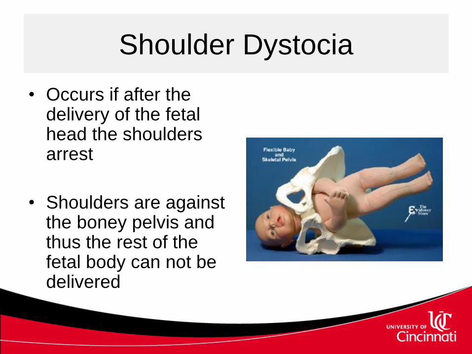
Shoulder Dystocia
• Occurs if after the delivery of the fetal head the shoulders arrest
• Shoulders are against the boney pelvis and thus the rest of the fetal body can not be delivered

Treatment
• Vaginal delivery
– Complete episiotomy
– McRoberts procedure
• Mother abducts legs > chest
– Suprapubic pressure
– Manual rotation of anterior shoulder
– Extraction of posterior arm
– Replacement of the head and C/S

Shoulder Dystocia
• Maternal Complications
– Uterine atony
• hemorrhage
– Uterine rupture
– Vaginal lacerations
• Fourth degree tear
– Genital tract trauma and infection

Multiple Births

Placenta Classification
Chorion -placenta
Amnion -amniotic sac

Placenta Classification
• Type of placentation determines likelihood
of vascular communications
– Vascular communications occur
• Nearly all monochorionic placentas
• Rare dichorionic placentas
– May result in
• Twin-twin transfusion syndrome
• Intrauterine fetal death

Maternal Physiologic Changes
• Increased uterine size
– decreased total lung capacity
– Decreased FRC
– Displaces stomach cephalad
• decreases competence of lower esophageal sphincter
• Weight gain > rate after 30 weeks
• Maternal blood volume (additional 500ml) &
CO increases
Fetal Risks
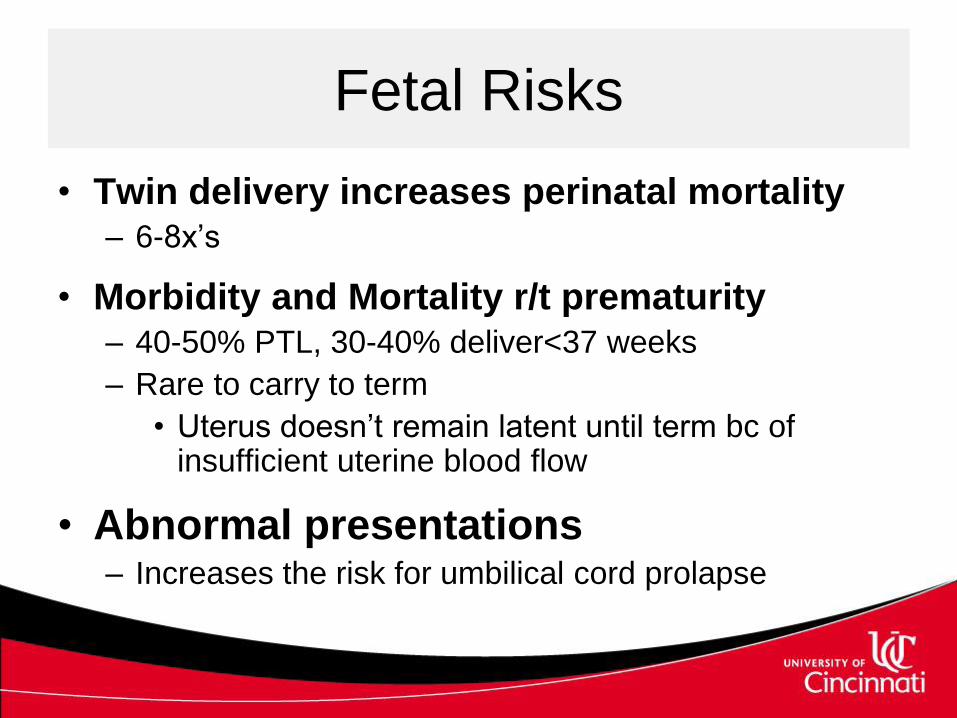
• Twin delivery increases perinatal mortality – 6-8x’s
• Morbidity and Mortality r/t prematurity – 40-50% PTL, 30-40% deliver<37 weeks
– Rare to carry to term
• Uterus doesn’t remain latent until term bc of insufficient uterine blood flow
• Abnormal presentations – Increases the risk for umbilical cord prolapse

Twin-to-Twin Transfusion
• Abnormal connection of chorionic blood vessels
in the placenta bt. two monochorionic twins
– one twin becomes the donor and the other
twin becomes the recipient
• Donor twin is smaller and is at risk for intrauterine
growth restriction and anemia
• Recipient twin is plethoric (excess amount of
blood) and is at risk for volume overload and
cardiac failure

Twin-to-Twin Transfusion
• Treatment
– Serial amnioreduction to control
polyhdramnios
– Selective feticide to allow the other fetus to
survive
– Selective fetoscopic laser photocoagulation of
the vascular anastomoses bt. the two twins
• Laser inserted either percutaneously or through a
maternal laparotomy

Anesthesia
TTTS Fetal Surgery
• Considerations
– Analgesia, amnesia and immobility
• Mom & Baby
– Preserve gas exchange and cardiovascular
stability of both
– Control uterine tone

Fetal Risks
• Second twin
– Increased morbidity and mortality than first
• FHR monitoring improves outcome
– After delivery of twin A the following reduce
intervillous blood flow and oxygenation to the 2nd
twin:
• Partial separation of placenta
• Reduced uterine size
• Clamping of the 1st umbilical cord

Twin Fetal Risks
• Malpresentation
– d/t growth retardation and polyhydramnios
• Placental problems
– Cord prolapse/entanglement d/t: – Malpresentation
– Malposition
– Premature rupture of membranes

Maternal Risks
• Physiologic risks
– Increased CO • an additional 15%
– Increased Supine Hypotensive Syndrome • a 32-week uterus of a twin gestation is as big as a term
uterus containing a single-fetus and gets progressively larger
• Anemia • occurs 2-4x more often

Maternal Risks
• Physiologic risks
– Hypoxia • Decrease in FRC
• Increase in closing volume and oxygen consumption
– Pulmonary edema • Use of tocolytics
– Difficult intubation
– Pulmonary aspiration

Obstetrical Risks
• PIH and preeclampsia – 5x more common
• Antepartum
hemorrhage
– Abruptio placenta &
placenta previa
• Postpartum
hemorrhage
– Uterine atony d/t over
distended uterus
– 2-3 x times increased risk
Obstetrical Risks
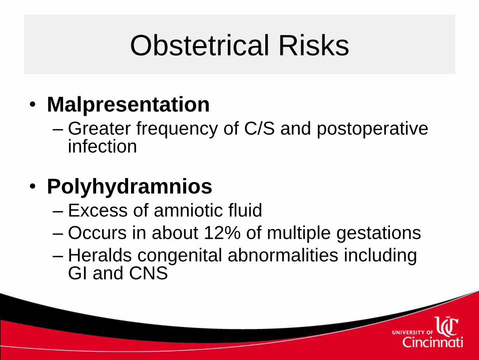
• Malpresentation – Greater frequency of C/S and postoperative
infection
• Polyhydramnios – Excess of amniotic fluid
– Occurs in about 12% of multiple gestations
– Heralds congenital abnormalities including GI and CNS

Obstetric Management
• Indications for C/S – Malpresentation of twin A
– Discordancy (twin B > twin A)
– Intrauterine death of one fetus
– Twin-twin transfusion
– Congenital deformities
– Decreased uteroplacental reserve
– Fetal cardiac decelerations
– Prematurity
– Three or more fetuses

Intrapartum Management
• Delivery should occur in the OR
– emergency abdominal delivery can be
performed
– Provide supplemental O2
– Have LA to extend epidural for C/S