some recent developments in immunocytochemistry
TRANSCRIPT
Histochemical Journal 15, 189-200 (1983)
R E V I E W
Some recent developments immunocytochemistry
in
FRED T. B O S M A N
Department of Pathology, University of Limburg, Biomedical Center, Beeldsnijdersdreef 101, Maastricht, The Netherlands
Received 31 March 1982 and in revised form 15 July 1982
Contents
S u m m a r y 189
Int roduct ion 190
Sensit ivity 190
Specificity, 194
Precision 196
Conclus ions 197
A c k n o w l e d g e m e n t s 197 References 198
Summary
lmmunocytochemistry has become an indispensable tool both in basic biomedical research and in diagnostic histophathology. Several recent innovations have led to improvements in the sensitiv- ity, specificity and precision of these techniques.
Numerous modifications of the original methods have been developed, many with increased sensitivity. In particular, methods using protein A or the avidin-biotin complex as second steps appear to be promising. New methods for the quantitative determination of sensitivity have become available.
The introduction of monoclonal antibodies as immunocytochemical reagents appears to be a major improvement. New methods of immunochemical analysis, such as immunoblotting and ;anmunospotting of antigens extracted from tissue specimens, allow the molecular composition of immunoreactive antigenic sites in a tissue to be analysed.
The accuracy of localization in immunoelectron microscopy has been improved significantly through the use of ultracryotomy of unfixed tissue in combination with colloidal gold particles as a label. In addition, gold particles can be counted and thus allow relatively simple quantification of the immune reaction. Using flow cytometry, especially in combination with monoclonal anti- bodies, quantitative immunofluorescence has become feasible.
0018-2214/83/030189-12503.94/0 �9 1983 Chapman and Hall Ltd.
190 BOSMAN
Introduction Immunocytochemical methods have been available now for about four decades and, in the last 15 years especially, have become an indispensable tool in many areas of biomedi- cal research and diagnostic histopathology (Taylor, 1978; Bosman & Nieuwenhuyzen Kruseman, 1979). Initially fluorescein was the only label available (Coons et aI., 1941). The introduction of enzyme labels, such as horseradish peroxidase (HRP) was a major step forwards (Nakane & Pierce, 1966; Avrameas, 1967). More recently, immunological methods have been developed to bind the label to the antibody (Masonet al., 1969). Of these unlabelled antibody methods, the peroxidase-antiperoxidase (PAP) complex method has become very popular (Sternberger et al., 1970). In addition, conjugation methods, which are used to link the antibody covalently to an enzyme label, have been improved (Wilson & Nakane, 1978; Boorsma & Streefkerk, 1979). Enzyme labels appear to be applicable for electron microscopy because the substrate for the most commonly used label, diaminobenzidine, results in a final reaction product which can be chelated with osmium and thus become electron-dense.
These methods have led to numerous important contributions in many areas of biomedical science. Several modifications of the original methods have been developed, usually to improve their performance. Most of these improvements concern method sensitivity, method specificity or the precision of immunolocalization. In this review, I shall discuss the more important developments in these crucial areas of immunocytochemistry.
Sensitivity
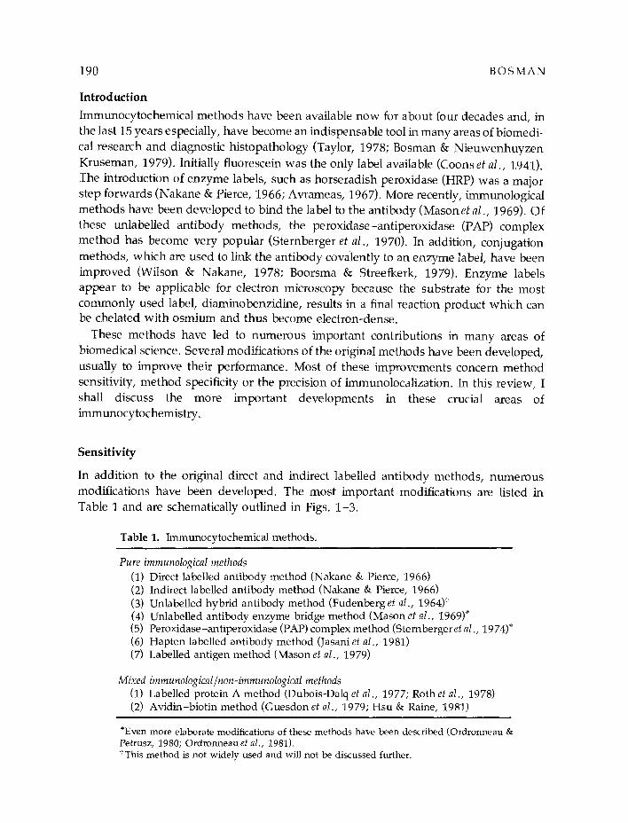
In addition to the original direct and indirect labelled antibody methods, numerous modifications have been developed. The most important modifications are listed in Table 1 and are schematically outlined in Figs. 1-3.
Table 1. Immunocytochemical methods.
Pure immunological methods (1) Direct labelled antibody method (Nakane & Pierce, 1966) (2) Indirect labelled antibody method (Nakane & Pierce, 1966) (3) Unlabelled hybrid antibody method (Fudenberg et al., 1964)t (4) Unlabelled antibody enzyme bridge method (Mason et al., 1969)* (5) Peroxidase-antiperoxidase (PAP) complex method (Sternbergeret al., 1974)* (6) Hapten labelled antibody method (Jasaniet al., 1981) (7) Labelled antigen method (Mason et al., 1979)
Mixed immunological~non-immunological methods (1) Labelled protein A method (Dubois-Dalq et al., 1977; Roth et al., 1978) (2) Avidin-biotin method (Guesdon et al., 1979; Hsu & Raine, 1981)
"Even more elaborate modifications of these methods have been described (Ordronneau & Petrusz, 1980; Ordronneauet al., 1981). tThis method is not widely used and will not be discussed further.

Developments in immunocytochemistry 191
The sensitivity of these variants has rarely been tested in absolute terms. In most studies of the sensitivity of immunoperoxidase procedures, tissue sections have been used as the test substrate (Vaccaet al., 1975; Ordronneau & Petrusz, 1980; Ordronneau et al., 1981; Naritoku & Taylor, 1982). Recently, artificial substrates have been advo-
cated for testing sensitivity (and also for specificity). Larsson (1981) has described the use of spots of antigen on filter paper, which can then be stained immunocytochemi- cally. Herbrink et al. (1982) reported the use of antigen spots on dibenzylmethoxybenzyl paper or nitrocellulose paper for the same purpose. A similar method and a solid-phase
~ antigen _ _ first antibody ,~ label
second antibody ~ protein A
~ b i o t i n
_ _ anti-hapten antibody avidin
~ anti-peroxidase antibody
Figs. 1-3. Key to symbols.
A 1
2 2 3 4
Fig. 1. Schematic outline of immunocytochemical procedures based on the use of conjugates. In all these methods, both fluorochromes and enzymes can be used as the label. (1) Single-step direct labelled antibody method. The first antibody itself is labelled. (2) Two-step indirect labelled antibody method. The first antibody is unlabelled and serves as the antigen for the labelled second antibody. (3) Two-step labelled antigen method. The unlabelled antibody is applied in excess; as a result, antigen binding sites remain free to bind the subsequently applied labelled antigen. (4) Two-step hapten-antihapten method. The first antibody is conjugated to the hapten (such as dinitrophenol, DNP), which can be detected with a labelled antihapten antibody.

192 B O S M A N
1 2
Fig. 2. Schematic outline of unlabelled antibody methods. In these methods only enzymes have been used. (1) Four-step unlabelled antibody enzyme bridge method. Unlabelled first antibody can react with second antibody, which is applied in excess. The third step consists of anti-enzyme antibody, which is derived from the same species as the first antibody. The enzyme label is applied in the final step. (2) Three-step enzyme-antienzyme complex method. As horseradish peroxidase is the enzyme which is most often employed, this technique is commonly referred to as the peroxidase-antiperoxidase or PAP technique. The procedure is similar to the enzyme bridge method but antienzyme antibody and enzyme label are applied together in the form of an immune complex. In both unlabelled antibody methods, the number of enzyme molecules per antigen binding site in the tissue can be increased by repeating the final two or three steps of the incubation sequence.
1 2 3
Fig. 3. Immunocytochemical techniques in which part of the sequence is non-immunological. In these methods various labels have been employed. (1) Two-step protein A method. Unlabelled first antibody is detected with labelled protein A. (2, 3) Two- and three-step methods using the avidin-biotin interaction. In both variants, the first antibody is conjugated with biotin. In variant 2, the second step consists of labelled avidin. In variant 3, unlabelled avidin is used as a bridge to bind labelled biotin. Several other variants of the avidin-biotin system have been reported.

Developments in immunocytochemistry 193
enzyme-linked immunoassay have also been applied by Bosman et aI. (1982) for the testing of sensitivity.
Theoretically, the sensitivity of immunoenzyme methods can be improved by: (1) increasing the number of enzyme molecules bound per antigen binding site; (2) increas- ing the staining intensity obtained with a given amount of enzymatic activity; and (3) increasing image contrast by reducing non-specific background staining without affect- ing specific staining intensity.
The number of enzyme molecules per antigen binding site has been claimed to be higher in the PAP complex method (Sternberger et al., 1970) than in the indirect labelled-antibody method (Nakane & Pierce, 1966). Consequently, it has been claimed that the PAP method has a superior sensitivity (Taylor, 1978; Sternberger, 1979) but a recent study has shown that the PAP method is not significantly more sensitive (Bosman et aI., 1982). However, the elaborate multistep-antibody enzyme-bridge and PAP complex methods (Vacca et al., 1975; Ordronneau & Petrusz, 1980; Ordronneau et al., 1981) are probably more sensitive than the more simple procedures, although they have not been tested on defined substrates.
The staining intensity arising from the enzyme reaction is a reflection of the amount of reaction product and varies for different 'chromogens'. Diaminobenzidine (DAB) is the 'chromogen' most frequently employed, but other substances, such as pyrocatechol- phenylenediamine (Hankeret al., 1977) have been claimed to be at least as sensitive but with the advantage of not being carcinogenic (Morrell et al., 1981). Recently, several means of enhancing the intensity of the DAB stain have been reported, such as by impregnation with silver salts (Gallyas et al., 1982) after the DAB reaction or by the addition of 0.01 M imidazole to the DAB substrate solution (Straus, 1982), In our hands, the first method has not proven useful, whereas the addition of imidazole certainly increases the intensity of staining about ten-fold (Bosman, unpublished results).
Non-specific background staining is primarily caused by non-specific adsorption of serum proteins to tissue sections. Therefore, non-specific background staining can be reduced by replacing the second antibody step in indirect procedures with non- immunological reagents. Protein A, with its high affinity for the Fc fragment of IgG of most species, was one of the first substances to be used in this way (Fig. 3). It has been labelled with fluorescein (Dorval et al., 1974), HRP (Dubois-Dalq et aI., 1977) and colloidal gold. For immuno-electron microscopy, labelled protein A has rapidly become popular. The advantages of protein A for routine use in immunoperoxidase methods are low background, versatility due to the bivalence for Fc fragments, and speed due to the fact that saturation of binding sites is reached rapidly (Notani et aI., 1979). However, care must be taken to prevent protein A binding to Fc fragments, particularly in unfixed sections of tissue. Another disadvantage of protein A procedures is that the antibody efficiency is comparatively low (Celio et aI., 1979).
In recent years, avidin, which can bind four molecules of biotin with an extremely high affinity (Bayeret al., 1979) has been advocated as a non-immunological second step reagent (Guesdon et al., 1979). As with protein A, non-specific background binding is

194 BOSMAN
very low and thus theoretically should, in combination with the high binding affinity, result in high sensitivity. In addition, the tetravalence of avidin for biotin, which might increase the number of label molecules per antigen binding site, should contribute towards a high sensitivity. The avidin-biotin system is also very versatile. Avidin can be fluoresceinated (Heggeness & Ash, 1977) or labelled with an enzyme (Guesdon et al., 1979) and these conjugates can be used in combination with biotinylated antibodies (Fig. 3). Alternatively, enzyme labels can be used as unlabelled bridging components (Hsu & Raine, 1981). In a recent study, Naritoku & Taylor (1982) compared the charac- teristics of the avidin-biotin methods and the conventional PAP method in combination with monoclonal antibodies. The avidin-biotin method appeared to be more sensitive than a three-step PAP method. This finding indicates that, in terms of sensitivity, the avidin-biotin method may not be a revolutionary improvement. However, background staining was low with avidin-biotin and, as with protein A, the method required less time to complete. On the other hand, avidin tends to bind lectins in some tissues (for example, neural tissues) which may result in background problems or even in false positive staining. These problems can be overcome with newly developed blocking procedures (Wood & Warnke, 1981; Naritoku & Taylor, 1982). The use of avidin and biotin for immunocytochemistry is clearly still in a developmental stage and more studies are required before a final judgement of their applicability can be reached.
Specificity Strict specificity of an immunochemical reaction is one of the most important prerequis- ites for reliable immunocytochemistry. Specificity is determined by adequate control procedures. Reagent testing ascertains the specificity of the antibody for the appropriate antigen. Method controls are essential to exclude the possibility that tissue staining is the result of unwan ted interactions of the reagent with tissue components. Originally, reagent specificity was mostly tested with immunodiffusion and immunoelectro- phoresis but these methods are far less sensitive than immunocytochemistry. In these respects, radio- or enzyme immunoassays are more suitable. We have found that the enzyme-linked immunosorbent assay is very suitable for specificity testing of antisera to polypeptide hormones. The immunospot t ing test, as described earlier, can also be used for this purpose (Larsson, 1981; Bosman et al., 1982; Herbrink et al., 1982).
The most important recent development in antibody specificity is the monoclonal technique (for a recent review, see Diamond et al., 1981). A monoclonal antibody reacts with only one single antigenic determinant and this characteristic should theoretically guarantee absolute specificity. However, if an antigenic determinant occurs on several different macromolecules, a monoclonal antibody specific for that determinant will not differentiate between those antigensl In addition, it has been reported recently that some monoclonal antibodies react wi th antigens that are not even remotely related to the original immunogen (Dulbecco et al., 1981). Monoclonal antibodies, therefore, may not be the final answer to problems of antibody specificity. An additional important
Developments in immunocytochemistry 195
advantage of monoclonal antibodies is their potential unlimited supply. A stable, antibody-producing cell line can be maintained practically without limit and, particu- larly when ascites tumours are induced with hybridoma cells, will yield enormous quantities of pure antibody. Universal use of sets of well-characterized monoclonal antibodies would make immmlocytochemical studies much more comparable and would reduce the occurrence of conflicting results that may be caused partly by the use of non-standardized reagents.
Monoclonal antibodies also have some disadvantages when applied in immunocytochemistry. They may be less sensitive than conventional antibodies because with a monoclonal antibody an antigen will bind only one antibody molecule, whereas, with a conventional antibody, which contains many different antibody molecules reactive for multiple determinants, several antibody molecules will be bound. However, studies comparing the immunocytochemical sensitivity of monoclonal anti- bodies and conventional antisera have not been reported as yet and, therefore, this theoretical consideration may not be a serious practical problem. A second disadvantage of monoclonal antibodies is related to the fact that only one determinant per antigen molecule is detected. It is conceivable that in a tissue section, due to factors such as tissue fixation or the packing of the antigen molecules in the cell, the relevant determin- ant is not immunoreactive. In that case, immunocytochemistry with a monoclonal anti- body would give a negative result, whereas that with a conventional antibody, which also contains antibody molecules reactive for other determinants, would give a positive result. Such a phenomenon has been reported for monoclonal antibodies to prostate acid phosphatase (Narikotu & Taylor, 1982). My colleagues and I have had similar experiences with monoclonal antibodies to prolactin (Bosman unpublished results). Both the above mentioned disadvantages could be overcome by using a well-defined mixture of several monoclonal antibodies. Such a reagent would combine the advantages of conventional antisera and monoclonal antibodies.
Method specificity is conventionally tested by including a standard set of control sections with each staining sequence (Bosman & Nieuwenhuyzen Kruseman, 1979; Sternberger, 1979). In addition, so-called blocking of immunostaining by pre- incubation of the antibody with the antigen under investigation is regarded as an import- ant specificity control (Grube, 1980). Even when these controls are adequately per- formed, false positive results may be obtained, due to traces of contaminants in the antigen used for immunization, which may result in unwanted reactivities (Sundler et el., 1981). In addition, non-immunological binding of immunoglobulin molecules to tissue components can occur (Grube & Weber, 1980).
All these specificity tests leave one problem unsolved. Many substances, such as small peptide hormones, occur in multiple molecular forms, larger polypeptide chains often constituting the (usually biologically inactive) precursor molecule. An example of a complex group of related polypeptides, which are derived from one single precursor is the ACTH family. When an antibody is prepared against the naturally occurring form of ACTH (a 39-aminoacid polypeptide chain) and the specificity for this molecule has been

196 BOSMAN
proved, immunoreactivity with this antibody may be partly due to the occurrence of larger precursor molecules containing the sequence of interest. This problem cannot be solved by antibody testing or by blocking controls. An adequate solution can be obtained by a biochemical analysis of tissue extracts containing the antigen(s) under investigation. Van Raamsdonk et aI. (1977) developed a method for the immunoenzy- matic analysis of proteins, which were separated by electrophoresis in polyacryl- amide gels. Frozen sections of these gels could be hnmunostained and, in this way, combined information about the physico-chemical properties (such as molecular weight and isoelectric point) and immunochemical characteristics could be obtained. An easier solution to this problem might be immuno-blotting (Towbin et al., 1979). With this technique, proteins, separated in polyacrylaminde gels, are transferred to nitrocellulose paper by electrophoresis. The replica of the protein bands on nitrocellulose can be stained by immunoenzyme or immunofluorescence methods.
Precision
The most important aspect of the precision of immunocytochemical methods is the accuracy of localization. Immunoreactive sites ideally reflect the localization of the anti- gen under investigation in living cells and tissues. However, during fixation and tissue processing, displacement of tissue antigens may occur, as recently stressed by Novikoff (1980). Studies on the topographical distribution of different proteins produced by hepatocytes, have been complicated by this phenomenon (Feldman et al., 1972; Nayak & Mital, 1979). Moreover, different methods of fixation may selectively mask immunoreactive sites in some tissues while leaving sites in other tissues intact (Orstik et aI., 1981). Techniques for tissue freezing and ultracryomicrotomy have significantly improved recently (Terracio & Schwabe, 1981) and, therefore, application of these methods may partly circumvent the above complications, although they may also intro- duce new artifacts. Results of immunocytochemical studies should, therefore, be inter- preted cautiously and preferably be corroborated by other methods.
The type of label which is used may also influence the accuracy of localization. When enzyme-labels are used for immuno-electron microscopy it should be kept in mind that the final reaction product is often distributed in a fairly wide radius around the site of the enzyme (for the DAB-peroxidase reaction, this radius has been found to be at least 20 nm; Ordronneau et al., 1981). This phenomenon limits the topographical resolution of immunoenzyme methods in electron microscopy. The use of colloidal gold particles as labels in immuno-electron microscopy has improved the topographical resolution (Roth et al., 1978). Colloidal gold particles easily bind to proteins such as protein A and, so far, the protein A-gold technique appears to be a very important addition to the techniques available for immuno-electron microscopy. Gold particles have another advantage; they can be prepared in different sizes (Slot & Geuze, 1981), which makes simultaneous labelling of two different antigens in the same section feasible (Geuze et

Developments in immunocytochemistry 197
al., 1981). Furthermore, the particles can easily be counted and thus allow simple quantification of the immuno-reaction.
The second aspect of precision is quantification. A linear relationship between the amount of label and the local amount of antigen would be desirable. Attempts have been made to quantify immunofluorescence intensity but several difficulties have been met (Haaijman et al., 1975), most of which were related to the inherent characteristics of conventional antibodies. These problems have been overcome largely by the introduc- tion of monoclonal antibodies whose specificity and binding affinity can be determined accurately. The use of these reagents, in combination with flow cytometry, has rapidly" become an area of promising developments (Hoffrnan et al., 1980).
Although flow cytometers are not at the moment as sensitive as the human eye, they can measure fairly weak fluorescence signals in single cells at a very high speed (>103 cells per second). In this way, small numbers of labelled cells can be detected amongst a majority of negative cells. Furthermore, the amount of immunoreactivity per cell can be determined accurately. With these instruments, even multiple parameters, such as immunofluorescence staining for two antigens (using two different fluorochromes and DNA staining combined with immunofluorescence and/or light scatter, which is related to cell size) can be measured (Zeile, 1980; Steen et al., 1982). Using this approach, reliable analyses of different lymphocyte subpopulations in peripheral blood has become an almost routine procedure (Coshni et al., 1981). An interesting recent applica- tion is the detection of DNA replication after incorporation of BrdU, which can be detected immunologically with an anti-BrdU antibody (Gratzner & Leif, 1981). This method has been reported to give results comparable with conventional autoradiogra- phy but is very easily quantifiable and much more rapid.
Conclusions
From this review of the evolution of immunocytochemistry in the last decade, it is clear that many technical innovations have extended enormously the applicability of immunocytochemistry. Consequently, immunocytochemical investigations have resulted in a vast amount of new information about the morphology and function of cells and tissues. The evolutionary process, however, is still going on. Improved methods of tissue preservation and preparation are being developed. The explosive development of hybridoma technology has already resulted in numerous interesting monoclonal antibodies which can be applied to relevant biological problems. Better labels and labelling methods will improve the sensitivity and precision of immunocytochemical methods still further.
Acknowledgements
Professor Mels van der Ploeg read the manuscript critically. Expert secretarial assistance was provided by Marjo van Rooden and Claire Bollen.

198 B O S M A N
R e f e r e n c e s
AVRAMEAS, S. (1967) Coupl ing of enzymes to proteins wi th glutaraldehyde. Use of conjugates for the detect ion of antigen and antibodies. Immunochemistny 6, 43-52.
BAYER, E., SKUTELSKY, E. & WILCHEK, M. (1979) The av id in-b io t in complex in affinity cytochemistry. Meth. EnzymoI. 62, 308-16.
BOORSMA, D. M. & STREEFKERK, J. G. (1979) Periodate or glutaraldehyde for prepar ing perox- idase conjugates. J. Immunoi. Meth. 30, 245-55.
BOSMAN, F. T., CRAMER-KRIJNENBURG, P. F. & VAN BERGEN HENEGOUW, J. (1982) Effi- ciency and sensitivity of indirect immunoperoxidase methods. Histochemistry (in press).
BOSMAN, F. T. & NIEUWENHUYZEN KRUSEMAN, A. C. (1979) Clinical applications of the enzyme labelled ant ibody method. Immunoperoxidase methods in diagnostic histopathol- ogy. J. Histochem, Cytochem. 27, 1140-7.
CELLO, M. R., LUTZ, H. & BINZ, H. (1979) Protein A in immunoperoxidase techniques. J. Histochem. Cytochem. 27, 691-3.
COONS, A. H., CREECH, H. J. & JONES, R. N. (1941) Immunological propert ies of an ant ibody containing a fluorescent group. Proc. Soc. exp. Biol. 47, 200-2.
COSIMI, A. B., COLVIN, R. B., BURTON, R. C., RUBIN, R. H., GOLDSTEIN, G., KUNG, P. C., HANSEN, W. P., DELMONICO, F. L. & RUSSELL, P. S. (1981) Monoclonal ant ibodies for immunologic moni tor ing and t reatment in recipients of renal allografts. N. Engl. J. Med. 305,
308-14. DIAMOND, B. A., YELTON, D. E. & SCHARFF, M. D. (1981) Monoclonal antibodies. A new
technology for producing serologic reagents. N. Engl. J. Med. 304, 1344-9. DORVAL, G., WELSH, K_. I. & W1GZELL, H. (1974) Labeled staphylococcal protein A as an
immunological probe in the analysis of cell surface markers. Scand. J. hnmunol. 3, 405-11. DUBOIS-DALQ, M., McFARLAND, H. & McFARLIN, D. (1977) Protein A peroxidase: a valuable
tool for the localization of antigens. J. Histochem. Cytochem. 25, 1201-6. DULBECCO, R., UNGER, M., BOLOGNA, M., BATTIFORA, H., SYKA, P. & OKADA, S. (1981)
Cross reactivity be tween thy-1 and a component of intermediate filaments demonst ra ted using a monoclonal ant ibody. Nature 292, 772-4.
FELDMAN, G., PENAUD-LAURENCIN, J., CRASSONS, J. & BENHAMON, J. P. (1972) Albumin synthesis by human liver cells: its morphological demonstrat ion. Gastroenterology 63, 1036-48.
FUDENBERG, H. H., DREWS, M. D. & NISONOFF, A. (1964) Serologic demonstra t ion of dual specificity of rabbit bivalent thyroid ant ibody. J. Exp. Med. 119, 151-66.
GALLYAS, F., GORCS, T. & MERCKENTHALER, I. (1982) High grade intensification of the end product of the d iaminobenzidine reaction for peroxidase histochemistry. J. Histochem, Cytochem. 30, 183-4.
GEUZE, H. J., SLOT, J. W., SCHEFFER, R. C. T. & VAN DER LEY, P. A. (1981) Use of colloidal gold particles in double- label ing immunoelectron microscopy of ultrathin frozen tissue sections. J. Cell Biol. 89, 653-65.
GRATZNER, H. G. & LEIE, R. C. (1981) An immunofluorescence method monitor ing D N A synth- esis by flow cytometry. Cytometry 1, 385-9.
GRUBE, D. (1980) Immunoreactivit ies of Gastrin (G-)cells. II. Non-specific binding of immuno- globulins to G-cells by ionic interactions. Histochemistry 66, 149-67.
GRUBE, D. & WEBER, E. (1980) Immunoreactivi t ies of gastrin (G-)cells. I. Dilution dependen t staining of G-cells by antisera and non- immune sera. Histochemistry 65, 223-37.
GUESDON, J. L., TERNYNCK, T. & AVRAMEAS, S. (1979) The use of av id in-b io t in interaction in immunoenzymat ic techniques. J. Histochem. Cytochem. 27, 1131-9.
D e v e l o p m e n t s in i m m u n o c y t o c h e m i s t r y 199
HAAIJMAN, J. J., BLOEMMEN, F. J. & HAM, C. M. (1975) Microfluorometric immoassays with antigens bound to Sepharose beads. Ann. N.Y. Acad. Sci. 254, 137-50.
HANKER, J. S., YATES, P. E., METZ, C. B. & RUSTONI, A. (1977) A new specific sensitive and non-carcinogenic reagent for the demonstration of horseradish peroxidase. Histochem. J. 9, 789-92.
HEGGENESS, M. H. & ASH, J. F. (1977) Use of the avidin-biotin complex for the localization of actin and myosin with fluorescence microscopy, J. Cell. Biol. 73, 783-8.
HERBRINK, P., VAN BUSSEL, F. J. & WARNAAR, S. O. (1982) The antigen spot test (AST): a highly sensitive assay for the detection of antibodies. J. Immunol. Meth, 48, 293-8.
HOFFMAN, R. A., KUNG, P. C., HANSEN, W, P. & GOLDSTEIN, G. (1980) Simple and rapid measurement of human T-lymphocytes and their subclasses in peripheral blood. Proc. Natl. Acad. Sci. U.S.A. 77, 4914-7.
HSU, S.-M. & RAINE, L. (1981) Protein A, avidin and biotin in immunohistochemistry. ). Histochem. Cytochem. 29, 1,349-53.
JASANI, B., WYNFORD THOMAS, D. & WILLIAMS, E. D. (1981) Use of monoclonal anti-hapten antibodies for immunolocalization of tissue antigen. J. Clin. Pathol. 34, 1000-2.
LARSSON, L. I. (1981) A novel immunocytochemical model system for specificity and sensitivity screening of antisera against multiple antigens. J. Histochem. Cytochem. 29, 4 0 8 - 1 0 . .
MASON, T. E., PHII~ER, R. F., SPICER, S. S. & SWALOW, R. A. (1969) An immunoglobul in-enzyme bridge method for localizing tissue antigens. J. Histochem, Cytochem. 17, 563-9.
MASON, D. Y. & SAMMONS, R. E. (1979) The labeled antigen method of immunoenzymatic staining. J. Histochem. Cytochem. 27, 832-40.
MORRELL, J. I., GREENBERGER, L. M. & PFAFE, W. (1981) Comparison of horseradish peroxidase visualization methods: quantitative results and further technical specifics. ]. Histochem. Cytochem, 29, 903-16.
NAKANE, P. K. & PIERCE, G. B. (1966) Enzyme labeled antibodies: preparation and application for the localization of antigens. J. Histochem. Cytochem. 14, 929-31.
NARITOKU, W. Y. & TAYLOR, C. R. (1982) A comparative study of the use of monoclonal anti- bodies using three different immunochemical methods: an evaluation of monoclonal and polyclonaI antibodies against human prostatic acid phosphatase. ]. Histochem. Cytochem. 3{), 253-60.
NAYAK, N. C. & MITAL, I. (1977) The dynamics of ~-fetoprotein and albumin synthesis in human and rat liver during normal ontogeny. Amer. J. Pathol. 86, 359-74.
NOTANI, G. W., PARSONS, J. A. & ERLANDSSEN, S. L. (1979) Versatility of staphylococcus protein A in immunocytochemistry. Use in unlabelled antibody enzyme system and fluores- cent methods. J. Histochem. Cytochem. 27, 1438-44.
NOVIKOFF, A. B. (1980) DAB cytochemistry: artifact problems in its current uses. J. Histochem. Cytochem. 28, 1036-7.
ORDRONNEAU, P., L I N D S T R O N , P. B.-M. & PETRUSZ, P. (1981) Four unlabeled antibody bridge techniques: A comparison. J. Histochem. Cytochem. 29, 1397-404.
ORDRONNEAU, P. & PETRUSZ, P. (1:980) Immunocytochemical demonstration of anterior pitu- itary hormones in the pars tuberalis of long-term hypophysectomized rats. Amer, J. Anat. 158, 491-8.
ORSTIK, T. B., BRANDZEAG, P., NUSTAD, K. & PrERCE, J. V. (1981) Effects of different tissue processing methods on the immunohistochemical localization of kallikrein in the pancreas. J. Histochem. Cytochem. 29, 985-8.
ROTH, J., BENDAYAN, M. & ORCI, L. (1978)Ultrastructural localization of intracellular antigens by the use of protein A-gold complex. J. Histochem. Cytochem. 26. 1074-81.

200 B O S M A N
SLOT, J. W. & GEUZE, H. J. (1981) Sizing of protein A-colloidal gold probes for hnmunoelectron microscopy. J. Cell. Biol. 90, 533-6.
STEEN, H. B., BOYE, E., S K A R S T A D , K. , B L O O M , H. , G O D A L , T. & M U S T A F A , S. (1982) Appli- cations of flow cytometry on bacteria: cell cycle kinetics, drug effects and quantitation of antibody binding. Cytometry 2, 249-57.
STERNBERGER, L. A: (1979)Immunocytochemistry, 2nd edn. New York: J. Wiley. S T E R N B E R G E R , LG~A.~,~HARDY J~, p. H. , C U C U L I S , J. ]. & MEYER, H. G. (1970) The unlabeled
antibody enzyme method for immunohistochemistry. Preparation and properties of soluble antigen-antibody complex (horseradish peroxidase-anti-horseradish peroxidase) and its use in identification of spirochetes. J. Histochem. Cytochem. 22, 315-33.
s TRAUS, W. (1982) Imidazol increases the sensitivity of the cytochemical reaction for peroxidase with diaminobenzidine at neutral pH. J. Histochem. Cytochem. 30, 491-3.
S U N D L E R , E., A L U M E T S , J . , E K M A N , R., H A K A N S O N , R. & VAN W I E M E R S M A G R E I D A N U S ,
TJ. B. (1981) lmmunoreactive adrenocorticotropic hormone (ACTH) in porcine gut and pan- creas: fact or artifact?J. Histochem. Cytochem. 29, 1328-35.
TAYLOR, C. R. (1978) Immunoperoxidase techniques. Practical and theoretical aspects. Arch. Pathol. Lab. Med. 102, 113-21.
TERRACIO, L. & SCHWABE, K. G. (1981) Freezing and drying of biological tissue for electron microscopy. J. Histochem. Cytochem. 29, 1021-8.
T O W B I N , H. , S T A E H E L I N , T. & G O R D O N , J. (1979) Electrophoretic transfer of proteins from polyacrylamide gels to nitrocellular sheets: procedure and some applications. Proc. natn. Acad. Sci., U.S.A. 76, 4350-4.
V A C C A , L. L. , R O S A R I O , S. L., Z I M M E R M A N , E. A. , T H O M A S H E F S K Y , P. , NG, P . -Y. & HSU,
K. C. (1975) Application of immunoperoxidase techniques to localize horseradish peroxidase tracer in the central nervous system. J. Histochem. Cytochem. 23, 208-15.
VAN R A A M S D O N K , W. , P O O L , C. W. & H E Y T I N G , C. (1977) Detection of antigens and antibodies by an immuno-peroxidase method applied on thin longitudinal sections of SDS-poly- acrylamide gels. ]. Immunol. Meth. 17, 337-48.
WILSON, M. B. & NAKANE, P. K. (1978) Recent developments in the periodate method of con- jugating horseradish peroxidase (HRPO) to antibodies. Inlmmunofluorescence and Related Stain- ing Techniques (edited by KNAPP, W., HOLUBAR, K. & WICK, G.), pp. 215-29. Amsterdam: Elsevier/North Holland.
WOOD, G. S. & WARNKE, R. (1981) Suppression of endogenous avidin-binding activity in tissues and its relevance to biotin-avidin detection systems. J. Histochem, Cytochem. 29, 1196-204.
ZE ILE, G. (1980) Intracytoplasmic immunofluorescence in multiple myeloma. Cytometry 1, 37-41.