somatostatina - giuseppe di bella · l’azione della somatostatina è mediata da recettori ad alta...
TRANSCRIPT

SOMATOSTATINA - OCTREOTIDE
IN
MALATTIE LINFOPROLIFERATIVE

Introduzione
La somatostatina a 14 aminoacidi (aa) è stata scoperta nel 1973 (Brazeau, Vale et al. 1973).
L’azione della somatostatina è mediata da recettori ad alta affinità (SSTR1-5), localizzati sulla
membrana plasmatica di molti tipi di cellule (Lahlou, Guillermet et al. 2004; Olias, Viollet et al.
2004).
Le azioni biologiche della somatostatina si possono così sintetizzare:
1) Sull’ipofisi anteriore inibizione dell’attività incretoria di:
• Ormone somatotropo (GH)
• Prolattina (PRL)
• Ormone tireotropo (TSH)
• Ormone corticotropo (ACTH)
• (Ormone Luteinizzante) (LH)
• (Ormone Follicolo Stimolante) (FSH)
2) Sul tratto genitourinario:
• Inibizione della renina
3) Sull’apparato gastroenterico:
a) Inibizione delle secrezioni endocrine
• Insulina
• Glucagone
• Peptide intestinale vasoattivo
• Gastrina
• Secretina
• Colecistochinina
• Motilina
b) Inibizione delle secrezioni esocrine
• Acidi gastrici
• Secrezione e svuotamento gastrico
• Flusso ematico gastrointestinale
• Trasporto di acqua VIP-stimolato
• Assorbimento e motilità intestinale
• Fattore intrinseco
• Pepsina
• Enzimi e bicarbonato pancreatici
• Fluido colico

• Flusso biliare
• Svuotamento gastrico

Analoghi della somatostatina
Essendo l’emivita plasmatica della somatostatina a 14 aa molto breve (meno di 3 minuti), sono
stati sintetizzati vari analoghi con una aumentata stabilità. L’Octreotide è un analogo sintetico
ad 8 aa, che ha effetti farmacologici simili alla somatostatina, ma possiede una durata d’azione
molto più lunga (Weckbecker, Raulf et al. 1993; Harris 1994). Viene di seguito riportato un
elenco di analoghi della somatostatina (Dasgupta 2004):
Nome Selettività SSTR Struttura

Effetti della somatostatina ed analoghi su cellule neoplastiche.
• Inibizione del rilascio di vari ormoni come GH, PRL, LH, FSH, TSH, e vari fattori di
crescita tra cui EGF, IGF, FGF, NGF, PDGF, TGF, VEGF, che svolgono un ruolo
determinante nell’induzione della proliferazione e disseminazione neoplastica (Robbins
1996; Dasgupta 2004).
• La somministrazione di octreotide migliora significativamente la sopravvivenza ed è
un’alternativa valutabile nel trattamento del carcinoma epatocellalure in operabile
(Kouroumalis, Skordilis et al. 1998; Kouroumalis 2001).
• Trattamento di cancri epatocellulari con analoghi della somatostatina (Raderer, Hejna et
al. 1999; Raderer, Hejna et al. 2000).
• Induzione di apoptosi in cellule di epatoma umano (Chen, Liu et al. 2001).
• Inibizione della crescita di cancri del colon (Szepeshazi, Schally et al. 2002; Tejeda,
Gaal et al. 2003).
• Riduzione dell’attività proliferativa delle cellule tumorali e dei livelli sierici di IGF-I in
pazienti con cancro colo-rettale (Cascinu, Del Ferro et al. 1997).
• Inibizione della crescita di cellule di cancro ovarico epiteliale umano (Yano, Radulovic et
al. 2000).
• Inibizione della crescita di tumori gliali (Feindt, Mentlein et al. 1997; Held-Feindt, Krisch
et al. 1999; Held-Feindt, Krisch et al. 2000; Held-Feindt, Forstreuter et al. 2001).
• Inibizione della crescita di cellule di cancro endometriale umano (Mishima, Yano et al.
1999).
• Inibizione della proliferazione di cellule di carcinoma polmonare umano a piccole cellule
ed anche a non piccole cellule (Cattaneo, Amoroso et al. 1996; Szereday, Schally et al.
2003).
• Inibizione della proliferazione cellulare in neuroblastoma umano (Cattaneo, Amoroso et
al. 1996; Cattaneo, Scita et al. 1999; Cattaneo, Taylor et al. 2000).
• Inibizione della proliferazione di cellule di leucemia umana (Ishihara, Hassan et al.
1999; Tejeda, Gaal et al. 2003).
• Inibizione della crescita di cellule di carcinoma prostatico umano (Brevini-Gandolfi, Cillo
et al. 2001; Tejeda, Gaal et al. 2003).
• Inibizione della crescita di cellule di cancro del seno (Dolan, Miltenburg et al. 2001;
Tejeda, Gaal et al. 2003).
• Inibizione della crescita di cellule di melanoma (Szende, Horvath et al. 2003; Tejeda,
Gaal et al. 2003).
• Effetto antiproliferativo in cellule pancreatiche normali e tumorali (Damge and Hajri
1998; Douziech, Calvo et al. 1999; Zalatnai 1999; Charland, Boucher et al. 2001).

• Inibizione dei percorsi del pentosio fosfato ossidativi e non ossidativi (Boros, Brandes et
al. 1998).
• Riduzione in cellule tumorali dei recettori del fattore di crescita epidermico EGFR
(Szepeshazi, Halmos et al. 1999).
• Potenziamento dell’attività dei chemioterapici nei tumori (Tesei, Ricotti et al. 2000).
• Effetto proapoptotico e antiproliferativo sinergico con Melatonina (Melen-Mucha,
Winczyk et al. 1998).

Somatostatina ed analoghi in malattie linfoproliferative
Recettori per la somatostatina (SSTR) sono espressi in organi e cellule linfoidi umane e
animali, indicando che tali recettori possono avere importanti ruoli nei processi di attivazione,
sviluppo e/o tumorigenesi delle cellule di origine linfoide (Nakamura, Koike et al. 1987; Aguila,
Dees et al. 1991; Tsutsumi, Takano et al. 1997; Ferone, van Hagen et al. 1999; Ferone, van
Hagen et al. 2004).
Poiché cellule linfoidi normali e patologiche esprimono SSTR, analoghi radiomarcati sono stati
utilizzati per visualizzare neoplasie linfoidi in vivo. Nei linfomi maligni, sia di Hodgkin che non-
Hodgkin, è risultato che la scintigrafia con analoghi SSTR radiomarcati può rivelare siti di
malattia attiva, non rilevati con altri metodi convenzionali (van den Anker-Lugtenburg,
Lowenberg et al. 1996). Inoltre, la scintigrafia SSTR permette una più accurata stadiazione del
linfoma di Hodgkin (Lugtenburg, Krenning et al. 2001). Questo permette una migliore diagnosi
determinando così una scelta terapeutica più efficace per il paziente. Inoltre, la scintigrafia
SSTR può aiutare a distinguere una patologia neoplastica (es. un linfoma maligno cutaneo) da
altre patologie (es. una linfoadenopatia dermatopatica) (van den Anker-Lugtenburg, Heule et
al. 1996). Ciò rende la scintigrafia SSTR un utile strumento diagnostico nei linfomi maligni.
È stato mostrato che l’octreotide inibisce la proliferazione in una linea cellulare di leucemia a
cellule-T, indicando che tale analogo della somatostatina può avere un potenziale terapeutico
in questo tipo di leucemia linfoide (Giannetti, Enjalbert et al. 2000). Anche l’analogo della
somatostatina TT-232 ha mostrato effetti antiproliferativi in cellule neoplastiche di origine
linfoide, sia in modelli in vitro che in vivo (Tompa, Jakab et al. 2000; Tejeda, Gaal et al. 2005).
La somatostatina regola negativamente la crescita di varie cellule normali e tumorali. Questo
effetto può essere mediato oltre che direttamente, mediante SSTR presenti sulle cellule
tumorali, anche indirettamente, attraverso SSTR presenti su cellule non tumorali (Robbins
1996; Ferjoux, Bousquet et al. 2000).
L’effetto antiproliferativo della somatostatina può essere mediato attraverso l’inibizione della
secrezione di fattori che promuovono la crescita tumorale. Per esempio, sono stati riportati
possibili effetti dell’ormone della crescita (GH) nello sviluppo di leucemia linfoblastica acuta e
linfoma di Hodgkin (Rogers, Komp et al. 1977; Magnavita, Teofili et al. 1996; Jeay,
Sonenshein et al. 2002) . La somatostatina essendo un inibitore della secrezione dell’ormone
GH potrebbe avere un effetto benefico nel trattamento di tali neoplasie.

Un altro effetto antiproliferativo indiretto della somatostatina può essere mediato attraverso
l’inibizione dell’angiogenesi, un processo fondamentale nello sviluppo e nella disseminazione
tumorale (Garcia de la Torre, Wass et al. 2002; Woltering 2003; Dasgupta 2004). È stato
mostrato che la somatostatina e vari suoi analoghi inibiscono l’angiogenesi in vari modelli
sperimentali, in vitro ed in vivo (Patel, Barrie et al. 1994; Danesi and Del Tacca 1996; Danesi,
Agen et al. 1997; Lawnicka, Stepien et al. 2000). Tale effetto anti-angiogenetico è mediato da
recettori SSTR presenti sulle cellule endoteliali proliferanti (Florio, Morini et al. 2003; Adams,
Adams et al. 2005). Quindi, la somatostatina o i suoi analoghi inibiscono, a livello delle cellule
endoteliali, la formazione di nuovi vasi indotti da fattori pro-angiogenetici, come il VEGF,
prodotti dalle cellule tumorali.
Uno studio clinico di fase II ha valutato l’effetto di un analogo della somatostatina in pazienti
con malattie lifoproliferative (Witzig, Letendre et al. 1995). I risultati hanno indicato che la
somatostatina alla dose di 150 microgrammi ogni 8 ore è ben tollerata ed ha attività nei
linfomi non-Hodgkin a basso grado: 10 pazienti su 28 valutabili hanno avuto una risposta
parziale.
Un altro studio clinico ha valutato la somatostatina in combinazione con ciclofosfamide,
bromocriptina, retinoidi, melatonina e ACTH nel trattamento di linfomi non-Hodgkin a basso
grado in fase avanzata (Todisco, Casaccia et al. 2001). In questo studio, è stata valutata la
tossicità e l’efficacia di un regime terapeutico conosciuto come multiterapia Di Bella, che risulta
dall’associazione di un agente chemioterapico (ciclofosfamide) con altre molecole non
mielosoppressive (somatostatina, bromocriptina, retinoidi, melatonina e ACTH). Su 20 pazienti
valutabili per risposta e tossicità il 70% (14 su 20) ebbe una risposta parziale; il 20% (4 su
20) ebbe una malattia stabile ed il 10% (2 su 20) ebbe progressione di malattia. Continuando
con la terapia, nessuno dei 14 pazienti con una risposta parziale ebbe una progressione della
malattia (tempo di controllo medio di 21 mesi, intervallo da 7 a 25) ed il 50% di questi pazienti
ebbe una risposta completa. Dei 4 pazienti con malattia stabile, il 25% (1 di 4) ebbe una
risposta parziale ed il 75% (3 di 4) progredirono con la terapia (tempo medio di progressione
14.3 mesi, intervallo da 7 a 21). La tossicità era molto modesta, gli effetti collaterali più
comuni furono: sonnolenza, diarrea e iperglicemia.

BIBLIOGRAFIA
Adams, R. L., I. P. Adams, et al. (2005). "Somatostatin receptors 2 and 5 are preferentially
expressed in proliferating endothelium." Br J Cancer 92(8): 1493-8.
Aguila, M. C., W. L. Dees, et al. (1991). "Evidence that somatostatin is localized and
synthesized in lymphoid organs." Proc Natl Acad Sci U S A 88(24): 11485-9.
Boros, L. G., J. L. Brandes, et al. (1998). "Inhibition of the oxidative and nonoxidative pentose
phosphate pathways by somatostatin: a possible mechanism of antitumor action." Med
Hypotheses 50(6): 501-6.
Brazeau, P., W. Vale, et al. (1973). "Hypothalamic polypeptide that inhibits the secretion of
immunoreactive pituitary growth hormone." Science 179(68): 77-9.
Brevini-Gandolfi, T. A., F. Cillo, et al. (2001). "Somatostatin up-regulates topoisomerase II
alpha expression and affects LNCaP cell cycle." Mol Cell Endocrinol 176(1-2): 103-10.
Cascinu, S., E. Del Ferro, et al. (1997). "Inhibition of tumor cell kinetics and serum insulin
growth factor I levels by octreotide in colorectal cancer patients." Gastroenterology
113(3): 767-72.
Cattaneo, M. G., D. Amoroso, et al. (1996). "A somatostatin analogue inhibits MAP kinase
activation and cell proliferation in human neuroblastoma and in human small cell lung
carcinoma cell lines." FEBS Lett 397(2-3): 164-8.
Cattaneo, M. G., G. Scita, et al. (1999). "Somatostatin inhibits PDGF-stimulated Ras activation
in human neuroblastoma cells." FEBS Lett 459(1): 64-8.
Cattaneo, M. G., J. E. Taylor, et al. (2000). "Selective stimulation of somatostatin receptor
subtypes: differential effects on Ras/MAP kinase pathway and cell proliferation in
human neuroblastoma cells." FEBS Lett 481(3): 271-6.
Charland, S., M. J. Boucher, et al. (2001). "Somatostatin inhibits Akt phosphorylation and cell
cycle entry, but not p42/p44 mitogen-activated protein (MAP) kinase activation in
normal and tumoral pancreatic acinar cells." Endocrinology 142(1): 121-8.
Chen, X., Z. Liu, et al. (2001). "Antineoplastic mechanism of Octreotide action in human
hepatoma." Chin Med J (Engl) 114(11): 1167-70.
Damge, C. and A. Hajri (1998). "Effect of the gastrin-releasing peptide antagonist BIM 26226
and lanreotide on an acinar pancreatic carcinoma." Eur J Pharmacol 347(1): 77-86.
Danesi, R., C. Agen, et al. (1997). "Inhibition of experimental angiogenesis by the
somatostatin analogue octreotide acetate (SMS 201-995)." Clin Cancer Res 3(2): 265-
72.
Danesi, R. and M. Del Tacca (1996). "The effects of the somatostatin analog octreotide on
angiogenesis in vitro." Metabolism 45(8 Suppl 1): 49-50.

Dasgupta, P. (2004). "Somatostatin analogues: multiple roles in cellular proliferation,
neoplasia, and angiogenesis." Pharmacol Ther 102(1): 61-85.
Dolan, J. T., D. M. Miltenburg, et al. (2001). "Treatment of metastatic breast cancer with
somatostatin analogues--a meta-analysis." Ann Surg Oncol 8(3): 227-33.
Douziech, N., E. Calvo, et al. (1999). "Inhibitory and stimulatory effects of somatostatin on
two human pancreatic cancer cell lines: a primary role for tyrosine phosphatase SHP-1."
Endocrinology 140(2): 765-77.
Feindt, J., R. Mentlein, et al. (1997). "Time-dependent influence of the somatostatin analogue
octreotide on the proliferation of rat astrocytes and glioma cells." Brain Res 746(1-2):
309-13.
Ferjoux, G., C. Bousquet, et al. (2000). "Signal transduction of somatostatin receptors
negatively controlling cell proliferation." J Physiol Paris 94(3-4): 205-10.
Ferone, D., P. M. van Hagen, et al. (1999). "Somatostatin receptors in the thymus." Ann Med
31 Suppl 2: 28-33.
Ferone, D., P. M. van Hagen, et al. (2004). "Somatostatin receptor distribution and function in
immune system." Dig Liver Dis 36 Suppl 1: S68-77.
Florio, T., M. Morini, et al. (2003). "Somatostatin inhibits tumor angiogenesis and growth via
somatostatin receptor-3-mediated regulation of endothelial nitric oxide synthase and
mitogen-activated protein kinase activities." Endocrinology 144(4): 1574-84.
Garcia de la Torre, N., J. A. Wass, et al. (2002). "Antiangiogenic effects of somatostatin
analogues." Clin Endocrinol (Oxf) 57(4): 425-41.
Giannetti, N., A. Enjalbert, et al. (2000). "Somatostatin analog SMS 201995 inhibits
proliferation in human leukemia T-cell line: relevance of the adenylyl cyclase
stimulation." J Cell Biochem 78(4): 666-73.
Harris, A. G. (1994). "Somatostatin and somatostatin analogues: pharmacokinetics and
pharmacodynamic effects." Gut 35(3 Suppl): S1-4.
Held-Feindt, J., F. Forstreuter, et al. (2001). "Influence of the somatostatin receptor sst2 on
growth factor signal cascades in human glioma cells." Brain Res Mol Brain Res 87(1):
12-21.
Held-Feindt, J., B. Krisch, et al. (2000). "Somatostatin receptors in gliomas." J Physiol Paris
94(3-4): 251-8.
Held-Feindt, J., B. Krisch, et al. (1999). "Molecular analysis of the somatostatin receptor
subtype 2 in human glioma cells." Brain Res Mol Brain Res 64(1): 101-7.
Ishihara, S., S. Hassan, et al. (1999). "Growth inhibitory effects of somatostatin on human
leukemia cell lines mediated by somatostatin receptor subtype 1." Peptides 20(3): 313-
8.
Jeay, S., G. E. Sonenshein, et al. (2002). "Growth hormone can act as a cytokine controlling
survival and proliferation of immune cells: new insights into signaling pathways." Mol
Cell Endocrinol 188(1-2): 1-7.

Kouroumalis, E., P. Skordilis, et al. (1998). "Treatment of hepatocellular carcinoma with
octreotide: a randomised controlled study." Gut 42(3): 442-7.
Kouroumalis, E. A. (2001). "Octreotide for cancer of the liver and biliary tree." Chemotherapy
47 Suppl 2: 150-61.
Lahlou, H., J. Guillermet, et al. (2004). "Molecular signaling of somatostatin receptors." Ann N
Y Acad Sci 1014: 121-31.
Lawnicka, H., H. Stepien, et al. (2000). "Effect of somatostatin and octreotide on proliferation
and vascular endothelial growth factor secretion from murine endothelial cell line
(HECa10) culture." Biochem Biophys Res Commun 268(2): 567-71.
Lugtenburg, P. J., E. P. Krenning, et al. (2001). "Somatostatin receptor scintigraphy useful in
stage I-II Hodgkin's disease: more extended disease identified." Br J Haematol 112(4):
936-44.
Magnavita, N., L. Teofili, et al. (1996). "Hodgkin's lymphoma in a cyclist treated with growth
hormone." Am J Hematol 52(1): 65-6.
Melen-Mucha, G., K. Winczyk, et al. (1998). "Somatostatin analogue octreotide and melatonin
inhibit bromodeoxyuridine incorporation into cell nuclei and enhance apoptosis in the
transplantable murine colon 38 cancer." Anticancer Res 18(5A): 3615-9.
Mishima, M., T. Yano, et al. (1999). "Inhibition of human endometrial cancer cell growth in
vitro and in vivo by somatostatin analog RC-160." Am J Obstet Gynecol 181(3): 583-
90.
Nakamura, H., T. Koike, et al. (1987). "Identification of lymphoid cell lines bearing receptors
for somatostatin." Immunology 62(4): 655-8.
Olias, G., C. Viollet, et al. (2004). "Regulation and function of somatostatin receptors." J
Neurochem 89(5): 1057-91.
Patel, P. C., R. Barrie, et al. (1994). "Postreceptor signal transduction mechanisms involved in
octreotide-induced inhibition of angiogenesis." Surgery 116(6): 1148-52.
Raderer, M., M. H. Hejna, et al. (1999). "Successful treatment of an advanced hepatocellular
carcinoma with the long-acting somatostatin analog lanreotide." Am J Gastroenterol
94(1): 278-9.
Raderer, M., M. H. Hejna, et al. (2000). "Treatment of hepatocellular cancer with the long
acting somatostatin analog lanreotide in vitro and in vivo." Int J Oncol 16(6): 1197-
201.
Robbins, R. J. (1996). "Somatostatin and cancer." Metabolism 45(8 Suppl 1): 98-100.
Rogers, P. C., D. Komp, et al. (1977). "Possible effects of growth hormone on development of
acute lymphoblastic leukaemia." Lancet 2(8035): 434-5.
Szende, B., A. Horvath, et al. (2003). "Effect of a novel somatostatin analogue combined with
cytotoxic drugs on human tumour xenografts and metastasis of B16 melanoma." Br J
Cancer 88(1): 132-6.
Szepeshazi, K., G. Halmos, et al. (1999). "Growth inhibition of experimental pancreatic cancers
and sustained reduction in epidermal growth factor receptors during therapy with
hormonal peptide analogs." J Cancer Res Clin Oncol 125(8-9): 444-52.
Szepeshazi, K., A. V. Schally, et al. (2002). "Targeted cytotoxic somatostatin analogue AN-238
inhibits somatostatin receptor-positive experimental colon cancers independently of
their p53 status." Cancer Res 62(3): 781-8.
Szereday, Z., A. V. Schally, et al. (2003). "Effective treatment of H838 human non-small cell
lung carcinoma with a targeted cytotoxic somatostatin analog, AN-238." Int J Oncol
22(5): 1141-6.
Tejeda, M., D. Gaal, et al. (2003). "The antitumor activity of the somatostatin structural
derivative (TT-232) on different human tumor xenografts." Anticancer Res 23(5A):
4061-6.
Tejeda, M., D. Gaal, et al. (2005). "Growth inhibitory effect of the somatostatin structural
derivative (TT-232) on leukemia models." Anticancer Res 25(1A): 325-30.
Tesei, A., L. Ricotti, et al. (2000). "Lanreotide-induced modulation of 5-fluorouracil or
mitomycin C cytotoxicity in human colon cancer cell lines: a preclinical study." J
Chemother 12(5): 421-30.
Todisco, M., P. Casaccia, et al. (2001). "Cyclophosphamide plus somatostatin, bromocriptin,
retinoids, melatonin and ACTH in the treatment of low-grade non-Hodgkin's lymphomas
at advanced stage: results of a phase II trial." Cancer Biother Radiopharm 16(2): 171-
7.
Tompa, A., M. G. Jakab, et al. (2000). "The somatostatin analogue peptide TT-232 induces
apoptosis and chromosome breakage in cultured human lymphocytes." Mutat Res
465(1-2): 61-8.
Tsutsumi, A., H. Takano, et al. (1997). "Expression of somatostatin receptor subtype 2 mRNA
in human lymphoid cells." Cell Immunol 181(1): 44-9.
van den Anker-Lugtenburg, P. J., F. Heule, et al. (1996). "Somatostatin receptor scintigraphy
in cutaneous malignant lymphomas." J Am Acad Dermatol 34(6): 985-93.
van den Anker-Lugtenburg, P. J., B. Lowenberg, et al. (1996). "The relevance of somatostatin
receptor expression in malignant lymphomas." Metabolism 45(8 Suppl 1): 96-7.
Weckbecker, G., F. Raulf, et al. (1993). "Somatostatin analogs for diagnosis and treatment of
cancer." Pharmacol Ther 60(2): 245-64.
Witzig, T. E., L. Letendre, et al. (1995). "Evaluation of a somatostatin analog in the treatment
of lymphoproliferative disorders: results of a phase II North Central Cancer Treatment
Group trial." J Clin Oncol 13(8): 2012-5.
Woltering, E. A. (2003). "Development of targeted somatostatin-based antiangiogenic therapy:
a review and future perspectives." Cancer Biother Radiopharm 18(4): 601-9.
Yano, T., S. Radulovic, et al. (2000). "Inhibition of human epithelial ovarian cancer cell growth
in vitro by somatostatin analog RC-160." Oncology 59 Suppl 1: 45-9.

Zalatnai, A. (1999). "Epidermal growth factor receptor, somatostatin and bcl-2 in human
pancreatic tumor xenografts. An immunohistochemical study." Pathol Oncol Res 5(2):
146-51.

Cancer Biother Radiopharm. 2001 Apr;16(2):171-7.
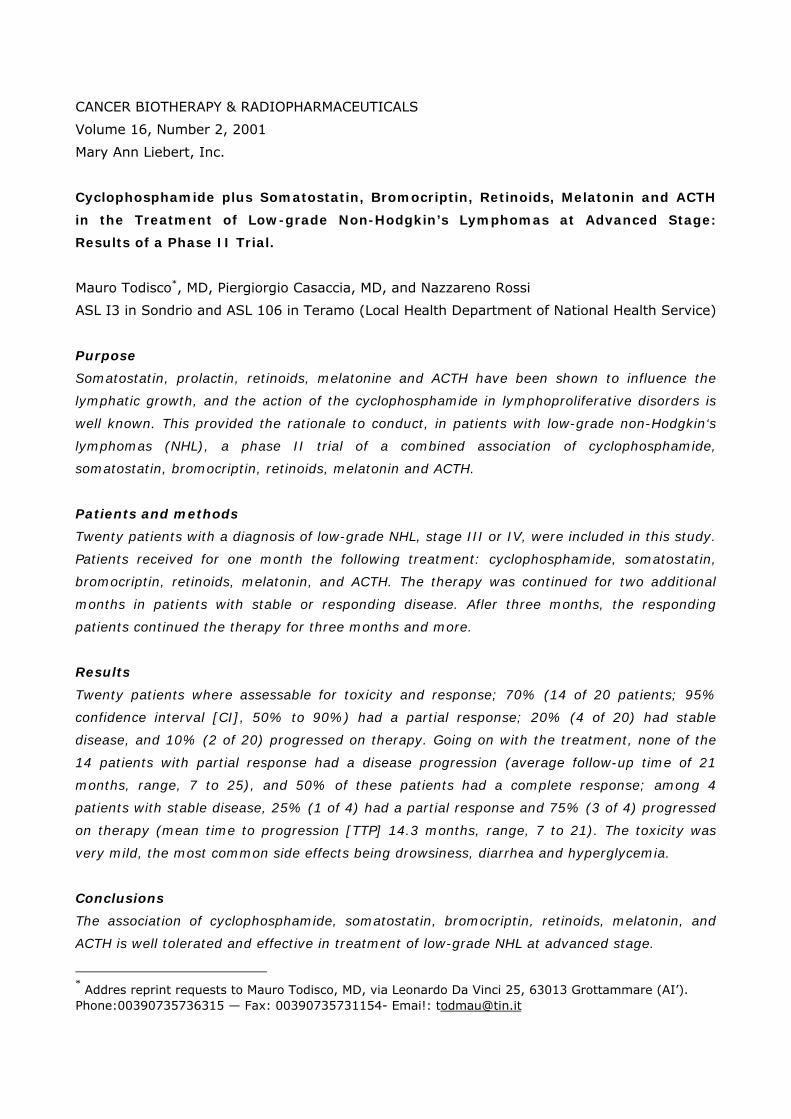
Cyclophosphamide plus somatostatin, bromocriptin, retinoids, melatonin and ACTH in the
treatment of low-grade non-Hodgkin's lymphomas at advanced stage: results of a
phase II trial.
Todisco M, Casaccia P, Rossi N.
PURPOSE: Somatostatin, prolactin, retinoids, melatonin and ACTH have been shown to
influence the lymphatic growth, and the action of the cyclophosphamide in
lymphoproliferative disorders is well known. This provided the rationale to conduct, in
patients with low-grade non-Hodgkin's lymphomas (NHL), a phase II trial of a combined
association of cyclophosphamide, somatostatin, bromocriptin, retinoids, melatonin and
ACTH. PATIENTS AND METHODS: Twenty patients with a diagnosis of low-grade NHL,
stage III or IV, were included in this study. Patients received for one month the
following treatment: cyclophosphamide, somatostatin, bromocriptin, retinoids,
melatonin, and ACTH. The therapy was continued for two additional months in patients
with stable or responding disease. After three months, the responding patients
continued the therapy for three months and more. RESULTS: Twenty patients were
assessable for toxicity and response; 70% (14 of 20 patients; 95% confidence interval
[CI], 50% to 90%) had a partial response; 20% (4 of 20) had stable disease, and 10%
(2 of 20) progressed on therapy. Going on with the treatment, none of the 14 patients
with partial response had a disease progression (average follow-up time of 21 months,
range, 7 to 25), and 50% of these patients had a complete response; among 4 patients
with stable disease, 25% (1 of 4) had a partial response and 75% (3 of 4) progressed
on therapy (mean time to progression [TTP] 14.3 months, range, 7 to 21). The toxicity
was very mild, the most common side effects being drowsiness, diarrhea and
hyperglycemia. CONCLUSIONS: The association of cyclophosphamide, somatostatin,
bromocriptin, retinoids, melatonin, and ACTH is well tolerated and effective in
treatment of low-grade NHL at advanced stage.
CANCER BIOTHERAPY & RADIOPHARMACEUTICALS
Volume 16, Number 2, 2001
Mary Ann Liebert, Inc.
Cyclophosphamide plus Somatostatin, Bromocriptin, Retinoids, Melatonin and ACTH
in the Treatment of Low-grade Non-Hodgkin’s Lymphomas at Advanced Stage:
Results of a Phase II Trial.
Mauro Todisco*, MD, Piergiorgio Casaccia, MD, and Nazzareno Rossi
ASL I3 in Sondrio and ASL 106 in Teramo (Local Health Department of National Health Service)
Purpose
Somatostatin, prolactin, retinoids, melatonine and ACTH have been shown to influence the
lymphatic growth, and the action of the cyclophosphamide in lymphoproliferative disorders is
well known. This provided the rationale to conduct, in patients with low-grade non-Hodgkin‘s
lymphomas (NHL), a phase II trial of a combined association of cyclophosphamide,
somatostatin, bromocriptin, retinoids, melatonin and ACTH.
Patients and methods
Twenty patients with a diagnosis of low-grade NHL, stage III or IV, were included in this study.
Patients received for one month the following treatment: cyclophosphamide, somatostatin,
bromocriptin, retinoids, melatonin, and ACTH. The therapy was continued for two additional
months in patients with stable or responding disease. Afler three months, the responding
patients continued the therapy for three months and more.
Results
Twenty patients where assessable for toxicity and response; 70% (14 of 20 patients; 95%
confidence interval [CI], 50% to 90%) had a partial response; 20% (4 of 20) had stable
disease, and 10% (2 of 20) progressed on therapy. Going on with the treatment, none of the
14 patients with partial response had a disease progression (average follow-up time of 21
months, range, 7 to 25), and 50% of these patients had a complete response; among 4
patients with stable disease, 25% (1 of 4) had a partial response and 75% (3 of 4) progressed
on therapy (mean time to progression [TTP] 14.3 months, range, 7 to 21). The toxicity was
very mild, the most common side effects being drowsiness, diarrhea and hyperglycemia.
Conclusions
The association of cyclophosphamide, somatostatin, bromocriptin, retinoids, melatonin, and
ACTH is well tolerated and effective in treatment of low-grade NHL at advanced stage.
* Addres reprint requests to Mauro Todisco, MD, via Leonardo Da Vinci 25, 63013 Grottammare (AI’). Phone:00390735736315 — Fax: 00390735731154- Emai!: [email protected]

INTRODUCTION
Recently, several new agents bave been shown to inhibit the lymphatic growth. Among these,
especially the ones without bone marrow toxicity, as somatostatin, aroused great interest for
the possibility to be used with myelosuppressive chemotherapy regimens without determining
a further myelosuppression.
An association like this was first reported in 1979 by Di Bella et al [1], who reported to have
used the cyclophosphamide together with somatostatin, bromocriptin, retinoids, melatonin
and ACTH in several cancers, including non Hodgkin’s lymphomas (NHL).
On the basis of this pharmacological association and, in particular, of the use of somatostatin
and bromocriptin, there is the assumption, formulated by Di Bella, that growth hormone (GH)
and prolactin are involved in neoplastic growth. Such an assumption, in acute lymphoblastic
leukernia, was formulated by other authors also, in the same period, even if just for GH [2].
Subsequently, Payan et al demonstrated that somatostatin inhibits the growth of cultured
primary human liymphocytes and Molt-4 cells [3]; Nakamura et al identified somatostatin
receptors on the membrane of several lymphoid cell lines [4] and Hiruma et al found
somatostatin receptors on primary leukemia human cells [5] Furthermore, most of
lymphomatous lesions were shown to be identifiable with radiolabelled somatostatin analogs.
[6-10]
In agreement with the above mentioned results, Witzig et al in 1995 reported that octreotide,
a somatostatin analog, shows activity in patients with low-grade NHL [11] The influence on
lymphatic growth has been also demonstrated for prolactin, retinoids, melatonin and ACTH.
The prolactin has been shown to stimulate the growth of experimental lymphomas both in vivo
and in vitro, [12] and prolactin receptors are present on the surface of normal and neoplastic
lymphoid cells.[13-16] Matera et al demonstrated that prolactin is an autocrine growth factor
for a human leukemic cell line, [17] and Hooghe et al returned on the hypothesis, already
mantained by Di Bella, that prolactin and GH have an important role in lymphoma and
leukemia. [18] In hematology, the antitumor effect of retinoids is based on several evidences
reporting the effect of trans-retinoic acid in promyelocytic leukemia, [19-20] T-cell lymphoma
localized to the skin [21-26] and also in B- cell lymphomas. [27-29]
Melatonin inhibits thymidine incorporation in normal lymphocytes and in lymphoblastoid cell
lines [30] and inhibits the proliferative response to mitogens. [31-32] In addition, melatonin
carries on an antimyelodysplastic action [33] and decreases the bone marrow toxicity of
chemotherapeutic agents. [34] T and B lymphocytes have been shown to express the ACTH
receptor on their cell surface [35]; moreover, ACTH depresses the lymphocyte blastogenesis in
response to phytoemagglutinin and concanavallin A [36] and has a role in the modulation of
NK celi activity. [37]
These studies, together with the well known action of cyclophosphamide, provided the
rationale to design a phase II study to determine if a combined therapy based on

cyclophosphamide, somatostatin. brinocriptin, retinoids, melatonin and ACTH has activity in
patients with low-grade NHL at advanced stage.
PATIENTS AND METHODS
Patient Selection
Patients were selected on the basis of a clinical diagnosis of low-grade NHL, stage III or IV.
Additional criteria of selection weree: a performance status (PS) between 0 and 3, according to
the Eastern Cooperative Oncology Group, and the presence of bidimensionally measurable
lesion, as demonstrated by physical examination, chest radiograph, ecotomography, or
computed tomographic or magnetic resonance scans.
Patients who had received other treatment were included in this study only upon evidence that
the previous treatment was not effective. Patients receiving chemotherapy were asked to
suspend any drug admnistration for at least 15 days prior to the beginning of the combined
therapy.
Toxicity was evaluated using criteria developed by the World Health Organization.
Treatment
Patients received a combination of cyclophosphamide, somatostatin, bromocriptin, retinoids,
melatonin and ACTH. Cyclophosphamide was given orally at a dose of 75 mg/day (50 mg at 2
pm and 25 al 9 pm). Somatostatin was administreted subcutaneously (SQ) at a dose or 1.5
mg/day within 8 hours using a syringe pump. The administration started at least three hours
after dinner and those patients who were psychologically unable to accept this type of
administration received single SQ injection of octreotide (0,5 mg/day) three hours after dinner.
Bromocriptin was given orally at a dose of 2.5 mg/day (1.25 mg at 2 pm and at 9 pm).
Retinods: all-trans retinoic acid, vitamin A palmitate and beta-carotene were administered
orally, at 8 am, in 5 ml of vitarnin E, respectively at doses of 5 mg, 5000 UI and 20 mg/day.
Melatonin was given orally at a dose of 20 mg/day (10 mg at 2 pm and at 9 pm). ACTH was
administered intermuscularly at a dose of 1 mg/week.
All the patients were treated for at least one month. At the end of this period, those who had
stable disease or partial response received additional two months of treatment, and the ones
who responded after three months were treated for three months and more.
Criteria for Response
Complete response or remission was defined as the complete regression of all the measurable
lymphomatous lesions. Partial response was defined as a ≥ 50% reduction in the sum of the
products of the two diameters (the longest diameter and the one perpendicular to it) of one or
more lesions lasting at least 4 weeks.

Progression was defined by the increase in size of the pre-existing lesions of at least 25%, the
onset of new lesions or the increase of spleen or liver of at least 2 centimeters due to
lymphoma.
Those patients who could not be clearly placed in any of the described categories were defined
in stable condition. The assessment of the response was made after 1 month from the
beginning of the treatment. Such an assessment was carried on after another 2 months and,
later, every 3 months in patients going on with the treatment.
RESULTS
Twenty patients (ten males and ten females between 37 and 70 years old) with low-grade
histology were included in this study, and all were assessable for toxicity and response. Sixty
percent (12 of 20) had centroblastic-centrocytic histology and 10 were previously treated.
Twenty percent (4 of 20) had centrocytic histology, the remaining twenty percent had
lymphocytic histology and 3 of 4 in both groups had been treated prior to this study. Eighty
percent of the patients (16 of 20) were stage IV and 20% (4 of 20) were stage III. Fifty
percent (8 of 16) of the previously treated patients had a therapy-free time (TFT) ≥ 6 months
and were in relapse; 50% (8 of 16), wilh TFT ≤ 1.5 months, had a progression during the
therapy followed before being enrolled in this study. The results of the treatment after 1 month
are analitically described in Table 1.

Seventy percent of the patients (14 of 20; 95% CI, 50 to 90%) had a partial response; 20%
(4 of 20; 95% CI, 2.5% lo 37.5%) had stable disease and 10% (2 of 20) progressed on
therapy. The response after 1 month in patients subdivided according to the previously
received therapies is described in Table 2.
One hundred percent (4 of 4) of the previously untreated patients, 100% (8 of 8) of the
patients wilh TFT ≥ 6 months and 100% of the patients with TFT ≤ 1.5 months non-responding
or progressed during interferon therapy, had a partial response. Among the patients with TFT
≤ 15
months non-responding or progressed during single agent or combination chemotherapy,
66.6% (4 of 6; 95% CI, 28% to 104%) had stable disease and 33.3% (2 of 6) progressed. The
response to the treatment prosecution is analitically described in Table 3.

Among the 14 patients that after 1 month had partial response, there was no disease
progression (average follow-up time of 21 months, range, 7 to 25), and 50% of these patients
(7 of 14; 95% CI, 24% to 76%) obtained a complete response. Among the 4 patients that
after 1 month had stable disease, 25% (1 of 4) had a partial response and 75% (3 of 4)
progressed (mean time to progression 14.3 months, range, 7 to 21). The response to the
treatment prosecution in patients subdivided according to the previously received therapies is
described in Table 4.
All the 20 patients were evaluable for the toxicity. The must common side effects were
gastrointestinal signs. Twenty-five percent of the cases had diarrhea first grade (2 patients) or
second grade (3 patients); 20% (4 patients) had nausea or grade 1 vomit, and 5 patients had
loss of appetite and anorexia. These side effects did not require suspension of the therapy but
only all adjustment of the dose of somatostatin. Drowsiness was observed in 20% (4 of 20) of
patients, and required an adjustment of the daily schedule of administration of melatonin (20
mg/day were subdivided into three doses, instead of two, one of which at bedtime).
Twenty five percent of patients (5 of 20) had grade I hyperglycemia (≤ 160/dL) and 20% (4 of
20) showed ankle and/or face edema; in both these cases the dose of ACTH was reduced to
0.5 mg/week.
DISCUSSION
The low-grade NHL at advanced stage are stili incurable disease, whose treatment, just in
consequence of these therapeutic limits, is very disputed, coexisting single agent
chemotherapy, combination chemotherapy, combined radiotherapy- chemotherapy, high-dose
intensive chemotherapy with autologous bone marrow transplanation or autologous blood
progenitor cell transplantation.

In this study we evaluated toxicity and efficacy of a regimen, known as Di Bella’s multitherapy
(DBM), resulting by the association of a chemotherapeutic agent, the cyclophosphamide, with
other non-myelosuppressive substances (somatostatin, bromocriptin, retinoids, melatonin and
ACTH).
The results we obtained - seventy percent of partial responses after 1 month, fifty percent of
which became complete responses going on with the treatment - have been really better than
the ones described with single agent chemiotherapy with alkylants, with wiich Kimby et al
described 36% of global response with complete response in 5% of 132 previously untreated
patients, [38] or than the ones described by Witzig et al with somatostatin as single agent
(36% of partial) response in 28 previously treated and untreated patients). This demonstrates
the therapeutic superiority of such a pharmacological association versus its single costituents.
Moreover, the activity of the regimen depended on the kind of previous therapy and on the
TFT. We documented 100% of global response among the previously untreated patients, the
patients in first relapse with TFT ≥ 6 months and the patients with TFT ≤ 1.5 months non-
responding or progressed during interferon therapy.
This result, better than others obtained with widely used chemotherapy regimens [Kimby et al
described 60% of global response with CHOP in 127 previously untreated patients with
lowgrade NHL stage III and IV] [38], and the very good tolerance of DBM (all the patients
carried on the treatment at home, going on with their normal activities) suggest further clinical
trials using this regimen in NHL
With regard to this, the recent availability of depot formulations of somatostatin may allow a
better feasibility of this regimen, where the main discomfort was in the daily SQ injection of
somatostatin, especially if done with syringe pump.
REFERENCES
1. Di Bella L, Rossi MT, Scalera O. Perspectives in pineal functions. Prog Brain Res
1979;52:475-8.
2. Rogers PC, Komp D, Rogol A, Sabio H. Possible effects of growth hormone on development
of acute lymphoblastic leukaemia. Lancet 1977;2:434-5.
3. Payan DG, Hess CA, Goetzl EJ. lnhibition by somatostatin of the proliferation of T-
lymphocytes and Molt-4 lymphoblasts. Cell Immunol 1984;84:433-8.
4. Nakarnura H, Koike T, Hiruma K, Sato T, Tomioka H, Yoshida S. Identification of lyrnphoid
cell lines bearing receptors for somatostatin. Immunology 1987;62:655-8.
5. Hiruma K, Koike T, Nakamura H, Sumida T, Maeda T, Tomioka H, Yoshida S, Fujita T.
Somatostatin receptors on human lymphocytes and leukaemia cells. Immunology
1990;71:480-5.
6. Krenning EP, Kwekkeboom DJ, Reubi JC, Van Hagen PM, van Eijck CH, Oei HY, Lamberts
SW. In-111-octreotide scintigrapby in oncology. Metabolism 1992;41 (9 Suppl 2):83-6.

7. Reubi JC, Waser B, van Hagen M, Lamberts SW, Krenning EP, Gebbers JO, Laissue JA. In
vitro and in vivo detection of somatostatin receptors in human malignant lymphornas. Int J
Canc 1992;50:895-900.
8. Reubi JC, Waser 8, Horisberger U, Krenning E, Lamberts SW, Gebbers JO, Gersbach P,
Laissue JA. In vitro autoradiographic and in vivo scintigraphic localisation of somatostatin
receptors in human lymphatic tissue. Blood 1993; 82:2143-51.
9. Bares R, Galonska P, Dempke W, Handt S, Bull U, Osieka R. Somatostatin receptor
scintigraphy in malignant lymphoma: first results and comparison with glucose rnetabolism
measured by positron emission tomography. Horm Metab Res 1993;27:56-8.
10. Rettenbacher L, Galvan O. Differentiation between residual cancer and thymic hyperplasia
in malignant non Hodgkin’s lymphoma with somatostatin receptor scintigraphy. Clin Nucl Med
1994;19:64-5.
11. Witzig TE, Letendre L, Gerstner I, Schroeder O, Mailliard JA, Colon-Otero G, MarschKe RF,
Windschill HE. Evaluation of a somatostatin analog in the treatment of lymphoproliferative
disorders: results of a phase II North Central Cancer Treatment Group Trial. J Clin Oncol
1995;13:2012-5.
12. Friesen HG, Shiu RP, Elsholtz H, Simpson S, Hughes J. Prolactin and growth hormone
receptors. Ciba Found Symp 1982;90:263-78.
13. Shiu RP, Elsholtz HP, Tanaka T, Friesen HG, Gout PW, Beer CT, Noble RL. Receptor-
mediated mitogenic action of prolactin in a rat lymphoma cell line. Endocrinology 1983;
113:159-65.
14. O’Neal KD, Schwarz LA, Yu-Lee LY. Prolactin receptor gene expression in lymphoid cells.
Moll Cell Endocrinol 1991 ;82: 127-35.
15. Hooghe R, Delhase M, Vergani P. Malur A. Hooghe-Peters EL. Growth hormone and
prolactin are paracrine growth and differentiation factors in the haemopoietic system. Immunol
Today 1993; 14:212-214.
16. Leite De Moraes MC, Touraine P, Gagnerault MC, Savino W, Kelly PA, Dardenne M.
Prolactin receptors and the immune system. Ann Endocrinol (Paris) 1995 ;56:567-70.
17. Matera L, Cutufia M, Geuna M, Conlarini M. Buttiglieri S, Galin S, Fazzari A, Cavaliere C.
Prolactin is an autocrine growth factor for the Jurkat human T-leukemic cell line. J
Neuroimmunol 1997;79: 12-21.
18. Hooghe R, Merchav S, Gaidano G, Naessens F, Matera L. A role for growth hormone and
prolactin in leukaemia and lymphoma? Cell Mol Life Sci 1998;54: 1095-101.
19. Sacchi S, Russo D, Avvisati G, Dastoli G, Lazzarino M, Pelicci PG, Bonora MR, Visani G,
Grassi C, Iacona I, Luzzi L, Vanzanelli P. All-trans retinoic acid in haematological malignancies,
un update. GER (Gruppo Ematologico Retinoidi). Haematologica 1997; 82:106-21.
20. Kessler JF, Meyskens FL Jr, Levine N, Lynch PJ, Jones SE. Treatment of cutaneous T-cell
lymphoma (mycosis fungoides) with 13-cis-retinoic acid. Lancet 1983;I: 1345-7.

21. Neely SM, Mehlmauer M, Feinstein DI. The effect of isotretinoin in six patients with
cutaneous T-cell lymphoma. Arch lntern Med 1987;147:529-31.
22. Mahrle G, Thiele B. Retinoids in cutaneous T cell lymphomas. Dermatologica 1987; 175
(Suppl I): 145-50.
23. Zachariae H, Thestrup-Pedersen K. Interferon alpha and etretinate combination treatment
of cutaneous T-cell lymphoma. J Invest Dermatol 1990:95(6 Suppl):206-8.
24. Chow JM, Cheng AL, Su IJ, Wang CH. 13-cis-retinoic acid induces cellular differentiation
and durable remission in refractory cutaneous Ki-1 lymphoma. Cancer 1991;67:2490-4.
25. Su IJ, Cheng AL, Tsai TF, Lay JD. Retinoic acid-induced apoptosis and regression of a
refractory Epstein-Barr virus-containing T cell lymphoma expressing multidrug-resistance
phenotypes. Br J Haematol 1993; 85: 826-8.
26. Chou WC, Su IJ, Tien HF, Liang DC, Wang CH, Chang YC, Cheng AL. Clinicopathologic,
cytogenetic, and molecular studies of 13 Chinese patients with Ki-1 anaplastic large cell
lymphoma. Special emphasis in the tumor response to 13-cis retinoic acid. Cancer 1996;78:
1805-12.
27. Turley JM, Funakoshi S, Ruscetti FW, Kasper J, Murphy WJ, Longo DL, Birchenall-Roberts
MC. Growth inhibition and apoptosis of RL human B lymphoma cells by vitamin E succinate and
retinoic acid: role for transforming growth factor beta. Cell Growth Differ 1995; 6:655-63.
28. Cheng AL, Chuang SE, Su IJ. Factors associated with the therapeutic efficacy of retinoic
acids on malignant lymphomas. J Formos Med Assoc 1997;96:525-34.
29. Sundaresan A, Claypool K, Mehta K, Lopez-Berestein G, Cabanillas F, Ford RJ Jr. Retinoid-
rnediated inhibition of cell growth with stimulation of apoptosis in aggressive B-cell
lymphomas. Cell Growth Differ 1997;8: 107 1-82.
30. Persengiev SP, Kyurkchiev S. Selective effect of the proliferation of lymphoid cells. Int J
Biochem, 1993; 25:441-4.
31. Konakchieva R, Kyurkchiev S, Kehayov I, Taushanova P, Kanchev L. Selective effect of
methoxyndoles on the lymphocyte proliferation and melatonin binding to activated human
lymphoid cells. J Neuroimmunol 1995; 63: 125-32.
32. Vijayalaxmi, Reiter RJ, Leal BZ, Meltz ML Effect of melatonin on mitotic and proliferation
indices, and sister chromatid exchange in human blood lymphocytes. Mutat Res
1996;351:187-92.
33. Viviani S, Negretti E. Orazi A, Sozzi G, Santoro A, Lissoni P, Esposti O, Fraschini F.
Preliminary studies on melatonin in the treatment of myelodislastic syndromes following cancer
chemotherapy. J Pineal Res 1990; 8:347-54.
34. Rapozzi V, Zorzet S, Comelli M, Mavelli I, Perissin L, Giraldi T. Melatonin decreases bone
marrow and lymphatic toxicity of adriamicin in mice bearing TLX5 lymphoma. Life Sci 1998;63:
1701-13.
35. Clarke BL. Bost KL. Differential expression of functional adrenocorticotropic hormone
receptors by subpopulations of lymphocytes. J Immunol 1989;143:464- 469.

36. Roth JA, Kaeberle ML, Hsu WH. Effects of ACTH administration on bovine
polymorphonuclear leukocyte function and lymphocyte blastogenesis. Am J Vet Res 1982 ;43:4
12-4 16.
37. Gatti O, Masera RG, Pallavicini L, Sartori ML. Staurenghi A, Orlandi F, Angeli A. Interplay in
vitro between ACTH, beta-endorphin, and glucocorticoids in the modulation of spontaneous
and lymphokine-inducible human natural killer (NK) cell activity. Brain Behav Immun
1993;7:16-28.
38. Kimby E, Bjorkholm M, Gahrton G, Glimelius B, Hagberg H, Johansson B, Johansson H,
Juliusson G, Jarnmark M, Lofvenberg E. Chlorambucil/prednisone vs. CHOP in symptomatic
low-grade non Hodgkin’s lymphomas: a randomized trial from the Lymphoma Group of Central
Sweden. Ann Oncol 1994;5 (Suppl 2):67-71.

Mutat Res. 2000 Feb 16;465(1-2):61-8.
The somatostatin analogue peptide TT-232 induces apoptosis and chromosome breakage in
cultured human lymphocytes.
Tompa A, Jakab MG, Major J, Idei M, Bocsi J, Mihalik R, Szende B, Keri G.
Somatostatin receptors are supposed to be important in the regulation of apoptosis. In this
study, we measured apoptosis occurring spontaneously, or induced by the synthetic
somatostatin analogue, the peptide TT-232. We examined isolated human peripheral
blood lymphocytes (PBL) from 32 nurses exposed bedside to cytostatic drugs, 12
chronic lymphoid leukaemia (CLL) patients prior to treatment, and 19 unexposed,
healthy donors without anamnestic occupational exposure to genotoxic agents. Cells
were stimulated by phytohaemagglutinin-P (PHA) and cultured for 69 h with or without
15 microg/ml TT-232, respectively. Cell kinetic parameters and apoptosis were
determined by flow cytometry after staining with FITC-labeled anti-BrdU and propidium
iodide (PI) and the results on spontaneous and peptide-induced apoptosis were
compared with the obtained chromosome aberration frequencies (CA). The peptide TT-
232 unexpectedly induced chromosome breakage in addition to apoptosis. The mean
spontaneous apoptotic fractions were 6.65+/-0.89%, 6.46+/-0. 53%, and 3.07+/-
0.57%, and the mean CA yields in the samples without TT-232 were 1.74+/-0.46%,
2.44+/-0.40%, and 4.50+/-1.05%, for healthy subjects, nurses, and CLL patients,
respectively. A total of 15 microg/ml TT-232 treatment in healthy subjects increased
the mean CA frequency (10.38+/-1.57%), as well as the apoptotic cell fraction
(2.63+/-0.45 times higher than the corresponding untreated sample). In TT-232-
treated PBLs of nurses, CA remained unchanged and the mean apoptotic cell fraction
showed only a slight increase (1.24+/-0.11 times higher than the untreated). Among
CLL patients, TT-232 treatment significantly increased both CA (up to 17.83+/-4.04%)
and the ratio of apoptotic cells (21.78+/-11.00 times higher than the untreated). These
results demonstrated significant differences in apoptosis sensitivity in controls, nurses
and CLL donors, after 15 microg/ml TT-232 treatment. Data also indicate that the
induced CA yields in CLL donors with high CA are in correlation with TT-232-induced
apoptosis.

Cell Immunol. 1997 Oct 10;181(1):44-9.
Expression of somatostatin receptor subtype 2 mRNA in human lymphoid cells.
Tsutsumi A, Takano H, Ichikawa K, Kobayashi S, Koike T.
We analyzed the mRNA expression of somatostatin receptor subtypes 1 to 5 (SSTR1-5) in
human lymphoid cell lines, human peripheral blood lymphocytes (PBL), and human
lymphatic leukemia cells, using the reverse transcription-polymerase chain reaction
method. In human lymphoid cell lines, SSTR2 mRNA expression was clearly detectable,
and there was no evidence of SSTR1 mRNA expression. SSTR2 mRNA was barely
detectable in PBL from healthy individuals but was clearly detectable in EB virus-
transformed lymphocytes. Lymphocytes from some of the leukemic patients showed
elevated SSTR2 mRNA expression. SSTR2 mRNA expression in PBL was upregulated
upon stimulation by PHA. SSTR3 mRNA was also observed in all the cell lines examined,
although in one cell line, the expression was weak. Some cell lines showed little or no
SSTR4 or 5 mRNA expression. The expression pattern of SSTR2 mRNA suggests that
this receptor may have some important roles in lymphocyte activation, development,
and/or tumorgenesis.

J Am Acad Dermatol. 1996 Jun;34(6):985-93.
Somatostatin receptor scintigraphy in cutaneous malignant lymphomas.
van den Anker-Lugtenburg PJ, Heule F, Vanhagen PM, van Joost T, Oei HY, Lowenberg B,
Krenning EP.
BACKGROUND: Lymphoid cells may express somatostatin receptors (SS-Rs) on their cell
surface. Therefore radiolabeled somatostatin analogues may be used to visualize SS-R-
positive lymphoid neoplasms in vivo. Exact staging is the basis for treatment decisions
in cutaneous malignant lymphoma. We considered the possibility that SS-R scintigraphy
might offer a clinically useful method of diagnostic imaging in patients with cutaneous
malignant lymphoma. OBJECTIVE: We evaluated SS-R scintigraphy in comparison with
conventional staging methods in the staging of cutaneous malignant lymphoma.
METHODS: We conducted a prospective study in 14 consecutive patients with
histologically proven cutaneous malignant lymphoma. SS-R scintigraphy was compared
with physical, radiologic, and bone marrow examinations. Lymph node excisions were
performed in patients with palpable lymph nodes. RESULTS: SS-R scintigraphy was
positive in the lymph nodes in all four patients with malignant lymph node infiltration
and negative in the three patients with dermatopathic lymphadenopathy. In two
patients, previously unsuspected lymphoma localizations were visualized by SS-R
scintigraphy. In only three patients all skin lesions were visualized by SS-R
scintigraphy; these three patients had not been treated with topical corticosteroids. SS-
R scintigraphy failed to detect an adrenal mass in one patient and bone marrow
infiltration in two patients. CONCLUSION: SS-R scintigraphy may help distinguish
dermatopathic lymphadenopathy from malignant lymph node infiltration in patients with
cutaneous malignant lymphoma.

Metabolism. 1996 Aug;45(8 Suppl 1):96-7. Related Articles, Links
The relevance of somatostatin receptor expression in malignant lymphomas.
van den Anker-Lugtenburg PJ, Lowenberg B, Lamberts SW, Krenning EP.
Somatostatin (SRIF) receptor (sst) expression on lymphoid cells may be related to activation
or proliferation of these cells. We investigated the effectiveness of sst scintigraphy in
the staging of malignant lymphomas compared with conventional methods. One
hundred twenty-six patients with newly diagnosed, histologically proven malignant
lymphoma (54 with Hodgkin's disease [HD] and 72 with non-Hodgkin's lymphoma
[NHL]) received 111In-labeled DTPA-octreotide (> 200 MBq 111In) and were assessed
by planar total-body scintigraphy and single-photon emission computed tomography
(SPECT) images of the upper abdomen. The sst scintigraphy was positive in 98% of HD
patients. Compared with conventional methods, additional lymphomas were detected in
37%, while lesions escaped detection in 7% (all located in the abdomen); 10 HD
patients were downgraded and one was upgraded. The sst scintigraphy was positive in
85% of NHL patients, but positivity did not correlate with the degree of malignancy.
Additional lesions were detected in 21% of NHL patients, with false-negatives in 7%
and upgrading in 13 NHL patients. The results indicate that sst scintigraphy is sensitive
in patients with HD and NHL and may reveal sites of active disease undetected by
conventional methods, making it a useful diagnostic tool for malignant lymphomas.
Further studies should define its value in clinical management.

Pharmacol Ther. 1993 Nov;60(2):245-64. Related Articles, Links
Somatostatin analogs for diagnosis and treatment of cancer.
Weckbecker G, Raulf F, Stolz B, Bruns C.
Somatostatin (SRIF) is a cyclic tetradecapeptide hormone initially isolated from ovine
hypothalami. It inhibits endocrine and exocrine secretion, as well as tumor cell growth,
by binding to specific cell surface receptors. Its potent inhibitory activity, however, is
limited by its rapid enzymatic degradation and the consequent short plasma half-life.
Octreotide is a short SRIF analog with increased duration of action compared to SRIF.
Octreotide is approved for the treatment of acromegaly, amine precursor uptake and
decarboxylation-omas, complications of pancreatic surgery and severe forms of
diarrhea. Preclinical studies have focussed on the anticancer effects of octreotide and
the related SRIF analogs BIM 23014 and RC-160. In vitro at nanomolar concentrations,
these analogs inhibit the growth of tumor cells that express high affinity SRIF receptors.
Accordingly, SRIF analogs, such as octreotide, potently inhibit the growth of SRIF
receptor-positive tumors in various rodent models, and, in particular, xenotransplanted
human tumors in nude mice. The range of cancers susceptible to octreotide and related
SRIF analogs includes mammary, pancreatic, colorectal and lung malignancies.
Moreover, an indirect antiproliferative effect of SRIF analogs is achievable in SRIF
receptor-negative tumors, whose growth is driven by factors (gastrin, insulin-like
growth factor-1, etc.) that are downregulated by SRIF. The use of radiolabeled
somatostatin analogs represents a new diagnostic approach. [111In-DTPA]octreotide
was developed for gamma camera imaging of SRIF receptor-positive malignancies, such
as gasteroenteropancreatic tumors. Visualization of SRIF receptor-positive tumors in
humans is emerging as an important methodology, both in tumor staging and
predicting therapeutic response to octreotide. Recently, five SRIF receptor subtypes
(SSTR1-5) have been cloned, all of which bind SRIF with high affinity. In contrast, SRIF
receptor subtypes 1-5 have different binding profiles for short SRIF analogs. Octreotide,
SSTR5, show moderate affinity for SSTR3 and fail to bind with high affinity to the other
subtypes (SSTR1 and 4). Accordingly, the oncological profile of these three analogs is
apparently similar. In conclusion, somatostatin analogs are a promising class of
compounds for diagnosis and treatment of cancer. Current work is focussed on the
identification of further SRIF receptor subtype-selective analogs with potential in
oncology.

J Clin Oncol. 1995 Aug;13(8):2012-5.
Evaluation of a somatostatin analog in the treatment of lymphoproliferative disorders: results
of a phase II North Central Cancer Treatment Group trial.
Witzig TE, Letendre L, Gerstner J, Schroeder G, Mailliard JA, Colon-Otero G, Marschke RF,
Windschitl HE.
PURPOSE: Malignant cells from non-Hodgkin's lymphomas (NHL) have been shown to express
the somatostatin receptor on their cell surface and most NHL are visible on
somatostatin radioscintigraphy scans. This provided the rationale to conduct a phase II
trial of a somatostatin analog in patients with B- and T-cell lymphoproliferative
disorders. PATIENTS AND METHODS: Sixty-one patients with measurable or assessable
lymphoproliferative disorders (31 stage III or IV low-grade NHL; 21 chronic lymphocytic
leukemia [CLL]; and nine cutaneous T-cell NHL [CTCL]) were enrolled. Patients were
treated with somatostatin 150 micrograms subcutaneously (SQ) every 8 hours for 1
month. Patients with stable or responding disease received 2 additional months of
therapy; those who responded after 3 months were treated for an additional > or = 3
months. RESULTS: Sixty patients were assessable for toxicity and 56 for response.
There were no complete remissions. In the low-grade NHL group, 36% (10 of 28
patients; 95% confidence interval [CI], 19% to 56%) had a partial remission. Forty-
four percent (four of nine; 95% CI, 14% to 79%) of patients with CTCL had a partial
response. No patients with CLL had a partial remission. Among 45 patients with stable
disease or a partial remission, the mean time to progression (TTP) was 10.9 months
(median, 6.2; range, 1.6 to 48.5). The drug was well tolerated, with the most common
side effects being diarrhea and hyperglycemia. CONCLUSION: Somatostatin at a dose of
150 micrograms every 8 hours is well tolerated and has activity in low-grade NHL.

Cancer Biother Radiopharm. 2003 Aug;18(4):601-9.
Development of targeted somatostatin-based antiangiogenic therapy: a review and future
perspectives.
Woltering EA.
Angiogenesis, the development of new blood vessels, is a critical determinant of tumor growth
and the dissemination of metastasis. A number of antiangiogenic therapies have been
introduced into clinical trials, though few of these are targeted therapies. Somatostatin
analogs may be an excellent candidate to develop as targeted antiangiogenic agents
alone, or in combination with cytotoxic or cytostatic compounds. Somatostatin analog
inhibition of angiogenesis has been demonstrated in the chicken chorioallantoic
membrane (CAM) model, the human umbilical vein endothelial cell (HUVEC)
proliferation model, and the human placental vein angiogenesis model (HPVAM). This
inhibition appears to be the result of a unique upregulation of somatostatin receptor
subtype 2 (sst 2) during the angiogenic switch from resting to proliferating
endothelium. The distinct overexpression of this receptor provides a unique target for
these somatostatin analogs or somatostatin analog conjugates. This manuscript reviews
the development of somatostatin analogs as antiangiogenics in both their unlabeled and
radiolabeled forms and postulates on future developments in this field.

Oncology. 2000;59 Suppl 1:45-9.
Inhibition of human epithelial ovarian cancer cell growth in vitro by somatostatin analog RC-
160.
Yano T, Radulovic S, Osuga Y, Kugu K, Yoshikawa H, Taketani Y, Schally AV.
In this study, we investigated the effects of somatostatin analog RC-160 on the growth of the
OV-1063 human epithelial ovarian cancer cell line in vitro. RC-160 inhibited cell
proliferation, as measured by cell number, and [(3)H]thymidine incorporation into DNA
at 10(-9)-10(-5) M. In OV-1063 cells, (125)I-labeled RC-160 was bound to one class of
specific, saturable binding sites with high affinity (K(d) = 0.2 +/- 0.03 nM) and low
capacity (5,500 binding sites per cell). (125)I-labeled RC-160 could be displaced by
unlabeled RC-160. Ligand binding was dependent on time and temperature. Receptor
internalization assay showed that the ligand-receptor complex was internalized at 37
degrees C, which indicates the presence of biologically active somatostatin receptors on
OV-1063 cells. These results suggest that somatostatin analog RC-160 can suppress
the growth of OV-1063 human epithelial ovarian cancer cells by a direct action and that
the inhibitory effect of somatostatin analog is mediated through the high-affinity
somatostatin receptors.

Pathol Oncol Res. 1999;5(2):146-51.
Epidermal growth factor receptor, somatostatin and bcl-2 in human pancreatic tumor
xenografts. An immunohistochemical study.
Zalatnai A.
Xenografted human pancreatic tumors (5 ductal adenocarcinomas, 1 leiomyosarcoma,
altogether 26 samples) were investigated about their immunohistochemical expression
of epidermal growth factor receptor (EGFR), somatostatin (SS) and bcl-2 protein. The
expression of the EGFR varied from tumor to tumor. One originally negative carcinoma
became immunoreactive during passagings, one tumor has lost its early positive
expression, and in 3 cancer lines a phenotypically constant pattern was seen. SS
immunoreactivity was practically absent in all tumor samples. Concerning bcl-2
expression, different staining patterns were observed among the carcinomas, but the
leiomyosarcoma has retained its strong positivity during xenograftings. In the PZX-5
carcinoma line that was originally negative, the one month Sandostatin treatment
induced the strong expression of bcl-2 protein suggesting a development of an acquired
resistance against programmed cell death in this tumor.

Peptides. 1999;20(3):313-8.
Growth inhibitory effects of somatostatin on human leukemia cell lines mediated by
somatostatin receptor subtype 1.
Ishihara S, Hassan S, Kinoshita Y, Moriyama N, Fukuda R, Maekawa T, Okada A, Chiba T.
Reverse transcription polymerase chain reaction analysis revealed that only somatostatin
receptor (SSTR) 1 mRNA was expressed in Ball-1 B-, Jurkat T-, and HL60 leukemia cell
lines. In contrast, human normal mononuclear cells expressed the mRNA of all five
subtypes of SSTR, although the expression level of SSTR1 was the highest. A binding
study, revealed that [125I]-somatostatin bound specifically to HL60 cells and this
binding was inhibited concentration-dependently by unlabeled somatostatin (SS). A
[3H]thymidine incorporation study showed that SS significantly and concentration-
dependently inhibited HL60 and BALL-1 leukemia cell growth. Furthermore, this
inhibition of leukemia cell growth was associated with reduces c-fos gene expression.
These data indicate that leukemia cells express SSTR1 and SS reduce c-fos gene
expression with resultant suppression of leukemia cell growth, possibly mediated by the
SSTRI.

Mol Cell Endocrinol. 2002 Feb 25;188(1-2):1-7.
Growth hormone can act as a cytokine controlling survival and proliferation of immune cells:
new insights into signaling pathways.
Jeay S, Sonenshein GE, Postel-Vinay MC, Kelly PA, Baixeras E.
While growth hormone (GH) is classically defined as a peptide hormone, recent evidence
supports a role for GH acting as a cytokine in the immune system under conditions of
stress, counteracting immunosuppression by glucocorticoids. Lymphoid cells express
the GH receptor, which belongs to the cytokine receptor superfamily, and GH can be
produced by immune tissues, suggesting an autocrine/paracrine mode of action of GH.
GH can act as a cytokine, promoting cell cycle progression of lymphoid cells and
preventing apoptosis. These effects of GH were shown to be mainly mediated by the PI-
3 kinase/Akt pathway and the transcription factor NF-kappaB. Expression of several cell
cycle mediators, as well as Bcl-2, c-Myc and cyclin proteins were found to be regulated
by GH. Survival of immune cells under conditions of stress was promoted by NF-
kappaB. Thus, GH acts not only as a hormone but also as a cytokine, playing a
potentially important role in immune system cells. Lastly, in this mini-review, we will
discuss whether the discovery of these molecules in GH signaling pathways offers new
insights into additional mechanisms of action whereby GH regulates apoptosis,
proliferation and neoplastic transformation of cells of the immune system.

Gut. 1998 Mar;42(3):442-7.
Treatment of hepatocellular carcinoma with octreotide: a randomised controlled study.
Kouroumalis E, Skordilis P, Thermos K, Vasilaki A, Moschandrea J, Manousos ON.
BACKGROUND: Standard treatment of inoperable hepatocellular carcinoma has not been
established. Somatostatin has been shown to possess antimitotic activity against a
variety of non-endocrine tumours. AIMS: To assess the presence of somatostatin
receptors in human liver and to treat advanced hepatocellular carcinoma with the
somatostatin analogue, octreotide. METHODS: Somatostatin receptors were measured
in liver tissue homogenates from patients with acute and chronic hepatitis, cirrhosis,
and hepatocellular carcinoma. Fifty eight patients with advanced hepatocellular
carcinoma were randomised to receive either subcutaneous octreotide 250 micrograms
twice daily, or no treatment. Groups were comparable with respect to age, sex, Okuda
classification, presence of cirrhosis, and liver biochemistry and virology. RESULTS:
Various amounts of somatostatin receptors were identified in liver tissue of all patients
including those with hepatocellular carcinoma. Treated patients had an increased
median survival (13 months versus four months, p = 0.002, log rank test) and an
increased cumulative survival rate at six and 12 months (75% versus 37%, and 56%
versus 13% respectively). Octreotide administration significantly reduced alpha
fetoprotein levels at six months. When a multivariable Cox's proportional hazards model
was fitted, variables associated with increased survival were: treatment administration,
absence of cirrhosis, increased serum albumin, and small tumours. Treated patients
clearly had a lower hazard (0.383) in the multivariate analysis. CONCLUSIONS:
Octreotide administration significantly improves survival and is a valuable alternative in
the treatment of inoperable hepatocellular carcinoma.
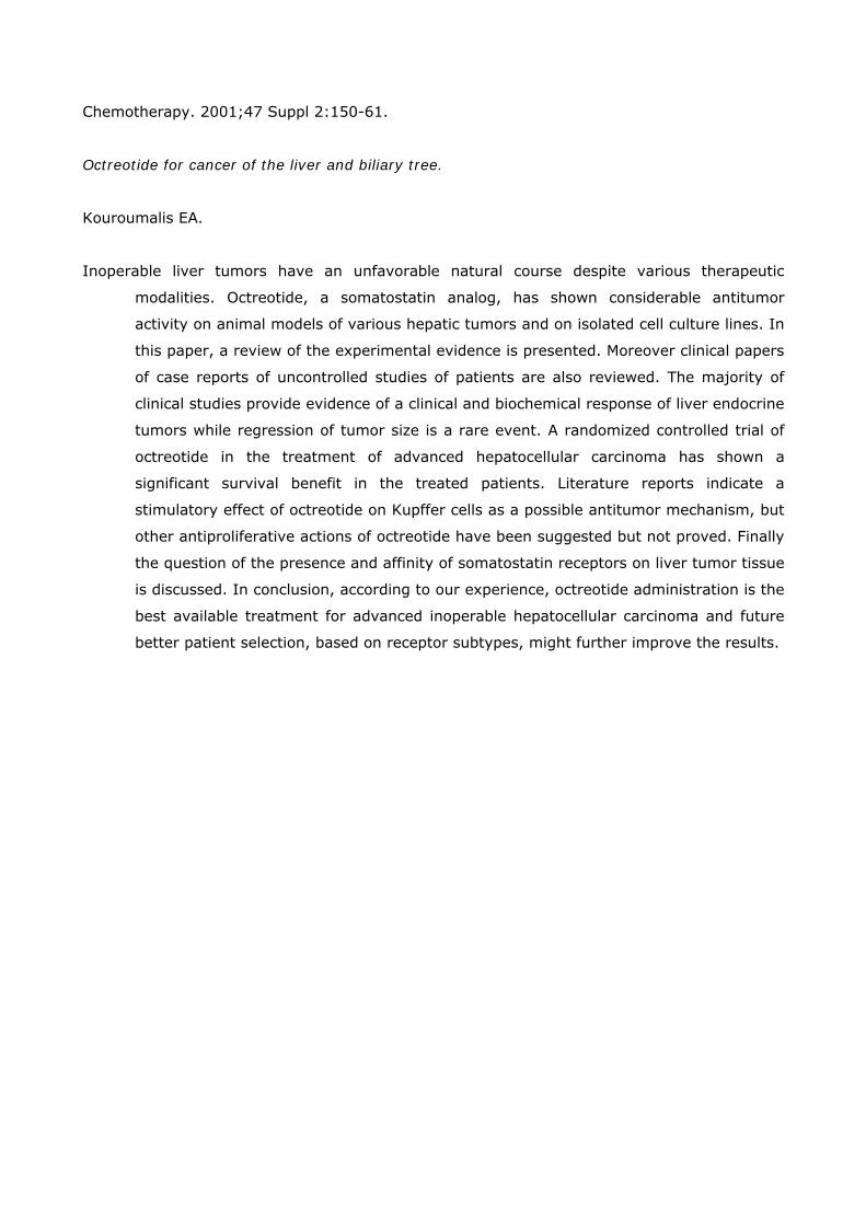
Chemotherapy. 2001;47 Suppl 2:150-61.
Octreotide for cancer of the liver and biliary tree.
Kouroumalis EA.
Inoperable liver tumors have an unfavorable natural course despite various therapeutic
modalities. Octreotide, a somatostatin analog, has shown considerable antitumor
activity on animal models of various hepatic tumors and on isolated cell culture lines. In
this paper, a review of the experimental evidence is presented. Moreover clinical papers
of case reports of uncontrolled studies of patients are also reviewed. The majority of
clinical studies provide evidence of a clinical and biochemical response of liver endocrine
tumors while regression of tumor size is a rare event. A randomized controlled trial of
octreotide in the treatment of advanced hepatocellular carcinoma has shown a
significant survival benefit in the treated patients. Literature reports indicate a
stimulatory effect of octreotide on Kupffer cells as a possible antitumor mechanism, but
other antiproliferative actions of octreotide have been suggested but not proved. Finally
the question of the presence and affinity of somatostatin receptors on liver tumor tissue
is discussed. In conclusion, according to our experience, octreotide administration is the
best available treatment for advanced inoperable hepatocellular carcinoma and future
better patient selection, based on receptor subtypes, might further improve the results.

Ann N Y Acad Sci. 2004 Apr;1014:121-31.
Molecular signaling of somatostatin receptors.
Lahlou H, Guillermet J, Hortala M, Vernejoul F, Pyronnet S, Bousquet C, Susini C.
Somatostatin is a neuropeptide family that is produced by neuroendocrine, inflammatory, and
immune cells in response to different stimuli. Somatostatin acts as an endogenous
inhibitory regulator of various cellular functions including secretions, motility, and
proliferation. Its action is mediated by a family of G-protein-coupled receptors (called
sst1-sst5) that are widely distributed in the brain and periphery. The five receptors bind
the natural peptides with high affinity, but only sst2, sst5, and sst3 bind the short
synthetic analogs used to treat acromegaly and neuroendocrine tumors. This review
covers the current knowledge in somatostatin receptor biology and signaling.

Biochem Biophys Res Commun. 2000 Feb 16;268(2):567-71.
Effect of somatostatin and octreotide on proliferation and vascular endothelial growth factor
secretion from murine endothelial cell line (HECa10) culture.
Lawnicka H, Stepien H, Wyczolkowska J, Kolago B, Kunert-Radek J, Komorowski J.
Angiogenesis, development of new blood vessels, is required for normal tissue repair and also
for tumor cell proliferation, extracellular matrix invasion, and hematogenous
metastases. Vascular endothelial growth factor (VEGF) is an endothelial cell-specific
mitogen that has been shown to play a key role in neovascularization. Inhibition of
angiogenesis in vitro and in vivo was documented by administration of native
neuropeptide somatostatin and its analog octreotide. We have studied the effect of
somatostatin-14 (SRIF) and ocreotide (sandostatin) on proliferation activity and VEGF
release from cultured murine endothelial cells HECa10 in vitro. SRIF in concentrations
from 10(-9) to 10(-5) M and ocreotide in concentrations from 10(-9) to 10(-5) M
diminished the proliferative activity of cultured cells vs controls. SRIF and ocreotide in
concentrations from 10(-14) to 10(-6) M did not change the release of VEGF into
supernatants of 24 or 72 h endothelial cell cultures. Although we showed the
antiproliferative effect of SRIF and ocreotide on mouse endothelial cells, we were
unable to demonstrate the inhibitory effect of tested peptides on VEGF secretion in
vitro.

Br J Haematol. 2001 Mar;112(4):936-44.
Somatostatin receptor scintigraphy useful in stage I-II Hodgkin's disease: more extended
disease identified.
Lugtenburg PJ, Krenning EP, Valkema R, Oei HY, Lamberts SW, Eijkemans MJ, van Putten WL,
Lowenberg B.
Somatostatin receptor (SS-R) scintigraphy successfully shows primary cancers and metastases
in patients with a variety of SS-R-positive tumours. In vitro studies have shown that
SS-Rs are present in lymph nodes from patients with Hodgkin's disease (HD). We
performed a prospective study in 126 newly diagnosed patients with HD and compared
the results of SS-R scintigraphy with conventional staging procedures, i.e. physical
examination, computerized tomography (CT) scanning and other imaging techniques.
We report positive scintigraphy in all patients. The lesion-related sensitivity was 94%
and varied from 98% for supradiaphragmatic lesions to 67% for infradiaphragmatic
lesions. In comparison with CT scanning and ultrasonography, SS-R scintigraphy
provided superior results for the detection of Hodgkin's localizations above the
diaphragm. In the intra-abdominal region, the CT scan was more sensitive than the SS-
R scan. A false-positive scan was rarely seen. In stages I and II supradiaphragmatic HD
patients, SS-R scintigraphy detected more advanced disease in 18% (15 out of 83) of
patients, resulting in an upstaging to stage III or IV, thus directly influencing patient
management. Our data would support the validity of SS-R scanning as a powerful
imaging technique for the staging of patients with HD.

Anticancer Res. 1998 Sep-Oct;18(5A):3615-9.
Somatostatin analogue octreotide and melatonin inhibit bromodeoxyuridine incorporation into
cell nuclei and enhance apoptosis in the transplantable murine colon 38 cancer.
Melen-Mucha G, Winczyk K, Pawlikowski M.
There is much evidence of the antiproliferative activity of somatostatin (SS) and melatonin
(Mel) upon the normal and neoplastic tissues. It has also been found, that both
substances are able to alter, under certain conditions, apoptotic processes. Recently, it
has been postulated that apoptosis plays a pivotal role in the control of tumour growth.
So far, there is no data about the effect of SS analogue--octreotide (Sandostatin, SMS)
and Mel on the apoptosis of colon cancer cells. The aim of this study is to examine the
effects of SMS and Mel administered separately or together on apoptosis,
bromodeoxyuridine incorporation and weight of tumours in the murine transplantable
Colon 38 cancer. The male mice were implanted subcutaneously (s.c.) with a
suspension of Colon 38 cells. After 6 days, the animals were subcutaneously injected
with SMS, Mel, SMS and Mel together (once daily at 6-8 p.m., for 6 days). The
incorporation of bromodeoxyuridine (BrDU) into cell nuclei was used as an index of cell
proliferation (labelling index-LI). The in situ labelling of nuclear DNA fragmentation
according to TUNEL method was considered as an apoptotic index (AI). Given
separately, both SMS and Mel significantly decreased the LI and increased the AI.
However, we have not observed any additive effect of SMS and Mel on either BrDU
incorporation or apoptosis. The mean AI in the group treated jointly with SMS and Mel
was significantly lower than in groups treated separately with SMS or Mel. It was also
found, that the proliferation/apoptosis ratio were significantly lower in the group treated
with SMS or MEL, which means that the imbalance between these two processes
changed in favour of cell death. Possibly, the observed antitumour effects of these two
substances could be due to this alteration.

Am J Obstet Gynecol. 1999 Sep;181(3):583-90.
Inhibition of human endometrial cancer cell growth in vitro and in vivo by somatostatin analog
RC-160.
Mishima M, Yano T, Jimbo H, Yano N, Morita Y, Yoshikawa H, Schally AV, Taketani Y.
OBJECTIVE: Our purpose was to investigate the effect of the somatostatin analog RC-160 on
the growth of the HEC-1 human endometrial cancer cell line in vivo and in vitro. STUDY
DESIGN: Nude mice bearing subcutaneous implanted HEC-1 tumors were treated for 25
days with RC-160 (100 microgram/d) delivered by osmotic minipumps. In cultured
HEC-1 cells radioreceptor assay of somatostatin was performed, and the expression of
messenger ribonucleic acid for somatostatin receptor subtypes (somatostatin receptors
1-5) was analyzed by reverse transcription-polymerase chain reaction. The effects of
RC-160 on epidermal growth factor-stimulated cell proliferation and tyrosine
phosphorylation of epidermal growth factor receptor were examined by colorimetric
assay and Western blotting, respectively. RESULTS: The treatment with RC-160
resulted in a significant decrease in tumor volume, tumor weight, and serum insulin-like
growth factor I levels compared with those values in control animals. The presence of
high-affinity somatostatin binding sites and the expression of somatostatin receptor 2
and somatostatin receptor 3 messenger ribonucleic acid were demonstrated in HEC-1
cells by radioreceptor assay and reverse transcription-polymerase chain reaction,
respectively. Epidermal growth factor-stimulated proliferation of HEC-1 cells was
inhibited by RC-160 in a dose-dependent manner. Western blotting revealed that
epidermal growth factor-induced tyrosine phosphorylation of epidermal growth factor
receptor was inhibited by RC-160, which suggests that the direct inhibitory effect of RC-
160 on HEC-1 cell growth might be mediated in part by interference with epidermal
growth factor receptor phosphorylation. CONCLUSION: These results indicate that
somatostatin analog RC-160 inhibits the growth of HEC-1 human endometrial cancer
cells, thus implying its potential clinical utility in treating endometrial cancer.

Immunology. 1987 Dec;62(4):655-8.
Identification of lymphoid cell lines bearing receptors for somatostatin.
Nakamura H, Koike T, Hiruma K, Sato T, Tomioka H, Yoshida S.
The MT-2, derived from an adult T-cell leukaemia (ATL) cell, the Molt-4F, a human T-cell line,
and the Isk, an EB virus-transformed B-cell line, were found to have high-affinity
receptors for somatostatin, a cyclic tetradecapeptide that inhibits the release of
substances such as growth hormone, TSH, glucagon, insulin, secretin, gastrin and
cholecystokinin. The quantity of radioactivity bound varied linearly with the number of
cells, and was displaced by non-radioactive somatostatin in a concentration-dependent
manner. Specific binding of 125I-somatostatin was time- and temperature-dependent
and at 22 degrees reached equilibrium within 120 min. Scatchard analysis
demonstrated one class of specific-binding sites on MT-2 cells, Isk cells and Molt-4F
cells that had respective densities and dissociation constants of 109 pM and 0.64 nM,
102 pM and 1.1 nM, and 5.8 pM and 0.22 nM.

J Neurochem. 2004 Jun;89(5):1057-91.
Regulation and function of somatostatin receptors.
Olias G, Viollet C, Kusserow H, Epelbaum J, Meyerhof W.
This review summarizes the latest advances that have been made to elucidate the
somatostatinergic system in respect to somatostatin receptor evolution, the
development of receptor agonists/antagonists, receptor regulation, signal transduction,
effects on cell proliferation, receptor-receptor or receptor-protein interactions and
receptor function.

Surgery. 1994 Dec;116(6):1148-52.
Postreceptor signal transduction mechanisms involved in octreotide-induced inhibition of
angiogenesis.
Patel PC, Barrie R, Hill N, Landeck S, Kurozawa D, Woltering EA.
BACKGROUND. Somatostatin analogues inhibit peptide release and cell growth through
multiple postreceptor signal transduction mechanisms (PRSTM), including G proteins
(GP), cyclic adenosine monophosphate (cAMP), calcium, protein kinase C (PKC), and
tyrosine phosphatase (TP). Octreotide acetate (OA), a somatostatin analogue, has been
shown to inhibit angiogenesis; however, the PRSTM involved are unknown. METHODS.
Fertilized chicken eggs were obtained and incubated. On day 3, embryos were removed
and placed in plastic wrap hammocks. On day 7, disks containing OA, test substances
that interfere with PRSTM, or combinations of OA plus a test substance were placed on
the developing chorioallantoic membrane. Blood vessel growth under each disk was
assessed at 24 hours. Data were evaluated by chi-squared analysis. RESULTS. OA's
ability to inhibit angiogenesis is significantly diminished when combined with calcium,
bradykinin (increases calcium), pertussis toxin (inhibits GP), or 3-isobutyl-1-
methylxanthine (increases cAMP). In contrast, no significant decrease is noted in OA's
ability to inhibit angiogenesis when combined with phorbol ester (activates PKC) or
vanadate (inhibits TP). CONCLUSIONS. OA-induced inhibition of angiogenesis is GP,
calcium, and cAMP dependent and is PKC and TP independent. Better understanding of
the PRSTM involved with OA-induced inhibition of angiogenesis may lead to
enhancement of OA's effect on angiogenesis.

Am J Gastroenterol. 1999 Jan;94(1):278-9.
Successful treatment of an advanced hepatocellular carcinoma with the long-acting
somatostatin analog lanreotide.
Raderer M, Hejna MH, Kurtaran A, Kornek GV, Valencak JB, Oberhuber G, Vorbeck F, Virgolini
I, Scheithauer W.
Treatment options for hepatocellular cancer apart from surgical resection are limited because
of the drug-refractory nature of this disease. Little is known about the role of
somatostatin-receptors in hepatocellular cancer, and somatostatin analogs have not
been investigated for treatment of this malignancy. We present the case of a 68-yr-old
male, who was successfully treated with the long-acting somatostatin analog lanreotide.

Int J Oncol. 2000 Jun;16(6):1197-201.
Treatment of hepatocellular cancer with the long acting somatostatin analog lanreotide in vitro
and in vivo.
Raderer M, Hejna MH, Muller C, Kornek GV, Kurtaran A, Virgolini I, Fiebieger W, Hamilton G,
Scheithauer W.
Based on the fact that somatostatin (SST) analogs have given promising results for treatment
of hepatocellular cancer, we performed both in vitro and in vivo investigations to define
the role of a depot formulation of the long acting SST-analog lanreotide (LAN). A
decrease of cells in the S-phase as compared to controls (p<0.03) followed by a
significant, dose-dependent induction of apoptosis could be demonstrated in Hep G2
cells along with a dose-dependent influence of the peptide on cellular proliferation.
Northern blotting demonstrated the presence of mRNA for SSTR subtypes 2, 3 and 4 in
Hep G2 cells, but only slight SSTR expression in normal liver tissue. In addition, 21
untreated patients with advanced HCC not amenable to surgery were administered 30
mg of LAN by deep intramuscular injection every 14 days until documented disease
progression. Fifteen of these patients also underwent scanning with commercially
available 111In-DTPA-D-Phe1-Octreotide (111In-OCT) to define the in vivo expression
of SSTR. No positive 111In-OCT scans were obtained, indicating the absence of relevant
amounts of functional SSTR2 in HCC. One patient (5%) showed a partial response to
treatment, 8 patients had stable disease (38%), while the remaining patients
progressed during treatment. The median survival was 4.2 months (range 1.2-13+),
and the median time to progression was 2.5 months (range, 1.5-7+). However, 4
patients (19%) had an increase in WHO performance status lasting between 2.5 and 6
months, 5 patients (24%) had an increase in body weight, while pain markedly
improved in 1 additional patient (5%). In total, 5 patients (24%) had a decrease in
serum-AFP levels by at least 30%. Our results clearly indicate the ability of LAN to
decrease the S-phase fraction along with induction of apoptosis in Hep G2 cells in a
dose-dependent manner. Our data suggest clinical potential of SST-analogs in HCC and
indicate that suboptimal doses of the peptide might have been administered in our
series.

Metabolism. 1996 Aug;45(8 Suppl 1):98-100.
Somatostatin and cancer.
Robbins RJ.
The potential role of somatostatin (SRIF) in the diagnosis and treatment of nonendocrine
human cancers is reviewed. There have been many reports of the growth-inhibitory
activity of SRIF on normal and transformed cells in vitro. Many processes involved in
malignant tumor growth depend on autocrine growth mechanisms, and somatostatin
receptors (ssts) are present on many human cancers. It is possible that mutations in
ssts result in a loss of check on proliferation in cancer cells. SRIF analogs may have a
number of roles in clinical oncology. Use of radiolabeled tracers enables imaging of
tumors bearing ssts; newer agents may enable positron emission tomography (PET)
analyses or may be used to deliver lethal radiation doses to cells bearing a unique
subset of sst. Although the ability of SRIF and its analogs to inhibit cellular proliferation
has been shown in vitro, it has yet to be demonstrated in humans with cancer. Clinical
improvements seen with SRIF or its analogs in cancer patients may be related to
indirect effects, such as pain relief, reduction of gastrointestinal side effects of
chemotherapeutic agents, effects on local production of growth factors, and inhibition of
tumoral angiogenesis. Thus, with regard to their potential therapeutic role, SRIF
analogs are likely to be used only in conjunction with other approaches, such as
radiation, immunotherapy, chemotherapy, and growth factor modulation. Further
research into the fundamental functions of each of the ssts and the intracellular actions
of SRIF analogs will be needed to assess the potential usefulness of the latter in slowing
the progression of human cancers.

Lancet. 1977 Aug 27;2(8035):434-5.
Possible effects of growth hormone on development of acute lymphoblastic leukaemia.
Rogers PC, Komp D, Rogol A, Sabio H.
Growth hormone (G.H.) or a G.H.-dependent somatomedin may be involved in the process of
acute lymphoblastic leukaemia (A.L.L.). Growth hormone has a trophic effect on
lymphoid tissue and also specific receptors on lymphocytes, most probably T cells.
Hypophycess. Resting concentrations of G.H. and somatomedin activity are raised in
some children with A.L.L. and may be reduced after remission is achieved. It is
suggested that control of G.H. and/or somatomedin concentrations may be necessary
for adequate treatment of some cases of A.L.L. in children.

Br J Cancer. 2003 Jan 13;88(1):132-6.
Effect of a novel somatostatin analogue combined with cytotoxic drugs on human tumour
xenografts and metastasis of B16 melanoma.
Szende B, Horvath A, Bokonyi G, Keri G.
A novel somatostatin analogue, TT-232 (which inhibits the proliferation of various cell cultures
and transplantable mouse tumours), was examined regarding its effect on human
melanoma and lymphoma xenografts as a single treatment or in combination with DTIC
(dacarbazine) and etoposide. TT-232 inhibited the growth of HT-18 melanoma
xenografts, a dose of 5 mg kg(-1) being the most effective. Combination of 1 mg kg(-1)
TT-232 with 30 or 60 mg kg(-1) DTIC (administered daily) resulted in a stronger
inhibitory effect compared to TT-232 or DTIC as a single modality. Antimetastatic effect
of TT-232 treatment combined with DTIC was studied using the B16 mouse melanoma
muscle - lung metastasis model. The number of lung metastases of B16 melanoma
could be decreased by the daily administration of 1 mg kg(-1) TT-232 or 60 mg kg(-1),
but not of 30 mg kg(-1) DTIC. TT-232, combined with 30 or 60 mg kg(-1) DTIC
decreased the lung metastasis number significantly lower than the control. Nearly 50%
growth inhibition of HT-58 lymphoma was achieved by daily treatment with 1 mg kg(-1)
TT-232. 5 mg kg(-1) etoposide, administered daily, resulted in a similar effect. The
combination of 1 mg kg(-1) TT-232 and 5 mg kg(-1) etoposide was significantly more
effective than TT-232 or etoposide as a single treatment. The very strong tumour
growth inhibitory effect of 10 mg kg(-1) etoposide could even be increased by
combination with TT-232. These experimental data suggest that TT-232 may be an
effective new tool in the combination chemotherapy of malignant tumours like
melanoma and lymphoma.

J Cancer Res Clin Oncol. 1999 Aug-Sep;125(8-9):444-52.
Growth inhibition of experimental pancreatic cancers and sustained reduction in epidermal
growth factor receptors during therapy with hormonal peptide analogs.
Szepeshazi K, Halmos G, Schally AV, Arencibia JM, Groot K, Vadillo-Buenfil M, Rodriguez-
Martin E.
Reduction in receptors for epidermal growth factor (EGF) in cancers appears to be one of the
principal mechanisms through which peptide hormone analogs can inhibit tumor
growth. In this study, hamsters with nitrosamine-induced pancreatic cancers were
treated for 8 weeks with bombesin/gastrin-releasing peptide (GRP) antagonist RC-3095,
somatostatin analog RC-160 or the luteinizing hormone-releasing hormone antagonist
Cetrorelix, using sustained delivery systems releasing 20, 35 and 20 microg analog/
day respectively. To establish the pattern of changes in the number and affinity of EGF
receptors on tumors, groups of animals were sacrificed at regular intervals during
therapy. Chronic treatment with RC-3095 or Cetrorelix resulted in an early (day 10) and
sustained reduction (71% or 69% respectively) in EGF receptors on pancreatic tumors.
In contrast, RC-160 decreased receptor concentration by 60% only after 20 days.
Among the histological characteristics of proliferation, the decrease in argyrophilic
nucleolar organizer regions, but not apoptotic and mitotic indices, showed a correlation
with the fall in EGF receptors. The concentration of the receptors returned to the control
level 4 days after cessation of chronic treatment with RC-3095. The effect of single
injections of RC-3095, RC-160 and Cetrorelix on EGF receptors was also investigated.
RC-160 decreased the number of EGF receptors on pancreatic cancers by 31% 3 h after
administration, but the receptors had returned to normal level at 6 h. RC-3095 and
Cetrorelix caused a 67% and 59% decline, respectively, in EGF receptors only 6 h after
injection and the concentration of receptors remained low for 24 h. Thus, the pattern of
downregulation of EGF receptors in pancreatic cancers appears to depend on the
peptide used for therapy. Since the antitumor effect may be the result of the fall in EGF
receptors in cancers, information on the time course of changes in these receptors
during treatment with these analogs may lead to an improvement in therapeutic
regimens.

Cancer Res. 2002 Feb 1;62(3):781-8.
Targeted cytotoxic somatostatin analogue AN-238 inhibits somatostatin receptor-positive
experimental colon cancers independently of their p53 status.
Szepeshazi K, Schally AV, Halmos G, Armatis P, Hebert F, Sun B, Feil A, Kiaris H, Nagy A.
The resistance of advanced colorectal cancers to therapy is often related to mutations in the
p53 tumor suppressor gene. Because somatostatin (SRIF) receptors (ssts) are present
in colorectal carcinomas, the treatment with targeted cytotoxic SRIF analogue AN-238,
consisting of 2-pyrrolinodoxorubicin (AN-201) linked to octapeptide SRIF carrier RC-
121, may overcome this resistance by producing a higher concentration of the cytotoxic
agent in the tumors. Four colon cancer cell lines, HCT-116 and LoVo expressing wild-
type p53, and HCT-15 and HT-29 with mutated p53, were investigated. HCT-116, HCT-
15, and HT-29, but not LoVo possess functional ssts. We analyzed changes in p53, p21,
and proliferating cell nuclear antigen (PCNA) concentrations in these cells in vitro by
immunoblotting after exposure to AN-238, its radical AN-201, or doxorubicin (DOX).
Equitoxic doses of AN-238, AN-201, or DOX affected p53, p21, and PCNA differently.
Analysis of the p21:p53 ratios revealed that DOX increased p53 levels, but most of p53
was mutated and inactive, whereas AN-238 produced smaller changes in p53
concentrations but enhanced its activity. In HCT-15 cells, PCNA:p21 ratios, which are
indicators of proliferation and repair processes, remained unchanged after exposure to
AN-238 but were increased by DOX. In vivo studies in nude mice demonstrated that
AN-238, AN-201, and DOX were equally effective on HCT-116 tumors that express wild-
type p53. However, AN-238 also inhibited the growth of HCT-15 and HT-29 cancers that
express mutant p53, whereas AN-201 and DOX showed no effect. None of the
compounds could suppress the proliferation of LoVo tumors that lack functional ssts. In
conclusion, cytotoxic SRIF analogue AN-238 inhibits the growth of experimental colon
cancers that express ssts, regardless of their p53 status.

Int J Oncol. 2003 May;22(5):1141-6.
Effective treatment of H838 human non-small cell lung carcinoma with a targeted cytotoxic
somatostatin analog, AN-238.
Szereday Z, Schally AV, Szepeshazi K, Bajo AM, Hebert F, Halmos G, Nagy A.
The accumulation of radioactive somatostatin analog [111In]pentetreotide in non-small cell
lung cancer (non-SCLC) during scintigraphy of patients provides a rationale for
investigating the efficacy of somatostatin receptor-based chemotherapy in non-SCLC.
Consequently, in this study, we evaluated the antitumor effects of cytotoxic
somatostatin analog AN-238 on H838 human non-SCLC xenografted into nude mice in
comparison with its cytotoxic radical, 2-pyrrolinodoxorubicin (AN-201). The expression
of messenger RNA (mRNA) for human somatostatin receptor subtypes 2 (hsst2) and 5
(hsst5) in H838 cells, and tumors was also investigated using reverse-transcription
polymerase chain reaction (RT-PCR). Somatostatin receptors on H838 tumors were
characterized by ligand competition assay using radiolabeled somatostatin analog, RC-
160. Three i.v. injections of AN-238 at 150 nmol/kg, given on days 1, 7 and 21,
resulted in a significant (p<0.05) tumor growth inhibition, the final tumor volume being
60% smaller than in the controls. The tumor doubling time was also extended
significantly (p<0.05) from 9.65+/-0.56 days in the controls to 17.52+/-3.3 days. Only
one of 8 mice died due to toxicity. In contrast, cytotoxic radical AN-201 was ineffective
and more toxic, killing 2 of 7 animals. mRNA for hsst2 was found in H838 xenografts,
but not in H838 cells from which the xenografts originated. Interestingly, H838 cells
grown in a special, serum-free medium did express mRNA for hsst2. mRNA for hsst5
was not found in any samples tested. Binding studies demonstrated the presence of
high affinity (K(d) = 7.3+/-1.2 nM) binding sites for RC-160 with a mean maximal
binding capacity (B(max)) of 953.3+/-45.3 fmol/mg protein. AN-238 at 3.14+/-0.93 nM
concentration displaced 50% of radiolabeled RC-160 binding to somatostatin receptors
in H838 tumors. Our results indicate that patients with inoperable non-SCLC may
benefit from chemotherapy targeted to somatostatin receptors based on AN-238.

Anticancer Res. 2003 Sep-Oct;23(5A):4061-6.
The antitumor activity of the somatostatin structural derivative (TT-232) on different human
tumor xenografts.
Tejeda M, Gaal D, Barna K, Csuka O, Keri G.
A somatostatin structural derivative (TT-232) has been developed in our laboratory with strong
antiproliferative effect but no GH- release inhibitory activity. TT-232 inhibited tyrosine
kinase activity of tumor cells lines and this inhibition correlated well with the inhibition
of cell proliferation. The antineoplastic activity of TT-232 has been found to be
associated with induction of programmed cell death (apoptosis) in tumor cell, resulting
in highly selective elimination of neoplastic tissue. The aim of this study was the
therapeutic efficacy of TT-232 on different human models: PC-3 prostate carcinoma,
MDA-MB-231 (ER-) and MCF-7 (ER+) breast carcinoma, HT-29 colon carcinoma, HT-18
melanoma, HL-60 promyelocytic leukemia. We studied the therapeutic efficacy of the
novel somatostatin analog, it for 30 days with intermittent injection once daily and for
14 days with s.c. infusion using the Alzet osmotic minipump (model 2002). The
antitumor activity of TT-232 was evaluated on the basis of survival time and tumor
growth inhibition. The tumor growth inhibitory effect of TT-232 on human tumor
xenografts proved to be significant, resulting in 30%-80% decrease in tumor volume
and in 20-60% tumor free animals. This antitumor efficacy of the novel somatostatin
analog was observable in almost all tumors investigated. These data suggest that the
novel somatostatin analog (TT-232) is an effective and promising antitumor agent.

Anticancer Res. 2005 Jan-Feb;25(1A):325-30.
Growth inhibitory effect of the somatostatin structural derivative (TT-232) on leukemia models.
Tejeda M, Gaal D, Csuka O, Keri G.
TT-232 is a structural derivative of the natural signal inhibitory peptide somatostatin, with
selective antiproliferative and anti-inflammatory properties. TT-232 activates SSTR
receptors (primarily the SSTR-1), which leads to irreversible cell cycle arrest, followed
by secondary induction of apoptosis. TT-232 has passed phase I clinical trials without
toxicity and significant side-effects. We examined the antiproliferative effect in vitro and
the antitumor effect in vivo of TT-232 on leukemia cell lines. During in vivo
experiments, we evaluated the therapeutic efficacy of TT-232 in various long-term
administration routes; traditional injection versus infusion treatment via an inserted
Alzet minipump on P-388 mice and HL-60 human leukemia models. Treatment with TT-
232 started after development of the disease. In vitro, TT-232 inhibited the proliferation
of P-388 mice lymphoid cells and HL-60 human promyelocytic leukemia cells in the
range of 46%-97% with 24-hour treatment and 82%-100% with 48-hour treatment.
Cells were treated with 30 microg/ml and 60 microg/ml dose of TT-232. With the same
in vivo models, the best results were achieved when TT-232 was applied by infusion
treatments. The infusion treatment with TT-232 produced 50%-80% inhibition of
growth and resulted in 20%-40% long-term and leukemia-free survivors. TT-232
showed dose-, time- and administration mode-dependent antileukemia activity in vitro
and in vivo, both on rodent and human models. Our results suggest that TT-232 is a
promising new antileukemia agent.

J Chemother. 2000 Oct;12(5):421-30.
Lanreotide-induced modulation of 5-fluorouracil or mitomycin C cytotoxicity in human colon
cancer cell lines: a preclinical study.
Tesei A, Ricotti L, De Paola F, Casini-Raggi C, Barzanti F, Frassineti GL, Zoli W.
The effect on growth of the long-acting somatostatin analogue lanreotide (LAN), alone or in
combination with 5-fluorouracil (5-FU) and mitomycin C (MIT), was investigated in
three human colon cancer lines. Cell survival inhibition induced by LAN alone, as
evaluated by sulforhodamine B assay, ranged from 20% to 40% as a function of cell
line and concentration. The IC50, the concentration inhibiting cell survival by 50%, was
never reached. The antiproliferative effect produced by a 48 h exposure to 5-FU or MIT
was synergistically enhanced in all cell lines by a subsequent 48 h exposure to LAN. The
synergistic interaction was not related to specific cell cycle perturbations or to the
somatostatin receptor 2 (sst2) mRNA abundance. In conclusion, our study seems to
indicate that LAN is a potentially useful modulating agent for enhancing 5-FU and MIT
activity in colorectal cancer patients.

Br J Cancer. 2005 Apr 25;92(8):1493-8.
Somatostatin receptors 2 and 5 are preferentially expressed in proliferating endothelium.
Adams RL, Adams IP, Lindow SW, Zhong W, Atkin SL.
Angiogenesis is characterised by activation, migration and proliferation of endothelial cells and
is central to the pathology of cancer, cardiovascular disease and chronic inflammation.
Somatostatin is an inhibitory polypeptide that acts through five receptors (sst 1, 2, 3,
4, 5). Sst has previously been reported in endothelium, but their role remains obscure.
Here, we report the expression of sst in human umbilical vein endothelial cells
(HUVECs) in vitro, during proliferation and quiescence. A protocol for culturing
proliferating and quiescent HUVECs was established, and verified by analysing cell cycle
distribution in propidium-iodide-stained samples using flow cytometry. Sst mRNA was
then quantified in nine proliferating and quiescent HUVEC lines using quantitative
reverse transcriptase-polymerase chain reaction. Sst 2 and 5 were preferentially
expressed in proliferating HUVECs. All samples were negative for sst 4. Sst 1 and 3
expression and cell cycle progression were unrelated. Immunostaining for sst 2 and 5
showed positivity in proliferating but not quiescent cells, confirming sst 2 and 5 protein
expression. Inhibition of proliferating cells with somatostatin analogues Octreotide and
SOM230, which have sst 5 activity, was found (Octreotide 10(-10)-10(-6) M: 48.5-
70.2% inhibition; SOM230 10(-9)-10(-6) M: 44.9-65.4% inhibition) in a dose-
dependent manner, suggesting that sst 5 may have functional activity in proliferation.
Dynamic changes in sst 2 and 5 expression during the cell cycle and the inhibition of
proliferation with specific analogues suggest that these receptors may have a role in
angiogenesis.

Proc Natl Acad Sci U S A. 1991 Dec 15;88(24):11485-9.
Evidence that somatostatin is localized and synthesized in lymphoid organs.
Aguila MC, Dees WL, Haensly WE, McCann SM.
Because several peptides originally found in the pituitary as within the central nervous system
have been localized in lymphoid tissues and because somatostatin (somatotropin-
release-inhibiting hormone, SRIH) can act on cells of the immune system, we searched
for this peptide in lymphoid organs. We demonstrated that SRIH mRNA exists in
lymphoid tissue, albeit in smaller levels than in the periventricular region of the
hypothalamus, the brain region that contains the highest level of this mRNA. SRIH
mRNA was found in the spleen and thymus of male rats and in the spleen, thymus, and
bursa of Fabricius of the chicken. Its localization in the bursa indicates that the peptide
must be present in B lymphocytes since this is the site of origin of B lymphocytes in
birds. The SRIH concentration in these lymphoid organs as determined by
radioimmunoassay was greater in the thymus than in the spleen of the rat. These
concentrations were 50 times less than those found in the periventricular region of the
hypothalamus, the site of the perikarya of SRIH-containing neurons. In the chicken, as
in the rat, the concentration of SRIH was greater in the thymus than in the spleen; it
was present in the bursa of Fabricius, also in higher concentration than in the spleen.
Fluorescence immunocytochemistry revealed the presence of SRIH-positive cells in
clusters inside the white pulp and more dispersed within the red pulp of the spleen of
both the rat and the chicken. The thymus from these species also contained SRIH-
positive cells within the medulla and around the corticomedullary junction. In the
chicken, there were large clusters of SRIH-positive cells in the medullary portion of
each nodule of the bursa of Fabricius. Preabsorption of the primary antiserum or
replacing this antiserum with normal rabbit serum verified the specificity of staining.
Sequential immunostaining of the same sections from rat spleen using first SRIH
antibody and subsequently a monoclonal antibody against a rat B-cell surface antigen
revealed the presence of SRIH immunoreactivity in some, but not all, B cells. Other cell
types in spleen not yet identified also stained positively with the SRIH antibody but
were not reactive to monoclonal antibodies to rat Thy-1.1, a marker for all the thymic T
lymphocytes. The possibility that SRIH is present in other populations of cells in the
spleen cannot be ruled out. Sequential immunostaining of the same sections of rat
thymus revealed the presence of SRIH immunoreactivity in a small population of T
lymphocytes in the medulla, as revealed by the Thy-1.1 marker. The SRIH-positive cells
were nonimmunoreactive when exposed to the B-cell marker; however, the possibility
that SRIH is present in other cells was not investigated. Thus, our results indicate that

SRIH is synthesized and stored in cells of the immune system. SRIH may be secreted
from these cells to exert paracrine actions that alter the function of immune cells in
spleen and thymus.

Med Hypotheses. 1998 Jun;50(6):501-6.
Inhibition of the oxidative and nonoxidative pentose phosphate pathways by somatostatin: a
possible mechanism of antitumor action.
Boros LG, Brandes JL, Yusuf FI, Cascante M, Williams RD, Schirmer WJ.
Long-acting somatostatin analogs have recently become supplemental drugs in the treatment
of neurofibroma because of their marked tumor growth inhibitory effect. Somatostatin
is currently under extended evaluation in other cancers as a possible supplemental drug
to the treatment protocols in use. The mode of action is not known. Somatostatin has
been shown to cause glucose intolerance by inhibiting glucose-6-phosphate
dehydrogenase (G6PD) in fish liver. Recent data generated in our laboratory indicate
that it is this pathway and the transketolase reactions of the pentose cycle (PC) which
are directly involved in the ribose synthesis process of pancreatic adenocarcinoma cells.
In cell culture, somatostatin alone inhibited glucose carbon recycling through the PC by
5.7%, which was increased to 19.8% in combination with oxythiamine, a competitive
inhibitor of transketolase. Oxythiamine produced strong apoptosis in in-vitro hosted
tumor cells. We hypothesize that somatostatin- and oxythiamine-induced
antiproliferative action is mediated by the inhibition of G6PD, transketolase, or both.

Mol Cell Endocrinol. 2001 May 15;176(1-2):103-10.
Somatostatin up-regulates topoisomerase II alpha expression and affects LNCaP cell cycle.
Brevini-Gandolfi TA, Cillo F, Favetta LA, Montagna A, Motta M.
mRNA differential display-PCR analysis was used to perform a systematic screening of
Somatostatin (SS)-regulated genes in the human prostatic carcinoma cell line LNCaP
(Lymph Node Carcinoma of the Prostate). A 170 bp fragment was shown to be up-
regulated by SS. Sequence analysis of this fragment revealed its homology with the
human Topoisomerase II Alpha gene. Up-regulation of Topoisomerase II Alpha was
confirmed by Northern blot hybridisation and was induced by the same dose of SS (1
nM) earlier demonstrated to inhibit LNCaP cell growth. Furthermore, SS possible effects
on timing, as well as concentration of Topoisomerase II Alpha along the different
phases of the cell cycle were investigated. To this purpose changes in the enzyme
protein concentration in response to SS were assessed in synchronised LNCaP cells. The
hormone was shown to exert a perturbing effect on both parameters considered,
possibly related to its inhibitory action on LNCaP cell replication.

Gastroenterology. 1997 Sep;113(3):767-72.
Inhibition of tumor cell kinetics and serum insulin growth factor I levels by octreotide in
colorectal cancer patients.
Cascinu S, Del Ferro E, Grianti C, Ligi M, Ghiselli R, Foglietti G, Saba V, Lungarotti F, Catalano
G.
BACKGROUND & AIMS: Octreotide was shown to inhibit the growth of colon cancer and to
reduce serum concentrations of tumor growth factors such as insulin-like growth factor
I (IGF-I) and epidermal growth factor (EGF) in vitro and in animal models. Effects of
octreotide on tumor cell kinetics and serum concentration of IGF-I and EGF in patients
with colorectal cancer were evaluated. METHODS: Seventy-five patients with colorectal
cancer were randomized to receive octreotide (200 micrograms daily) in the 2 weeks
before surgery or the usual medications. Samples of tumor tissue were taken at
endoscopy and at surgery. [3H]Thymidine labeling index and flow cytometry were used
to assess the S-phase fraction. In octreotide-treated patients, plasma levels of IGF-I,
EGF, and growth hormone were assessed before and after treatment. RESULTS: There
was a statistically significant reduction in the mean percentage of the S-phase fraction
as a result of octreotide treatment measured by both [3H]thymidine labeling index (P =
0.001) and flow cytometry (P = 0.001). No reduction in the percentage of the S-phase
fraction was observed in the control group patients. Serum values of IGF-I were
significantly reduced by octreotide, whereas EGF and growth hormone levels were not
affected. CONCLUSIONS: Octreotide reduces the proliferative activity of tumor cells and
the serum IGF-I levels in patients with colorectal cancer. This activity may have a role
in the treatment of colorectal cancer.

FEBS Lett. 1996 Nov 18;397(2-3):164-8.
A somatostatin analogue inhibits MAP kinase activation and cell proliferation in human
neuroblastoma and in human small cell lung carcinoma cell lines.
Cattaneo MG, Amoroso D, Gussoni G, Sanguini AM, Vicentini LM.
Somatostatin possesses antisecretory and antiproliferative activity on some human tumors. We
herein report that, in a human neuroblastoma cell line, the somatostatin analogue BIM
23014 inhibited mitogen-activated protein (MAP) kinase activity stimulated by either
insulin-like growth factor-1, whose receptor bears a tyrosine kinase, or carbachol,
which acts at a G-protein coupled receptor. In a human small cell lung carcinoma line
BIM inhibited serum-stimulated MAP kinase activation. These inhibitory actions occur in
a dose range quite similar to that observed for suppression of proliferation induced by
the analogue in the same cell lines. The decrease in cAMP elicited by the analogue in
the two cell lines is not responsible for its inhibitory action on MAP kinase and cell
growth. Moreover, the analogue did not modify intracellular [Ca2+] and pH. An
involvement of a phosphatase activity is suggested.

FEBS Lett. 1999 Oct 1;459(1):64-8.
Somatostatin inhibits PDGF-stimulated Ras activation in human neuroblastoma cells.
Cattaneo MG, Scita G, Vicentini LM.
The main physiological role of somatostatin (SST) is the control of hormone secretion.
Recently, SST has been shown to exert antiproliferative effects on some human tumors
via both direct and indirect mechanisms. We have previously found that in the human
neuroblastoma cell line SY5Y the SST analogue lanreotide (BIM 23014) inhibited serum-
stimulated cell proliferation and MAP kinase activity. Here, we examine the effect of
SST on PDGF-induced Ras activation. We found that SST suppressed PDGF-induced Ras
activation in a pertussis toxin (PTx)-independent and peroxovanadate-dependent
manner. Ras-specific GTPase activating protein (GAP) activities were not altered by SST
treatment. On the contrary, PDGF-induced PDGF receptor phosphorylation was
decreased by SST in a PTx-independent, peroxovanadate-dependent manner, likely
accounting for the SST-mediated inhibition of PDGF-induced Ras activation.

FEBS Lett. 2000 Sep 22;481(3):271-6.
Selective stimulation of somatostatin receptor subtypes: differential effects on Ras/MAP kinase
pathway and cell proliferation in human neuroblastoma cells.
Cattaneo MG, Taylor JE, Culler MD, Nisoli E, Vicentini LM.
In previous studies we have showed that somatostatin (SST) inhibits cell division, mitogen-
activated protein (MAP) kinase and Ras activity in the human neuroblastoma cell line
SY5Y. In the present study, we have assessed the role of a series of SST analogs, three
of which were selective for SSTR1, SSTR2 or SSTR5, in these cellular events. All the
analogs inhibited forskolin-induced cAMP accumulation. Selective stimulation of SSTR1
or SSTR2 but not of SSTR5 inhibited platelet-derived growth factor (PDGF)-induced
[(3)H]thymidine incorporation. The three analogs inhibited PDGF-stimulated MAP kinase
activity, at least at an early time. In contrast, none of the analogs used individually was
able to inhibit PDGF-stimulated Ras activity. A combined stimulation of SSTR2 and
SSTR5 was necessary to obtain a significant inhibitory effect, suggesting the possibility
of receptor heterodimerization. These results indicate that SST inhibition of Ras and
MAP kinase activities takes place via different pathways and that SST inhibition of
PDGF-induced cell proliferation occurs via a Ras-independent pathway.

Endocrinology. 2001 Jan;142(1):121-8.
Somatostatin inhibits Akt phosphorylation and cell cycle entry, but not p42/p44 mitogen-
activated protein (MAP) kinase activation in normal and tumoral pancreatic acinar cells.
Charland S, Boucher MJ, Houde M, Rivard N.
Somatostatin, or its structural analog SMS 201-995 (SMS), is recognized to exert a growth-
inhibitory action in rat pancreas, but the cellular mechanisms are not completely
understood. This study was undertaken to evaluate the effect of SMS on p42/p44 MAP
kinases and phosphatidylinositol 3-kinase activation and to analyze expression of some
cell cycle regulatory proteins in relation to pancreatic acinar cell proliferation in vivo (rat
pancreas), as well as in the well-established tumoral cell line AR4-2J. We herein report
that: 1) SMS inhibits caerulein-induced pancreatic weight and DNA content and
abolishes epidermal growth factor (EGF)-stimulated AR4-2J proliferation; 2) SMS only
moderately reduces the stimulatory effect of caerulein on p42/p44 MAP kinase activities
in pancreas and has no effect on EGF-stimulated MAP kinase activities in AR4-2J cells;
3) SMS repressed caerulein-induced Akt activity in normal pancreas; 4) SMS has a
strong inhibitory action on cyclin E expression induced by caerulein in pancreas and EGF
in AR4-2J cells and as expected, the resulting cyclin E-associated cyclin-dependent
kinase (cdk)2 activity, as well as pRb phosphorylation, are blunted by SMS treatment in
both models; and 5) SMS suppresses mitogen-induced p27(Kip1) down-regulation, as
well as marginally induces p21(Cip) expression. Thus, our data suggest that
somatostatin-induced growth arrest is mediated by inhibition of phosphatidylinositol 3-
kinase pathway and by enhanced expression of p21(Cip) and p27(Kip1), leading to
repression of pRb phosphorylation and cyclin E-cdk2 complex activity.

Chin Med J (Engl). 2001 Nov;114(11):1167-70.
Antineoplastic mechanism of Octreotide action in human hepatoma.
Chen X, Liu Z, Ai Z.
OBJECTIVES: To investigate whether apoptosis can be induced by Octreotide in human
hepatoma cells in vitro and elucidate the antineoplastic mechanism of Octreotide in
hepatoma. METHODS: A cultured human hepatoma cell line, BEL-7402, was exposed to
Octreotide and apoptosis was evaluated by cytochemical staining (Hochesst 33,258),
transmission electron microscopy, agarose gel electrophoresis and flow cytometry
(FCM). RESULTS: After exposure to 0.2 microgram/ml Octreotide, apoptosis with
nuclear chromatin condensation as well as fragmentation, cell shrinkage and the
formation of apoptotic bodies was observed using cytochemical staining and
transmission electron microscopy. A DNA ladder in agarose gel electrophoresis was also
displayed. FCM showed that the apoptotic cell number rose with an increase in the
concentration of Octreotide (0-2 micrograms/ml). There was a positive correlation
between Octreotide concentration and apoptotic rate in BEL-7402 cells (r = 0.809, P <
0.05). CONCLUSION: Apoptosis in human hepatoma cells can be induced by Octreotide,
which may be related to the mechanism of antineoplastic action of Octreotide in
hepatoma.

Eur J Pharmacol. 1998 Apr 17;347(1):77-86.
Effect of the gastrin-releasing peptide antagonist BIM 26226 and lanreotide on an acinar
pancreatic carcinoma.
Damge C, Hajri A.
The effects of a potent specific gastrin-releasing peptide receptor antagonist, BIM 26226 ([D-
F5 Phe6, D-Ala11] bombesin (6-13) OMe), and the long-acting somatostatin analogue,
lanreotide (BIM 23014), on the growth of an acinar pancreatic adenocarcinoma growing
in the rat or cultured in vitro were investigated. Lewis rats bearing a pancreatic
carcinoma transplanted s.c. in the scapular region, were treated with gastrin-releasing
peptide (30 microg/kg per day), BIM 26226 (30 and 100 microg/kg per day) and
lanreotide (100 microg/kg per day) alone or in combination for 14 successive days.
Chronic administration of BIM 26226 and lanreotide significantly inhibited the growth of
pancreatic tumours stimulated or not by gastrin-releasing peptide (GRP), as shown by a
reduction in tumour volume, protein, ribonucleic acid, amylase and chymotrypsin
contents. This effect was more pronounced with 100 microg/kg per day BIM 26226 than
with 30 microg/kg per day. However, BIM 26226 and lanreotide, given together, did not
exert any additive effect on GRP-treated and -untreated tumours. In cell cultures, both
BIM 26226 and lanreotide (10(-6) M) inhibited [3H]thymidine incorporation in tumour
cells induced or not by GRP, but no increased effect was observed after combined
treatment with both agents. Binding studies showed that BIM 26226 had a high affinity
for GRP receptors in tumour cell membranes (IC50 = 6 nM). These results from in vivo
and in vitro experiments suggest that BIM 26226 and lanreotide are able to reduce the
growth of an experimental acinar pancreatic tumour. Thus, these agents represent
interesting steps toward the development of new approaches for treatment of
pancreatic carcinomas.
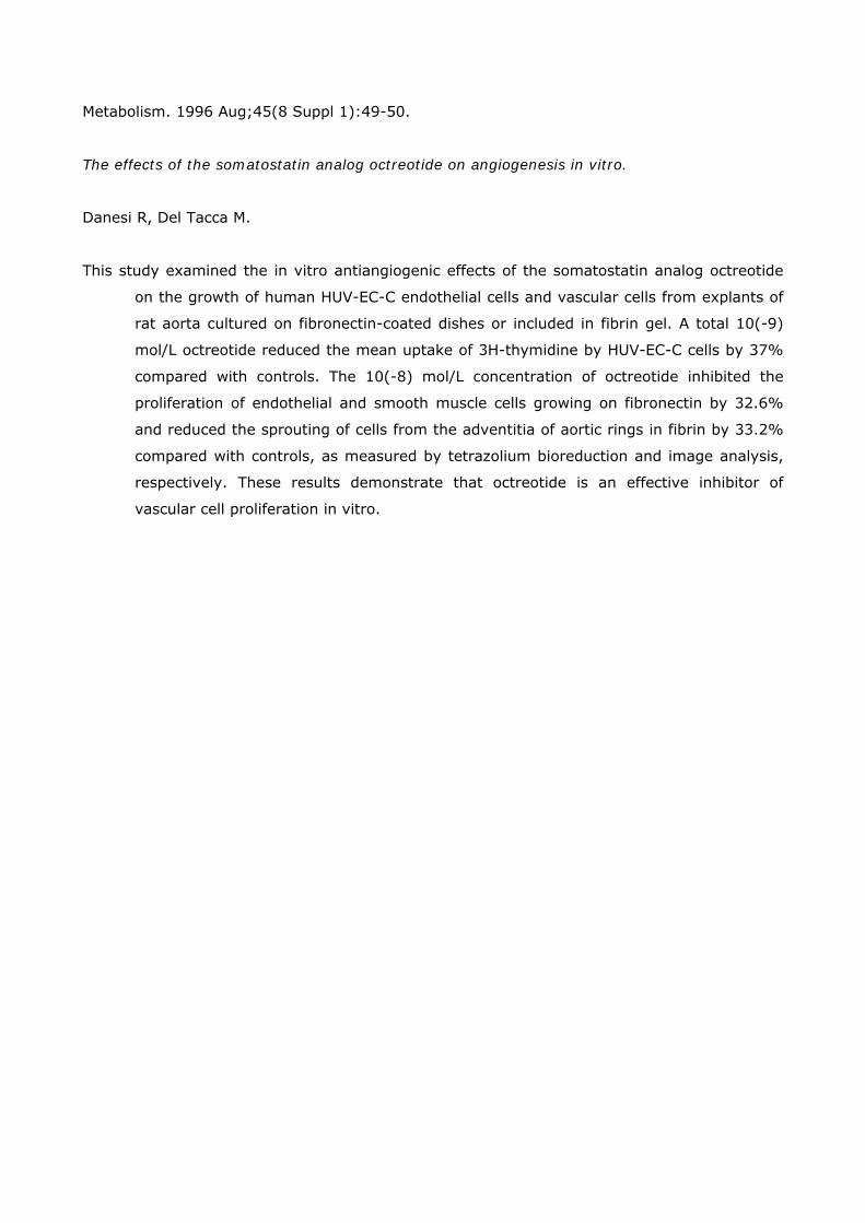
Metabolism. 1996 Aug;45(8 Suppl 1):49-50.
The effects of the somatostatin analog octreotide on angiogenesis in vitro.
Danesi R, Del Tacca M.
This study examined the in vitro antiangiogenic effects of the somatostatin analog octreotide
on the growth of human HUV-EC-C endothelial cells and vascular cells from explants of
rat aorta cultured on fibronectin-coated dishes or included in fibrin gel. A total 10(-9)
mol/L octreotide reduced the mean uptake of 3H-thymidine by HUV-EC-C cells by 37%
compared with controls. The 10(-8) mol/L concentration of octreotide inhibited the
proliferation of endothelial and smooth muscle cells growing on fibronectin by 32.6%
and reduced the sprouting of cells from the adventitia of aortic rings in fibrin by 33.2%
compared with controls, as measured by tetrazolium bioreduction and image analysis,
respectively. These results demonstrate that octreotide is an effective inhibitor of
vascular cell proliferation in vitro.
Clin Cancer Res. 1997 Feb;3(2):265-72.
Inhibition of experimental angiogenesis by the somatostatin analogue octreotide acetate (SMS
201-995).
Danesi R, Agen C, Benelli U, Paolo AD, Nardini D, Bocci G, Basolo F, Campagni A, Tacca MD.
The present study investigates the effect of the somatostatin analogue octreotide acetate (SMS
201-995) on experimental angiogenesis in vitro and in vivo. Octreotide reduced the
proliferation of human HUV-EC-C endothelial cells (mean, -45.8% versus controls at
10(-9) M; P < 0.05) as well as the density of the vascular network of the chick
chorioallantoic membrane (mean, -35.7% versus controls at 50 microgram; P < 0.05).
Furthermore, octreotide significantly inhibited chick chorioallantoic membrane
neovascularization by the human MCF-10Aint-2 mammary cells secreting the
angiogenic protein FGF-3. The proliferation of endothelial and smooth muscle cells from
rat aorta explants on fibronectin was reduced by octreotide 10(-8) M (mean, -32.6%
versus controls; P < 0.05), and a similar effect was produced on cells sprouting from
explants cultured in fibrin (mean, -52.9% versus controls; P < 0.05). Topical
administration of octreotide 10 microgram/day for 6 days inhibited rat cornea
neovascularization induced by AgNO3/KNO3 (mean, -50.6% versus controls; P < 0.05).
Octreotide 40 microgram/day i.p was tested on angiogenesis in rat mesentery obtained
by i.p. injections of compound 48/80, a mast cell degranulating agent, or conditioned
medium from MCF-10Aint-2 cells and was able to reduce the extent of
neovascularization (mean, -45.6 and -64.1%, respectively, versus controls; P < 0.05).
These data provide evidence that octreotide is an inhibitor of experimental angiogenesis
in vitro and in vivo.

Pharmacol Ther. 2004 Apr;102(1):61-85.
Somatostatin analogues: multiple roles in cellular proliferation, neoplasia, and angiogenesis.
Dasgupta P.
Angiogenesis, the development of new blood vessels is a crucial process both for tumor growth
and metastatic dissemination. Additionally, dysregulation in angiogenesis has been
implicated in the pathogenesis of cardiovascular disease, proliferative retinopathy,
diabetic nephropathy, and rheumatoid arthritis (RA). The neuropeptide somatostatin
has been shown to be a powerful inhibitor of neovascularization in several experimental
models. Furthermore, somatostatin receptors (sst) are expressed on endothelial cells;
particularly, sst2 has been found to be uniquely up-regulated during the angiogenic
switch, from quiescent to proliferative endothelium. The present manuscript reviews the
anti-angiogenic activity of somatostatin and its analogues in neoplastic and
nonneoplastic disease. The role of sst subtypes particularly sst2 in mediating its
angioinhibitory activity is described. Somatostatin agonists may also exert their anti-
angiogenic activity indirectly by inhibition of growth factors like vascular endothelial
growth factor (VEGF), basic fibroblast growth factor (bFGF), and the growth hormone
(GH)/insulin-like growth factor-I (IGF-I) axis or through its immunomodulatory effects.
However, the therapeutic utility of somatostatin agonists as anti-angiogenic drugs in
these diseases remains confusing because of conflicting results from different studies.
More basic research, as well as patient-oriented studies, is required to firmly establish
the clinical potential of somatostatin agonists in therapeutic angiogenesis. The currently
available somatostatin agonists have high affinity of sst2 with lower affinities for sst3
and sst5. The emergence of novel somatostatin agonists especially bispecific analogues
(agonists targeting multiple cellular receptors) and conjugates (synthesized by
chemically linking somatostatin analogues with other antineoplastic agents) with
improved receptor specificity signify a new generation of anti-angiogenics, which may
represent novel strategies in the treatment of neovascularization-related diseases.

Ann Surg Oncol. 2001 Apr;8(3):227-33.
Treatment of metastatic breast cancer with somatostatin analogues--a meta-analysis.
Dolan JT, Miltenburg DM, Granchi TS, Miller CC 3rd, Brunicardi FC.
BACKGROUND: Somatostatin analogues appear to have antiproliferative effects in breast
cancer by inhibiting various hormones. Several small phase 1 and 2 clinical trails have
evaluated the efficacy of somatostatin analogues, but the results are varied. The
purpose of this study was to use the technique of meta-analysis to determine the effect
of somatostatin analogues on tumor response, toxicity, and serum hormone levels in
women with metastatic breast cancer. METHODS: All published and unpublished trials
were reviewed. Meta-analysis was preformed by best linear unbiased estimate
regression with observations weighted inversely to their variance. Significance was
considered at P < .05. RESULTS: Fourteen studies (N = 210) were included. Positive
tumor response was reported in 87 patients (41.4%). Mean duration of response was
3.9 months. Response was best when somatostatin analogues were given as first-line
therapy (69.5% versus 28.5%, P < .006) and in patients with < or =2 metastases
(45.0% versus 5.6%, P = .3). Mild side effects occurred in 47 of 185 patients (25.4%).
Therapy was associated with a decrease in serum insulin-like growth factor (IGF-1) and
an increase in growth hormone. CONCLUSIONS: In patients with metastatic breast
cancer, treatment with somatostatin analogues was associated with a tumor response
of over 40% with few side effects. Best results were achieved when somatostatin
analogues were given as first-line therapy.

Endocrinology. 1999 Feb;140(2):765-77.
Inhibitory and stimulatory effects of somatostatin on two human pancreatic cancer cell lines: a
primary role for tyrosine phosphatase SHP-1.
Douziech N, Calvo E, Coulombe Z, Muradia G, Bastien J, Aubin RA, Lajas A, Morisset J.
Somatostatin (SS-14) and its structural analogue SMS 201-995 (SMS) are recognized as
physiological inhibitors of multiple organs and tissue functions through specific
membrane receptors (sst1-sst5). The effects of SS-14 and SMS in the growth control of
the pancreatic cancer cell lines MIA PaCa-2 and PANC-1 were investigated to identify
and clarify the intracellular events involved. In PANC-1 cells, SS-14 and SMS caused
inhibition of their basal growth, and that stimulated by epidermal growth factor, with a
maximal effect at 0.1-1 microM. To understand the inhibitory mechanisms, we
investigated the effects of SS-14 and SMS on phosphotyrosine phosphatase (PTPase)
activity and, more specifically, that of tyrosine phosphatase SHP-1 (PTP1C). SS-14 and
SMS caused significant increases in total cellular PTPase activity, and particularly SHP-
1, with maximal activation within 1 min. Inhibition of membrane tyrosine kinase and
p42 MAP kinase activities was also observed, in response to SS-14 and SMS. In MIA
PaCa-2 cells, SS-14 and SMS were associated with a positive growth response at 1-10
nM, after 4 days of culture in serum-free medium. Total cellular PTPase activity was
slightly increased, but SHP-1 activity could not be detected; its absence in this cell line
was confirmed by Western blot. Membrane tyrosine kinase activities were significantly
increased by SS-14 and SMS at concentrations needed for maximal growth. p44/p42,
which are constitutively active in this cell line, and p38 activities were not affected by
somatostatin. In conclusion, somatostatin can exert different effects on human
pancreatic cancer cell growth, depending upon the presence or absence of SHP-1. This
enzyme can play a key role in the control of cell proliferation, and its cellular presence
may determine the therapeutic potential of somatostatin in the control of cancer cell
growth.

J Physiol Paris. 2000 May-Aug;94(3-4):205-10.
Signal transduction of somatostatin receptors negatively controlling cell proliferation.
Ferjoux G, Bousquet C, Cordelier P, Benali N, Lopez F, Rochaix P, Buscail L, Susini C.
Somatostatin acts as an inhibitory peptide of various secretory and proliferative responses. Its
effects are mediated by a family of G-protein-coupled receptors (sst1-5) that can
couple to diverse signal transduction pathways such as inhibition of adenylate cyclase
and guanylate cyclase, modulation of ionic conductance channels, and protein
dephosphorylation. The five receptors bind the natural peptide with high affinity but
only sst2, sst5 and sst3 bind the short synthetic analogues. Somatostatin negatively
regulates the growth of various normal and tumour cells. This effect is mediated
indirectly through inhibition of secretion of growth-promoting factors, angiogenesis and
modulation of the immune system. Somatostatin can also act directly through sst
receptors present on target cells. The five receptors are expressed in various normal
and tumour cells, the expression of each receptor being receptor subtype and cell type
specific. According to the receptor subtypes, distinct signal transduction pathways are
involved in the antiproliferative action of somatostatin. Sst1, 4 and 5 modulate the MAP
kinase pathway and induce G1 cell cycle arrest. Sst3 and sst2 promote apoptosis by
p53-dependent and -independent mechanisms, respectively.

Ann Med. 1999 Oct;31 Suppl 2:28-33.
Somatostatin receptors in the thymus.
Ferone D, van Hagen PM, Colao A, Annunziato L, Lamberts SW, Hofland LJ.
The thymus is the primary lymphoid organ where different factors participate in regulating the
proliferation and differentiation of T cells. The thymic epithelium is the main cellular
component in driving the maturation of thymocytes through cell-to-cell and
extracellular matrix-mediated interactions. Thymic hormones and cytokines play a
critical role in the proliferation, differentiation and selection of precursor cells along the
T-cell lineage. However, other locally produced hormones and neuropeptides participate
in thymic functions in an autocrine and paracrine manner. Some of them have well-
characterized actions, whereas somatostatin (SS), although it has been identified, has
not been investigated in detail. SS inhibits hormone and exocrine secretion, modulates
neurotransmission and inhibits cell proliferation. The biological effects of SS are
mediated through five G protein-coupled membrane receptor subtypes (sst1-5). SS
receptors (SS-R) have been demonstrated in normal tissues and tumours at the protein
and mRNA levels. Sst2 mRNA has been detected in the murine thymus, whereas sst3
and sst4 mRNAs are expressed in the rat immune system. The significance of the
presence of specific SS-R subtypes remains to be clarified. Moreover, the activation of
lymphoid cells seems to modify their SS-R expression pattern. SS, sst1, sst2A and sst3
mRNAs have been found in normal human thymic tissue, whereas enriched cultured
thymic epithelial cells (TEC) selectively express SS, sst1 and sst2A mRNAs.
Furthermore, TEC respond in vitro to SS and octreotide by inhibiting cell proliferation.
Immunoreactivity for sst2A has been detected primarily in the medulla, where TEC,
dendritic cells and macrophages are the major components, in line with the
predominant binding of the sst2 receptor-preferring ligand [125I-Tyr3]-octreotide in
this region. The heterogeneous distribution of SS-R subtypes on specific cell subsets
indicates that SS may play a paracrine and/or autocrine role in regulating cell activities
in the thymus.
Dig Liver Dis. 2004 Feb;36 Suppl 1:S68-77.
Somatostatin receptor distribution and function in immune system.
Ferone D, van Hagen PM, Semino C, Dalm VA, Barreca A, Colao A, Lamberts SW, Minuto F,
Hofland LJ.
Somatostatin and cortistatin, a recently discovered endogenous neuropeptide relative of
somatostatin, have multiple modulatory effects on the immune system. The specific
somatostatin receptor distribution might in part explain the heterogeneity of effects of
somatostatin or its analogs on immunocytes. In fact, somatostatin receptor subtypes
are differentially expressed on specific cell subsets within the organs of the immune
system and the expression is dynamically regulated and seems to depend on the traffic
of these cells through and within lymphoid structure and homing in tissues.
Somatostatin effects on immune cells are mainly based on autocrine and paracrine
modes of action. In fact, activated cells producing somatostatin (or cortistatin) may
interact with other cells expressing the receptors. Here, we review the postulated
modes of action of somatostatin and somatostatin-like peptides, including the currently
available synthetic somatostatin analogs, in cells of the immune system. We also
discuss the wide distribution of somatostatin and its specific five receptor subtypes in
immune cell lines, as well as throughout animal and human lymphoid organs, in both
normal and pathological conditions.

Endocrinology. 2003 Apr;144(4):1574-84.
Somatostatin inhibits tumor angiogenesis and growth via somatostatin receptor-3-mediated
regulation of endothelial nitric oxide synthase and mitogen-activated protein kinase
activities.
Florio T, Morini M, Villa V, Arena S, Corsaro A, Thellung S, Culler MD, Pfeffer U, Noonan DM,
Schettini G, Albini A.
Somatostatin was reported to inhibit Kaposi's sarcoma (KS) cell (KS-Imm) xenografts through
an antiangiogenic activity. Here, we show that somatostatin blocks growth of
established KS-Imm tumors with the same efficacy as adriamycin, a clinically effective
cytotoxic drug. Whereas KS-Imm cells do not express somatostatin receptors (SSTRs),
endothelial cells express several SSTRs, in particular SSTR3. We investigated the
molecular mechanisms and receptor specificity of somatostatin inhibition of
angiogenesis. Somatostatin significantly inhibited angiogenesis in vivo in the matrigel
sponge assay; this inhibition was mimicked by the SSTR3 agonist L-796778 and
reversed by the SSTR3 antagonist BN81658, demonstrating involvement of SSTR3. In
vitro experiments showed that somatostatin directly affected different endothelial cell
line proliferation through a block of growth-factor-stimulated MAPK and endothelial
nitric oxide (NO) synthase (eNOS) activities. BN81658 reversed somatostatin inhibition
of cell proliferation, NO production, and MAPK activity, indicating that SSTR3 activation
is required for the effects of somatostatin in vitro. Finally in vivo angiogenesis assays
demonstrated that eNOS inhibition was a prerequisite for the antiangiogenic effects of
somatostatin, because high concentrations of sodium nitroprusside, an NO donor,
abolished the somatostatin effects. In conclusion, we demonstrate that somatostatin is
a powerful antitumor agent in vivo that inhibits tumor angiogenesis through SSTR3-
mediated inhibition of both eNOS and MAPK activities.

Clin Endocrinol (Oxf). 2002 Oct;57(4):425-41.
Antiangiogenic effects of somatostatin analogues.
Garcia de la Torre N, Wass JA, Turner HE.
Inhibition of angiogenesis has become a target for antineoplastic therapy and for treatment of
retinal neovascularization. The presence of somatostatin receptors on tumour cells and
on the proliferating vascular endothelium has led to several in vitro and in vivo studies
to investigate the antiproliferative and antiangiogenic effects of somatostatin
analogues. Currently available data suggest that somatostatin analogues might inhibit
angiogenesis directly through somatostatin receptors present on endothelial cells and
also indirectly through the inhibition of growth factor secretion such as IGF-I and
vascular endothelial growth factor (VEGF) and reducing monocyte chemotaxis.
However, beneficial effects on inhibition of neovascularization have been questioned by
some studies. More work is therefore required to firmly establish the role of
somatostatin analogues as potential antiangiogenic therapy. The currently available
somatostatin analogues have high affinity for somatostatin receptor subtype 2 (sst2)
and, to a lesser extent, sst5 and sst3. However, because vascular endothelial cells
express several types of somatostatin receptors, it will be important to investigate
somatostatin analogues with different receptor subtype affinities, which might increase
the spectrum of available therapy for tumours.

J Cell Biochem. 2000 Jun 12;78(4):666-73.
Somatostatin analog SMS 201995 inhibits proliferation in human leukemia T-cell line:
relevance of the adenylyl cyclase stimulation.
Giannetti N, Enjalbert A, Krantic S.
Octreotide SMS 201995 is a stable somatostatin (SRIF) analog with potent antiproliferative
actions in numerous cell types including normal T lymphocytes. It is currently used in
the clinical treatment of different malignancies. However, the possible beneficial actions
of octreotide in T-cell leukemia have not been addressed before, although these cells
express SRIF receptors. For instance, human leukemia Jurkat T cells have been shown
to express a single SRIF receptor isotype: sst3 that can be pharmacologically targeted
by octreotide. In this study, we therefore studied SMS 201995 effects on in vitro [(3)H-
CH3]thymidine incorporation in Jurkat T cells. Our data show that octreotide inhibits the
proliferation of Jurkat cells both in the absence and in the presence of mitogens. By
contrast, SRIF28, an endogenous SRIF analog sharing with SMS 201995 an almost
identical affinity for somatostatin sst3 receptors, increases [(3)H-CH3]thymidine uptake
in both mitogen-activated and nonactivated cells. To assess the mechanisms of the
opposite actions of these two analogs on leukemia T-cell proliferation, we next studied
their effects on adenylyl cyclase activity in whole Jurkat cells. At least in the presence of
mitogens, SMS 201995 significantly enhances the adenylyl cyclase activity whereas
SRIF28 inhibits it. Taken together these data are in accordance with the current
hypothesis according to which increase and decrease in cAMP production are required to
allow the inhibition and stimulation of T-cell proliferation, respectively. They also point
to a potential therapeutic benefit of SMS 201995 in the management of human T-cell
leukemia.
Gut. 1994;35(3 Suppl):S1-4.
Somatostatin and somatostatin analogues: pharmacokinetics and pharmacodynamic effects.
Harris AG.
Somatostatin is a 14 amino acid peptide that inhibits pancreatic exocrine and endocrine
secretion. Its clinical application has been limited by its very short half life,
necessitating continuous intravenous infusion. Octreotide is an 8 amino acid synthetic
analogue of somatostatin that possesses similar pharmacological effects. It has a much
longer duration of action, however, and can be given subcutaneously. Both the
intravenous and subcutaneous routes of injection of octreotide are well tolerated. Peak
serum concentrations occur within 30 minutes after subcutaneous administration and
within four minutes of a three minute intravenous infusion. Serum concentration
increases linearly with dose. Octreotide is distributed rapidly, mainly in the plasma,
where it is 65% protein bound. The elimination half life is about 1.5 hours and about
32% of a subcutaneous dose is excreted in the urine as unchanged octreotide.
Octreotide inhibits gastroenteropancreatic secretion, especially of insulin, glucagon,
pancreatic polypeptide, gastric inhibitory polypeptide, and gastrin. It also inhibits both
release of thyroid stimulating hormone and growth hormone secretion in response to
exercise, insulin induced hypoglycaemia, and argenine stimulation. Octreotide reduces
splanchnic blood flow in healthy volunteers and hepatic venous pressure in cirrhotic
patients. It can accelerate or delay gastric emptying, prolong transit time at moderate
to high doses, stimulate motility at low doses, and inhibit gall bladder emptying.
Octreotide considerably inhibits pentagastrin stimulated gastric acid secretion and
significantly diminishes exocrine pancreatic function (amylase, trypsin, lipase).
Octreotide increases intestinal transit time and decreases endogenous fluid secretion in
the jejunum and ileum, thus increasing the absorption of water and
electrolytes.(ABSTRACT TRUNCATED AT 250 WORDS)