somatostatin analogue predisposes enterocytes to apoptosis
TRANSCRIPT

Somatostatin Analogue Predisposes Enterocytes to Apoptosis
yen S. Thompson, M.D.
The somatostatin analogue octreotide impairs intestinal regeneration and the adaptive response to in- testinal resection and other stimuli. These effects are mediated in part by inhibition of enterocyte mi- gration and proliferation. The aim of this study was to determine whether octreotide promotes entero- cyte apoptosis. Twenty-four New Zealand white rabbits were studied including 18 animals that underwent patch enteroplasty in the distal ileum to stimulate the mucosa and six unoperated controls. The patched animals either received 100 kg or 1000 p,g of subcutaneous octreotide daily or served as operated control subjects. Normal ileal mucosa adjacent to the patch was evaluated at 7 days for villus height, crypt depth, crypt cell production rate (CCPR), and in situ end labeling of DNA fragmentation. Mean DNA frag- mentation was significantly greater in octreotide-treated animals (I’ co.05 Mann-Whitney rank test). Fragmentation scores ranged from 1 .O to 1.5 in controls and 1.1 to 2.65 in treated animals. Staining of en- terocytes was quite heterogenous, however, among the villi of individual treated animals. Staining was greater and cells with chromatin condensation were more prevalent near the tip of the villus. Octreotide increased apoptosis at the villus tip, lateral villus, and crypt. The two control groups had similar villus height, crypt depth, and CCPR. The two octreotide-treated groups had similar villus height and CCPR compared to control animals. However, crypt depth was significantly less in the octreotide-treated ani- mals (100 2 9 pm, and 90 f. 6 pm, 100 pg, and 1000 kg) compared to controls (I 2 1 + 10 p,rn and 117 t 10 pm, unoperated and operated; P ~0.05). Crypt depth but not villus height correlated with DNA fiag- mentation. Neither correlated with CCPR. The following conclusions were reached: (I) Octreotide treat- ment is associated with increased DNA fragmentation in enterocytes; (2) octreotide promotes apoptosis in both villus and crypt compartments; (3) predisposition to apoptosis may play a role in octreotides effects on intestinal regeneration and adaptation; and (4) the role of proliferation and apoptosis in determining the size of the enterocyte compartments remains unclear. (J GASTROINTEST SURG 1998;2:167-173.)
The somatostatin analogue octreotide impairs in- testinal regeneration and the adaptive response of the small intestine to intestinal resection and other stim- uli.1-3 These effects are mediated in part by inhibition of enterocyte migration and proliferation. However, intestinal growth is also regulated by the rate of cell death. Apoptosis (programmed cell death) can be in- duced by a variety of signaling pathways including growth factor withdrawal and other metabolic per- turbations.4 Since somatostatin inhibits release of a variety of regulatory gastrointestinal polypeptides and has direct effects on enterocytes, it might influence the rate of apoptosis in the intestinal epithelium.s Given the generally inhibitory effects of somatostatin on intestinal growth, an increase in the rate of apop-
tosis might be expected in response to somatostatin and its analogue. The aim of the present study was to determine whether octreotide promotes enterocyte apoptosis in stimulated intestinal epithelium.
MATERIALANDMETHODS
Twenty-four male new Zealand white rabbits (3 to 4 kg) were included in the study. Six unoperated ani- mals served as the control group (group 1). All other animals had 2 X 5 cm ileal defects patched with adja- cent colon serosal surface. Group 2 consisted of six an- imals undergoing patch enteroplasty alone. Groups 3 (n = 6) and 4 (n = 6) received octreotide, 100 pg/day and 1000 pg/day subcutaneously, in two divided equal
From the Surgical Service, Omaha VAMedical Center, and the Depanment of Surgery, University of Nebraska Medical Center, Omaha, Neb. Presented at the Thirty-Eighth Annual Meeting of The Society for Surgery of the Alimentary Tract, Washington, D.C., May 11-14, 1997 (poster presentation). Reprint requests: Jon S. Thompson, M.D., University of Nebraska Medical Center, Department of Surgery, 600 S. 42nd St., Omaha, NE 68198-3280.
167

168 Thompson Journal of
Gastrointestinal Surgery
doses, respectively. Animals were killed at 7 days and the ileum was studied 5 cm proximal to the patch. In- testinal structure was assessed by morphometric mea- surements on histologic sections. Proliferative activity of the mucosa was evaluated by measuring the mu- cosal crypt cell production rate (CCPR). Apoptosis was estimated by in situ end labeling of DNA frag- mentation and morphologic assessment.
Operative Procedure
Operations were performed after an overnight fast using sterile technique. Anesthesia was achieved with intramuscular ketamine (35 mg/kg) and xylazine (7 mg/kg) and maintained with halothane by inhalation. Intestinal patching was performed by making a 5 cm incision on the antimesenteric border of the ileum, be- ginning 20 cm proximal to the ileocecal junction. The serosal surface of adjacent colon was apposed to the cut edge of the defect with a continuous inverting 4-O silk suture, thus creating a 2 X 5 cm serosal patch exposed to the lumen of the small intestine. The animals re- ceived perioperative ampicillin and supplemental sub- cutaneous fluid until they resumed oral intake on post- operative day 2. The rabbits were active, ate normally, and maintained their body weight during the study.
xylene and taken through alcohol (100% to 70%). The sections were permeabilized with 40 pg/ml pro- teinase K in 10 moVL Tris (pH 8) for 60 minutes at room temperature and rinsed with Tris-buffered saline solution. Endogenous peroxidases were inacti- vated by incubation with 3% HzOz for 5 minutes. La- beling was carried out after a 30-minute incubation in diluted Klenow equilibration buffer (0.5 mol/L Tris [pH 81, 0.5 mol/L NaCl; and 0.1 mol/L MgClJ. Sixty microliters of labeling reaction mix (58.4 ~1 la- beling reaction mix with 1.6 l.~l Klenow enzyme) was applied to each specimen and the slides were incu- bated in a humidified chamber for 1.5 hours at 37” C. The reaction was terminated by incubation with 0.5 mol/L EDTA (pH 8) for 5 minutes. Sections were covered with 4% bovine serum albumin in phosphate- buffered saline solution for 10 minutes. Peroxidase strep&din conjugate was applied for visualization of the avidin-biotin complex and the section was incu- bated at room temperature for 30 minutes. Slides were rinsed and developed in 3.3 diaminobenzidine. Sections were lightly counterstained with methyl green. Positive and negative controls were evaluated for each sample. Positive controls were generated by incubation with Klenow DNAase for 20 minutes. Negative controls had the polymerase omitted from the labeling mixture.
Morphologic and Biochemical Measurements Quantitation of Apoptosis
Ileal segments were excised 5 cm proximal to the patched intestinal defect at sacrifice. Samples were processed histologically, and transverse sections were stained with hematoxylin and eosin. Villus height and crypt depth were measured at 10 sites around the cir- cumference with the aid of an eyepiece micrometer.
CCPR was determined using the metaphase arrest technique with vincristine sulfate.6 One milligram of vincristine sulfate was injected intraperitoneally 2 hours prior to sacrifice. The mucosal samples were fixed in Carnoy’s fixative, hydrolyzed in an acid at 60” C for 5 minutes, and stained with Schiff’s reagent. The crypts were dissected free using a dissecting mi- croscope. Samples of crypts were transferred to a glass slide in 15 % glacial acetic acid and squashed for de- termination of the number of metaphases per crypt in a minimum of 10 crypts. CCPR was calculated as- suming a linear accumulation for 2 hours.
In Situ End Labeling
In situ end labeling was performed using Klenow polymerase with detection of biotinylated nucleotides using a streptavidin-horseradish peroxidase conjugate (Frag EL, Oncogene Research Products, Cambridge, Mass.).’ Paraffin sections (3 pm) were dewaxed with
Apoptosis was evaluated by a blinded observer grading DNA fragmentation on the in situ end- labeled sections and quantitating cells with morpho- logic characteristics of apoptosis. Fragmentation was graded on a scale of 1 to 3 as follows: 1 = minimal staining and two or fewer densely stained nuclei/ villus; 2 = diffuse light staining and two or fewer densely stained nuclei/villus; and 3 = diffuse staining with more than five densely stained nuclei/villus (Fig. 1). Twenty consecutive villi were graded on each sec- tion. Under high-power microscopy, cell morphology was studied to determine the presence of chromatin condensation in the nucleus, separation of the cell from adjacent enterocytes, and the presence of apop- totic bodies. Intraepithelial lymphocytes were ex- cluded based on location and size. Enterocytes were counted in 10 consecutive axially oriented villi and apoptotic cells expressed as the number per 100 api- cal, lateral, and total villus cells. Ten crypt cross sec- tions were also examined to determine the number of apoptotic cells.
Statistical Analysis
Data are expressed as mean + standard deviation. Analysis of variance with the Bonferonni correction

Vol. 2, No. 2 1998 Somatostatin and Apoptosis 169
A
Fig. 1. DNA fragmentation scores ranged from 1 (A) to 3 (B) based on differences in overall staining and number of intensely stained cells at the villus tip. Staining was greater and cells with chromatin con- densation were more prevalent near the tip of the villus.
l
l e l
l
l
I I I I I
:~A,,’ GpN&” Group III Group IV
Control Ck%& de 7 O’ti%%e
Fig. 2. DNA fragmentation scores were significantly higher in the two octreotide-treated groups com- pared to normal ileum and ileum of patched control animals (P ~0.05).
and the Mann-Whitney rank test were used for com- parisons. Correlations were evaluated by linear re- gression analysis. Statistical significance was ascribed to Pvalues cO.05.
RESULTS
DNA fragmentation scores were significantly greater in the octreotide-treated animals compared to the unoperated and patched controls (P ~0.05) (Fig. 2). Fragmentation scores ranged from 1 .O to 1.5 in normal tissue and tissue from patched control ani-
mals. There was no significant difference between the two octreotide-treated groups. Staining of enterocytes was quite heterogenous, however, among the villi of the individual treated animals and scores ranged from 1.1 to 2.7. There was at least one grade 3 villus in five (84%) and six (100%) of the octreotide-treated ani- mals compared to one (16%) patched control and none of the unoperated control animals (P ~0.05). In general, staining was greater and cells with chromatin condensation were more prevalent near the tip of the villus in all animals.
The number of cells with chromatin condensation
Journal of 170 Thompson Gastrointestinal Surgery
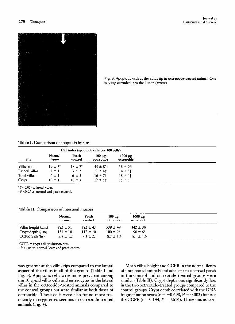
Fig. 3. Apoptotic cells at the villus tip in octreotide-treated animal. One is being extruded into the lumen (arrow).
Table I. Comparison of apoptosis by site
Cell index (apoptotic cells per 100 cells)
Normal Patch 100 ~g 1000 ~g Site i leum control octreotide octreotide
Villus tip 19 + 7* 18 -+ 7* 45 -+ 8*t 38 -+ 9*t Lateralvil lus 2 + 1 3 -+ 2 9 -+ 4 t 14 -+ 3t Total villus 6 -+ 3 6 -+ 3 16 _+ 7t 18 -+ 4 t Crypt 10 _+ 4 10 _+ 3 17 -+ 5t 15 _+ 5
*P <0.05 vs. lateral villus. tP <0.05 vs. normal and patch control.
Table II. Comparison of intestinal mucosa
Normal Patch 100 ~g 1000 ~g ileum control octreotide octreotide
Villus height (~m) 382 -+ 51 382 -+ 43 338 -+ 49 342 _+ 30 Crypt depth (~m) 121 _+ 10 117 + 10 100 -+ 9* 90 + 6* C C P R (cells/hr) 5.8 + 1.2 7.3 -+ 2.1 8.7 + 1.4 8.1 -+ 1.6
CCPR = crypt cell production rate. *P <0.05 vs. normal ileum and patch control.
was greatest at the villus tips compared to the lateral aspect of the villus in all of the groups (Table I and Fig. 3). Apoptotic cells were more prevalent among the 10 apical villus cells and enterocytes in the lateral villus in the octreotide-treated animals compared to the control groups but were similar at both doses of octreotide. These cells were also found more fre- quently in crypt cross sections in octreotide-treated animals (Fig. 4).
Mean villus height and CCPR in the normal ileum ofunoperated animals and adjacent to a serosal patch in the control and octreotide-treated groups were similar (Table II). Crypt depth was significantly less in the two octreotide-treated groups compared to the control groups. Crypt depth correlated with the DN A fragmentation score (r = -0.698, P = 0.002) but not the CCPR (r = 0.144, P = 0.616). There was no cor-

Vol. 2, No. 2 1998 Somatostatin and Apoptosis 171
Fig. 4. Apoptotic cell in crypt cross section (arrow).
relation between either the CCPR (r = 0.165, P = 0.651) or the DNA fragmentation score (r = 0.456, P -- 0.133) and mean villus height.
D I S C U S S I O N
Somatostatin has a variety of effects on the intesti- nal epithelium. In addition to its known antiprolifer- ative effect, somatostatin has been demonstrated to impair enterocyte migration and differentiation.l,s In the present study the somatostatin analogue octreotide predisposed enterocytes to apoptosis. Whereas we found previously that enterocyte migration and pro- liferation in regenerating epithelium was inhibited by 1000 ~g but not 100 ~g/day ofoctreotide, both doses had similar effects on apoptosis in the present study. Thus somatostatin appears to influence intestinal growth and structure via several different mecha- nisms.
The effect of octreotide on enterocyte apoptosis was studied in a model of stimulated epithelium (ad- jacent serosal patch) because previous studies have demonstrated that the antiproliferative effects of so- matostatin occur only in stimulated epithelium. Whereas somatostatin has no effect on proliferation of normal mucosa, it inhibits the increased prolifera- tion induced by intestinal resection and growth fac- tors including epidermal growth factor, growth hor- mone, and gastrin. 1-3,s,9-1° Creation of a serosal patch results in increased proliferation in the immediately adjacent intestinal epithelium. 11 However, villus height and crypt depth and the CCPR were similar in the normal ileum and ileum adjacent to the intestinal
patch in the present study so stimulation of mucosa was not as marked as with these other stimuli. Whether or not the somatostatin analogue would promote enterocyte apoptosis in normal intestinal epithelium remains unclear.
Apoptosis (programmed cell death) is a complex process that is still incompletely understood) 2 How- ever, it appears to he a highly regulated, genetically controlled process of cell deletion without any signs of inflammation or disruption of tissue architecture. Proliferation and programmed cell death are the im- portant regulators of the cell pool in tissue homeosta- sis. Although there appears to be a persistent low rate of spontaneous apoptosis in the intestine, there are a variety of positive and negative triggers of induced programmed cell death. 12 Growth factor deprivation appears to be an important initiating factor that re- suits in downregulation of survival genes and perhaps new gene expression leading to endonuclease activa- tion and ultimately cell death. 13 However, several mechanisms of injury, for example, irradiation and various other agents, may also trigger this process. Once initiated, the transduction of the cell death sig- nal also follows several different pathways. In the present study there were both increased DNA frag- mentation and morphologic changes of apoptosis in the somatostatin-treated animals.
The role of apoptosis in regulation of normal in- testinal homeostasis is unclear. Certainly apoptosis is triggered by normal physiologic stimuli. In the rat, apoptosis has a circadian rhythm and is increased by fasting. 14 However, the potential regulatory sites of apoptosis within the enterocyte compartment are con-

172 Thompson
troversial. Although some investigators suggest that apoptosis occurs at the villus tip with cells being ex- truded into the lumen, others only find DNA frag- mentation in the crypts. 15J6 Using a technique similar to that employed in the present study, Hall et a1.16 found that apoptotic cells could be found in most villi and crypts, being more frequent at the top of the vil- lus. Similarly, in the present study, apoptotic cells were more frequent at the tip of the villus compared to the lateral surface in the various control and experimental groups. Furthermore, in both operated and unoper- ated control animals there was a detectable rate of apoptosis on the lateral villus and in the crypts. A re- cent study in humans does suggest that apoptosis is an important mechanism for cell loss at the villus tip.17
The findings in the present study suggest that oc- treotide enhances apoptosis diffusely throughout the enterocyte compartment. A similar observation has been made in patients with celiac sprue where apop- tosis is not only increased but also more diffusely dis- tributed rather than occurring predominantly at the villus tip.‘8 However, in an in vitro study, Stange et a1.8 found no increase in DNA in shed medium from cell cultures exposed to somatostatin, which suggests that there is no enhanced cellular loss in the presence of somatostatin.
The mechanism of somatostatin-induced apopto- sis in the intestinal epithelium might be related to in- hibition of growth factor activity or release or via a direct stimulatory effect. Both direct and indirect ac- tions have a role in the antiproliferative effects of so- matostatin.5~8-10 Either explanation is plausible given existing knowledge about apoptosis. Furthermore, so- matostatin exerts its effects on tissue via a variety of different postreceptor signal transduction mecha- nisms, suggesting that it might also influence apopto- sis via more than one mechanism.19 Thus further studies would be necessary to elucidate the mecha- nism of somatostatin-induced apoptosis.
In the present study, mean crypt death but not vil- lus height correlated with apoptosis in the individual animals. Neither parameter correlated with the CCPR. Thus it is difficult to support a hypothesis that either cell proliferation or death is the dominant fac- tor determining the size of the villus compartment. However, there was a tendency toward reduced villus height in the octreotide-treated animals despite a comparable or greater CCPR. The diminished crypt depth in the octreotide-treated animals may be re- lated to enhanced apoptosis in the stem cell compart- ment, which would be expected to diminish the villus enterocyte compartment unless cell survival on the villus was prolonged. There was marked heterogenic-
Journal of Gastrointestinal Surgery
ity in DNA fragmentation, even among villi from the same histologic section, so that sampling error may be a problem in interpretation.
In summary, octreotide treatment is associated with increased DNA fragmentation in enterocytes, sug- gesting endonuclease activation. Enterocytes with morphologic characteristics of apoptosis were more prevalent in both villi and crypts. Thus predisposition to apoptosis may play a role in octreotide’s effects on intestinal regeneration and adaptation. Since there was only a correlation with crypt depth and apoptosis but not between villus height and either CCPR or apoptosis, the role of these factors in determining the size of the enterocyte compartment remains unclear.
REFERENCES
1.
2.
3.
4.
5.
6.
7.
8.
9.
10.
11.
12.
13.
14.
Thompson JS, Nguyen BLT, Harty RF. Somatostatin ana- logue inhibits intestinal regeneration. Arch Surg 1993;128: 38.5-389. Holmes SJK, Jaspan JB, Moosa AR. The effect of somatostatin on postresectional ileal hyperplasia. Endocrinology 1982;lll: 1397-1399. Bass BL, Fischer BA, Richardson C, Harmon JW. Somato- statin analogue treatment inhibits postresectional adaptation of the small bowel in rats. Am J Surg 1991;161:107-112. Thompson CB. Apoptosis in the pathogenesis and treatment of disease. Science 1995;267:1456-1462. Conteas CN, Nandimajumdar AI? The effects of gastrin, epi- dermal growth factor, and somatostatin on DNA synthesis in a small intestinal crypt cell line (IEC-6). Proc Sot Exp Biol Med 1987;184:307-311. Wright N, Watson A, Morely A, et al. The measurement of cell production rates in the crypts of Lieberkuhn. Virchows Arch 1974;364:311-323. Gavrieli Y, Sherman Y, Ben-Sassin SA. Identification of pro- grammed cell death in situ via specific labeling of nuclear DNA fragmentation. J Cell Biol 1992;119:493-501. Stange EF, Schneider A, Schuselziarra V, Ditschuneit H. In- hibitory effects of somatostatin on growth and differentiation in cultured intestinal mucosa. HormMetab Res 1987;16:74-78. Lehy T, Dubrasquet M, Bonfils S. Effect of somatostatin on normal and gastrin-stimulated cell proliferation in the gastric and intestinal mucosa of the rat. Digestion 1979;19:99-109. Konturek SJ, Brzozowski T, Dembinski A, Warzecha 2, Kon- turek PK, Yanaihara N. Interaction of growth hormone re- leasing factors and somatostatin on ulcer healing and mucosal growth in rats: Role of gastrin and epidermal growth factor. Digestion 1988;41:121-128. Saxena SK, Thompson JS, Joshi SS, Sharp JG. Extent and role of urogastrone in the adaptive response on the rat intestine to patching or a surgical defect in the ileum. J Invest Surg 1993;6:485-492. Binder C, Hiddemann W. Programmed cell death-Many questions still to be answered. Arm Hematol 1994;69:45-55. Collins MKL, Perkins GR, Rodriquez-Tarduchv G. Nieto MA, Lopez-R&as A. Growth factors as survival factors: Reg- ulation of auootosis. Bioessavs 1994;16:133-138. ._ __-. Iwakirii R, AwTY. Apoptosis in rat small intestine: Circadian rhythm and effect of feeding and fasting [abstr]. Gastroen- terology 1995;108:A292.

Vol. 2, No. 2 1998 Somatostatin and Apoptosis 173
15. Watson AJM. Manipulation of cell death-The development of novel strategies for the treatment of gastrointestinal dis- ease. Aliment Pharmacol Ther 1995;9:215-226.
16. Hall PA, Coates PJ, Ansari B, Hopwood D. Regulation of cell number in the mammalian gastrointestinal tract: The impor- tame of apoptosis. J Cell Sci 1994;107:3569-3577.
17. Shibahara T, Nakahara A, Fukutomi H. The fate of effete en- terocytes at the villas tips of human small intestine with spe- cial reference to their exfoliating process and apoptosis. Gas- troenterology 1996;l lO:A838.
18. Attia AL, Moss RF, Walters JRF, Wang S, Holt PR. Increased small bowel epithelial apoptosis reflects celiac sprue activity [abstr]. Gastroenterology 1995;10&4271.
19. Pate1 PC, Barrie R, Hill N, Laudeck S, Kurqzawa D, Wolter- ing EA. Postreceptor signal transduction mechanisms involved in octreotide-induced inhibition of angiogenesis. Surgery 1994;116:1148-1152.
BOUND VOLUMES
Bound volumes are available to subscribers only. The hardbound volume of six issues of the 1998 3 ownal of Gastrointestinal Surgery must be ordered by October 1, 1998, from Quality Medical Publishing, Inc., 11970 Borman Dr., Suite 222, St. Louis, MO 63 146. Payment of $75 in U.S. funds must accompany all orders.