somatic embryogenesis and morphoanatomy of … · somatic embryogenesis and morphoanatomy of ocotea...
TRANSCRIPT
Ci. Fl., v. 23, n. 4, out.-dez., 2013
Ciência Florestal, Santa Maria, v. 23, n. 4, p. 595-605, out.-dez., 2013ISSN 0103-9954
595
SOMATIC EMBRYOGENESIS AND MORPHOANATOMY OF Ocotea porosa SOMATIC EMBRYOS
EMBRIOGÊNESE SOMÁTICA E MORFOANATOMIA DE EMBRIÕES SOMÁTICOS DE Ocotea porosa
Luciana Luiza Pelegrini1 Luciana Lopes Fortes Ribas2 Erika Amano3 Marguerite Quoirin4
ABSTRACT
Ocotea porosa seeds have strong tegument dormancy, recalcitrant behavior, low and irregular germination and that makes its natural propagation difficult. The aim of this study was to establish a protocol of regeneration of Ocotea porosa from somatic embryogenesis. Immature embryonic axes were inoculated on WPM culture medium supplemented with 2.4-D (200 µM) combined or not with hydrolyzed casein or glutamine (0.5 or 1 g l-1), during 90 days. The repetitive embryogenesis was induced on medium with 2.4-D (22.62 µM) combined with 2-iP (2.46 µM) followed by transfer to culture medium with hydrolyzed casein or glutamine (1 g l-1) during 90 days. The maturation of somatic embryos was tested in culture medium containing NAA (0.5 μM) and 2-iP (5; 10 and 20 μM). The highest percentage of somatic embryos induction (8.3%) was observed in WPM culture medium containing 200 µM 2.4-D and 1 g L-1 hydrolyzed casein and the development of somatic embryos occurred indirectly. Repetitive somatic embryogenesis was promoted in WPM medium containing hydrolyzed casein or glutamine. However, the culture medium containing hydrolyzed casein promoted the maintenance of embryogenic capacity for more than two years. During the maturity phase, there was a low progression of globular embryos to cordiform and torpedo stages. The different ontogenetic stages of somatic embryos of Ocotea porosa were characterized by histological studies.Keywords: Embryogenic mass; hydrolyzed casein; glutamine; histology.
RESUMO
Ocotea porosa apresenta sementes com forte dormência tegumentar, comportamento recalcitrante, baixa germinação e irregularidade, o que dificulta a propagação natural. O objetivo deste trabalho foi estabelecer um protocolo de embriogênese somática para Ocotea porosa. Eixos embrionários zigóticos imaturos foram inoculados em meio de cultura WPM suplementado com 2,4-D (200 µM) combinado ou não com caseína hidrolisada ou glutamina (0,5 ou 1 g l-1) durante 90 dias. A embriogênese repetitiva foi induzida em meio contendo 2,4-D (22,62 µM) combinado com 2-iP (2,46 µM) seguido de transferência para meio de cultura com caseína hidrolisada ou glutamina (1 g l-1), durante 90 dias cada. A maturação dos embriões somáticos foi testada em meio de cultura contendo ANA (0,5 μM) e 2-iP (5, 10 ou 20 μM). A maior porcentagem de embriões somáticos induzidos (8,3%) foi observada em meio de cultura WPM contendo 200 µM de 2,4-D e 1 g l-1 de caseína hidrolisada e o desenvolvimento de embriões somáticos ocorreram de forma indireta. A embriogênese somática repetitiva foi promovida em meio de cultura WPM contendo caseína hidrolisada ou glutamina, no entanto, a caseína hidrolisada promoveu a manutenção da competência embriogênica por mais de dois anos. Na fase de maturação, houve baixa progressão dos embriões globulares para os estádios
1 Bióloga, Drª. pelo Programa de Pós-Graduação em Produção Vegetal, Setor de Ciências Agrárias, Universidade Federal do Paraná, CEP 81531-990, Curitiba (PR), Brasil. Bolsista Reuni. [email protected]
2 Bióloga, Drª, Professora Associada II do Departamento de Botânica, Setor de Ciências Biológicas, Universidade Federal do Paraná, CEP 81531-970, Curitiba (PR), Brasil. [email protected]
3 Bióloga, Drª, Professora Adjunto do Departamento de Botânica, Setor de Ciências Biológicas, Universidade Federal do Paraná, CEP 81531-980, Curitiba (PR), Brasil. [email protected]
4 Engenheira Agrônoma, Drª, Professora Associada do Departamento de Botânica, Setor de Ciências Biológicas, Universidade Federal do Paraná, CEP 81531-990, Curitiba (PR), Brasil. [email protected]
Recebido para publicação em 28/08/2011 e aceito em 28/08/2012
Ci. Fl., v. 23, n. 4, out.-dez., 2013
Pelegrini, L. L. et al.596
INTRODUCTION
Ocotea porosa (Liberato Barroso), known as “imbuia”, belongs to the Lauraceae family and is native to the Mixed Ombrophilous Forest (Araucaria Forest) where it was heavily exploited due to the high quality and worldwide value of its hardwood, that is exported in large quantities for luxury fur-nishing manufactures (CARVALHO, 2003). As a result, this species is an endangered one and is quot-ed in the official list of endangered Brazilian flora species, in the vulnerable category (IBAMA, 1992) and in the Red List of Parana, in the rare category (SEMA, 1995).
The sexual propagation of Ocotea porosa at its natural occurrence area is difficult, due to the strong tegumentary dormancy and its irregular ger-mination (CARVALHO, 2003). Moreover, the seed viability is short by virtue of being a recalcitrant spe-cies, showing high level of humidity (CARVALHO, 2003). Another limiting factor for its vegetative propagation is the low response of cuttings to the induction of adventitious roots (4%) (INOUE and PUTTON, 2007).
Somatic embryogenesis is a process by which the somatic cells, under inductive conditions, generate embryogenic cells, which undergo a series of morphological and biochemical alterations, re-sulting in formation of somatic embryos (SANTOS et al., 2002; KOMAMINE et al., 2005). Somatic embryogenesis is a model for the study of morpho-logical, physiological, molecular characteristics and biochemical events that occur during the embryo-genesis in higher plants. Beyond that, it has poten-tial biotechnological applications, such as artificial seed production and propagation of elite plants or genetically modified plants (SANTOS et al., 2002; QUEIROZ-FIGUEROA, 2006).
The maturation is considered a fundamental step in the development of the embryo for subse-quent conversion and for the seedling development (PERAN-QUESADA et al., 2004). During matura-tion, embryo cells undergo various physiological changes, which become evident by the deposit of storage materials, the repression of germination and
the acquisition of desiccation tolerance (JIMÉNEZ, 2005; VAHDATI et al., 2008). The cell expansion and accumulation of reserve substances are crucial to stimulate the conversion process (GARCIA-MARTIN et al., 2005). Although in some woody plants, the maturation and the germination of somat-ic embryos have been obtained in media which lack plant growth regulators (SANTA-CATARINA et al., 2001; CANHOTO et al., 2005; SHI et al., 2009), in most cases, the application of specific treatments is necessary (RIBAS, 2000). Abscisic acid and os-motic stresses are known to be important factors for the seed maturation in many angiosperms (RAI et al., 2011; VAHDATI et al., 2008). The addition of reduced nitrogen in the maturation medium is a re-quirement to ensure maturation of somatic embryos (MERKLE et al., 1995). Compared to other inor-ganic nitrogen sources the nitrogen in reduced form of amino acids such as glutamine, asparagine and arginine is rapidly absorbed and incorporated into the carbon skeletons for the metabolism and protein synthesis (LEA, 1993). Hydrolyzed casein is an ex-cellent source of reduced nitrogen containing a mix-ture of up to 18 amino acids, particularly glutamine (GEORGE and DE KLERK, 2008).
In somatic embryogenesis, the histologi-cal and morphological analysis of the plant mate-rial may be critical for the results interpretation and a help to increase the efficiency of protocols. Histological analysis can be used to identify and to characterize the formation of monopolar and bipolar structures as well as the cellular changes, analyz-ing the quality and development of these structures (PEREZ-NUÑEZ et al., 2006).
For many forest species, including several members of Lauraceae family, like Laurus nobilis (CANHOTO et al., 1999), Persea americana Mill (SANCHEZ-ROMERO, 2006), Ocotea catharinensis (VIANA and MANTELL, 1999) and Ocotea odorifera (SANTA-CATARINA et al., 2005), somatic embryogenesis induction proto-cols have been established. However, for Ocotea catharinensis, Santa-Catarina et al. (2005) found difficulties in optimizing the conversion stage of so-matic embryos into plants.
cordiforme e torpedo. Os diferentes estádios ontogenéticos de embriões somáticos de Ocotea porosa foram caracterizados pelo estudo histológico. Palavras-chave: massa embriogênica; caseína hidrolisada; glutamina; histologia.
Ci. Fl., v. 23, n. 4, out.-dez., 2013
Somatic embryogenesis and morphoanatomy of Ocotea porosa somatic embryos 597
The aim of this study was to establish a pro-tocol of somatic embryogenesis of Ocotea porosa from immature zygotic embryonic axes.
MATERIAL AND METHOD
Zygotic embryonic axes from immature Ocotea porosa seeds, collected at Campina Grande do Sul, Paraná state, Brazil, in February 2009, were used as explants. The seed disinfestation was per-formed in a laminar flow chamber, through immer-sion in ethanol 70% (v/v) for 5 min, followed by 20 min immersion in a sodium hypochlorite solution (NaOCl) 4% (v/v) supplemented with 0,1% Tween®
20. After that, the seeds were rinsed five times with sterile water.
Induction of somatic embryos
The immature embryonic axes (0.2 to 0.4 cm) were inoculated in an induction medium consist-ing of WPM “Woody Plant Medium” (LLOYD and McCOWN, 1980) vitamins and salts, supplemented with sucrose (20 g l-1), activated charcoal (1.5 g l-1), Vetec® agar (4 g l-1) and 2,4-D (2,4-dichlorophen-oxyacetic acid) (200 µM) alone or combined with hy-drolyzed casein or glutamine (0.5 and 1.0 g l-1). The explants were kept in the dark and, after 90 days, they were evaluated for embryogenic mass forming so-matic embryos, friable mass callus (not embryogen-ic), yellowish white callus, blackish callus (brown or black) and necrosis of immature zygotic embryonic axes. The evaluation was performed every 30 days.
Repetitive embryogenesis
The somatic embryos obtained in the experi-ment of induction were transferred to the same cul-ture medium supplemented with 2.4-D (22.62 µM) and 2-iP (2-isopentyladenine) (2.46 µM) for second-ary somatic embryogenesis induction. Three subcul-tures were performed at 30-day intervals. After this period, the cultures were transferred to WPM culture medium without plant growth regulator, supple-mented with sucrose (20 g l-1), agar Vetec® (3.5 g l-1) and hydrolyzed casein or glutamine (1 g l-1). Three subcultures were carried out at 30-day intervals and were evaluated after 90 days. The number of globu-lar, cordiform, torpedo and cotiledonary embryos was recorded. These cultures were maintained dur-ing three years, being transferred to a fresh medium every 30 days.
Maturation of somatic embryos
Somatic embryos formed through second-ary somatic embryogenesis in culture media con-taining hydrolyzed casein (1 g l-1) were transferred to WPM media supplemented with sucrose (20 g l-1), agar Vetec® (3.5 g l-1) and 2-iP (5, 10 e 20 µM) combined with NAA (naphthaleneacetic acid) (0.5 µM). After 30 days the percentage of callus de-veloping embryogenic mass and the average num-ber of globular, heart shaped and torpedo embryos was recorded.
Culture conditions
The pH of all culture media was adjusted to 5.8 prior to autoclaving. Test tubes (25 x 150 mm) containing 10 ml of culture media were used. The cultures were placed in the dark at 27±2ºC (day) and 18±2ºC (night).
Experimental design and statistical analysis
Experimental design was completely ran-domized with four replications and 10 embryonic axes per plot. The data were subjected to Kruskal-Wallis test at 5% of probability, using the STATISTICA program, version 7.1.
Morphoanatomic studies
Samples were fixed using glutaraldehyde (2%) and paraformaldehyde (4%) buffered with 0.2 M phosphate buffer, at pH 7.2 for 96 hours (KARNOVSKY, 1965). After that, they were de-hydrated in an alcoholic series with a duration of 60 min for each treatment. The samples were em-bedded in Historesin® (Leica, Heidelberg). Serial sections were performed on a rotation microtome with a thickness of 7μm. The sections were stained with toluidine blue O (O’BRIEN et al. 1965), ob-served under optical microscope and then photo-graphed with a digital camera.
RESULTS AND DISCUSSION
Induction of somatic embryos
Immature zygotic embryonic axes of Ocotea porosa showed different responses when cultured in vitro. The expression events of somatic embryogenesis occurred between the 30th and 40th
Ci. Fl., v. 23, n. 4, out.-dez., 2013
Pelegrini, L. L. et al.598
day after inoculation on the culture medium and the induction of somatic embryos occurred indirectly.
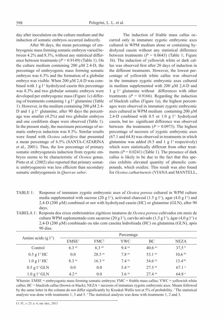
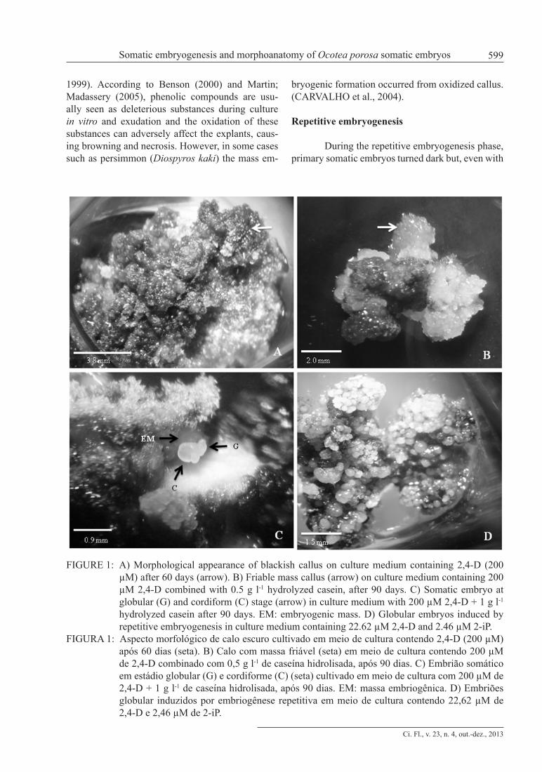
After 90 days, the mean percentage of em-bryogenic mass forming somatic embryos varied be-tween 4.2% and 8.3%, without any statistical differ-ence between treatments (P = 0.9149) (Table 1). On the culture medium containing 200 µM 2.4-D, the percentage of embryogenic mass forming somatic embryos was 6.3% and the formation of a globular embryo was visible. When 200 µM 2,4-D was com-bined with 1 g l-1 hydrolyzed casein this percentage was 8.3% and two globular somatic embryos were developed per embryogenic mass (Figure 1c) differ-ing of treatments containing 1 g l-1 glutamine (Table 1). However, in the medium containing 200 µM 2.4-D and 1 g l-1 glutamine, after 90 days the percent-age was smaller (4.2%) and two globular embryos and one cordiform shape were observed (Table 1). In the present study, the maximum percentage of so-matic embryos induction was 8.3%. Similar results were found with Ocotea odorifera that presented a mean percentage of 6.5% (SANTA-CATARINA et al., 2001). Thus, the low percentage of primary somatic embryogenesis induction from zygotic em-bryos seems to be characteristic of Ocotea genus. Pinto et al. (2002) also reported that primary somat-ic embryogenesis was less efficient than secondary somatic embryogenesis in Quercus suber.
The induction of friable mass callus oc-curred only in immature zygotic embryonic axes cultured in WPM medium alone or containing hy-drolyzed casein without any statistical difference between treatments (P = 0.0643) (Table 1; Figure 1b). The induction of yellowish white or dark cal-lus was observed first after 20 days of induction in the different treatments. However, the lowest per-centage of yellowish white callus was observed in the immature zygotic embryonic axes cultured in medium supplemented with 200 µM 2,4-D and 1 g l-1 glutamine without differences with other treatments (P = 0.9166). Regarding the induction of blackish callus (Figure 1a), the highest percent-ages were observed in immature zygotic embryonic axes cultured in WPM medium containing 200 µM 2,4-D combined with 0.5 or 1.0 g l-1 hydrolyzed casein, but no significant difference was observed between the treatments (P = 0.0973). The highest percentage of necrosis of zygotic embryonic axes (67.1 and 64.8) was observed in treatments in which glutamine was added (0.5 and 1 g l-1 respectively) which were statistically different from other treat-ments (P = 0.0241) (Table 1). The presence of dark callus is likely to be due to the fact that this spe-cies exhibits elevated quantity of phenolic com-pounds, which oxidize. This result was also found for Ocotea catharinensis (VIANA and MANTELL,
TABLE 1: Response of immature zygotic embryonic axes of Ocotea porosa cultured in WPM culture media supplemented with sucrose (20 g l-1), activated charcoal (1.5 g l-1), agar (4.0 g l-1) and 2,4-D (200 µM) combined or not with hydrolyzed casein (HC) or glutamine (GLN), after 90 days.
TABELA 1: Resposta dos eixos embrionários zigóticos imaturos de Ocotea porosa cultivados em meio de cultura WPM suplementado com sacarose (20 g l-1), carvão ativado (1,5 g l-1), ágar (4,0 g l-1) e 2,4-D (200 µM) combinado ou não com caseína hidrolisada (HC) ou glutamina (GLN), após 90 dias.
Amino acids (g l-1)Percentage
EMSE1 FMC2 YWC BC NEZAControl 6.3 ns 6.3 ns 9.4 ns 40.6 ns 37,5 b
0.5 g l-1 HC 0.0 28.5 ns 7.8 ns 53.1 ns 10,6 bc
1.0 g l-1 HC 8.3 ns 16.3 ns 7.4 ns 54.6 ns 13.4 bc
0.5 g l-1 GLN 0.0 0.0 5.4 ns 27.5 ns 67.1 a
1.0 g l-1 GLN 4.2 ns 0.0 3.6 ns 27.4 ns 64.8 a
Wherein: EMSE = embryogenic mass forming somatic embryos; FMC = friable mass callus; YWC = yellowish white callus; BC = blackish callus (brown or black); NEZA = necrosis of immature zygotic embryonic axes. Means followed by the same letter in the column do not differ significantly by Kruskal-Wallis test at 5% of probability. 1 The statistical analysis was done with treatments 1, 3 and 5. 2 The statistical analysis was done with treatments 1, 2 and 3.
Ci. Fl., v. 23, n. 4, out.-dez., 2013
Somatic embryogenesis and morphoanatomy of Ocotea porosa somatic embryos 599
1999). According to Benson (2000) and Martin; Madassery (2005), phenolic compounds are usu-ally seen as deleterious substances during culture in vitro and exudation and the oxidation of these substances can adversely affect the explants, caus-ing browning and necrosis. However, in some cases such as persimmon (Diospyros kaki) the mass em-
bryogenic formation occurred from oxidized callus. (CARVALHO et al., 2004).
Repetitive embryogenesis
During the repetitive embryogenesis phase, primary somatic embryos turned dark but, even with
FIGURE 1: A) Morphological appearance of blackish callus on culture medium containing 2,4-D (200 µM) after 60 days (arrow). B) Friable mass callus (arrow) on culture medium containing 200 µM 2,4-D combined with 0.5 g l-1 hydrolyzed casein, after 90 days. C) Somatic embryo at globular (G) and cordiform (C) stage (arrow) in culture medium with 200 µM 2,4-D + 1 g l-1 hydrolyzed casein after 90 days. EM: embryogenic mass. D) Globular embryos induced by repetitive embryogenesis in culture medium containing 22.62 µM 2,4-D and 2.46 µM 2-iP.
FIGURA 1: Aspecto morfológico de calo escuro cultivado em meio de cultura contendo 2,4-D (200 µM) após 60 dias (seta). B) Calo com massa friável (seta) em meio de cultura contendo 200 µM de 2,4-D combinado com 0,5 g l-1 de caseína hidrolisada, após 90 dias. C) Embrião somático em estádio globular (G) e cordiforme (C) (seta) cultivado em meio de cultura com 200 µM de 2,4-D + 1 g l-1 de caseína hidrolisada, após 90 dias. EM: massa embriogênica. D) Embriões globular induzidos por embriogênese repetitiva em meio de cultura contendo 22,62 µM de 2,4-D e 2,46 µM de 2-iP.
Ci. Fl., v. 23, n. 4, out.-dez., 2013
Pelegrini, L. L. et al.600
a necrotic appearance, the formation of new struc-tures in culture medium with 2,4-D (22,62 µM) and 2-iP (2,46 µM) was induced. After three months, on the calluses subcultured at 30-day intervals in me-dia with 2,4-D (22.62 µM) and 2-iP (2.46 µM), an average of 76.9 globular, 3.4 cordiform, 1.5 torpedo and one cotyledonary embryos was formed per cal-lus (Figure 1d). These results corroborate those of Moura-Costa et al. (1993) which reported the dark-ening of somatic embryos of Ocotea catharinensis that, after being subcultivated, formed new globular structures.
After this period, the embryos were sub-cultured in media containing 1 g l-1 of hydrolyzed casein or glutamine without plant growth regula-tors. The addition of 1 g l-1 hydrolyzed casein pro-moted an average formation of 61.8 new globular and 0.5 cordiform embryos per callus at the end of every subculture (P = 0.0433) (Table 2). When 1 g l-1 glutamine was added into the media, the in-duction of globular somatic embryos was lower (30.4 embryos) than the one in the presence of HC and other stages of development were not observed (Table 2).
In the present study, it was shown that hy-drolyzed casein was more effective than glutamine as a source of amino acids for the induction of new somatic embryos through repetitive embryogen-esis, as well as to maintain the embryogenic com-petence (dates not shown). The amino acids are important substances for primary assimilation of nitrogen and for protein synthesis (ORTIZ-LOPEZ et al., 2000). According to Morcillo et al. (1999) and Garin et al. (2000) the glutamine combined or not with hydrolyzed casein can be used at different stages of somatic embryogenesis. The addition of glutamine and/or hydrolyzed casein to the culture media could increase the level of proliferation of calluses and somatic embryos, as these substances
provide great amount of reduced organic nitrogen (GEORGE, 1996).
During repetitive embryogenesis the new somatic embryos are formed from others and some species keep this embryogenic competence for many years (PINTO et al., 2008). Embryogenic competence was observed, for example, in Quercus suber (HERNÀNDEZ et al., 2003), Vitis rupestris (MARTINELLI et al., 2001), Ocotea odorifera (SANTA-CATARINA et al., 2001), Ocotea catharinensis (MOURA-COSTA et al., 1993) and Aspidosperma polyneuron (RIBAS et al., 2000). Litz et al. (1995) and Tulecke et al. (1995) reported that the embryogenic capacity could be maintained in Mangifera indica and Juglans regia through repetitive embryogenesis for more than 5 and 9 years, respectively. After the evaluation period (90 days), the cultures remained in WPM culture medium supplemented with 1 g l-1 hydrolyzed ca-sein, showing that the embryogenic competence was maintained and the new embryos were multi-plied for more than one year. Considering the low induction of somatic embryos from zygotic em-bryonic axes, the repetitive embryogenesis may be an important system of mass propagation, as high production rates of embryos were reached.
Maturation of somatic embryos
The cultures, which consisted of globular somatic embryos obtained on culture media con-taining hydrolyzed casein and transferred to matu-ration media containing 2-iP (5, 10 and 20 µM) combined with NAA (0.5 µM), showed differ-ent morphogenetic responses (Table 3). The per-centage of embryogenic mass was similar in all treatments, without any statistical difference (P = 0.8446).The maturation media containing 2-iP combined with NAA promoted continuous prolif-
TABELA 2: Repetitive somatic embryogenesis from immature zygotic embryonic axes of Ocotea porosa during three subcultures.
TABLE 2: Embriogênese somática repetitiva a partir de eixos embrionários zigóticos imaturos de Ocotea porosa durante três subcultivos.
Treatments (1 g l-1)Average number of embryos/callus
Globular Cordiform Torpedo CotiledonaryHydrolyzed casein 61.8 a 0.5 0.0 0.0
Glutamine 30.4 b 0.0 0.0 0.0Means followed by same letter in column do not differ significantly by Kruskal-Wallis test at 5% of probability.
Ci. Fl., v. 23, n. 4, out.-dez., 2013
Somatic embryogenesis and morphoanatomy of Ocotea porosa somatic embryos 601
eration of embryogenic mass and globular somatic embryos.
During maturation phase, the somatic em-bryos develop from initial stages to final stages. However, in the present conditions of maturation, the percentage of embryos developing to cordiform and torpedo ontogenetic stages was low and no embryo at cotyledonary phase was seen, revealing an asynchronous development, once globular so-matic embryos were formed (Table 3). The average number of globular somatic embryos varied few and without any statistic difference between treat-ments (P =.6084); the same was observed for the average number of cordiform and torpedo somatic embryos (P = 0.2437 and P = 0.4835 respectively). According to Guerra et al. (1999), during the matu-ration phase, it is necessary to disrupt the repeti-tive cycles of cell division and to provide physio-logical, biochemical and environmental stimuli for cellular differentiation, then the cycles of develop-ment and maturation give rise to a large number of mature somatic embryos of high quality and ca-pable of turning into plants. However, in the pres-ent study, a combination of 2-iP and NAA was not effective for maturation of Ocotea porosa somatic embryos. Carvalho et al. (2004) also reported that the progression of somatic embryos of persimmon (Diospyros kaki) cultures in media containing 2-iP combined with IBA occurred in an asynchronous way. Guerra and Handro (1998), obtained ma-ture somatic embryos of Euterpe edulis in media containing 2-iP and NAA and, at the same time, managed to maintain the embryogenic competence culture. Santa-Catarina et al. (2001), observed the development of Ocotea odorifera somatic embry-os in culture media without plant growth regulator.
Preliminary test using hydrolyzed casein (1 g l-1) combined with NAA (0.5 µM) and 2-iP (5, 10 or 20 µM) was also tested in the maturation of
somatic embryos of Ocotea porosa. However, the results were similar to those found when the NAA and 2iP alone were utilized. Thus, it is recom-mended to test other concentrations of hydrolyzed casein or other amino acids in order to increase the percentage as well as synchronization of the so-matic embryo maturation.
In the maturation media, some abnormal somatic embryos were formed and, above them, globular somatic embryos. The formation of ab-normal somatic embryos during the secondary so-matic embryogenesis is probably due to the dura-tion of contact with the medium containing 2,4-D. Titon et al. (2007) also reported lack of synchro-nization in the development and the formation of abnormal somatic embryos of Eucalyptus grandis.
Morphoanatomical studies
Structures obtained in WPM medium con-taining 2,4-D (22.62 µM) combined with 2-iP (2.46 µM) presented histodifferentiation of somat-ic embryos (Fig. 2). In the Figure 2a, two distinct regions were observed: a region consisting of em-bryonic isodiametric cells, with dense cytoplasm, prominent nucleus and stained dark nucleoli show-ing the formation of pro-embryos (Fig. 2a arrow) with dividing cells (Fig 2b arrow). The inner re-gion consisted of cells with large vacuole content featuring blue-green color through the metachro-matic reaction with toluidine blue, indicating the presence of phenolic compounds. In Myrciaria aureana also, embryogenic explants were rich in phenolic compounds that had accumulated primar-ily in a sub-superficial zone between layers of cells of the peripheric region and storage parenchyma (MOTOIKE et al., 2007). As in the present study, the meristematic activity was observed near the zone rich in phenolic compounds where the cells
TABLE 3: Effect of 2-iP combined with NAA on the maturation of somatic embryos of Ocotea porosa after 30 days of culture on WPM culture media.
TABELA 3: Efeito do 2,iP combinado com ANA na maturação de embriões somáticos de Ocotea porosa após 30 dias de cultivo em meio de cultura WPM.
2-iP (µM) Embryogenic mass (%)
Average number of embryos/callusGlobular Cordiform Torpedo
5 85.0 ns 30.7 ns 0,5 ns 1,5 ns
10 92.5 ns 37.2 ns 1,0 ns 1,0 ns
20 90.0 ns 36.2 ns 2,8 ns 3,0 ns
Wherein: n.s. = not significant according to Kruskal-Wallis test at 5% probability.
Ci. Fl., v. 23, n. 4, out.-dez., 2013
Pelegrini, L. L. et al.602
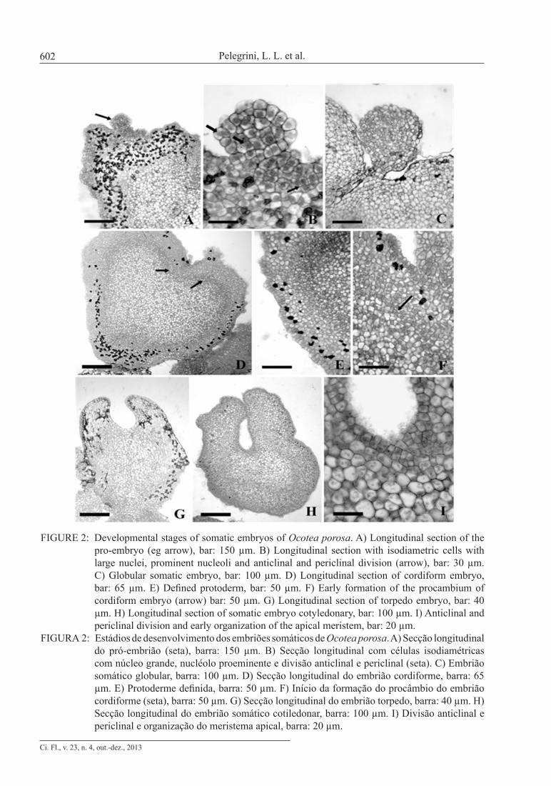
FIGURE 2: Developmental stages of somatic embryos of Ocotea porosa. A) Longitudinal section of the pro-embryo (eg arrow), bar: 150 µm. B) Longitudinal section with isodiametric cells with large nuclei, prominent nucleoli and anticlinal and periclinal division (arrow), bar: 30 µm. C) Globular somatic embryo, bar: 100 µm. D) Longitudinal section of cordiform embryo, bar: 65 µm. E) Defined protoderm, bar: 50 µm. F) Early formation of the procambium of cordiform embryo (arrow) bar: 50 µm. G) Longitudinal section of torpedo embryo, bar: 40 µm. H) Longitudinal section of somatic embryo cotyledonary, bar: 100 µm. I) Anticlinal and periclinal division and early organization of the apical meristem, bar: 20 µm.
FIGURA 2: Estádios de desenvolvimento dos embriões somáticos de Ocotea porosa. A) Secção longitudinal do pró-embrião (seta), barra: 150 µm. B) Secção longitudinal com células isodiamétricas com núcleo grande, nucléolo proeminente e divisão anticlinal e periclinal (seta). C) Embrião somático globular, barra: 100 µm. D) Secção longitudinal do embrião cordiforme, barra: 65 µm. E) Protoderme definida, barra: 50 µm. F) Início da formação do procâmbio do embrião cordiforme (seta), barra: 50 µm. G) Secção longitudinal do embrião torpedo, barra: 40 µm. H) Secção longitudinal do embrião somático cotiledonar, barra: 100 µm. I) Divisão anticlinal e periclinal e organização do meristema apical, barra: 20 µm.
Ci. Fl., v. 23, n. 4, out.-dez., 2013
Somatic embryogenesis and morphoanatomy of Ocotea porosa somatic embryos 603
divided rapidly, forming somatic embryos from cells of the peripheric region. Histological sections confirmed the formation of various stages of so-matic embryos of Ocotea porosa (Figure 2).
A globular structure with protoderm and a suspensor was observed, characterizing the first stage of somatic embryo development (Fig. 2c). The transition from globular to cordiform stage was marked by cellular elongation and cotyledon early growth (Fig. 2d). In this phase a defined protoderm (Fig. 2e) was observed, as well as cell division lead-ing to procambium formation (Fig. 2f arrow). In Figure 2g, the differentiation of procambial track with elongated and vacuolated cells and the elonga-tion of the cotyledons are visible, characterizing the torpedo stage. In this phase, the longitudinal section shows bilateral symmetry. At that stage, it was evi-dent that deposition of phenolic compounds in the outer layers caused the degradation of cells. Figure 2h shows a somatic embryo at cotyledonary stage. Organized meristematic tissues are also present, initiating the formation of a shoot apical meristem (Fig. 2i) but root apical meristem is not observed. Although it was possible to characterize the cotyle-donary stage of the embryo, the presence of the en-tire vascular system was not confirmed. According to Dodeman et al. (1997), in order to complete so-matic embryogenesis, the formation of meristems must occur. However, histological analysis showed that somatic embryos of Ocotea porosa did not fin-ish their development. Although somatic embryos have been characterized up to cotyledonary stage, these structures are not considered bipolar, since no difference was observed at the root apical meristem. Ribas et al. (2000), also observed the absence of dif-ferentiation of the meristem in somatic embryos of Aspidosperma polyneuron.
CONCLUSIONS
For the first time, we demonstrated that so-matic embryogenesis of Ocotea porosa can be in-duced from immature zygotic embryonic axes on WPM medium containing 200 µM 2,4-D alone or combined with 1 g l-1 hydrolyzed casein. Somatic embryos were maintained and proliferated on hor-mone-free WPM medium supplemented with hy-drolyzed casein (1 g l-1) via repetitive embryogen-esis. The maturation of the embryos was low and asynchronous. Thus, the results of this study indi-cate that somatic embryogenesis of Ocotea porosa is feasible and this repetitive embryogenesis system
will provide a source of somatic embryo production, ensuring the optimization of a complete protocol for the regeneration of this species.
ACKNOWLEDGMENTS
The authors thank REUNI (Reestruturação e Expansão das Universidades Federais, Brazil) pro-gram for the PhD grant to L.P. and Danielle Ferreira for help in translating the manuscript.
REFERENCES
BENSON, E. E. In vitro plant recalcitrance: an introduction. In Vitro Cellular Developmental Biology - Plant, Wallinghord, v. 36, n3, p. 141-148, maio/jun. 2000.CARVALHO, P. E. R. Espécies arbóreas brasileiras. Brasília: Embrapa Informação Tecnológica; Colombo, PR: Embrapa Florestas, 2003, 1039 p.CARVALHO, D. C. et al. Embriogênese somática do caquizeiro. Revista Brasileira de Fruticultura, Jaboticabal, v. 26, n. 2, p. 280-283, ago. 2004.CANHOTO, J. M. et al. Somatic embryogenesis in Bay Laurel (Laurus nobilis). In: JAIN, S. M.; GUPTA, P. K.; NEWTON, R. J. Somatic embryogenesis in woody plants. Dordrecht: Kluwer Academic Publishers, 1999. v. 4, p. 341-367.CANHOTO, J. M. et al. Protocol for somatic embryogenesis: tamarillo (Cyphomandra betacea (Cav.) Sendt.). In: JAIN, S. M.; GUPTA, P. K. (eds). Protocol for somatic embryogenesis in woody plants. Dordrecht: Springer, 2005, p 379–389.DODEMAN, V. L. et al. Zygotic embryogenesis versus somatic embryogenesis. Journal of Experimental Botany, v. 48, p. 1493-1509, 1997.GARCIA-MARTIN, G. et al. Effects of exogenous ABA on embryo maturation and quantification of endogenous levels of ABA and IAA in Quercus suber somatic embryos. Plant Cell, Tissue and Organ Culture, v. 80, p. 171-177, 2005.GARIN, E. et al. Effect of sugars, amino acids, and culture technique on maturation of somatic embryos of Pinus strobus on medium with two gellan gum concentration. Plant Cell, Tissue and Organ Culture, Dordrecht, v. 62, n. 1, p. 27-37, jul. 2000.GEORGE, E. F. Plant propagation by tissue culture. 2nd ed. London: Exegetics Limited, 1996. v. 2.
Ci. Fl., v. 23, n. 4, out.-dez., 2013
Pelegrini, L. L. et al.604
GEORGE, E. F.; DE KLERK, G. J. The Components of Plant Tissue Culture Media I : Macro- and Micro-Nutrients. IN: GEORGE, E.F.; HALL, M.A.; DE KLERK, G.J. Plant propagation by tissue culture. The Background, 2008. v. 1GUERRA, P. M. et al. Embriogênese somática e semente sintética. In: TORRES, A.; CALDAS L. S.; BUSO, J. A. Técnicas e aplicações da cultura de tecidos de plantas. Brasília: ABCTP/EMBRAPA – CNPH, p. 533-568, 1999.GUERRA, M. P.; HANDRO, W. Somatic embryogenesis and plant regeneration in different organs of Euterpe edulis Mart. (Palmae): control and structural features. Journal of Plant Research, Tokyo, v. 111, n. 1, p. 65-71, mar. 1998.HERNÁNDEZ, I. et al. Vegetative propagation of Quercus suber L. by somatic embryogenesis: II. Plant regeneration from selected cork oak trees. Plant Cell Reports, Berlin, v. 21, n.8, p. 765–770, abr. 2003.IBAMA – Instituto Brasileiro do Meio Ambiente. Portaria n. 37-N, de 3 de abril de 1992. Lista oficial de espécies da flora brasileira ameaçadas de extinção.INOUE, M. T.; PUTTON, V. Macropropagação de 12 espécies arbóreas da floresta ombrófila mista. Floresta, Curitiba, v. 37, n. 1, p. 55-61, jan./abr. 2007.JIMÉNEZ, V. M. Involvement of plant hormones and plant growth regulators on in vitro somatic embryogenesis. Plant Growth Regulator, v. 47, p. 91-110, 2005.KARNOVSKY, M. J. A. formaldehyde-glutaraldehyde fixative of high osmolality for use in eletron microscopy. Journal of Cell Biology, New York, v. 27, n. 10, p. 137-138, 1965.KOMAMINE, A. et al. Mechanisms of somatic embryogenesis in carrot suspension cultures – morphology, physiology, biochemistry, and molecular biology. In vitro Cellular Developmental Biology - Plant, Wallinghord, v. 41, n. 1, p. 6-10, Jan./Feb. 2005. LEA, P. J. Nitrogen metabolism. In: LEA, P. J.; LEEGOOD, R. C (eds). Plant Biochemistry and Molecular Biology. New York: Wiley & Sons, 1993. p 155–180.LITZ, R. E. et al. Somatic embryogenesis in mango (Mangifera indica L.). In: JAIN, S. M.; GUPTA, P. K.; NEWTON, R. J. (ed) Somatic embryogenesis in woody plants, Angiosperms. Dordrecht: Kluwer Academic Publishers, 1995. v. 2, p. 341-357.LLOYD, G.; MCCOWN, B. Commercially feasible
micropropagation of mountain laurel, Kalmia latifolia, by use of shoot tip culture. Proceedings of International Plant Propagation Society, Ashville, v. 30, p. 421-427, 1980.MARTIN, K. P.; MADASSERY, J. Direct and indirect somatic embryogenesis on cotyledon explants of Quassia amara L., an antileukaemia drug plant. In vitro Cellular Developmental Biology - Plant, Wallinghord, v. 41, n. 1, p. 54-57, Jan./Feb. 2005.MARTINELLI, L. et al. Morphogenic competence of Vitis rupestris S. secondary somatic embryos with a long culture history. Plant Cell Reports, Berlin, v. 20, n.4, p. 279-284, June 2001.MERKLE, S. A. et al. Morphogenic aspects of somatic embryogenesis. In: THORPE, T.A. In vitro embryogenesis in plants. Dordrecht: Kluwer Academic, 1995. p. 155-203.MORCILLO, F. et al. Differential effects of glutamine and arginine on 7S globulin accumulation during the maturation of Oil Palm somatic embryos. Plant Cell Reports, Berlin, v. 18, n. 10, p. 868-872, June/July 1999.MOTOIKE, S. Y. et al. Somatic embryogenesis of Myrciaria aureana (Brazilian grape tree). Plant Cell, Tissue and Organ Culture, v. 89, p. 75 – 81, 2007.MOURA-COSTA, P. H. et al. In vitro plantlet regeneration of Ocotea catharinensis, an endangered Brazilian hardwood forest tree. Plant Cell, Tissue and Organ Culture, Dordrecht, v. 35, p. 279-286, 1993.O’BRIEN, T. P. et al. Polychromatic Staining of Plant Cell Walls by Toluidine Blue O. Protoplasma, v. 59, n. 2, p. 368-373, 1965.ORTIZ-LOPEZ, A. et al. Amino acid transporters in plants. Biochimica et Biophysica Acta, v. 1465, n. 1-2, p. 275-280, maio 2000.PERÁN-QUESADA, R. et al. Factors affecting maturation of avocado somatic embryos. Scientia Horticulturae., v. 102, p. 61-73, 2004.PEREZ-NUÑEZ, M. T. et al. Improved somatic embryogenesis from Cocos nucifera (L) plumule explants. In Vitro Cellular Development Biology - Plant, Wallinghord, v. 42, n.1, p. 37-43, Jan./Feb. 2006.PINTOS, B. et al. Synthetic seed production from encapsulated somatic embryos of cork oak (Quercus suber L.) and automated growth monitoring. Plant Cell, Tissue and Organ Culture, v. 95, n.2, p. 217–225, Nov. 2008.PINTO, G. et al. Somatic embryogenesis in leaf
Ci. Fl., v. 23, n. 4, out.-dez., 2013
Somatic embryogenesis and morphoanatomy of Ocotea porosa somatic embryos 605
callus from a mature Quercus suber L. tree. In Vitro Cellular Development Biology - Plant, Wallinghord, v. 38, n.6, p. 569–572, Nov./Dec. 2002.QUEIROZ-FIGUEROA, F. R. et al. Embryo production through somatic embryogenesis can be used to study cell differentiation in plants. Plant Cell, Tissue and Organ Culture, v. 86, n.3, p.285-301, Sept. 2006.RAI, M.K. et al. The role of abscisic acid in plant tissue culture: a review of recent progress. Plant Cell, Tissue Organ and Culture, v. 106, p. 79 - 190, 2011.RIBAS, L. L. F. et al. Somatic embryogenesis in Aspidosperma polyneuron Mull. Arg. In: JAIN, S. M.; GUPTA, P. K.; NEWTON, R. J. (ed). Somatic embryogenesis in woody plants. Dordrecht: Springer, 2000. v. 6, p. 509-537, 2000.SANCHEZ-ROMERO, C. et al. Somatic and zygotic embryogenesis in Avocado. In: MEYIB, A.; SAMAY, J. (ed) Somatic embryogenesis. Plant Cell Monographs, v. 2, p. 271-284, 2006.SANTA-CATARINA, C. et al. Germinação in vitro e embriogênese somática a partir de embriões imaturos de canela sassafrás (Ocotea odorifera Mez). Revista Brasileira de Botânica, São Paulo, v. 24, n. 4, p. 501-510, dez. 2001. SANTA-CATARINA, C. et al. Protocol of somatic embryogenesis: Ocotea catharinensis Mez (Lauraceae). In: JAIN, S. M.; GUPTA, P. K. (ed) Protocol for somatic embryogenesis in woody plants. Dordrecht: Springer, 2005. p. 427-443.SANTOS, A. L. et al. Somatic embryogenesis
in parana pine (Araucaria angustifolia (Bert.) O. Kuntze). Brazilian Archives of Biology and Technology, v. 45, n. 1, p. 97-106, Mar. 2002.SEMA - Secretaria de Estado do Meio Ambiente. Lista vermelha das plantas ameaçadas de extinção no Estado do Paraná. Curitiba: SEMA: GTZ, 1995.SHI, X. et al. Enhancement of somatic embryogenesis in camphor tree (Cinnamomum camphora L.): osmotic stress and other factors affecting somatic embryo formation on hormone-free medium. Trees, v. 23, p. 1033-1042, 2009.StatSoft, Inc. (2005). STATISTICA (data analysis software system), version 7.1.www.statsoft.com.TITON, M. et al. Efeito dos reguladores de crescimento dicamba e picloram na embriogênese somática em Eucalyptus grandis. Revista Árvore, Viçosa, v. 31, n. 003, p. 417-426, maio./jun. 2007.TULECKE, W. et al. Somatic embryogenesis in walnut (Juglans species). In: BAJAJ, Y. P. S. (ed). Biotechnology in agriculture and forestry, somatic embryogenesis and synthetic seed. Springer-Verlag, 1995. v. 30, p. 370-377.VAHDATI, K. et al. Effect of exogenous ABA on somatic embryo maturation and germination in Persian walnut (Juglans regia L.). Plant Cell, Tissue and Organ Culture, v. 93, p. 163–171, 2008.VIANA, A. M.; MANTELL, S. H. Somatic embryogenesis of Ocotea catharinensis, an endangered tree of the mata atlântica (South Brazil). In: JAIN, S. M.; GUPTA, P. K.; NEWTON, R. J. (ed). Somatic embryogenesis in woody plants. Dordrecht: Kluwer Academic Publishers, 1999. v. 5, p. 197-214.