solution structure of a novel α-conotoxin with a distinctive loop spacing pattern
TRANSCRIPT

ORIGINAL ARTICLE
Solution structure of a novel a-conotoxin with a distinctive loopspacing pattern
Bingbing Zhang • Feijuan Huang • Weihong Du
Received: 27 May 2011 / Accepted: 19 September 2011 / Published online: 4 October 2011
� Springer-Verlag 2011
Abstract a-Pharmacological conotoxins are among the
most selective ligands of nicotinic acetylcholine receptors
with typical cysteine frameworks. They are characterized by
the intercysteine loop and classified into various subfamilies,
such as a3/5 and a4/7 conotoxins. A novel a-conotoxin,
Pu14a (DCPPHPVPGMHKCVCLKTC), with a distinct
loop spacing pattern between cysteines was reported
recently. Pu14a belongs to the Cys framework 14 (–C–C–C–C)
family containing four proline residues in the loop 1 region.
Similar to another framework 14 conotoxin Lt14a
(MCPPLCKPSCTNC-NH2), Pu14a has C1–C3/C2–C4
disulfide linkage, and can inhibit some subtypes of nicotinic
acetylcholine receptors. In this study, the solution structure
of Pu14a was investigated using 1H nuclear magnetic reso-
nance spectroscopy to understand the structure-activity
relationship of this conotoxin. 20 converged structures of this
conopeptide, with RMSD value of 0.77 A, were obtained
based on distance constraints, dihedral angles and disulfide
bond constraints. The three-dimensional structure of Pu14a
showed remarkable difference from typical a-conotoxins
because of a large intercysteine loop between C2 and C13, as
well as a 310-helix near the C-terminal. Furthermore, four
proline residues in Pu14a adopted the trans conformation
that may correlate with the large loop configuration and the
biological activity of this conopeptide. The distinct structural
characteristics of Pu14a will be very useful for studying the
structure-activity relationship of a-conotoxins.
Keywords a-Conotoxin � Pu14a � Solution structure �Proline
Introduction
Cone snail venoms are a rich source of peptides that target
various neuroreceptors, ion channels, and transporters
(Terlau and Olivera 2004; Oliveral et al. 2008). A number
of studies on conotoxins have shown that these peptides,
which are mostly small (typically 10–40 amino acids) and
disulfide-rich, can serve as valuable probes for neuro-
physiological studies (Olivera and Cruz 2001; Janes 2005;
Olivera et al. 1985) and potential tools for drug discovery
(McIntosh et al. 2000; Craik and Adams 2007). Some
classification schemes are used to describe aspects of
conotoxins: ‘‘gene superfamily,’’ ‘‘cysteine framework,’’
and ‘‘pharmacological family’’ schemes (Kaas et al. 2010).
Based on the targeted receptor and the type of interac-
tion with the receptor, conopeptides are classified into 11
pharmacological families, such as the a-, x-, d-, l-, j-
pharmacological family, and 21 cysteine frameworks by
various cysteine residue patterns in the sequence (Kaas
et al. 2008). Conopeptides that target the nicotinic acetyl-
choline receptor (nAChR) are designated to the most
populated family, the a-pharmacological family (Janes
2005). Defining the subfamily, according to the number of
residues between two consecutive cysteines, is a very
useful classification in investigating the structure-function
relationship between a-conotoxins and different nAChR
subtypes. For example, a-conotoxins MI from Conus
magus (McIntosh et al. 1982) and GI from C. geographus
(Gray et al. 1981) belong to the subfamily of a3/5; these
species show a high binding affinity toward the a/d subtype
of nAChR. PnIA from C. pennaceus (Luo et al. 1999) and
Electronic supplementary material The online version of thisarticle (doi:10.1007/s00726-011-1093-x) contains supplementarymaterial, which is available to authorized users.
B. Zhang � F. Huang � W. Du (&)
Department of Chemistry, Renmin University of China,
Beijing 100872, China
e-mail: [email protected]
123
Amino Acids (2012) 43:389–396
DOI 10.1007/s00726-011-1093-x
AnIA from C. anemone (Loughnan et al. 2004) belong to
the subfamily of a4/7; these species are selective for a3b2
or a3b4-containing receptors. a-Conotoxin ImI, the first
neuronally active toxin and identified as a4/3, block the
neuromuscular a7 subtype of nAChR and is potent toward
the a9 subtype of nAChR (McIntosh et al. 1994). These
studies show that the loop spacing pattern has a remarkable
influence on the biological functions of conopeptides.
Different nAChR subtypes are implicated in learning,
pain sensation, and disease states, including Parkinson’s
disease and nicotine addiction. Studies indicate that the
nAChR-binding site is composed of a hydrophobic region
containing conserved aromatic residues from both a and bsubunits (Jensen et al. 2005). The different concentration-
responses of F119 on wild-type a3b2 nAChR and mutation
receptors of T59K, V111I, and F119Q reveal that F119 has
relatively considerable effects on the binding interaction
between ligands and nAChR subunits (Luo et al. 2010). In
addition, hydrophobic residues, such as Leu and Val,
occupying special positions in conopeptide sequences are
significant in enhancing the affinity of a conopeptide to
nAChR (Nicke et al. 2003; Lopez-Vera1 et al. 2007). The
mutation of K7A in Lt14a exhibits higher activity than in
native Lt14a, indicating that charged residues may play a
vital role in target recognition (Peng et al. 2006; Sun et al.
2010). As for the residue in the C-terminal of neuromus-
cular-selective a-conotoxins, the side chain with positive
charge gives rise to greater potency than a neutral one in
that position (Janes 2005).
Pu14a, a novel conotoxin with two disulfide bonds and
multiple proline residues, was identified from C. pulicarius
(Peng et al. 2010). This conotoxin belongs to the 14th
cysteine framework (C–C–C–C) similar to Lt14a and
shares high sequence identity with ts14a. Moreover, Pu14a
has the same disulfide linkage (C1–C3, C2–C4) with that of
numerous a-conotoxins, such as PnIA and EI (Park et al.
2001) (Table 1). Recent study reported that 1uM of Pu14a
strongly inhibited the mouse neuromuscular a1b1cd sub-
type of nAChR, blocking *82% of the Ach-evoked cur-
rent. At the same toxin concentration, the Ach-evoked
current was blocked by *55% in a6a3b2 subtype of the rat
neuronal nAChR, then rapidly dissociated from the
receptor. While 10 lM Pu14a exhibited *51% block
effect on a3b2 subtype of the rat neuronal nAChR. How-
ever, this toxin had no inhibitory effect on potassium
channels in mouse superior cervical ganglion neurons
(Peng et al. 2010) Furthermore, Pu14a has a distinct loop
spacing pattern with C(X10)C(X1)C(X3)C, which is not
observed in a-conotoxins. However, the structural feature
of Pu14a remains unexplored. Among the identified
framework 14 conopeptides, Lt14a can also inhibit the
neuronal-type nAChR. Despite the differences in sequen-
ces and loop patterns between Pu14a and Lt14a, these
conopeptides may have similar structural characteristics
between them to target nAChR. In this study, we reported
the solution structure of Pu14a using two-dimensional (2D)1H nuclear magnetic resonance (NMR) method. Moreover,
we re-determined the structure of Lt14a because its coor-
dinates were not released. A comparison between Pu14a
and Lt14a showed that Pu14a has a unique loop size and
secondary structure elements different from those of Lt14a.
The results of this study provide valuable information on
the structure-activity relationship of a-conotoxins.
Materials and methods
Peptide synthesis
A sample of Pu14a was synthesized and identified as
reported previously (Peng et al. 2010). To obtain enough
NMR samples, the peptides, including Pu14a and Lt14a,
were further chemically synthesized by SBS Co. Ltd.
(Beijing, China). Furthermore, the peptides were identified
by high performance liquid chromatography and mass
spectrometry with more than 95% certainty.
NMR experiments
The NMR samples were prepared by dissolving Pu14a and
Lt14a in 500 lL of either 99.99% D2O (Cambridge Isotope
Lab, MA, USA) or 9:1 (v/v) H2O/D2O with 0.01% triflu-
oroacetic acid (St. Louis, USA) at pH 3.0. The final peptide
concentration was approximately 4.0 mM.
Table 1 Several conotoxins and their pharmacological activities
name Primary structureDisulfide Connectivity
Specificity Target Reference
Pu14a -----DCPPHPVPGMHKCV-----------CLKT----C C1-C3, C2-C4 C(X10)C(X1)C(X3)C α1β1γδ, α6α3β2, α3β2 Peng et al. 2010 and this work
Lt14a -----MCPPL-------CKPS---------CTN-----C* C1-C3, C2-C4 C(X3)C(X3)C(X2)C n-AChRPeng et al. 2006; Sun et al. 2010 and this work
LtIA -----GC----------CARAA--------CAGIHQELC* C1-C3, C2-C4 α4/7 α3β2 Pi et al. 2006PnIA -----GC----------CSLPP--------CAANNPDYC* C1-C3, C2-C4 α4/7 α3β2, α3β4 Luo et al. 1999
ImI -----GC----------CDPR---------CAWR----C* C1-C3, C2-C4 α4/3 α7Gehrmann et al. 1999 and Armishaw et al. 2006
EI ---RDOC----------CYHPT--------CNMSNPQIC* C1-C3, C2-C4 α4/7 α1δ Park et al. 2001ts14a ----DGCPPHPVPGMHPCM-----------CTNT----C C1-C3, C2-C4 C(X10)C(X1)C(X3)C - Peng et al. 2010Pl14a FPRPRICNLA-------CRAGIGHKYPF--CH------CR* C1-C3, C2-C4 C(X3)C(X10)C(X1)C Kv1.6, α3β4, α1β1εδ Imperial et al. 2006Vil14a -GGLGRCIYN-------CMNSGGGLSFIQ-CKTM----CY C1-C4, C2-C3 C(X3)C(X11)C(X3)C K+ channels Möller et al. 2005
390 B. Zhang et al.
123
NMR measurements were performed using standard
pulse sequences and phase cycling on Bruker Avance 400
and 600 MHz NMR spectrometer (Germany) at 293 K.
Proton DQF-COSY, NOESY and TOCSY spectra were
acquired with the transmitter set at 4.70 ppm and a spectral
window of 6,000 Hz. All 2D NMR spectra were acquired
in a phase-sensitive mode using time-proportional phase
incrementation for quadrature detection in the t1 dimen-
sion. Presaturation during the relaxation delay period was
used to suppress the solvent resonance. NOESY spectra
were acquired with mixing time of 350 ms. TOCSY
spectra were acquired with a spin lock of 120 ms. The
sample lyophilized from the H2O solution was redissolved
in D2O to identify the slow exchange of backbone amide
protons. One-dimensional (1D) 1H spectra were measured
after 5 min and every 10 min thereafter up to 5 h. All
chemical shifts were referenced to the methyl resonance of
4,4-dimethyl-4-silapentane-1-sulfonic acid (DSS) used as
internal standard. The spectra were processed using Bruker
Topspin 2.1 and analyzed by Sparky 3.0. Final matrix sizes
were usually 4,096 9 2,048 real points.
The structural information on Lt14a was reported in
a previous study (Sun et al. 2010). Nevertheless, we
re-determined the solution structure of Lt14a to obtain
more detailed information. The acquired NOESY spectrum
for Lt14a was undesirable because the small molecular
weight of this peptide resulted in numerous missing NOE
cross-peaks. The ROESY experiment was performed for
this peptide to obtain a sufficient number of NOE con-
straints and calculate the solution structure. The ROESY
spin lock was set at 200 ms.
Distance restraints and structure calculations
A set of distance constraints was derived from the NOESY
spectrum of Pu14a and the ROESY spectrum of Lt14a.
Distance constraints, representing unambiguously assigned
dipolar couplings, were used for structural calculations by
Cyana 2.1. Dihedral angle restraints were determined based
on the 3JHN-Ha coupling constants derived from the DQF-
COSY spectral analysis. The u angle constraints for some
residues were set to -120 ± 40� for 3JHN-Ha [ 8.0 Hz (C2,
V7, G9, H11, and C15 for Pu14a; C2, C6, C10, and C13 for
Lt14a) and -65±25� for 3JHN-Ha \ 5.5 Hz (none was found
in the NMR spectra). Backbone dihedral constraints were not
applied for 3JHN-Ha values between 5.5 and 8.0 Hz. Distance
constraints of the hydrogen bond were added as target
values of 0.18–0.22 nm for the HN(i)—O(j) bond and
0.28–0.32 nm for the HN(i)—O(j) bond. The C-terminal
amidation was produced in the Cyana library as a new resi-
due to calculate the structures of Lt14a because C-terminal
amidation has a significant influence on the folding tendency
and the biological activity of a conotoxin (Kang et al. 2005).
100 random structures were generated to fit covalent and
spatial requirements according to the primary sequence. The
outcomes of Pu14a and Lt14a were a set of 20 lowest energy
structures. The outcomes were used for structural quality
analysis by MOLMOL software (Koradi et al. 1996).
Results
Sequence-specific resonance assignments
Two-dimensional NMR spectroscopy was used to examine
the 3D structure of conotoxin Pu14a in aqueous solution at
pH 3.0. The sequence-specific resonance assignments were
achieved using the traditional visual analysis method
(Wuthrich 1986). The spin systems of most amino acids
were resolved by the TOCSY and DQF-COSY spectra.
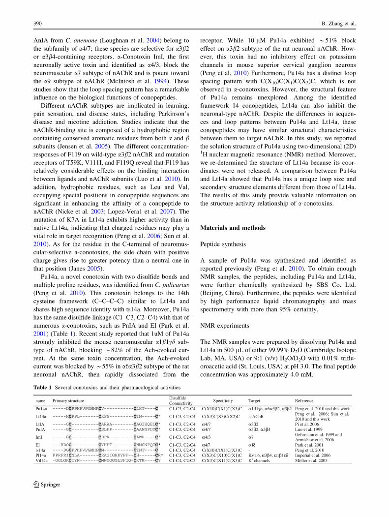
Figure 1 shows a representative portion of the Pu14a
TOCSY spectrum in H2O. Thirteen expected cross peaks
between the amide proton and Ha were observed. Other spin
systems were found in the fingerprint region of the TOCSY
spectrum, and their assignments were verified in the fin-
gerprint region of the DQF-COSY spectrum. The sequential
assignments of amino acids in the primary sequence started
with the unique residues G9, L16, and T18. A NOESY walk
identified the residues from P4 to G9 toward the N-terminus
and the residues from G9 to C19 toward the C-terminus.
Owing to the missing amide proton of D1 and C2, the first
three residues at the N-terminal were finally assigned based
on the three spin systems, the cross peak between the Ha of
C2 and Hb of C15, and the cross peak between the Ha of C2
and Hd of P3. The amide proton of D1 disappeared in the
H2O spectrum possibly because of its special position at the
N-terminal and fast exchange in water.
Twelve of the 13 spin systems were found in the fin-
gerprint region of the 120 ms Lt14a TOCSY spectrum. The
Fig. 1 HN(x-axis)-aliphatic(y-axis) region of the H-H TOCSY spec-
trum for Pu14a at 298 K, pH 3.0
Solution structure of a novel a-conotoxin 391
123

sequential assignments of amino acids in the primary
sequence started with the unique residues L5, K7, and T11.
Hence, NOE sequential walks along the N-terminus were
identified. P8 was assigned by its spin system and the NOE
from its d-proton to the a-proton of G7. The methionine
residue at position 1 was finally assigned according to the
NOEs from the a- and b-protons of Met1 to the amide
proton of C2. NOE connectivities from T11 to the C-ter-
minus were also identified. In addition, a minor component
of this synthesized conotoxin with a proportion of 15% was
observed (see supporting material Fig. 1S).
The NOESY data acquired for the two conopeptides
showed a number of NOEs, suggesting that the structures
of the peptides were sufficiently restrained for distance-
geometry calculation. All Pu14a chemical shifts are listed
in Table 2. The final chemical shifts of Lt14a (data not
shown) were basically similar to the chemical shifts
reported recently (Sun et al. 2010).
Structure calculation and evaluation
The constraints for determining Pu14a structure were
obtained from a survey of NMR data. A total of 179 dis-
tance constraints were used and the NOE root mean square
violation was no more than 0.2 A. The major NOE con-
nectivity is shown in Fig. 2. Moreover, five u angle con-
straints and two disulfide bond constraints from C2 to C15
and C13 to C19 for Pu14a were input for the molecular
modeling protocol of the Cyana algorithm. Although the
H-bond constraints were not found by H–D exchange
experiments, existing constraints were enough for the
structural calculation of such a small-sized peptide. As for
Lt14a, 134 distance constraints, 4 dihedral restraints, 2
H-bond restraints from H–D exchange experiments, and 2
disulfide bonds from C2 to C10 and C6 to C13 were used to
build up the structures. The structure of Lt14a was calcu-
lated using the data from a major conformer, and a 15%
minor component was not considered at this stage.
First, we computed 20 solution structures to evaluate the
folding of the peptidic chain using medium-distance con-
straints that have more than two bond intervals |i–j| [ 2.
Subsequently, all distance constraints and dihedral
restraints were used. Then, H-bond constraints were
introduced into the calculations. The simulated annealing
calculations were started with 100 random structures.
Finally, the ensemble of the 20 best resulting models with
the lowest residual target function and the minimum root
mean square deviation (RMSD) were selected. The
resulting conformers contained no significant violation of
any constraint. The Ramachandran plots were chosen to
represent the 3D folding of Pu14a and Lt14a in a solution
at pH 3.0. A summary of statistics for the converged
structures evaluated in terms of structural parameters are
listed in Table 3. The NMR constraints and obtained
coordinates files were submitted to the BMRB database
with ID code 21015 for Pu14a and 21014 for Lt14a.
Solution structure of Pu14a
An overlay of the backbone atoms for the 20 structures of
Pu14a is shown in Fig. 3a. The overall average backbone
RMSD reported for the final 20 structures of Pu14a was
0.77 ± 0.26 A. The N-terminal of these structures poorly
converged because of the few constraints at this region.
The refined structure of conotoxin Pu14a contains a larger
loop between C2 and C13, rather than a x-type twist in the
Table 2 Proton resonance assignments (ppm) for Pu14a
Residue HN a b Other
Asp1 4.27 2.68, 2.81
Cys2 5.00 3.20, 2.91
Pro3 4.73 2.34 c: 1.93, 2.00; d: 3.84, 3.67
Pro4 4.27 2.20 c: 1.69; d: 3.73
His5 7.79 5.02 3.09, 3.19 d: 7.24; e: 8.46
Pro6 4.42 2.21,1.90 c: 1.96, 2.03; d: 3.60, 3.73
Val7 8.56 4.52 2.14 c: 1.00
Pro8 4.34 2.34,1.91 c: 2.00, 2.12; d: 3.72,3.93
Gly9 8.65 3.73, 4.14
Met10 7.86 4.45 2.00 c: 2.45, 2.51; e: 2.19
His11 7.99 4.61 3.23, 3.33 d: 7.24; e: 8.29
Lys12 8.54 4.22 1.87 c: 1.46 d: 1.70; e: 3.00
Cys13 8.91 4.46 3.14, 3.22
Val14 7.99 3.94 2.20 c: 1.00, 1.07
Cys15 8.56 4.70 3.13, 3.33
Leu16 7.85 4.30 1.82 c: 1.66; d: 0.86
Lys17 8.04 4.21 2.08, 2.01 c: 1.44; d: 1.72; e: 3.04
Thr18 8.09 4.52 4.46 c: 1.12
Cys19 7.83 4.44 3.02, 3.45
Fig. 2 Summary of the sequential NOE connectivities involving the
NH, Ha, and Hb protons measured at 293 K and mixing time of
350 ms for Pu14a
392 B. Zhang et al.
123
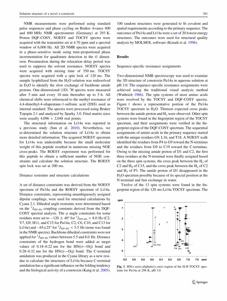
center of typical a-conotoxins, such as PnIA and EI.
Although three intercysteine loops of Pu14a could be
described by the sequence, only one real loop existed
between C2 and C13. The real loop corresponded to the
intercysteine loop 1 containing 10 residues. Interestingly,
in the so-called intercysteine loop 2 (with one residue V14)
and loop 3 (with 3 residues L16, K17 and T18) regions, the
secondary structural elements of 310-helix and turns were
obtained (Fig. 4a). Inspection of NOE constraints revealed
that the NOE connections for V14Ha-K17HN (i–i ? 3),
V14Ha-L16HN (i–i ? 2) and C13Ha-L16Hb (i–i ? 3)
were observed in Pu14a. The folding of residues C13 to
L16 tended to form a 310-helix, though no H-bond con-
straints were added in the calculation process. On the other
hand, the side chains of most residues, except P3 and V14,
were oriented outside (Fig. 3b).
Comparison of Pu14a with other a-conotoxins
The structural feature of Pu14a was quite different from
those of typical a-conotoxins, such as PnIA. A comparison
of the backbone structures of Pu14a, Lt14a, ImI, and PnIA
is shown in Fig. 4. The illustrated structure of Lt14a was
based on our determination in the present study. The
average backbone RMSD reported for the final 20 struc-
tures of Lt14a was 0.46 ± 0.18 A with 2 H-bond con-
straints and 0.32 ± 0.19 A without H-bond constraints,
besides other constraints used. It indicated that the struc-
tures of Lt14a were well resolved. Except for the N-ter-
minal residue M1, the refined structure of Lt14a contained
two turns similar to that reported in a previous study (Sun
et al. 2010). Moreover, the loop region found in Lt14a was
smaller than that in Pu14a.
As shown in Fig. 4, the backbone structure of Pu14a was
distinct from that of PnIA, although they both inhibited the
a3b2 subtype of nAChR. Interestingly, the backbone
structure of Lt14a exhibited remarkable resemblance to
that of PnIA despite their distinct secondary structure ele-
ments. Albeit in the same cysteine framework of 14, both
Pu14a and Lt14a are the ligands of different subtypes of
nAChR, and their conformational profiles are dissimilar.
Table 3 Structural statistics for 20 structures of Pu14a and Lt14a
Assigned NOE cross peaks Pu14a Lt14a
Intra-residue 112 68
Sequential 49 21
Medium range 14 6
Long range 4 1
H bond constraints 0 2
Dihedral constraints 5 4
RMSD to mean coordinates
Mean global backbone atoms RMSD 0.77 ± 0.26 0.46 ± 0.18
Mean global heavy atoms RMSD 1.11 ± 0.24 0.69 ± 0.24
Rachandran statistics from
PROCHECK_NMR
Most favored regions (%) 53.3 36.4
Additional allowed regions (%) 44.6 54.3
Generously allowed regions (%) 0 9.3
Disallowed regions (%) 2.1 0.0
Fig. 3 a Overlay of the
backbone atoms for the 20
converged structures of
conotoxin Pu14a. The
N-terminal D1 is poorly
resolved. b Inward side chain
orientation of P3 and V14 in
Pu14a
Fig. 4 Mean structures of conotoxins illustrated in tube mode.
a Pu14a, b Lt14a, c ImI (PDB ID: 1NCL) and d PnIA (PDB ID:
1PEN). The disulfide bond is shown in stick. Underlying secondary
structure features were analyzed by MOLMOL. The images are
generated by ViewerLite software
Solution structure of a novel a-conotoxin 393
123
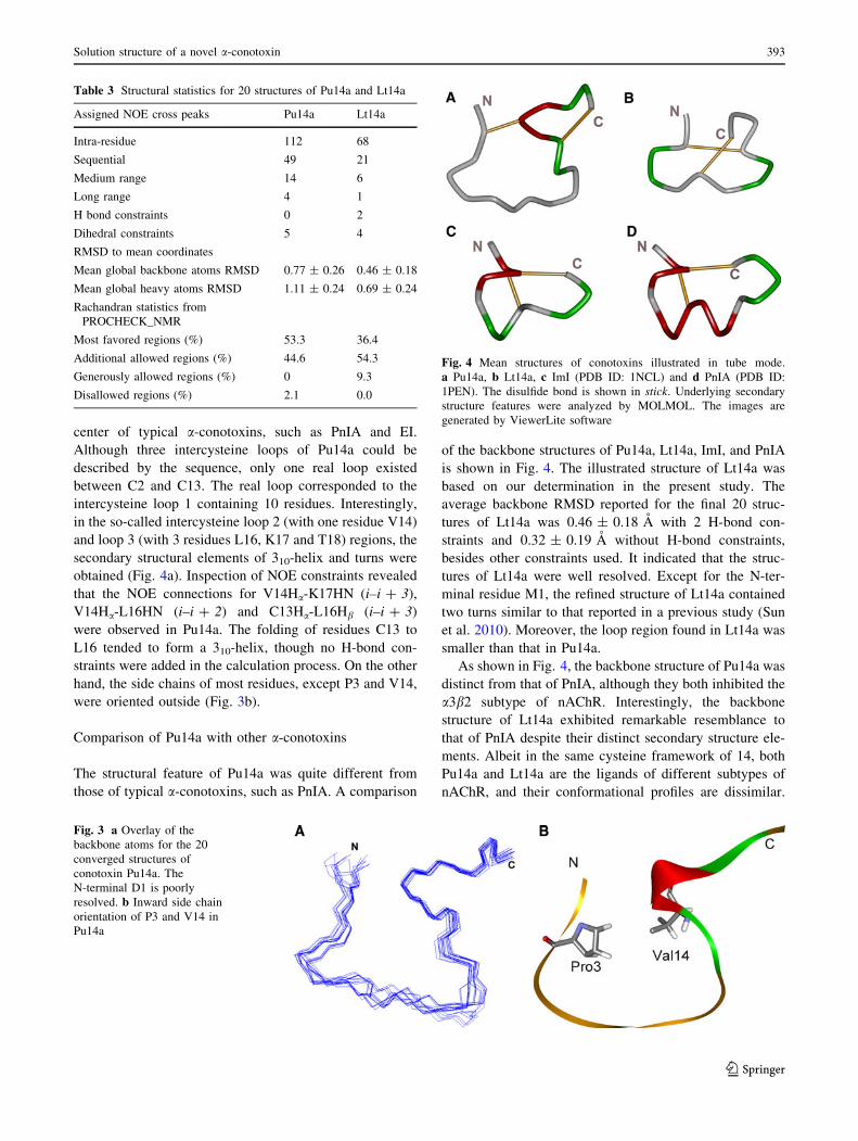
Pu14a has a relatively larger volume than Lt14a (see sup-
porting material Fig. 2S). Furthermore, the loop of Pu14a
was large enough to form a pore in the loop center, which
can be displayed by the structural surfaces from the front
and back views (Fig. 5).
Discussion
Distinct loop configuration of Pu14a
The present study describes the solution structure of a
novel a-conotoxin Pu14a and compares its structure with
that of another framework 14 conotoxin (Lt14a) and other
a-conotoxins. a-Conotoxins, the largest pharmacological
family in conotoxins, are extensively investigated. Most
a-conotoxins are classified into different subfamilies as
previously mentioned. Nevertheless, the loop spacing pat-
tern of Pu14a is unique from that of other a-conotoxins.
The loop pattern is C(X10)C(X1)C(X3)C for Pu14a,
whereas it is C(X3)C(X3)C(X2)C for Lt14a. Hence, the
distinct loop spacing pattern possibly results in various
backbone foldings and their corresponding effects on the
biological functions of a-conotoxins. In addition, without a
typical x-type twist in the loop center, Pu14a also inhibits
a3b2-containing receptors such as PnIA. The relatively
large loop 1 that existed in Pu14a was determined based on
all of the constraints from the NMR spectra; moreover, the
refined structure was credible because long-range distance
constraints were used (see supporting material Table 1S).
This result indicates that Pu14a may have a rather flexible
conformation in solution, and it may be transformed when
targeting nAChR.
Tran X-P peptide bonds in Pu14a
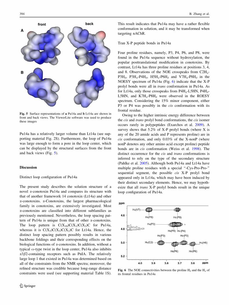
Four proline residues, namely, P3, P4, P6, and P8, were
found in the Pu14a sequence without hydroxylation, the
popular posttranslational modification in conotoxins. By
contrast, Lt14a has three proline residues at positions 3, 4,
and 8. Observations of the NOE crosspeaks from C2Ha-
P3Hd, P3Ha-P4Hd, H5Ha-P6Hd and V7Ha-P8Hd in the
NOESY spectrum of Pu14a (Fig. 6) indicate that the X-P
prolyl bonds were all in trans conformation in Pu14a. As
for Lt14a, only those crosspeaks from P4Ha-L5HN, P4Hd-
L5HN, and K7Ha-P8Hd were observed in the ROESY
spectrum. Considering the 15% minor component, either
P3 or P4 was possibly in the cis conformation with its
frontal residue.
Owing to the higher intrinsic energy difference between
the cis and trans prolyl bond conformations, the cis isomer
occurs rarely in polypeptides (Exarchos et al. 2009). A
survey shows that 5.2% of X-P prolyl bonds (where X is
any of the 20 amide acids and P represents proline) are in
cis conformation, and only 0.03% of the X-nonP (where
nonP denotes any other amino acid except proline) peptide
bonds are in cis conformation (Weiss et al. 1998). The
distinct occurrence for the cis and trans conformations is
inferred to rely on the type of the secondary structure
(Pahlke et al. 2005). Although both Pu14a and Lt14a have
multiple proline residues with a special ‘‘-Cys-Pro-Pro-’’
sequential segment, the possible cis X-P prolyl bond
appeared only in Lt14a, which may have been induced by
their distinct secondary elements. Hence, we may hypoth-
esize that all trans X-P prolyl bonds result in the unique
loop configuration of Pu14a.
Fig. 5 Surface representations of a Pu14a and b Lt14a are shown in
front and back views. The ViewerLite software was used to produce
these images
Fig. 6 The NOE connectivities between the proline Hd and the Ha of
its frontal residues in Pu14a
394 B. Zhang et al.
123
Structure-activity relationship of Pu14a
The structure of Pu14a is characterized by a large loop and
a distinct 310-helix. Pu14a can inhibit the mouse neuro-
muscular a1b1cd subtype, the rat neuronal a6a3b2 and
a3b2 subtypes of nAChR. Interestingly, Pl14a, another
framework 14 conotoxin with the structural feature of an
a-helix and two 310-helices, can inhibit the Kv1.6, neuronal
a3b4, and neuromuscular a1b1ed subtypes of nAChR
(Imperial et al. 2006). The 310-helix is also observed in one
of the most studied a-conotoxin, ImI, which exhibits full
bioactivity on the a7 subtype of nAChR (Gehrmann et al.
1999). Pu14a, Lt14a, Pl14a, and ImI have the same disul-
fide linkages with C1-C3 and C2-C4 connectivities;
nevertheless, the differences among the aforementioned
conopeptides in terms of loop pattern and secondary
structure elements reflect the relationship between their
structures and bioactivities. The side chains of most resi-
dues in Pu14a, except P3 and V14, were oriented outside
(Fig. 3b). However, all of the side chains in Lt14a were
generally oriented outside. The inside orientation of non-
polar residues, including P3 and V14 in Pu14a, may
improve the hydrophobic interaction between ligands and
nAChR.
Recently, Luo’s work elucidated an energetically favor-
able hydrophobic interaction between Lt1a-L15 and
b2-F119 (Luo et al. 2010). Due to the block effect on a3b2
for Pu14a, L16 was proposed to contribute to the energy
minimization of the interaction and further stabilize the
conformation of Pu14a-a3b2. Based on Pu14a sequence,
K17, a positively charged residue in the C-terminal region of
a-conotoxin, gave rise to greater neuromuscular-selectivity
than a neutral residue, as reported by Janes (Janes 2005).
Another framework 14 conopeptide, vil14a, has a
Lys/Tyr dyad separated by approximately 6 A, which is a
conserved structural feature in K channel blockers (Moller
et al. 2005). The pl14a sequence shows contiguous Lys/Tyr
dyad. The Lys/X motif of pl14a is also distinct from that of
vil14a. A Lys/X motif has a significant effect on the for-
mation of H-bond between conotoxins and their targets.
The great difference between Pu14a and vil14a in terms of
their bioactivities may be related to their sequences, sec-
ondary structures, and Lys/X motifs.
In summary, the solution structure of Pu14a is distinct
from other a-conotoxins because of its special loop spacing
pattern. Compared with Lt14a, Pu14a has a larger volume
and an additional proline residue that correlates with var-
ious secondary structural elements and X-P bond confor-
mations. Pu14a conformation reflects the structural
diversity and flexibility in a-conotoxins. These preliminary
studies will help to understand the structure and activity
relationships among different kinds of a-conotoxins. The
distinctive loop spacing pattern and proline-rich sequence
will be very beneficial for chemical modification of cono-
toxins to improve their stability and bioactivity, further for
the design and development of potential peptide drugs.
Acknowledgments We thank Prof. Chunguang Wang and Prof.
Chengwu Chi from Tongji University for supplying the sample of
Pu14a friendly. This work was supported by the National Basic
Research Program (No. 2011CB808503), and the Fundamental
Research Funds for the Central Universities and the Research Funds
of Renmin University of China (No. 10XNJ011).
Conflict of interest None.
References
Armishaw CJ, Daly NL, Nevin ST, Adams DJ, Craik DJ, Alewood PF
(2006) a-Selenoconotoxins, a new class of potent a7 neuronal
nicotinic receptor antagonists. J Biol Chem 281:14136–14143
Craik DJ, Adams DJ (2007) Chemical modification of conotoxins to
improve stability and activity. ACS Chem Biol 2:457–468
Exarchos KP, Exarchos TP, Papaloukas C, Troganis AN, Fotiadis DI
(2009) Detection of discriminative sequence patterns in the
neighborhood of proline cis peptide bonds and their functional
annotation. BMC Bioinformatics 10:113
Gehrmann J, Daly NL, Alewood PF, Craik DJ (1999) Solution
structure of a-Conotoxin ImI by 1H nuclearmagnetic resonance.
J Med Chem 42:2364–2372
Gray WR, Luque A, Olivera BM, Barrett J, Cruz LJ (1981) Peptide
toxins from Conus geographus venom. J Biol Chem 256:
4734–4740
Hu SH, Gehrman J, Guddat LW, Alewood PF, Craik DJ, Martin JL
(1996) The 1.1 A crystal structure of the neuronal acetylcholine
receptor antagonist, a-conotoxin PnIA from Conus pennaceus.
Structure 4:417–423
Imperial JS, Bansal PS, Alewood PF, Daly NL, Craik DJ, Sporning A,
Terlau H, Lopez-Vera E, Bandyopadhyay PK, Olivera BM
(2006) A novel conotoxin inhibitor of Kv1.6 channel and
nAChR subtypes defines a new superfamily of conotoxins.
Biochemistry 45:331–8340
Janes RW (2005) a-Conotoxins as selective probes for nicotinic
acetylcholine receptor subclasses. Curr Opin Pharmacol
5:280–292
Jensen AA, Frølund B, Liljefors T, Krogsgaard-Larsen P (2005)
Neuronal nicotinic acetylcholine receptors: structural revela-
tions, target identifications, and therapeutic inspirations. J Med
Chem 48:4705–4745
Kaas Q, Westermann JC, Craik DJ (2010) Conopeptide characteriza-
tion and classifications: an analysis using ConoServer. Toxicon
55:1491–1509
Kaas Q, Westermann JC, Halai R, Wang CK, Craik DJ (2008)
Conoserver, a database for conopeptide sequences and struc-
tures. Bioinformatics 24:445–446
Kang TS, Vivekanandan S, Jois SDS, Kini RM (2005) Effect of
C-terminal amidation on folding and disulfide-pairing of
a-conotoxin ImI. Angew Chem Int Ed 117:6491–6495
Koradi R, Billeter M, Wuthrich K (1996) MOLMOL: a program for
display and analysis of macromolecular structures. J Mol
Graph 14(51–55):29–32
Lopez-Vera1 E, Aguilar MB, Schiavon E, Marinzi C, Ortiz E,
Cassulini RR, Batista CVF, Possani LD, de la Cotera EPH, Peri
F, Becerril B, Wanke E (2007) Novel a-conotoxins from conus
Solution structure of a novel a-conotoxin 395
123
spurius and thea-conotoxin EI share high-affinity potentiation
and low-affinity inhibition of nicotinic acetylcholine receptors.
FEBS J 274:3972–3985
Luo SL, Akondi KB, Zhangsun D, Wu Y, Zhu XP, Hu YY,
Christensen S, Dowell C, Daly NL, Craik DJ, Wang CA, Lewis
RJ, Alewood PF, McIntosh JM (2010) A typical a-conotoxin
ltIA from conus litteratus targets a novel microsite of the a3b2
nicotinic receptor. J Biol Chem 285:12355–12366
Luo SL, Nguyen TA, Cartier GE, Olivera BM, Yoshikami D,
McIntosh JM (1999) Single-residue alteration in alpha-conotoxin
PnIA switches its nAChR subtype selectivity. Biochemistry
38:14542–14548
Loughnan ML, Nicke A, Jones A, Adams DJ, Alewood PF, Lewis RJ
(2004) Chemical and functional identification and characteriza-
tion of novel sulfated a-conotoxins from the cone snail Conusanemone. J Med Chem 47:1234–1241
McIntosh JM, Corpuz GO, Layer RT, Garrett JE, Wagstaff JD, Bulaj
G, Vyazovkina A, Yoshikami D, Cruz LJ, Olivera BM (2000)
Isolation and characterization of a novel Conus peptide with
apparent antinociceptive activity. J Biol Chem 275:32391–32397
McIntosh JM, Cruz LJ, Hunkapiller MW, Gray WR, Olivera BM
(1982) Isolation and structure of a peptide toxin from the marine
snail Conusmagus. Arch Biochem Biophys 5:280–292
McIntosh JM, Yoshikami D, Mahe E, Nielsen DB, Rivier JE, Gray
WR, Olivera BM (1994) A nicotinic acetylcholine receptor
ligand of unique specificity, alpha- conotoxin ImI. J Biol Chem
269:16733–16739
Millard EL, Daly NL, Craik DJ (2004) Structure-activity relationships
and a-conotoxins targeting neuronal nicotinic acetylcholine
receptors. Eur J Biochem 271:2320–2326
Moller C, Rahmankhah S, Lauer-Fields J, Bubis J, Fields GB, Marı F
(2005) A novel conotoxin framework with a helix loop helix (Cs
a/a) fold. Biochemistry 44:15986–15996
Nicke A, Loughnan ML, Millard EL, Alewood PF, Adams DJ, Daly
NL, Craik DJ, Lewis RJ (2003) Isolation, structure, and activity
of GID, a novel a4/7-conotoxin with an extended N-terminal
sequence. J Biol Chem 278:3137–3144
Olivera BM, Cruz LJ (2001) Conotoxins, inretrospect. Toxicon
39:7–14
Olivera BM, Gray WR, Zeikus R, McIntosh JM, Varga J, Rivier J, de
Santos V, Cruz LJ (1985) Peptide neurotoxins from fish-hunting
cone snails. Science 230:1338–1343
Oliveral BM, Quik M, Vincler M, Mclntosh JM (2008) Subtype-
selective conopeptides targeted to nicotinic receptors: concerted
discovery and biomedical applications. Channels 2:143–152
Pahlke D, Freund C, Leitner D, Labudde D (2005) Statistically
significant dependence of the Xaa-Pro peptide bond conforma-
tion on secondary structure and amino acid sequence. BMC
Struct Biol 5:8
Park KH, Suk JE, Jacobsen R, Gray WR, McIntosh JM, Han KH
(2001) Solution conformation of a-conotoxin EI, a neuromus-
cular toxin specific for the a1/d subunit interface of Torpedonicotinic acetylcholine receptor. J Biol Chem 276:49028–49033
Peng C, Tang SJ, Pi CH, Liu JL, Wang F, Wang L, Zhou WL, Xu AL
(2006) Discovery of a novel class of conotoxin from Conus
litteratus, Lt14a, with a unique cysteine pattern. Peptides
27:2174–2181
Peng C, Ye MY, Wang YF, Shao XX, Yuan DD, Liu J, Hawrot E,
Wang CG, Chi CW (2010) A new subfamily of conotoxins
belonging to the A-superfamily. Peptides 31:2009–2016
Pi CH, Liu JL, Peng C, Liu Y, Jiang XH, Zhao Y, Tang SJ, Wang L,
Dong ML, Chen SW, Xu AL (2006) Diversity and evolution of
conotoxins based on gene expression profiling of Conuslitteratus. Genomics 88:809–819
Sun DD, Ren ZH, Zeng XY, You YW, Pan WG, Zhou MJ, Wang L,
Xu AL (2010) Structure–function relationship of conotoxin
Lt14a, a potential analgesic with low cytotoxicity. Peptides
32:300–305
Terlau H, Olivera BM (2004) Conus venoms: a rich source of novel
ion channel targeted peptides. Physiol Rev 84:41–68
Weiss MS, Jabs A, Hilgenfeld R (1998) Peptide bonds revisited. Nat
Struct Biol 5:676
Wuthrich K (1986) NMR of proteins and nucleic acids. Wiley, New
York
396 B. Zhang et al.
123