solution nmr study of the interaction between ntf2 and nucleoporin fxfg repeats
TRANSCRIPT

Solution NMR Study of the Interaction Between NTF2and Nucleoporin FxFG Repeats
Jonathan Morrison, Ji-Chun Yang, Murray Stewart andDavid Neuhaus*
MRC Laboratory of MolecularBiology, Hills Road, CambridgeCB2 2QH, UK
Interactions with nucleoporins containing FxFG repeat cores are crucialfor the nuclear import of RanGDP mediated by nuclear transport factor 2(NTF2). We describe here a solution NMR-based study that identifiesprimary and secondary FxFG-binding sites on NTF2 and accounts for arange of observations on the rate of NTF2 nuclear trafficking. We usedthree complementary NMR methods, namely amide group chemical shifttitrations, NOE and cross-saturation measurements, to show that themajor FxFG-binding site on the dimeric rat NTF2 (rNTF2) molecule iscentred on Trp7 and is formed by residues from both NTF2 chains. A sec-ondary FxFG-binding site is located at the rNTF2 hydrophobic cavity andthese two sites, together with a surface hydrophobic cluster centred onTrp112, merge into an elongated hydrophobic stripe on the rNTF2 surface.The primary site centred on Trp7 is lost in the rNTF2-W7A mutant thathas been shown to bind FxFG nucleoporins with greatly reduced affinity,whereas the secondary site at the rNTF2 hydrophobic cavity is retained.The interface between NTF2 and FxFG nucleoporins detected by NMR ismore extensive than that detected by X-ray crystallography, and the pre-sence of a secondary site at the NTF2 hydrophobic cavity accounts forthe unexpectedly rapid nuclear import of rNTF2-W7R recently observedby others. The structure of the binding interfaces on these transport fac-tors provides a rationale for the specificity of their interactions withnucleoporins that, combined with their weak binding constants, facilitatesrapid translocation through NPCs during nuclear trafficking.
q 2003 Elsevier Ltd. All rights reserved.
Keywords: nuclear trafficking/NTF2/FxFG repeats; protein–proteininteraction; nuclear magnetic resonance; intermolecular NOE; cross-saturation*Corresponding author
Introduction
Macromolecules traffic between the nuclear andcytoplasmic compartments through nuclear porecomplexes (NPCs), huge proteinaceous structuresthat perforate the nuclear envelope (reviewed by
Weis,1 Kuersten,2 Bayliss3 and Stewart4). Traffickingis mediated by shuttling carrier molecules thatbind their cargo in one compartment and release itin the other. For example, the importin-a/bcomplex mediates nuclear import of proteins con-taining a classical nuclear localization sequence(NLS). Once the cargo-carrier complex has beentranslocated through the NPC to the nucleus,RanGTP binds to importin-b and dissociates thecomplex to release the cargo. Importin-b isrecycled to the cytoplasm complexed withRanGTP, where RanGAP promotes hydrolysis ofthe GTP bound to Ran, dissociating the importin-Ran complex and thus freeing importin-b foranother round of nuclear protein import (reviewedby Gorlich,5 Kuersten2 and Bayliss3). CytoplasmicRanGDP is re-imported and recharged with GTP
0022-2836/$ - see front matter q 2003 Elsevier Ltd. All rights reserved.
Supplementary data associated with this article can befound at doi: 10.1016/j.jmb.2003.08.050
E-mail address of the corresponding author:[email protected]
Abbreviations used: NTF2, nuclear transport factor 2;yNTF2, Saccharomyces cerevisiae NTF2; rNTF2, rat NTF2(human has identical amino acid sequence); NPC,nuclear pore complex; NOE, nuclear Overhauser effect;NOESY, NOE spectroscopy; HSQC, heteronuclear singlequantum coherence; TOCSY, total correlatedspectroscopy.
doi:10.1016/j.jmb.2003.08.050 J. Mol. Biol. (2003) 333, 587–603

by the nuclear exchange factor RCC1. The importof RanGDP is mediated by NTF2,6,7 which providesa simple and specialized system in which to studythe molecular principles underlying translocation(reviewed by Stewart8 and Ribbeck9).
NPCs are constructed from multiple copies ofproteins termed collectively nucleoporins.10,11 Bothyeast and vertebrate NPCs contain approximately30 different nucleoporins10,12 and many havesequences containing large regions of characteristicFG tandem repeats based on hydrophobic Phe-richcores separated by hydrophilic linkers.11,13 Thehydrophobic cores of FG repeats are highly con-served and contain one or two Phe residues. Themost common repeat core sequence motifs arebased on FG, GLFG or FxFG (where x is usually asmall residue such as Ser, Gly or Ala). By contrast,the hydrophilic spacers that link successive coresare highly variable both in length and sequence,although they are generally rich in charged andpolar side-chains and have few hydrophobic resi-dues. Nucleoporins frequently have domains thatcontain many copies of these repeats. For example,there are 19 FxFG repeats in Nsp1p and 33 GLFGrepeats in Nup116.13
Although the precise mechanism by whichcargo-carrier complexes move through NPCsremains controversial, there is an emergingconsensus that interactions between carriers andFG-nucleoporins are involved directly.3,4,9,11,14 – 16
Many carrier molecules bind FG-nucleo-porins.11,14,17,18 Different carriers appear to bindspecific nucleoporins and/or classes of nucleo-porin preferentially.14,18 For example, importin-b,its yeast homologue Kap95, and the mRNA exportfactor, TAP, bind both FxFG and GLFGnucleoporins,14,16,17,19,20 whereas NTF2 binds pri-marily FxFG nucleoporins.21 – 23 Mutants of NTF2or importin-b in which the strength of FxFG-repeatbinding is reduced exhibit a correspondinglyreduced rate of nuclear import.9,15,16,19,24,25
NTF2 has been a powerful model system inwhich to study the molecular basis of translocationthrough NPCs on account both of its simplicity andspecialization,8,9 as well as the availability of crystalstructures of the molecule itself26 and its complexeswith RanGDP27 and FxFG peptides.15 The interactionbetween NTF2 and FxFG-nucleoporins, which iscrucial to mediate the nuclear import of RanGDP,15,24
has been studied using both the vertebrate (rNTF2)and Saccharomyces cerevisiae (yNTF2) proteins. Theyeast and rat proteins share 40% sequence identityand rNTF2 can functionally replace the essentialyNTF2 protein.28 Previous studies have identified asurface hydrophobic patch, centred on Trp7 onrNTF2, that interacts with FxFG repeats.15,24,25 Site-directed mutagenesis in combination with protein–protein interaction studies and functional assaysboth in vivo and in vitro have established the import-ance of this hydrophobic patch in the interactionwith FxFG nucleoporins.9,15,24,25 However, althoughthe crystal structure of the yNTF2–FxFG complexshowed the general position of the FxFG-binding
site, the electron density due to the peptide wasvery weak and, because of its high temperaturefactor, it was difficult to define the interaction sitecompletely. This problem was compounded by asubstantial level of disorder in the N terminus of theyNTF2 chains in these crystals, so that the role ofthe residues in this region (where, for examplePhe5, the yeast equivalent of Trp7 in rNTF2, waslocated) was difficult to evaluate. Thus, althoughthese studies have established a number of keyresidues in the NTF2 nucleoporin-binding site, thecomplete structural basis for the interaction has notbeen defined. Moreover, Ribbeck & Gorlich9 foundthat wild-type rNTF2 passed through NPCs 125times faster than Green fluorescent protein, amolecule of similar size with no known role innuclear trafficking, and suggested that additionalareas of the surface of rNTF2 have hydrophobic,translocation-promoting properties. Although theyshowed that Trp7 contributed to translocation, it didnot appear to be the sole determinant, because theW7R mutant of rNTF2 still passed through NPCs 30times faster than Green fluorescent protein. Consist-ent with this observation, mutation of the corre-sponding residue, Phe5, in yNTF2 has only a smalleffect on the in vivo function of yNTF2.25 Moreover,mutations that alter NTF2–nucleoporin interactionshave been described that do not lie near or withinthe hydrophobic patch. These include the dominantnegative mutants D23A29 and N77Y,25 both of whichblock nuclear transport when overexpressed. Thus,it appears likely that, although Trp7 is a componentof the rNTF2 FxFG-binding site, the site itself ismore extensive.
To address the nature of the NTF2–FxFG inter-action in more detail, we employed three comp-lementary NMR approaches (amide groupchemical shift titrations, nuclear Overhauser effect(NOE) and cross-saturation measurements) tostudy the interaction between rNTF2 and an FxFGpeptide in solution. We have thus shown that theFxFG-binding interface on NTF2 is indeed moreextensive than that indicated by X-raycrystallography15 and mutagenesis,24,25 and haveidentified two FxFG-binding sites on rNTF2: ahigher-affinity site, containing Trp7 that corre-sponds to the site identified in crystals and bymutagenesis; and a lower-affinity site located atthe hydrophobic cavity to which Ran Phe72 binds.These sites have enabled us to evaluate thedetailed manner in which FxFG nucleoporin coresinteract with this nuclear transport factor and forits unusual behaviour in nuclear protein importassays.
Results and Discussion
Titration of Nsp1-P30 against [15N]rNTF2 and[15N]W7A-rNTF2
We initially used chemical shift changes during atitration against a nucleoporin-derived peptide to
588 NTF2-FxFG Interaction

identify the rNTF2 residues involved in the bind-ing of FxFG nucleoporin cores. Titration of anunlabelled ligand into a solution of a 15N-labelledprotein generally causes chemical shift changes inthe 15N and 1H resonances of backbone amidegroups of the protein close to the ligand-bindingsite, which can be followed by recording a seriesof (15N,1H) heteronuclear single quantum coher-ence (HSQC) spectra for samples with increasingligand to protein ratios.30 Although the detailedorigins of these chemical shift changes at the bind-ing site are almost never analyzed, they are likelyto include small changes in the local protein back-bone conformation and hydrogen-bonding patternwithin the immediate vicinity of the binding site,changes due to small movements of nearbyaromatic side-chains on the protein, proximity ofaromatic moieties on the ligand and, to a lesserextent, proximity of charges on the ligand.31,32,33
However, if there are allosteric changes in the pro-tein conformation upon ligand binding, these maycause additional chemical shift changes in reson-ances due to amide groups distant from theligand-binding site, which cannot rigorously bedistinguished a priori from primary effects.
The peptide we used (referred to here as Nsp1-
P30) was a 30 residue synthetic peptide derivedfrom the sequence of FG-nucleoporin Nsp1p andcontaining two FxFG cores. When this was titratedinto a solution of 15N-labelled rNTF2, backboneamide group chemical shift changes appeared pro-gressively for a number of residues as the concen-tration of peptide was increased. The chemicalshift changes were generally quite small, makingit relatively straightforward to follow movementsof individual peaks (see Figure 1). However, in par-ticularly crowded portions of the HSQC spectrum,one or two assignment ambiguities were unavoid-able, so that, for example, it was not readilypossible to determine reliably the chemical shiftchange for Gln47 in these spectra. The measuredchemical shift changes for 1H and 15N were com-bined into a composite chemical shift displacementvector DCS (see Materials and Methods):
DCS ¼ ððDd1HÞ2 þ ðDd15N=10Þ2Þ1=2
and this statistic plotted against the rNTF2sequence (Figure 2). Data for successive values ofthe peptide:protein ratio were then mapped ontothe molecular surface to indicate which parts ofthe rNTF2 surface are affected predominantly by
Figure 1. HSQC chemical shift titration of the Nsp1-P30 peptide against wild-type rat NTF2. The free rNTF2 proteinspectrum is shown in black, and overlaid on this are the spectra of a 0.5:1 mixture of peptide and rNTF2 (magenta; fewmagenta peaks are visible, as the shift changes relative to free protein are small), a 1:1 mixture of peptide and rNTF2(blue) and a 2:1 mixture of peptide and rNTF2 (red). Expansions of two regions are shown, with indicative signalassignments. See the text for further discussion.
NTF2-FxFG Interaction 589

Figure 2 (legend opposite)
590 NTF2-FxFG Interaction

interaction with the peptide (Figure 3). We chose0.01 ppm as the threshold for DCS values thatwould be represented in this Figure. About 20% ofthe residues show larger changes than this at the0.5:1 titration point, with an increasing proportionaffected at higher ratios; the overall pictureobtained using other ratios (e.g. in the range0.008–0.012) does not change significantly.
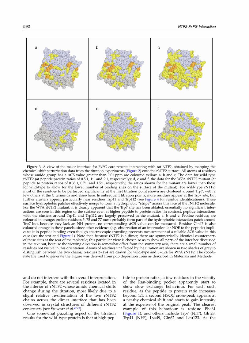
Data for early titration points (see Figure 3a)showed that, initially, the peptide binds principallyto a pronounced cluster of surface hydrophobicresidues centred on Trp7, consistent with earlierwork that implicated this residue as a key com-ponent of the FxFG nucleoporin binding site onrNTF2.24 Residues in this cluster that show shiftchanges early in the titration include Trp7, Glu8,Ala36, and Phe46. It is likely that three nearbyproline residues (Pro5, Pro75 and Pro77) contributeto this hydrophobic surface cluster, but anyinvolvement of these in the peptide interaction isnecessarily undetectable in the chemical shifttitration because proline residues lack an amideproton. In addition to the cluster of residuescentred on Trp7 that showed chemical shifts evenat low stoichiometry, a small number of chemicalshift changes started to become apparent elsewhereon the surface, most notably in the vicinity of theNTF2 hydrophobic cavity and the C terminus,both of which are implicated in bindingRanGDP,27,34 and in a surface hydrophobic regionon the opposite side of the Trp7 site, close toTrp112. However, the involvement of these otherresidues developed more substantially at laterpoints in the titration, suggesting that there wassome preference for the Trp7 site to be occupiedfirst, with other interactions developing morestrongly once a higher proportion of peptide waspresent (Figure 3b). At a peptide to protein ratioof 1:1, further chemical shift changes appeared inthe vicinity of the Ran-binding pocket, and aroundTrp112. These three regions effectively mergetogether to form a hydrophobic “stripe” acrossone face of the protein, indicating that the peptideinteraction interface in solution is probably signifi-cantly more extensive than that detected by X-raycrystallography of the complex of an Nsp1 peptidewith yeast NTF2.15 Other residues involved in the
vicinity of the Trp7 patch include Gly30 and Lys49(above the stripe, as shown in Figure 3), andAsn16, Tyr19 and Asp23 (below the stripe). By thetime the peptide to protein ratio reached 2:1, quitea large proportion of the protein surface hadstarted to show appreciable effects (Figure 3c),suggesting that now much of the interaction wasnon-specific. Figure 4 highlights some of the keyresidues in each of the three surface hydrophobicclusters referred to.
Previous biochemical studies demonstrated thatthe interaction between FxFG repeats and rNTF2was reduced dramatically by the W7A-rNTF2mutation.24 When the titration experiments wererepeated using W7A-rNTF2, it was immediatelyapparent that, as expected, the primary site centredon Trp7 had been ablated. In the case of W7A-rNTF2, chemical shift changes at early titrationpoints were concentrated mainly on the Ran-bind-ing pocket, with some other shifts appearing nearthe C terminus and Trp112 site (Figure 3d–f). It isnot surprising that these chemical shift changesoccur earlier during the titration of the W7Amutant than they did for the wild-type protein,since the Trp7 site is effectively non-existent inthis mutant, leaving just the secondary sites tointeract.
Both these titration experiments show clearlythat there are additional regions of the rNTF2 sur-face capable of interacting with the Nsp1 peptidebeyond those detected previously by eithercrystallography15 or mutagenesis techniques.24,25
Although it is not clear whether some of theseadditional chemical shift changes may have beendue to allosteric changes upon peptide binding tosecondary sites, it is extremely unlikely that theywere due to indirect effects of peptide binding tothe primary site centred on Trp7. This followsfrom the observation that the additional shifts inthe titration of wild-type rNTF2 appeared largelyafter the Trp7 site had already been occupied and,still more convincingly, from the observation thatan essentially identical set of shifts due to bindingat secondary sites appears in the W7A rNTF2titration, where effectively the site centred on Trp7has been ablated. Some allosteric effects areevident in the results, but these are quite limited
Figure 2. a, A histogram showing the value of the chemical shift displacement vector DCS ¼ ððDd1HÞ2 þðDd15N=10Þ2Þ1=2 as a function of the rNTF2 sequence and across the titration with Nsp1-P30 peptide against wild-typerNTF2. For each residue, the value of DCS is plotted cumulatively for three titration points 0.5:1 (black), 1:1 (grey)and 2:1 (open) (i.e. the height of each black bar represents DCS for that residue at a 1:0.5 peptide to protein ratio, thetotal height to the top of each grey bar represents DCS for that residue at a 1:1 peptide to protein ratio and the totalheight to the top of the open bar represents DCS for that residue at a 2:1 peptide to protein ratio). The thresholdvalue for DCS used as a criterion for significance (0.01 ) is marked using a red line. b, A similar histogram for titrationof the same peptide against the W7A rNTF2 mutant; here, the three titration points shown are 0.35:1 (black), 0.7:1(grey) and 1.5:1 (open). c, A histogram showing the intensity ratios observed in the cross-saturation experiment usinga 1:1 mixture of rNTF2 and Nsp1-P30, and a saturation time of 360 ms. The open bars correspond to the residueswhere significant effects (intensity ratio .0.1) were observed in the corresponding control experiment with the freeprotein, as described in the text. The threshold value for intensity ratio used as a criterion for significance (0.1) ismarked using a red line.
NTF2-FxFG Interaction 591
and do not interfere with the overall interpretation.For example, there are several residues located inthe interior of rNTF2 whose amide chemical shiftschange during the titration, most likely due to aslight relative re-orientation of the two rNTF2chains across the dimer interface that has beenobserved in crystal structures of different rNTF2constructs (see Stewart et al.27,35).
One somewhat puzzling aspect of the titrationresults for the wild-type protein is that at high pep-
tide to protein ratios, a few residues in the vicinityof the Ran-binding pocket apparently start toshow slow exchange behaviour. For each suchresidue, as the peptide to protein ratio increasesbeyond 1:1, a second HSQC cross-peak appears ata nearby chemical shift and starts to gain intensityat the expense of the original peak. The clearestexample of this behaviour is residue Phe61(Figure 1), and others include Trp7 (NHe), Gln28,Trp41 (NHe), Lys49, Gln62 and Leu123. As the
Figure 3. A view of the major interface for FxFG core repeats interacting with rat NTF2, obtained by mapping thechemical shift perturbation data from the titration experiments (Figure 2) onto the rNTF2 surface. All atoms of residueswhose amide group has a DCS value greater than 0.01 ppm are coloured yellow. a, b and c, The data for wild-typerNTF2 (at peptide:protein ratios of 0.5:1, 1:1 and 2:1, respectively); d, e and f, the data for the W7A rNTF2 mutant (atpeptide to protein ratios of 0.35:1, 0.7:1 and 1.5:1, respectively; the ratios shown for the mutant are lower than thosefor wild-type to allow for the lower number of binding sites on the surface of the mutant). For wild-type rNTF2,most of the residues to be perturbed significantly at the first titration point shown are clustered around Trp7, with afew others at the C terminus and elsewhere. In subsequent titration points, more residues appear at the Trp7 site, butfurther clusters appear, particularly near residues Trp41 and Trp112 (see Figure 4 for residue identifications). Thesesurface hydrophobic patches effectively merge to form a hydrophobic “stripe” across this face of the rNTF2 molecule.For the W7A rNTF2 mutant, it is clearly apparent that the Trp7 site has been ablated; essentially no significant inter-actions are seen in this region of the surface even at higher peptide to protein ratios. In contrast, peptide interactionswith the clusters around Trp41 and Trp112 are largely preserved in the mutant. a, b and c, Proline residues arecoloured in orange; proline residues 5, 75 and 77 most probably form part of the hydrophobic interaction patch aroundTrp7 but, because they lack an NH proton, no corresponding DCS value can be measured. Residue Gln47 is alsocoloured orange in these panels, since other evidence (e.g. observation of an intermolecular NOE to the peptide) impli-cates it in peptide binding even though spectroscopic crowding prevents measurement of a reliable DCS value in thiscase (see the text and Figure 1). Note that, because rNTF2 is a dimer, there are symmetrically identical counterpartsof these sites at the rear of the molecule; this particular view is chosen so as to show all parts of the interface discussedin the text but, because the viewing direction is somewhat offset from the symmetry axis, there are a small number ofresidues not visible in this orientation. Atoms of residues unaffected by the titration are shown in two shades of grey todistinguish between the two chains; residues 2–124 are drawn for wild-type and 5–124 for W7A rNTF2. The coordi-nate file used to generate the Figure was derived from pdb deposition 1oun as described in Materials and Methods.
592 NTF2-FxFG Interaction

chemical shifts involved are only a few tens of Hzapart, it is clear that any process responsible forsuch slow exchange must take place with a rateno faster than about 10 s21. While we do not havequantitative data on the kinetics of complex for-mation between NTF2 and the Nsp1-P30 peptide,it seems virtually certain that any process occur-ring at a rate of (less than) 10 s21 is a much slowerprocess than dissociation of the complex. We mustconclude therefore that any slow exchange processresponsible for these effects is associated with asubtle change in the structure of the protein itself(most probably in the immediate vicinity of theRan-binding pocket). Even though the lifetime ofthe complex may be short, the presence of the pep-tide could influence the position of such a slowequilibrium between two protein forms (forinstance by binding more strongly to one than tothe other), and so cause the observed behaviour.
Alternatively, it is possible that these resonancesare not actually in slow exchange at all. It hasbeen pointed out recently that, for a system in fastexchange where binding proceeds via a low popu-lation of one or more intermediate states, reson-ances may exhibit behaviour somewhat similar tothat expected for slow to intermediate exchangerates. This is because near the mid-point of thetitration intensity is distributed over severalrelatively weak resonances, rather than being con-centrated into a single peak at the average of thefree and bound states as expected for a simpletwo-state fast exchange.36 – 38 Either of theseexplanations would be consistent with the obser-vation that many of the other peaks in the HSQCspectrum show only steadily changing shifts
through the titration, i.e. fast exchange behaviour.In the case of the W7A mutant, no residue showsslow exchange behaviour in the titration withNsp1-P30.
The chemical shift changes seen across thesetitrations are, in general, rather small. This lendsfurther support to the argument that any allostericchanges in the conformation of rNTF2 upon pep-tide binding are themselves small. It suggests alsothat the peptide adopts a large number of locallydifferent interaction geometries across theensemble present in solution. Since the effectsresponsible for chemical shift perturbations aremainly very local in origin, it would be expectedthat even if the peptide always interacts at or nearthe same patch on the surface, variations of theconformation of the peptide across the ensembleof complexes present in solution would tend toaverage the chemical shift perturbations towardslow values. Ultimately, it is the clustering of mostof the observed chemical shift perturbations toform distinct patches on the surface of rNTF2 thatlends the most powerful support to their interpret-ation, in conjunction with the fact that a consistentinterpretation emerges from the combined use ofall three NMR techniques employed (see below).
Intermolecular NOEs define the nature of theinteraction at the Trp7 site in solution
We supplemented the 15N chemical shift data bymeasuring intermolecular NOEs associated withFxFG nucleoporin binding. Unlike chemical shiftchanges during a titration that generally definejust a relatively low-resolution interaction patch,intermolecular NOE enhancements report on indi-vidual interatomic contacts between hydrogenatoms on the ligand and on the protein. For anappreciable intermolecular NOE enhancement tobe observable, it is the instantaneous ensembleaveraged value of the corresponding interatomicdistance (actually the value kr26l21=6; where r is theinteratomic distance and the brackets representthe ensemble average) that must be short.39,40 Sinceit is an ensemble average rather than a time aver-age of the interatomic distance that controls thedevelopment of the NOE, there is effectively nolower limit (apart from the correlation time foroverall molecular tumbling) on the residence timeof the ligand for an intermolecular NOE enhance-ment to be observed, provided the concentrationof complex is high enough. For instance, the firstintermolecular NOE enhancement ever reportedwas a strong positive effect (þ31%) betweenchloroform and cyclohexane, where the residencetime was presumably in the order of nanosecondsor less.41 However, it is common experience thatintermolecular NOE enhancements are quite oftendifficult or impossible to detect for weak com-plexes between biological macromolecules. Ingeneral, this is likely to be because in such com-plexes the precise interaction geometry may notbe defined uniquely, leading to significantly longer
Figure 4. View of the rNTF2 surface showing key resi-dues involved in the three hydrophobic clusters basedaround Trp7 (black labels), Trp41 (red labels) andTrp112 (green labels). His124 is also indicated (bluelabel) to show the position of the group of C-terminalresidues whose shifts are affected by peptide addition.The view used in this Figure is identical with that inFigure 3b. The coordinate file used to generate the Figurewas derived from pdb deposition 1oun as described inMaterials and Methods.
NTF2-FxFG Interaction 593

values for particular intermolecular distances oncethe different contributing interaction geometrieshave been appropriately averaged across theensemble.
In the case of the interaction between the rNTF2protein and the Nsp1-P30 peptide, we observedexamples both of interactions that do yield inter-molecular NOE enhancements, and others that donot. A 1:1 mixture of rNTF2 and the peptide wasstudied using filtered NOESY experiments (seeMaterials and Methods), including particularly afiltered 3D-13C-HSQC-NOESY designed to observeselectively just intermolecular NOE cross-peakslinking 13C-bound, non-aromatic protons onrNTF2 to (any) protons on the peptide, and whereeach intermolecular NOE peak is characterized bythe 13C and 1H shifts of the proton on rNTF2 andthe 1H shift of the proton on the peptide. For opti-mal sensitivity, this spectrum was acquired over aperiod of approximately four days using a
500 MHz spectrometer equipped with a cryoprobe.Analysis of this spectrum quickly showed that allof the detectable intermolecular NOE cross-peaksinvolved the aromatic ring protons of the Phe resi-dues of the Nsp1-P30 peptide. By extracting the2D plane corresponding to these protons from thefull 3D dataset, it was therefore possible in thiscase to present a 2D sub-spectrum containing allof the detected intermolecular contacts, fullyresolved from one another, and in a format thatallowed ready assignment of the contributingproton on rNTF2 for each (Figure 5). The NOEinteractions that we observed are illustrated onthe structure of rNTF2 in Figure 6. Note thatresidue-specific chemical shifts were not resolvedfor the aromatic ring protons of any of the fourdifferent Phe residues on the peptide, so theobserved intermolecular NOE cross-peaks poten-tially represent a combination of contributionsfrom all four of these rings. The relative sizes of
Figure 5. Filtered NOE experiments reveal specific details of the interaction between 13C, 15N labelled rNTF2 and aro-matic protons of the Phe residues of unlabelled Nsp1-P30 peptide (measured using a 1:1 peptide to protein mixture). a,A strip from a two-dimensional double-half-filtered (1H, 1H) NOESY spectrum that contains most of the detectableintermolecular cross-peaks involving aromatic protons of Phe residues in the peptide; these signals occur at approxi-mately 7.3 ppm in the F1 dimension. The F2 chemical shifts of these intermolecular cross-peaks each correspond to aparticular proton on rNTF2 involved in an intermolecular contact. b, The corresponding F2; F3 slice (that is, the sliceat F1 ¼ 7:3 ppm) from a three-dimensional F1-filtered 13C-NOESY-HSQC spectrum of the same sample. For each ofthe cross-peaks shown in a, the chemical shift of the 13C atom bonded to the interacting proton on rNTF2 is revealedby the corresponding cross-peak in b, allowing the assignments indicated to be made unambiguously in every case.In addition to the peaks in the region shown, there are clear cross-peaks linking the peptide aromatic protons at7.3 ppm to the Ha proton of Ile10 and to the Ha and Hb protons of Ser13.
594 NTF2-FxFG Interaction

the individual contributions from the four rings arenot experimentally accessible (without differentialisotopic labelling of the individual rings indifferent samples).
Analysis of these results showed clearly that themajor intermolecular interactions that give rise todetectable NOE cross-peaks are those that connectthe aromatic rings of the Phe residues on the pep-tide to residues clustered around Trp7 on rNTF2.The strongest NOE cross-peaks were clusteredaround the side-chains of Ile6, Ile10, Ser13 andIle15 (interactions involving the aromatic ring ofTrp7 were not detected in this experiment, sincearomatic 13C shifts were not within the frequencybandwidth of the half-filters used (see Materialsand Methods; experiments designed specifically todetect intermolecular NOE interactions involvingaromatic ring protons on rNTF2 were inconclu-
sive). In addition, there were some weaker NOEcross-peaks involving Cys38, Gln45 and Gln47. Itis tempting to speculate that each of these twoclusters of NOE cross-peaks corresponds to aninteraction site for one of the Phe residues in asingle FxFG repeat, so that overall the FxFG repeatcore would span the dimer interface. Such a bind-ing mode would be broadly consistent with thatobserved in the crystal structure of the complex ofyeast NTF2 with Nsp1 peptide that contained asingle FxFG repeat core15 in which only the Pherings of the FxFG peptide made intimate contactswith yNTF2 and the peptide backbone was poorlydefined. However, it is in the nature of the NMRresults that the NOE cross-peaks represent anaverage over all interactions, so one may concludewith certainty only that, on average, aromaticrings from the peptide have a high degree of
Figure 6. Mapping the NOE interactions onto the structure of rNTF2. Individual atoms receiving an intermolecularNOE from the aromatic ring protons of the Phe residues of the peptide are coloured red (for relatively strong NOEcross-peaks) or yellow (for weaker NOE cross-peaks). The majority of NOE cross-peaks are seen into residues of theTrp7 surface hydrophobic cluster, particularly around Ile6 and Ile10, but also to residues on the opposite chain suchas Gln45, Gln47 and Cys38. Two weak NOEs are seen to the secondary binding site (to Met97 and Ile96). a, A surfacerepresentation analogous to that shown in Figure 3, while b and c use cartoon representations to show the positionsof affected side-chain atoms in greater detail. a and b have the same orientation as that used for Figures 3, 4 and 7,while in c the molecule is rotated to a view in which overlap is reduced. In these views, in order to represent therelationships more clearly, we have shown only one copy of each residue involved, in each case choosing a copy toform the best defined cluster (this is in contrast to Figures 3, 4 and 7, where both copies of each participating residueare shown). In the cartoon views, an additional translucent sphere is added to emphasize each atom receiving anNOE. The coordinate file used to generate the Figure was derived from pdb deposition 1oun as described in Materialsand Methods.
NTF2-FxFG Interaction 595

occupancy in these hydrophobic pockets on therNTF2 surface.
Our results clearly reflect the fact that NOE inter-actions involving methyl groups are usually intrin-sically easier to detect than others, due to thesharper and more intense resonances characteristicof methyl groups. This is not unexpected. How-ever, the implication is that although these proteinmethyl groups must, on average, be quite close tothe aromatic protons of the peptide, there maywell be other, non-methyl, protein protons that arestill closer to the peptide yet escape detection inthese experiments. This particularly needs to beborne in mind when interpreting the two smallNOE enhancements observed in the secondarybinding site near Trp41 (see Figure 6). These effectsare much weaker than those seen in the Trp7 site,but they are clearly present (Figure 5), and theypresumably owe their detection to the intrinsicsharpness and intensity of the methyl signals onwhich they appear.
The absence of further NOE cross-peaks linkingflanking residues on the peptide to surface resi-dues adjacent to these hydrophobic pockets onrNTF2 does not necessarily mean that interactionsinvolving such contacts do not occur. However, itdoes imply that any such interactions do not havea well-conserved geometry across the ensemble,and consequently individual NOE cross-peaksfrom these more diffuse interactions do not achieveappreciable intensities because their correspondingensemble averaged distances ðkr26l21=6
Þ are toolong. This is consistent with the interpretation ofthe chemical shift results described earlier, wherechemical shift changes do extend well beyond theTrp7 site even at a 1:1 peptide to protein ratio. Asimilar explanation may be invoked to explain theabsence of any detectable intermolecular NOEcross-peaks in a filtered NOESY experiment with a1:1 mixture of W7A-rNTF2 and Nsp1-P30 peptide.Here, because the Trp7 site is missing, only themore diffuse interactions involving the secondarysites are present, and no sufficiently well-definedinteraction geometry predominates in any of theseinteractions for a measurable NOE cross-peak todevelop.
Cross-saturation extends the results fromintermolecular NOE experiments
The cross-saturation method recently proposedby Takahashi et al.42 combines some of the advan-tages both of chemical shift mapping and of atom-specific NOE measurements. In this technique aperdeuterated, 15N labelled sample of protein(here rNTF2) is mixed with ligand (here Nsp1-P30peptide) at natural isotopic abundance, and thenan HSQC (or transverse relaxation optimized spec-troscopy (TROSY)) experiment carried out inwhich the most highfield populated spectral region(here around 0.9 ppm, where only side-chainprotons of the peptide resonate) is irradiated selec-tively. Spin diffusion then carries the resultant
saturation from ligand protons onto interfacialamide resonances of the protein, thereby decreas-ing their intensity. By taking the difference betweenthis spectrum and a second where irradiation isapplied well off resonance, the intensity changesfor interfacial signals are observed selectively. Likechemical shift mapping, this technique highlightsthe whole interaction patch, but unlike chemicalshift mapping, it does not suffer from interferencefrom secondary conformational changes in the pro-tein. Although it gives less detailed informationthan measurements of specific interproton NOEcross-peaks, it is, at least in principle, intrinsicallymore sensitive than the latter because the buildupof cross-saturation is driven continuously through-out an evolution period, and because the amideprotons of the protein are starved of cross-relax-ation partners other than ligand protons, since theprotein is perdeuterated.
In the case of the rNTF2 protein–Nsp1-P30 pep-tide interaction, cross-saturation experiments werecarried out at a peptide to protein ratio of 1:1. Thecross-saturation effects that we detected in theexperiment using the shortest pre-saturationperiod (360 ms) are plotted as a histogram inFigure 2c and are shown mapped onto the mol-ecular surface of rNTF2 in Figure 7, using a valueof 0.1 as the threshold for significance. Unfortu-nately, the extent of deuteration of the rNTF2 waslimited to approximately 97% due to the methodof preparation (see Materials and Methods). Thisled to unwanted intensity reductions of rNTF2amide signals in the cross-saturation experiments,arising from effects mediated by saturation of theresidual aliphatic protons in rNTF2 itself andunrelated to ligand binding. So as to exclude sucheffects from interpretation, we carried out cross-saturation experiments in which no ligand waspresent, and used these data to identify the fewresidues for which this unwanted contribution tointensity reduction of the amide signal was par-ticularly large (intensity ratio .0.1); data for theseresidues are indicated by the open boxes inFigure 2c, and all such residues were omittedfrom the list of those mapped to the rNTF2 surfaceshown in Figure 7. Very similar results overallwere obtained by interpreting the differencebetween intensity ratios measured in the cross-saturation experiments with and without ligand,choosing a threshold for significance (0.04) thatresulted in selection of approximately the samenumber of residues (see Supplementary Material).
The overall distribution of residues for whichcross-saturation effects are observed is somewhatsimilar to that for the intermolecular NOE inter-actions, although there are a number of differences.The clearest cross-saturation effects are seen forresidues in the region of Trp7, including Trp7 itself,as well as Glu8, Gln9, Ile10, Phe14, Ile15, Gln16,Cys38 and Gln71. Further effects are seen to resi-dues near to Trp112, including residues Ser79,Lys106 and Asn107. A few of the residues forwhich cross-saturation effects are seen (Ile10 and
596 NTF2-FxFG Interaction

Ile15) show clear intermolecular NOE cross-peaksinvolving their methyl signals, whereas inter-molecular NOE cross-peaks are not seen for mostof the others, perhaps in part because their reson-ances are more complex and therefore less intensethan methyl resonances. Another possible reasonwhy more residues appear in the cross-saturationthan in the NOE results is that the detectable inter-molecular NOE cross-peaks all involve just the res-onances of the aromatic rings of the Phe residues ofthe peptide, whereas in the cross-saturation experi-ments it is the aliphatic protons of the peptide thatreceive saturation directly, and these may reporton flanking interactions involving a larger area ofthe rNTF2 surface. Because saturation is continu-ously driven throughout the pre-irradiation periodof the cross-saturation experiment, it may matterless that the ensemble averaged distances for suchinteractions are longer, making the cross-saturationexperiment better able to detect these somewhatmore diffuse interactions than is the intermolecularNOE experiment.
Interestingly, clearly distinguishable cross-
saturation effects are not seen for residues nearthe Trp41 site, even though weak cross-peaks (tothe methyl signals of Ile96 and Met97) are presentin the data from the supposedly less sensitiveintermolecular NOE experiments. However, thisapparent discrepancy actually highlights animportant difference between the two approaches.In the case of the NOE experiment, the ability todetect cross-peaks is linked directly to the signal-to-noise ratio of the data. Thus, we were able toincrease greatly the utility of the NOE experimentsby running these for an extended period on aninstrument equipped with a cryoprobe. In contrast,while the signal-to-noise ratio of the NMR data isclearly important in the case of cross-saturationexperiments, the ability of such experiments to dis-tinguish interfacial from non-interfacial residuesdepends strongly on additional factors unrelatedto the signal-to-noise ratio. In our experiments it isclear that spin-diffusion effects involving residualprotons in the rNTF2 itself eroded the discrimina-tory power of the technique; it is likely that if amore highly deuterated sample had beenaccessible the number of clearly interpretablecross-saturation effects would have been higher.
Nature of the binding sites
Results from all three of the NMR techniquesused in this study confirm the importance of theinteraction of the aromatic rings of the FxFG corerepeats with the large hydrophobic patch centredon Trp7. In particular, the observation that it isonly these moieties on the peptide that showdetectable intermolecular NOE cross-peaks to therNTF2, and that all but two of the effects seeninvolve residues involved in the Trp7 clustershows that, overall, these are the most consistentlypopulated features of the interaction across theensemble present in our experiments. This obser-vation strongly suggests a higher affinity orspecificity for the Trp7 site in preference to thesecondary FxFG repeat binding seen at the hydro-phobic cavity of rNTF2 where RanGDP binds.27
Further support for this argument comes from theobservation, already described, that it is predomi-nantly the residues of the Trp7 site that are per-turbed during the early part of the titrationexperiment (prior to 1:1), and that even at a 1:1ratio, perturbations in the region of Trp7 stilllargely dominate (Figures 2 and 3a and b). Com-bining the evidence from the three NMRapproaches, the residues involved in this large siteare Ile6, Trp7, Glu8, Ile10, Ser13, Phe14, Ile15,Gln16, Ala36, Cys38, Gln45, Phe46, Gln47 andIle81, and it is likely that the contiguous nearbysurfaces of Pro5, Pro75 and Pro77 also contribute.As illustrated in Figure 3, this major binding sitefor the FxFG peptide involves the interfacebetween the two chains of the rNTF2 dimer at astoichiometry of one FxFG core per symmetry-related site, equivalent to one FxFG core perrNTF2 chain. The location of this major FxFG
Figure 7. Mapping of the cross-saturation results ontothe surface of rNTF2, using the same viewing orientationas in Figure 3. All atoms of residues that had an intensityratio of greater than 0.1 in the cross-saturation experi-ment (with 360 ms saturation time) are coloured inyellow. Residues coloured in purple correspond to caseswhere chemical shift degeneracy in the (1H, 15N) HSQCspectrum leaves an ambiguity as to which of tworesidues received a significant transfer of saturation;these ambiguities involve the pairs Thr76/Asp110 andAla51/Phe126. The distribution of residues detected bythe cross-saturation experiment is somewhat similar tothat for the intermolecular NOE enhancements observedin the filtered NOESY experiments (Figures 5 and 6). Asin Figure 3a–c, proline residues are coloured orange,and the two chains are distinguished by colouring intwo shades of grey. The coordinate file used to generatethe Figure was derived from pdb deposition 1oun asdescribed in Materials and Methods.
NTF2-FxFG Interaction 597

binding site on NTF2 was largely consistent withprevious work using X-ray crystallography15 andmutagenesis.15,24,25
In addition to this evidence concerning theprimary interaction site based on Trp7, there isclear evidence from the chemical shift pertur-bations at higher peptide to protein ratios, fromthe chemical shift perturbations in the titrationswith the W7A mutant, from the weak intermolecu-lar NOE cross-peaks to Ile96 and Met97 and fromthe cross-saturation experiments, that other regionsof the rNTF2 surface do interact with the Nsp1-P30peptide. The two principal additional sitessuggested by our data are positioned on eitherside of the main Trp7 site, and they tend to amalga-mate into one large “stripe”. The first additionalsite corresponds to the region around the hydro-phobic pocket into which the aromatic ring of RanPhe72 inserts in the complex between NTF2 andRanGDP,27 and involves particularly residuesTrp41, Gly43, Ala70, Phe 119, Arg120 and Leu121.The second is a region on the opposite side of theTrp7 site, centred on Trp112 and involvingprimarily residues Ser79, Cys80, Lys106, Asn107,Ile108 and Trp112. Because these sites are relativelyclose together in space, it may be that a contribut-ing factor to their becoming occupied at higherpeptide to rNTF2 levels is that binding of one ofthe two FxFG repeat cores on the peptide to theTrp7 site produces an increase in the local concen-tration of FxFG repeats. However, the observationthat the secondary sites are occupied in the W7A-rNTF2 mutant, where the Trp7 site is missing,shows that any such effect is not essential.
Additional significant chemical shift changes areobserved in the rNTF2 C-terminal residuesHis124–Phe126. In crystal structures of NTF2alone or complexed with FxFG repeats,15,23,24,26
these residues usually have very high temperaturefactors and are often poorly defined, consistentwith a very high degree of internal mobility forthis portion of the free protein. However, whenNTF2 binds to RanGDP, these C-terminal residuesof NTF2 become more ordered, consistent withtheir interacting with RanGDP.27 It may be that asimilar but less pronounced effect takes place inthe present experiments, whereby the C-terminalresidues could make intermittent contact with theNsp1-P30 peptide as it starts to occupy the regionof the hydrophobic cavity that forms part of theRanGDP binding site on the rNTF2 surface, result-ing in comparatively high chemical shift changesfor these residues.
Since nucleoporins contain multiple FxFGrepeats, we examined the effects of using peptidescontaining different numbers of FxFG repeats inthe chemical shift titration experiments (data notshown). The peptides used contained either asingle FxFG repeat (peptide Nsp1-P12, sequenceSKSAFSFGSKPT, obtained by chemical synthesis)or five repeats (peptide FF5, comprising residues497–608 of Nsp1 with short C and N extensionsand obtained by overexpression in Escherichia coli
as described19). The results obtained were broadlyconsistent with those described for the two-repeatpeptide Nsp1-P30, although there were a fewpoints of difference. In summary, addition of eitherthe one-repeat peptide Nsp1-P12 or the five-repeatpeptide FF5 caused a similar pattern of chemicalshift changes in the (15N, 1H) HSQC spectrum ofrNTF2 to that seen for addition of the two-repeatpeptide. In the case of the single-repeat peptideNsp1-P12, the observed shifts were systematicallysmaller than those seen for the two-repeat peptide(Nsp1-P30) by very roughly a factor of 2. In thecase of the five-repeat peptide FF5, addition ofpeptide caused progressive and substantial broad-ening of the cross-peaks in the HSQC spectrum(as well as chemical shift movements), to the extentthat after addition of about two equivalents of FF5the HSQC spectrum of the rNTF2 was largelydestroyed. We consider that this effect is mostlikely to be due to aggregation involving a networkof NTF2–FxFG interactions to form a weaklylinked mesh-like polymeric arrangement. Such anarrangement is possible in the NTF2-FF5 casebecause both the peptide and the NTF2 have morethan one binding site, but may be precluded inthe case of the interaction between NTF2 and thetwo-repeat peptide Nsp1-P30 because the linkerbetween the two FxFG repeats in this peptidemight be too short to participate in such a poly-meric arrangement.
Comparison of NMR and crystallography data
The different types of information generated bythe three NMR techniques discussed above extendX-ray crystallography data,15 with which they arebroadly consistent, and give additional insightinto this interaction and its functional significancein nuclear trafficking. Although the X-ray crystallo-graphy data extended to high resolution (1.9 A)and the electron density due to the yNTF2 chainswas very well defined, the difference electron den-sity for the bound peptide in the complex wasmuch weaker and less well defined. The bestresolved portions of the peptide were the aromaticrings of the two interacting Phe residues. Oftenonly the three central residues (FSF) of the peptidecore could be resolved, while there was no inter-pretable electron density for more distant parts ofthe peptide.15 This appearance was consistent withthe primary interaction between the FxFG coresand NTF2 being dominated by contributions fromthe two aromatic rings of the Phes, with onlyminor contributions from the remainder of thepeptide. Thus, the conclusions drawn from crystal-lography are broadly similar to those from theNOE experiments described here, albeit in thecrystal structure it was possible to trace the chainconnecting the two aromatic rings consistent withboth rings of a single FxFG core repeat beingcapable of binding simultaneously to the siteformed by the two chains at the NTF2 dimer inter-face. Of course, this observation in the crystal does
598 NTF2-FxFG Interaction

not necessarily imply that all such interactions insolution involve two aromatic rings simul-taneously, as it may well be that the constraints ofproducing an ordered array within the crystalhave selected for this arrangement. As expected,many of the residues of yNTF2 that are observedto contact the peptide in the crystal structure arethe same as those detected in the NOE experiments(allowing for the change of numbering in the yeastsequence). For instance, in the yNTF2–peptidecomplex crystal structure, the aromatic ring of thefirst Phe residue of the peptide (F1) contactsresidues Gln43, Gln45, Phe5 and Met36 (corre-sponding to residues Gln45, Gln47, Trp7 andCys38 in rNTF2), whereas the second Phe (F2)interacts with Met36, Phe115, Pro73 and Glu34(corresponding to residues Cys38, Thr117, Pro75and Ala36 in rNTF2); most of these residues arepresent amongst those for which we observe inter-molecular NOE enhancements in the presentstudy. A few NOE enhancements are observed torNTF2 side-chains that lie somewhat further awayfrom the sites occupied by aromatic rings in thecrystal structure (for instance, Ile15, Leu105 andMet118 of rNTF2); this could reflect a small amountof spin-diffusion during the NOE mixing time inour experiments or, perhaps more likely, it couldsuggest that the aromatic rings genuinely exchangebetween a number of subtly different bindingmodes in the complex in solution, allowing therings to make a set of NOE contacts not whollyconsistent with a single interaction geometry. Thismust almost certainly be the case for the weakNOE effects seen at Ile96 and Met97.
Overall, both crystallography and the NOEresults emphasize the importance of the interactionof the aromatic rings of the Phe residues of theFxFG core repeats with the Trp7 hydrophobic siteon the NTF2 surface. However, the NMR study sig-nificantly extends the crystallography data bydetecting the more extensive nature of the inter-action site. This is most evident from the chemicalshift perturbation results, but is seen also in thecross-saturation and NOE data, as discussedearlier. Clearly, both X-ray crystallography andNOE experiments have severe difficulties in detect-ing parts of an interaction interface for which anensemble of significantly different interactiongeometries may be present simultaneously. In con-trast, the cross-saturation approach and, still moreso, the chemical shift perturbation approach isbetter able to detect such weak interactions. Theresolution of the information revealed in suchcases is necessarily lower, but this is essentiallyunavoidable if the interface is intrinsically variable.As observed earlier, another consequence of thepresence of an ensemble of different interactiongeometries is that the magnitude of the chemicalshift changes observed in our titration experimentsis generally rather small. In addition, crystal pack-ing contacts may have prevented the crystallo-graphic study from detecting the binding of FxFGcores at the hydrophobic cavity of NTF2. One of
the common contacts between different yNTF2molecules in the crystal structure involves thearomatic ring of the side-chain of Phe77 from onemolecule of yNTF2 inserting into the hydrophobicpocket of a neighbouring molecule of yNTF2. Itseems likely that this would effectively preventbinding of an FxFG core repeat in this location inthe crystal, since the amount of binding energyavailable from such a peptide–protein interactionwould presumably be insufficient to allow thecrystal to adopt a different packing arrangement.This constraint is, of course, not applicable in thesolution NMR study.
FxFG-nucleoporins in nuclear trafficking
The NMR study offers new insight into the wayin which FxFG nucleoporins interact with NTF2and offers an explanation for some puzzlingfeatures observed in the rates of nuclear import ofwild-type NTF2 and the W7R mutant.
Although the interaction between NTF2 andFxFG repeat cores is relatively weak (Kd of theorder of micromolar), the high concentration ofFxFG repeats in the central transport channel ofNPSs (probably of the order of 10 mM24) combinedwith a local NTF2 concentration of approximately20 mM35 implies that the interactions observedhere using NMR do take place in vivo. Indeed,because NTF2 probably moves successively fromone FxFG nucleoporin to the next as it traversesthe NPC, it is crucial that its binding be weak sothat the off-rate for the interaction is sufficientlyrapid to enable transport rates of the order of100 s21.8,9,24,35 The concentration of NTF2 withinNPCs (of the order of 20 mM) is sufficiently highto ensure that it is dimeric when interacting withFxFG nucleoporins, although in the cytoplasm andnucleoplasm its concentration is much lower andit has been proposed that dissociation of the NTF2dimer may contribute to termination of its nucleartransport.35
Previous work has indicated that the rate ofnuclear import of NTF2 mutated at residue 7 isunexpectedly high and has led to the suggestionthat the FxFG binding site may be more extensive.9
Thus, whereas the W7R mutation reduced theimport rate by approximately fourfold, this valuewas still approximately 30 times faster thanobserved for other molecules of comparable sizeto NTF2.9 The more extensive FxFG nucleoporinbinding site indicated by the combination of sol-ution NMR studies reported here is consistentwith this hypothesis and identifies the hydro-phobic cavity of NTF2 as a secondary interactionsite together with perhaps some further contri-bution from the cluster of surface hydrophobicresidues centred on Trp112. The involvement ofthe hydrophobic cavity in binding especially thePhe rings of FxFG repeat cores is not unexpected,since X-ray crystallography of the yNTF2–RanGDP complex has shown that the ring ofPhe72 of RanGDP binds intimately at this site.
NTF2-FxFG Interaction 599

However, in addition to the burying of the ring ofRan Phe72 in the NTF2 hydrophobic cavity, thereis also a major contribution made by salt-bridgesto the NTF2–ranGDP interaction27 accounting forits being considerably stronger than the binding ofFxFG nucleoporins to NTF2, even at the primarysite centred on Trp7.24,35 Because the binding ofRanGDP to the NTF2 hydrophobic cavity is somuch stronger than the binding of FxFG nucleo-porin cores, it is unlikely that the latter interactionis functionally important in the NTF2-mediatednuclear import of RanGDP. However, this inter-action may well be important in the recycling ofNTF2 to the cytoplasm when it is not bound toRanGDP.
The decreased growth-rate and Ran mislocaliza-tion seen in yeast in which the Q43/45D mutantreplaced wild-type yNTF215 confirmed the physio-logical importance of the NTF2–nucleoporin inter-action. However, the interaction interface betweenthe FxFG core and NTF2 as revealed by bothNMR and crystallography is comparatively small.Indeed, the interactions seem to serve primarily toshield the exposed hydrophobic surfaces of NTF2and the FxFG-repeats from bulk solvent. However,to enable nuclear trafficking to proceed sufficientlyrapidly, it is crucial that the interaction betweennucleoporins and transport factors beweak.3,4,9,24,25,35 The small size of the interface raisesthe question of how transport factors specificallyrecognize nucleoporin FxFG-repeats rather thangeneral hydrophobic surfaces. First, the FG-bind-ing sites have evolved to recognize a Phe side-chain in either the context of a tight turn wherethe Gly adopts a conformation forbidden to allother amino acids (in the case of importin-b andTAP-NTF2), or in the context of the sequence FxFin the case of yNTF2 (see Bayliss et al.15,16). Afurther enhancement to specificity arises from themultiple independent FxFG binding sites on eachtransport factor that complement the many FxFG-repeats in each nucleoporin domain. Multipleweak sites may contribute to more efficient traffick-ing, because, to pass from one nucleoporin to thenext, a transport factor must initially release anFxFG-repeat from only one of its sites, which isfreed to bind an adjacent nucleoporin, whilst stillmaintaining an interaction with FG-repeats at itsother site(s). Thus the activation energy of movingfrom one nucleoporin to the next is minimized, acrucial prerequisite for trafficking to occur atalmost diffusion-limited rates.9
In summary, we have used a variety of NMRtechniques to probe the interface between rNTF2and an FxFG peptide, and we have provided astructural basis for understanding this interaction,which is fundamental for the function of NTF2 inthe nuclear import of RanGDP. The FxFG-bindingsite on NTF2 appears to be more extensive thanthat observed by X-ray crystallography15 and con-tains residues from both chains of the dimericmolecule. Overall, the structure of the bindinginterface provides a rationale for the specificity of
the interaction with nucleoporins, and provides abasis for the rapid translocation required to movetransport factors and cargoes through the NPC inthe course of nuclear transport.
Materials and Methods
Expression of 15N, (13C, 15N) and (2H, 15N)-labelled rNTF2
The expression plasmid encoding wild-type rNTF2has been described.23,25,34 The 15N, (13C, 15N)-labelled and(2H, 15N)-labelled NTF2 were expressed in E. coli strainBL21 (DE3) Codonplus RIL (Stratagene). Fresh trans-formants were picked into 10 ml aliquots of Martek 9 N(for 15N protein), Silantes OD2 13C15N (for (13C, 15N)-labelled protein) or Silantes OD2 2H15N (for (2H, 15N)-labelled protein), containing 75 mg/ml of ampicillin and34 mg/ml of chloramphenicol. The degree of isotopicenrichment for the Silantes media was .98% for allisotopes (manufacturer’s data). The culture was grownovernight (24 hours for 2H, 15N-labelled protein) at37 8C, then 10 ml was used to inoculate a larger volumeof medium containing antibiotics and warmed to 37 8C.The large culture was grown to an absorbance of 0.7–0.8 at 600 nm and then induced with 1 mM IPTG. Proteinwas expressed for four to five hours (eight to ten hoursfor 2H, 15N-labelled protein) before cells were harvestedby centrifugation at 6000g for 20 minutes at 4 8C, thenthe pellets were frozen and stored in 25% (w/v) sucrose,50 mM Tris (pH 8.0), 5 mM MgCl2, 1 mM EGTA, 0.1 mMPMSF at 220 8C. Despite investigation of a number ofvariables, attempts to obtain protein grown from mini-mal medium (M9) were repeatedly unsuccessful.
Purification
Cells were lysed by French pressing. The supernatantafter centrifugation was dialysed overnight against20 mM NaPO4 (pH 8.0), 1 mM DTT, 0.1 mM PMSF andone tablet of Complete Protease Inhibitor (Boehringer).The dialysed supernatant was then applied to DE52ion-exchange resin buffered in dialysis buffer and elutedusing a 0–400 mM NaCl gradient. rNTF2 containingfractions were pooled and concentrated using AmiconCentriprep YM-10. The protein was then further purifiedby gel-filtration on a Sephacryl S100 column or SephadexS75, adapting the protocol described for wild-typerNTF2.34 The purified protein was over 95% pure asmeasured using HPLC or SDS-PAGE with Coomassiebrilliant blue staining.43
Peptide
The 30 residue peptide Nsp1-P30 (KPAFSFGAKANEKKESDESKSAFSFGSKPT; 3208.5 Da) correspondingto residues 528–557 of the sequence of the FxFG nucleo-porin Nsp1, was synthesized chemically (PeptideSynthesis Unit IBMS, University of Southampton).Lyophilized peptide was resuspended in water.
NMR spectroscopy
NMR spectra were recorded on Bruker Avance 800,DMX 600 and DRX 500 spectrometers, equipped witheither a 5 mm triple-resonance (1H/15N/13C) cryoprobe
600 NTF2-FxFG Interaction

(500 MHz), a 5 mm quadruple-resonance (1H/15N/13C/31P) probe (500 MHz) or a 5 mm triple-resonance(1H/15N/13C) probe (600 MHz and 800 MHz). All datawere acquired at 27 8C, with sample concentrations inthe range of approximately 0.4–1 mM rNTF2 dimer.
Homonuclear experiments included 2D (1H, 1H)NOESY, TOCSY and 2Q correlation experiments (spectralwidths of 8992.18 Hz in both dimensions, except for F1 inthe 2Q experiment, which was set to 12002.66 Hz). Thesewere used mainly for assigning resonances of the freepeptide, but also for assigning aromatic proton reson-ances of the rNTF2. Heteronuclear experiments (forlabelled rNTF2) included 2D 15N-HSQC, 2D 13C-HSQC,3D HNCACB, 3D CBCA(CO)NH, 3D HNHAHB, 3DHBHA(CO)NH, 3D 15N NOESY-HSQC, 3D 13C NOESY-HSQC, 3D HCCH-TOCSY and 3D H(CCCO)NH-TOCSY(3D experiments recorded for free protein only). In the3D through-bond experiments, acquired at 500 MHz,spectral widths were generally 7002.80 Hz for 1H,1618.12 Hz for 15N, and 7788.16 Hz for 13C. For NOE-based experiments, acquired at 800 MHz, spectral widthswere generally 12,019.23 Hz for protons, 2762.47 Hz for15N, while for 13C dimensions they were 13,297.87 Hz.The mixing time for NOE-based experiments was set to150 ms. The following half-filtered experiments wereacquired using a sample of rNTF2 (0.4 mM) and Nsp1-P30 peptide (0.4 mM) in 20 mM NaPO4 and 500 mMNaCl at pH 6.5: (i) a 2D NOESY (tm ¼ 150 ms) run at800 MHz with half-filters set to reject 13C- and 15N-coupled protons during t1 and to accept 13C(aliphatic),13C(aromatic) and 15N-coupled protons during t2; andwith heteronuclear decoupling applied during t2 but notduring t1 (total duration of this experiment was approx.two days). (ii) A 3D 13C NOESY-HSQC experiment(tm ¼ 150 ms) run at 500 MHz (using a cryoprobe) with afilter set to reject 13C(aliphatic), 13C(aromatic) and15N-coupled protons during t1 and no heteronucleardecoupling applied during t1 (total duration of this experi-ment was approximately four days). By omittingdecoupling during t1 in both these experiments, recog-nition of breakthrough artifacts is facilitated, since all suchartifacts should show a characteristic splitting in F1 due toa large heteronuclear one-bond coupling, whereas genuineintermolecular cross-peaks should not. The cross-satur-ation experiment was carried out essentially as described.42
Selective irradiation of high-field aliphatic signals wasachieved using a train of WURST2 inversion pulses, eachhaving B1max/2p ¼ 178 Hz, Q0 (adiabaticity function) ¼ 1,length 15 ms, and offset 0.91 ppm; this train comprisedeither 24, 56, 104, 168 or 248 such pulses, giving irradiationperiods of 0.36, 0.84, 1.56, 2.52 and 3.72 s.
All spectra were acquired in phase-sensitive modeand frequency discrimination in indirect dimensionswas achieved using either TPPI, States-TPPI or echo-anti-echo (for 15N or 13C dimensions with gradient selection).Water suppression was achieved by using the WATER-GATE sequence44 or (for the 2D double-half-filteredexperiment) selective irradiation during the relaxationdelay and during the mixing time. 1H, 13C and 15Nchemical shifts were referenced following the methoddescribed by Wishart et al.,45 using sodium 3,3,3-tri-methylsilylpropionate (TSP) as internal 1H reference.Data were processed using the program XWIN-NMR(Bruker GmbH, Karlsruhe, Germany) and analysedusing the programs XWIN-NMR and SPARKY†.
Assignments
The bulk of the NMR backbone resonances of rNTF2were assigned by analysing a combination of multi-dimensional triple resonance experiments, comprisingHNCACB, CBCA(CO)HN, HNHAHB and HBHA-(CO)HN datasets. This approach yielded approx. 80% ofthe total assignments for amide group, Ca, Cb, Ha andHb signals. Aliphatic side-chain signals were assignedusing HCCH-TOCSY and H(CCCO)NH spectra in com-bination with 2D homonuclear NOESY and 3D 13CHSQC-NOESY data, resulting in assignments for mostmethylene and methine signals, and essentially allmethyl signals. Resonances in aromatic rings wereassigned using a combination of a high-resolution full-spectral-width 2D 13C-HSQC spectrum, together withhomonuclear (1H,1H) TOCSY, 2Q correlation andNOESY spectra, which resulted in an essentially com-plete set of assignments for these signals except forhighly overlapped signals from Phe residues. Remainingassignments of backbone and side-chain signals weremade largely using NOE-based experiments, including2D (1H, 1H) NOESY, 3D 15N-HSQC-NOESY and 3D 13C-HSQC-NOESY. Assignments for the corresponding sig-nals (nearly all amide group signals and a limited num-ber of carbon-bound protons) in complexes of rNTF2with peptide were made by following individual cross-peaks through a titration series. Assignments for thefree Nsp1-P30 peptide were made using homonuclear2D TOCSY and NOESY spectra, using standardmethods.46
NMR titrations
Successive additions of Nsp1-P30 peptide were madeto a solution containing 0.4 mM rNTF2 in 20 mM NaPO4
(pH 6.5) and 500 mM NaCl, and the HSQC spectrumrecorded at each step. Data were acquired at peptide toprotein ratios of 0.3:1, 0.5:1, 1.0:1, 1.25:1, 1.6:1, 2.0:1,2.3:1, 2.6:1 and 3.0:1 (wild-type rNTF2) and 0.35:1, 0.7:1,1.0:1, 1.2:1, 1.4:1, 1.65:1 and 1.9:1 (W7A mutant). rNTF2concentrations were derived from the A280 measuredunder denaturing conditions (6 M GuHCl, pH 6.5),using an extinction coefficient calibrated by determiningthe concentration by amino acid analysis on a PharmaciaBiochrom 20 amino acid analyser after 18 hours hydroly-sis in 6 M HCl at 110 8C. Peptide concentrations weredetermined by amino acid analysis. After each titration,the pH was re-measured and the integrity of the peptidechecked using HPLC (in one or two cases experimentshad to be repeated because the peptide was degraded,presumably by proteases co-purified with the rNTF2).Shift displacements were measured using a home-written macro within the SPARKY program†, using thebuilt-in peak peaking utility to locate peak centres.
Figures
In the crystal structures of wild-type and W7A rNTF2,crystal packing results in one of the two chains (the Bchain) of the dimer being less well defined than theother. To generate Figures, we accordingly used NTF2dimers constructed by duplicating the A chain, super-posing one A-chain copy on the corresponding residuesof the B-chain and then deleting the B chain. For wild-type rNTF2, residues Gly2–Phe126 were used, while forW7A, residues Pro5–Phe126 were used. Figures were† http://www.cgl.ucsf.edu/home/sparky/
NTF2-FxFG Interaction 601

produced using MOLSCRIPT47 or BOBSCRIPT48 withrendering using Raster3D.49
Supplementary material
Coordinate files for both wild-type and W7A rNTF2 asused to generate Figures 3, 4, 6 and 7, and a histogramshowing intensity ratios measured in cross-saturationexperiments for: (a) rNTF2 protein alone, (b) a 1:1 mix-ture of rNTF2 and Nsp1-P30 peptide (identical withFigure 2c), and (c) the differences (a)–(c).
Data deposition
Assignments have been deposited at BioMagResBankunder accession numbers 5887 (1H, 15N and 13C assign-ments for wild-type NTF2), 5888 (1H, 15N, 13Ca and 13Cb
assignments for the W7A NTF2 mutant) and 5889 (1Hassignments for the Nsp1-P30 peptide).
Acknowledgements
We are most grateful to our many colleagues inCambridge and Atlanta, and especially to RichardBayliss and Trevor Littlewood, for their many help-ful suggestions, comments and criticisms. Wethank David Owen for peptide synthesis and car-rying out the HPLC analyses, Wayne Boucher forinterest in the project and for writing the SPARKYmacro, and Ulrich Gunther and Tanja Mittag foruseful discussions. This work was supported, inpart, by grants from the Human Frontiers ScienceProgram (RGP 0386/2001-MR).
References
1. Weis, K. (2003). Regulating access to the genome:nucleocytoplasmic transport throughout the cellcycle. Cell, 112, 441–452.
2. Kuersten, S., Ohno, M. & Mattaj, I. W. (2001). Nucleo-cytoplasmic transport: Ran, beta and beyond. TrendsCell Biol. 11, 497–503.
3. Bayliss, R., Corbett, A. H. & Stewart, M. (2000). Themolecular mechanism of transport of macro-molecules through nuclear pore complexes. Traffic,1, 448–456.
4. Stewart, M., Baker, R. P., Bayliss, R., Clayton, L.,Grant, R. P., Littlewood, T. & Matsuura, Y. (2001).Molecular mechanism of translocation throughnuclear pore complexes during nuclear proteinimport. FEBS Letters, 498, 145–149.
5. Gorlich, D. & Kutay, U. (1999). Transport between thecell nucleus and the cytoplasm. Annu. Rev. Cell Dev.Biol. 15, 607–660.
6. Smith, A., Brownawell, A. & Macara, I. G. (1998).Nuclear import of Ran is mediated by the transportfactor NTF2. Curr. Biol. 8, 1403–1406.
7. Ribbeck, K., Lipowsky, G., Kent, H. M., Stewart, M.& Gorlich, D. (1998). NTF2 mediates nuclear importof Ran. EMBO J. 17, 6587–6598.
8. Stewart, M. (2000). Insights into the molecular mech-anism of nuclear trafficking using nuclear transportfactor 2 (NTF2). Cell Struct. Funct. 25, 217–225.
9. Ribbeck, K. & Gorlich, D. (2001). Kinetic analysis oftranslocation through nuclear pore complexes.EMBO J. 20, 1320–1330.
10. Rout, M. P., Aitchison, J. D., Suprapto, A., Hjertaas,K., Zhao, Y. & Chait, B. T. (2000). The yeast nuclearpore complex: composition, architecture, and trans-port mechanism. J. Cell Biol. 148, 635–652.
11. Ryan, K. J. & Wente, S. R. (2000). The nuclear porecomplex: a protein machine bridging the nucleusand cytoplasm. Curr. Opin. Cell Biol. 12, 361–371.
12. Cronshaw, J. M., Krutchinsky, A. N., Zhang, W.,Chait, B. T. & Matunis, M. J. (2002). Proteomicanalysis of the mammalian nuclear pore complex.J. Cell Biol. 158, 915–928.
13. Rout, M. P. & Wente, S. R. (1994). Pores for thought:nuclear pore complex proteins. Trends Cell Biol. 4,357–365.
14. Allen, N. P. C., Huang, L., Burlingame, A. & Rexach,M. (2001). Proteomic analysis of nucleoporin inter-acting proteins. J. Biol. Chem. 276, 29268–29274.
15. Bayliss, R., Leung, S. W., Baker, R. P., Quimby, B. B.,Corbett, A. H. & Stewart, M. (2002). Structural basisfor the interaction between NTF2 and nucleoporinFxFG repeats. EMBO J. 21, 2843–2853.
16. Bayliss, R., Littlewood, T., Strawn, L. A., Wente, S. R.& Stewart, M. (2002). GLFG and FxFG nucleoporinsbind to overlapping sites on importin-beta. J. Biol.Chem. 277, 50597–50606.
17. Rexach, M. & Blobel, G. (1995). Protein import intonuclei: association and dissociation reactions involv-ing transport substrate, transport factors, andnucleoporins. Cell, 83, 683–692.
18. Shah, S. & Forbes, D. J. (1998). Separate nuclearimport pathways converge on the nucleoporinNup153 and can be dissected with dominant-negative inhibitors. Curr. Biol. 8, 1376–1386.
19. Bayliss, R., Littlewood, T. & Stewart, M. (2000).Structural basis for the interaction between FxFGnucleoporin repeats and importin-beta in nucleartrafficking. Cell, 102, 99–108.
20. Strawn, L. A., Shen, T. & Wente, S. R. (2001). TheGLFG regions of Nup116p and Nup100p serve asbinding sites for both Kap95p and Mex67p at thenuclear pore complex. J. Biol. Chem. 276, 6445–6452.
21. Paschal, B. M. & Gerace, L. (1995). Identification ofNTF2, a cytosolic factor for nuclear import that inter-acts with nuclear pore complex protein p62. J. CellBiol. 129, 925.
22. Clarkson, W. D., Kent, H. M. & Stewart, M. (1996).Separate binding sites on nuclear transport factor 2(NTF2) for GDP-Ran and the phenylalanine-richrepeat regions of nucleoporins p62 and Nsp1p.J. Mol. Biol. 263, 517–524.
23. Clarkson, W. D., Corbett, A. H., Paschal, B. M., Kent,H. M., McCoy, A. J., Gerace, L. et al. (1997). Nuclearprotein import is decreased by engineered mutantsof nuclear transport factor 2 (NTF2) that do not bindGDP-Ran. J. Mol. Biol. 272, 716–730.
24. Bayliss, R., Ribbeck, K., Akin, D., Kent, H. M.,Feldherr, C. M., Goerlich, D. & Stewart, M. (1999).Interaction between NTF2 and xFxFG-containingnucleoporins is required to mediate nuclear importof RanGDP. J. Mol. Biol. 293, 579–593.
25. Quimby, B. B., Leung, S. W., Bayliss, R., Harreman,M. T., Thirumala, G., Stewart, M. & Corbett, A. H.(2001). Functional analysis of the hydrophobic patchon nuclear transport factor 2 involved in interactionswith the nuclear pore in vivo. J. Biol. Chem. 276,38820–38829.
602 NTF2-FxFG Interaction

26. Bullock, T. L., Clarkson, W. D., Kent, H. M. &Stewart, M. (1996). The 1.6 A resolution crystal struc-ture of nuclear transport factor 2 (NTF2). J. Mol. Biol.260, 422–431.
27. Stewart, M., Kent, H. M. & McCoy, A. J. (1998). Struc-tural basis for molecular recognition between nucleartransport factor 2 (NTF2) and the GDP-bound formof the Ras-family GTPase Ran. J. Mol. Biol. 277,635–646.
28. Corbett, A. H. & Silver, P. A. (1996). The NTF2 geneencodes an essential, highly conserved protein thatfunctions in nuclear transport in vivo. J. Biol. Chem.271, 18477–18484.
29. Lane, C. M., Cushman, I. & Moore, M. S. (2000).Selective disruption of nuclear import by a func-tional mutant nuclear transport carrier. J. Cell Biol.151, 321–332.
30. Grzesiek, S., Bax, A., Clore, G. M., Gronenborn, A. M.,Hu, J. S., Kaufman, J. et al. (1996). The solution struc-ture of HIV-1 Nef reveals an unexpected fold andpermits delineation of the binding surface for theSH3 domain of Hck tyrosine protein kinase. NatureStruct. Biol. 3, 340–345.
31. Xu, X. P. & Case, D. A. (2002). Probing multipleeffects on 15N,13Ca, 13Cb, and 13C0 chemical shifts inpeptides using density functional theory. Biopolymers,65, 408–423.
32. Osapay, K. & Case, D. A. (1991). A new analysis ofproton chemical shifts in proteins. J. Am. Chem. Soc.113, 9436–9444.
33. Asakura, T., Taoka, K., Demura, M. & Williamson,M. P. (1995). The relationship between amide protonchemical shifts and secondary structure in proteins.J. Biomol. NMR, 6, 227–236.
34. Kent, H. M., Clarkson, W. D., Bullock, T. L. &Stewart, M. (1996). Crystallization and preliminaryX-ray diffraction analysis of nuclear transport factor2. J. Struct. Biol. 116, 326–329.
35. Chaillan-Huntington, C., Butler, P. J. G., Huntington,J. A., Akin, D., Feldherr, C. & Stewart, M. (2001).NTF2 monomer–dimer equilibrium. J. Mol. Biol.314, 465–477.
36. Gunther, U., Mittag, T. & Schaffhausen, B. (2002).Probing Src homology 2 domain ligand interactionsby differential line broadening. Biochemistry, 41,11658–11669.
37. Mittag, T., Schaffhausen, B. & Gunther, U. (2003).Tracing structural intermediates in ligand binding.Eur. Biophys. J. 32, 243.
38. Ladbury, J. E., Hensmann, M., Panayotou, G. &Campbell, I. D. (1996). Alternative modes of tyrosylphosphopeptide binding to a Src family SH2domain: implications for regulation of tyrosinekinase activity. Biochemistry, 35, 11062–11069.
39. Abragam, A. (1961). The Principles of Nuclear Magnetic
Resonance, Oxford University Press, Londonpp. 272–274..
40. Neuhaus, D. & Williamson, M. P. (2000). The NuclearOverhauser Effect, Structural and ConformationalAnalysis, 2nd edit., Wiley, New York pp. 167–174and 599–609.
41. Kaiser, R. (1965). Intermolecular nuclear Overhausereffect in liquid solutions. J. Chem. Phys. 42,1838–1839.
42. Takahashi, H., Nakanishi, T., Kami, K., Arata, Y. &Shimada, I. (2000). A novel NMR method for deter-mining the interfaces of large protein–proteincomplexes. Nature Struct. Biol. 7, 220–223.
43. Laemmli, U. K. (1971). Cleavage of structuralproteins during the assembly of the head of bacterio-phage T4. Nature, 227, 680–685.
44. Sklenar, V., Piotto, M., Leppik, R. & Saudek, V. (1993).Gradient-tailored water suppression for 1H–15NHSQC experiments optimized to retain full sensi-tivity. J. Magn. Res. 102, 241–245.
45. Wishart, D. S., Bigam, C. G., Yao, J., Abildgaard, F.,Dyson, H. J., Oldfield, E. et al. (1995). 1H, 13C and15N chemical shift referencing in biomolecular NMR.J. Biomol. NMR, 6, 135–141.
46. Wuthrich, K. (1986). NMR of Proteins and NucleicAcids, Wiley, New York.
47. Kraulis, P. J. (1991). MOLSCRIPT: a program toproduce both detailed and schematic plots of proteinstructures. J. Appl. Crystallog. 24, 946–950.
48. Esnouf, R. M. (1997). An extensively modifiedversion of MolScript that includes greatly enhancedcoloring capabilities. J. Mol. Graph. Model. 15,132–134.
49. Merritt, E. A. & Bacon, D. J. (1997). Raster3D: photo-realistic molecular graphics. Methods Enzymol.505–524.
Edited by P. Wright
(Received 16 June 2003; received in revised form21 August 2003; accepted 22 August 2003)
Supplementary Material comprising coordinatefiles and one Figure is available on Science Direct
NTF2-FxFG Interaction 603