solŒgel synthesis of nanosized lanthanide-doped …lfd/lfz/471/12/ljp47112.pdf · lithuanian...
TRANSCRIPT

Lithuanian Journal of Physics, Vol. 47, No. 1, pp. 75–80 (2007)
SOL–GEL SYNTHESIS OF NANOSIZED LANTHANIDE-DOPEDMIXED-METAL GARNETS
S. Šakirzanovas and A. KareivaFaculty of Chemistry, Vilnius University, Naugarduko 24, LT-03225 Vilnius, Lithuania
E-mail: [email protected]
Received 14 December 2006
Lanthanide-doped mixed-metal garnets RE : Y3Sc2AlGa2O12 (RE = Nd, Ho, Er, Tb, Dy, Yb) have been synthesized usingan aqueus sol–gel synthesis route. Results obtained by different characterization techniques (FTIR, XRD, ICP, SEM, TEM)revealed no influence of a doping element on the morphology and phase purity of the synthesized samples. However, elec-tron diffraction images showed streakings which indicated that different point or planar defects occured in the structure ofsynthesized garnets.
Keywords: sol–gel, mixed-metal garnet, rare-earth metals
PACS: 42.70.-a, 61.46.-w, 68.37.Hk, 68.37.Lp
1. Introduction
The specific properties of metal oxide ceramics arehighly sensitive to the host material, dopant compo-sition and concentration, host stoichiometry, and pro-cessing conditions [1–3]. Thus, the physical propertiesof garnets are usually controlled by varying their com-position with the aim of preparing isomorphous solidsolutions by choosing appropriate conditions of syn-thesis [4]. In the context of doped materials, incor-poration of homogeneously distributed nanosized sec-ondary phases into a host matrix can be realized by themolecular level fabrication of new materials. Subse-quent treatment of the materials in such a way leads tothe formation of specific point defects and crystallineordering or, in other words, this ensures a particular dis-tribution of ions and defects over crystallographicallynonequivalent positions in the garnet structure [5].
In order to combine the favourable properties ofglasses (i. e., broad emission bandwidth) and crys-tals (i. e., good thermo-mechanical properties), therare-earth (RE) doped disordered ceramic materialsRE : Y3Sc2AlGa2O12 (YSAGG) were synthesized us-ing an aqueous sol–gel method. These materials aresolid solutions of Y3Al5O12 (YAG), Y3Sc2Al3O12
(YSAG), and Y3Ga5O12 (YGG). The ceramic materi-als have the advantage in comparison to a single crys-tal: a flexible fabrication route and homogenous incor-
poration of trivalent rare-earth doping ion into a hostmatrix [6].
There are several reasons for examining the proper-ties of RE : Y3Sc2AlGa2O12 in greater detail. The dis-tribution coefficient for RE in YSAGG is much higherthan in YAG, making it possible to increase the RE con-centration in YSAGG more than in YAG. Replacementof Al3+ ions with larger Sc3+ ions increases the dis-tance between dodecahedral lattice sites (substitutionsites for RE ions in the garnet structure). Any increasein separation between neighbouring RE ions tends toreduce the relatively strong ion–ion interaction in ahost matrix, which reduces the concentration quench-ing of RE fluorescence [7]. For example, the spectro-scopic properties of Nd : Y3ScxAl5−xO12 ceramics ex-hibited five times broader emission bandwidth than thatof Nd : YAG with a composition parameter of x = 1 [8].The main aim of this study was the sol–gel prepara-tion and detailed characterization of lanthanide-dopedY3Sc2AlGa2O12.
2. Experimental
The Nd : Y3Sc2AlGa2O12, Ho : Y3Sc2AlGa2O12,Er : Y3Sc2AlGa2O12, Tb : Y3Sc2AlGa2O12, Dy :Y3Sc2AlGa2O12, and Yb : Y3Sc2AlGa2O12 garnetswere prepared by the sol–gel method. The Y(RE)-Sc-Al-Ga-O precursor gels were prepared using stoi-chiometric amounts of the following analytical grade
c© Lithuanian Physical Society, 2007c© Lithuanian Academy of Sciences, 2007 ISSN 1648-8504
76 S. Šakirzanovas and A. Kareiva / Lithuanian J. Phys. 47, 75–80 (2007)
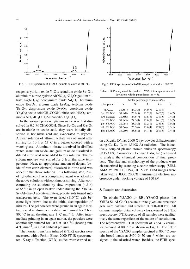
Fig. 1. FTIR spectrum of YSAGG sample calcined at 800 ◦C.
reagents: yttrium oxide Y2O3; scandium oxide Sc2O3;aluminium nitrate hydrate Al(NO3)3·9H2O; gallium ni-trate Ga(NO3)3; neodymium oxide Nd2O3; holmiumoxide Ho2O3; erbium oxide Er2O3; terbium oxideTb4O7; dysprosium oxide Dy2O3; ytterbium oxideYb2O3; acetic acid CH3COOH; nitric acid HNO3; am-monia NH3·4H2O; 1,2-ethanediol C2H6O2.
In the sol–gel process, yttrium oxide was first dis-solved in 0.2 M CH3COOH. Since Sc2O3 and Ga2O3
are insoluble in acetic acid, they were initially dis-solved in hot nitric acid and evaporated to dryness.A clear solution of yttrium acetate was obtained afterstirring for 10 h at 65 ◦C in a beaker covered with awatch glass. Aluminum nitrate dissolved in distilledwater, scandium oxide, and gallium oxide dissolved indiluted nitric acid were added to this solution. The re-sulting mixture was stirred for 3 h at the same tem-perature. Next, an appropriate amount of dopant (ox-ide of rare-earth element) dissolved in nitric acid wasadded to the above solution. In a following step, 2 mlof 1,2-ethanediol as a complexing agent was added tothe above solutions with continuous stirring. After con-centrating the solutions by slow evaporation (∼8 h)at 65 ◦C in an open beaker under stirring the Y(RE)–Sc–Al–Ga–O acetate-nitrate-glycolate sols turned intotransparent gels. The oven dried (110 ◦C) gels be-came light brown due to the initial decomposition ofnitrates. The gel powders were ground in an agate mor-tar, placed in alumina crucibles, and burned for 2 h at800 ◦C in air (heating rate 1 ◦C min−1). After inter-mediate grinding in an agate mortar, the powders wereadditionally sintered for 10 h at 1000 ◦C (heating rate4 ◦C min−1) in air at ambient pressure.
The Fourier transform infrared (FTIR) spectra weremeasured with a Perkin Elmer 2000 FT-IR spectrome-ter. X-ray diffraction (XRD) studies were carried out
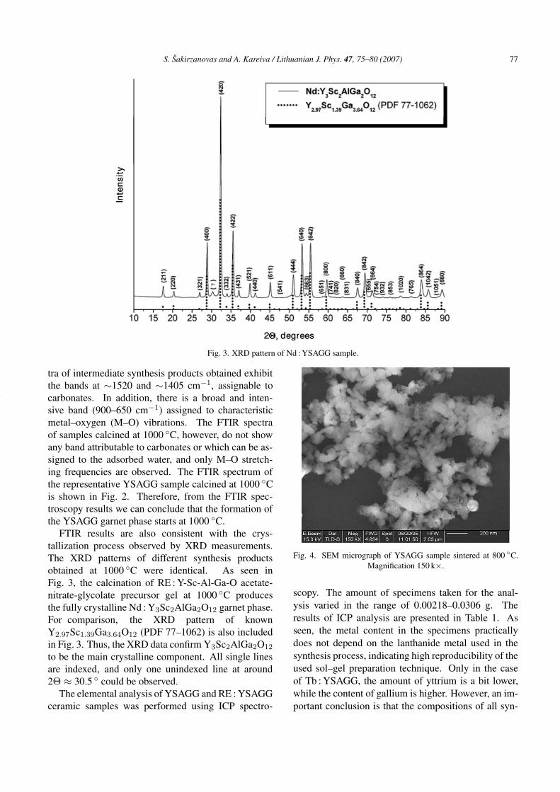
Fig. 2. FTIR spectrum of YSAGG sample sintered at 1000 ◦C.
Table 1. ICP analysis of the final RE : YSAGG samples (standarddeviations within parentheses; n = 3).
Molar percentage of metals (%)Compound Y Sc Al Ga RE
YSAGG 37.5(7) 24.7(5) 14.0(7) 23.8(4) –Dy : YSAGG 37.8(8) 23.9(5) 13.7(5) 24.2(5) 0.4(2)Er : YSAGG 37.5(6) 24.5(7) 13.8(6) 23.8(5) 0.4(3)Ho : YSAGG 37.8(5) 24.3(8) 13.6(7) 24.1(5) 0.2(2)Yb : YSAGG 37.0(4) 25.3(5) 13.2(9) 23.6(5) 0.9(5)Nd : YSAGG 37.6(4) 25.7(6) 13.6(4) 22.8(5) 0.3(1)Tb : YSAGG 34.2(9) 25.5(8) 14.1(4) 25.8(5) 0.4(4)
on a Rigaku D/max-2000 X-ray powder diffractometerusing Cu Kα (λ = 1.5408 Å) radiation. The induc-tively coupled plasma atomic emission spectroscopy(ICP-AES, Plasma-Spec, Leeman Labs, Inc.) was usedto analyse the chemical composition of final prod-ucts. The size and morphology of the products werecharacterized by scanning electron microscopy (SEM,AMARY 1910FE, USA) at 15 kV. TEM images weretaken with a JEOL 200CX transmission electron mi-croscope under working voltage of 160 kV.
3. Results and discussion
To obtain YSAGG or RE : YSAGG phases theY(RE)-Sc-Al-Ga-O acetate-nitrate-glycolate precursorgels were calcined and sintered at 800–1000 ◦C. Allceramic samples obtained were characterized by FTIRspectroscopy. FTIR spectra of all samples were qualita-tively the same regardless of the nature of substitution.The representative FTIR spectrum of YSAGG ceram-ics calcined at 800 ◦C is shown in Fig. 1. The FTIRspectra of the YSAGG samples calcined at 800 ◦C con-tain broad bands at 3450–3470 cm−1 which are as-signed to the adsorbed water. Besides, the FTIR spec-
S. Šakirzanovas and A. Kareiva / Lithuanian J. Phys. 47, 75–80 (2007) 77
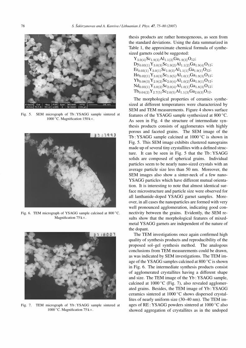
Fig. 3. XRD pattern of Nd : YSAGG sample.
tra of intermediate synthesis products obtained exhibitthe bands at ∼1520 and ∼1405 cm−1, assignable tocarbonates. In addition, there is a broad and inten-sive band (900–650 cm−1) assigned to characteristicmetal–oxygen (M–O) vibrations. The FTIR spectraof samples calcined at 1000 ◦C, however, do not showany band attributable to carbonates or which can be as-signed to the adsorbed water, and only M–O stretch-ing frequencies are observed. The FTIR spectrum ofthe representative YSAGG sample calcined at 1000 ◦Cis shown in Fig. 2. Therefore, from the FTIR spec-troscopy results we can conclude that the formation ofthe YSAGG garnet phase starts at 1000 ◦C.
FTIR results are also consistent with the crys-tallization process observed by XRD measurements.The XRD patterns of different synthesis productsobtained at 1000 ◦C were identical. As seen inFig. 3, the calcination of RE : Y-Sc-Al-Ga-O acetate-nitrate-glycolate precursor gel at 1000 ◦C producesthe fully crystalline Nd : Y3Sc2AlGa2O12 garnet phase.For comparison, the XRD pattern of knownY2.97Sc1.39Ga3.64O12 (PDF 77–1062) is also includedin Fig. 3. Thus, the XRD data confirm Y3Sc2AlGa2O12
to be the main crystalline component. All single linesare indexed, and only one unindexed line at around2Θ ≈ 30.5 ◦ could be observed.
The elemental analysis of YSAGG and RE : YSAGGceramic samples was performed using ICP spectro-
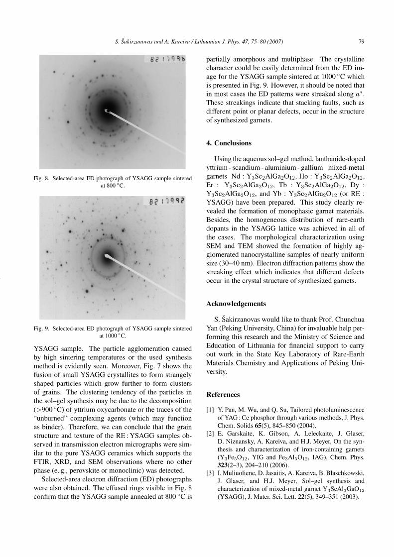
Fig. 4. SEM micrograph of YSAGG sample sintered at 800 ◦C.Magnification 150 k×.
scopy. The amount of specimens taken for the anal-ysis varied in the range of 0.00218–0.0306 g. Theresults of ICP analysis are presented in Table 1. Asseen, the metal content in the specimens practicallydoes not depend on the lanthanide metal used in thesynthesis process, indicating high reproducibility of theused sol–gel preparation technique. Only in the caseof Tb : YSAGG, the amount of yttrium is a bit lower,while the content of gallium is higher. However, an im-portant conclusion is that the compositions of all syn-
78 S. Šakirzanovas and A. Kareiva / Lithuanian J. Phys. 47, 75–80 (2007)
Fig. 5. SEM micrograph of Tb : YSAGG sample sintered at1000 ◦C. Magnification 150 k×.
Fig. 6. TEM micrograph of YSAGG sample calcined at 800 ◦C.Magnification 75 k×.
Fig. 7. TEM micrograph of Yb : YSAGG sample sintered at1000 ◦C. Magnification 75 k×.
thesis products are rather homogeneous, as seen fromthe standard deviations. Using the data summarized inTable 1, the approximate chemical formula of synthe-sized garnets could be suggested:
Y3.0(4)Sc1.9(3)Al1.1(2)Ga1.9(3)O12;Dy0.03(1)Y3.0(3)Sc1.9(2)Al1.1(2)Ga1.9(4)O12;Er0.03(1)Y3.0(1)Sc1.9(3)Al1.1(1)Ga1.9(1)O12;Ho0.02(1)Y3.0(3)Sc1.9(5)Al1.0(1)Ga1.9(3)O12;Yb0.08(3)Y2.9(2)Sc2.0(4)Al1.0(2)Ga1.9(1)O12;Nd0.03(1)Y3.0(2)Sc2.0(4)Al1.0(1)Ga1.8(3)O12;Tb0.04(2)Y2.7(5)Sc2.0(5)Al1.1(3)Ga2.0(3)O12.
The morphological properties of ceramics synthe-sized at different temperatures were characterized bySEM and TEM measurements. Figure 4 shows surfacefeatures of the YSAGG sample synthesized at 800 ◦C.As seen in Fig. 4 the structure of intermediate syn-thesis products consists of agglomerates with highlyporous and faceted grains. The SEM image of theTb : YSAGG sample calcined at 1000 ◦C is shown inFig. 5. This SEM image exhibits clustered nanograinsmade up of several tiny crystallites with a defined struc-ture. It can be seen in Fig. 5 that the Tb : YSAGGsolids are composed of spherical grains. Individualparticles seem to be nearly nano-sized crystals with anaverage particle size less than 50 nm. Moreover, theSEM images also show a sinter-neck of a few nano-YSAGG particles which have different mutual orienta-tion. It is interesting to note that almost identical sur-face microstructure and particle size were observed forall lanthanide-doped YSAGG garnet samples. More-over, in all cases the nanoparticles are formed with verywell pronounced agglomeration, indicating good con-nectivity between the grains. Evidently, the SEM re-sults show that the morphological features of mixed-metal YSAGG garnets are independent of the nature ofthe dopant.
The TEM investigations once again confirmed highquality of synthesis products and reproducibility of theproposed sol–gel synthesis method. The analogousconclusions from TEM measurements could be drawn,as was indicated by SEM investigations. The TEM im-age of the YSAGG samples calcined at 800 ◦C is shownin Fig. 6. The intermediate synthesis products consistof agglomerated crystallites having a different shapeand size. The TEM image of the Yb : YSAGG sample,calcined at 1000 ◦C (Fig. 7), also revealed agglomer-ated grains. Besides, the TEM image of Yb : YSAGGceramics sintered at 1000 ◦C shows dispersed crystal-lites of nearly uniform size (30–40 nm). The TEM im-ages of RE : YSAGG powders sintered at 1000 ◦C alsoshowed aggregation of crystallites as in the undoped
S. Šakirzanovas and A. Kareiva / Lithuanian J. Phys. 47, 75–80 (2007) 79
Fig. 8. Selected-area ED photograph of YSAGG sample sinteredat 800 ◦C.
Fig. 9. Selected-area ED photograph of YSAGG sample sinteredat 1000 ◦C.
YSAGG sample. The particle agglomeration causedby high sintering temperatures or the used synthesismethod is evidently seen. Moreover, Fig. 7 shows thefusion of small YSAGG crystallites to form strangelyshaped particles which grow further to form clustersof grains. The clustering tendency of the particles inthe sol–gel synthesis may be due to the decomposition(>900 ◦C) of yttrium oxycarbonate or the traces of the“unburned” complexing agents (which may functionas binder). Therefore, we can conclude that the grainstructure and texture of the RE : YSAGG samples ob-served in transmission electron micrographs were sim-ilar to the pure YSAGG ceramics which supports theFTIR, XRD, and SEM observations where no otherphase (e. g., perovskite or monoclinic) was detected.
Selected-area electron diffraction (ED) photographswere also obtained. The effused rings visible in Fig. 8confirm that the YSAGG sample annealed at 800 ◦C is
partially amorphous and multiphase. The crystallinecharacter could be easily determined from the ED im-age for the YSAGG sample sintered at 1000 ◦C whichis presented in Fig. 9. However, it should be noted thatin most cases the ED patterns were streaked along a∗.These streakings indicate that stacking faults, such asdifferent point or planar defects, occur in the structureof synthesized garnets.
4. Conclusions
Using the aqueous sol–gel method, lanthanide-dopedyttrium - scandium - aluminium - gallium mixed-metalgarnets Nd : Y3Sc2AlGa2O12, Ho : Y3Sc2AlGa2O12,Er : Y3Sc2AlGa2O12, Tb : Y3Sc2AlGa2O12, Dy :Y3Sc2AlGa2O12, and Yb : Y3Sc2AlGa2O12 (or RE :YSAGG) have been prepared. This study clearly re-vealed the formation of monophasic garnet materials.Besides, the homogeneous distribution of rare-earthdopants in the YSAGG lattice was achieved in all ofthe cases. The morphological characterization usingSEM and TEM showed the formation of highly ag-glomerated nanocrystalline samples of nearly uniformsize (30–40 nm). Electron diffraction patterns show thestreaking effect which indicates that different defectsoccur in the crystal structure of synthesized garnets.
Acknowledgements
S. Šakirzanovas would like to thank Prof. ChunchuaYan (Peking University, China) for invaluable help per-forming this research and the Ministry of Science andEducation of Lithuania for financial support to carryout work in the State Key Laboratory of Rare-EarthMaterials Chemistry and Applications of Peking Uni-versity.
References
[1] Y. Pan, M. Wu, and Q. Su, Tailored photoluminescenceof YAG : Ce phosphor through various methods, J. Phys.Chem. Solids 65(5), 845–850 (2004).
[2] E. Garskaite, K. Gibson, A. Leleckaite, J. Glaser,D. Niznansky, A. Kareiva, and H.J. Meyer, On the syn-thesis and characterization of iron-containing garnets(Y3Fe5O12, YIG and Fe3Al5O12, IAG), Chem. Phys.323(2–3), 204–210 (2006).
[3] I. Muliuoliene, D. Jasaitis, A. Kareiva, B. Blaschkowski,J. Glaser, and H.J. Meyer, Sol–gel synthesis andcharacterization of mixed-metal garnet Y3ScAl3GaO12
(YSAGG), J. Mater. Sci. Lett. 22(5), 349–351 (2003).
80 S. Šakirzanovas and A. Kareiva / Lithuanian J. Phys. 47, 75–80 (2007)
[4] O.Yu. Goncharov, Evaluation of the parameters ofcation distribution in Y3A5−zBzO12 solid solutions,Crystallography Rep. 48(1), 1–7 (2003) [in Russian:Kristallografiya 48(1), 7–13 (2003)].
[5] F. Maglia, V. Buscaglia, S. Gennari, P. Ghigna, M. Dapi-aggi, A. Speghini, and M. Bettinelli, Incorporation oftrivalent cations in synthetic garnets A3B5O12 (A = Y,Lu–La, B = Al, Fe, Ga), J. Phys. Chem. B 110(13),6561–6568 (2006).
[6] J. Saikawa, Y. Sato, T. Taira, and A. Ikesue, Pas-sive mode locking of a mixed garnet Yb : Y3ScAl4O12
ceramic laser, Appl. Phys. Lett. 85(24), 5845–5847(2004).
[7] T.H. Allik, C.A. Morrison, J.B. Gruber, and M.R. Kokta,Crystallography, spectroscopic analysis, and lasingproperties of Nd3+ : Y3Sc2Al3O12, Phys. Rev. B 41(1),21–30 (1990).
[8] J. Saikawa, Y. Sato, T. Taira, and A. Ikesue, Absorption,emission spectrum properties, and efficient laser perfor-mances of Yb : Y3ScAl4O12 ceramics, Appl. Phys. Lett.85(11), 1898–1900 (2004).
MIŠRIU METALU GRANATU SU LANTANOIDU PRIEMAIŠOMIS NANODALELIU SINTEZEZOLIU IR GELIU METODU
S. Šakirzanovas, A. Kareiva
Vilniaus universiteto Chemijos fakultetas, Vilnius, Lietuva
SantraukaVandeniniu zoliu ir geliu metodu buvo susintetinti ivairus
mišrus metalu granatai (RE : Y3Sc2AlGa2O12, RE = Nd, Ho, Er,Tb, Dy, Yb). FTIR ir XRD tyrimais irodyta, kad 1000 ◦C tempe-raturoje susidaro vienfazis mišrus metalu granatas. Nuodugnesni
tyrimai (SEM, TEM, ICP) parode, kad priemaišinio elemento pri-gimtis neturi itakos gautu granatu morfologijai ir faziniam grynu-mui, nors elektronu difrakcijos nuotraukos rodo ivairius kristalinesgardeles defektus.