soft tissue phantoms for realistic needle insertion: a ... · soft tissue phantoms for realistic...
TRANSCRIPT
Soft Tissue Phantoms for Realistic Needle Insertion: A Comparative
Study
ALEXANDER LEIBINGER,1 ANTONIO E. FORTE,1 ZHENGCHU TAN,1 MATTHEW J. OLDFIELD,1 FRANK BEYRAU,2
DANIELE DINI,1 and FERDINANDO RODRIGUEZ Y BAENA1
1Department of Mechanical Engineering, Imperial College London, Exhibition Road, South Kensington, London SW7 2AZ,UK; and 2Lehrstuhl fur Technische Thermodynamik, Otto-von-Guericke-Universitat Magdeburg, Magdeburg, Germany
(Received 24 September 2015; accepted 25 November 2015)
Associate Editor K. A. Athanasiou oversaw the review of this article.
Abstract—Phantoms are common substitutes for soft tissues inbiomechanical research and are usually tuned to match tissueproperties using standard testing protocols at small strains.However, the response due to complex tool-tissue interactionscan differ depending on the phantom and no comprehensivecomparative study has been published to date, which could aidresearchers to select suitable materials. In this work, gelatin, acommon phantom in literature, and a composite hydrogeldeveloped at Imperial College, were matched for mechanicalstiffness to porcine brain, and the interactions during needleinsertionswithin themwere analyzed. Specifically, we examinedinsertion forces for brain and the phantoms; we also measureddisplacements and strains within the phantoms via a laser-basedimage correlation technique in combination with fluorescentbeads. It is shown that the insertion forces for gelatin and brainagree closely, but that the composite hydrogel bettermimics theviscous nature of soft tissue. Both materials match differentcharacteristics of brain, but neither of them is a perfectsubstitute. Thus, when selecting a phantom material, both thesoft tissue properties and the complex tool-tissue interactionsarising during tissue manipulation should be taken intoconsideration. These conclusions are presented in tabular formto aid future selection.
Keywords—Minimally invasive surgery, Brain, Tool-tissue
interactions, Digital image correlation, Strain imaging, Soft
tissue biomechanics, Gelatin.
INTRODUCTION
Working with real specimens of soft tissue forexperimental studies in the biomechanical field pre-sents a number of difficulties; in addition to ethicalregulations and scarce availability, the data obtained is
often unreliable or inconsistent due to complex tissueproperties and testing protocols. Brain, especially, hasproperties that vary between in vivo and in vitro con-ditions2 and is highly sensitive to factors such as post-mortem time, sample preparation or mechanical his-tory.6 Soft tissue specimens are also cumbersome to fixand constrain, leading to complex experimentaldesigns7,12 and varying conditions between experi-ments. These problems have led to large deviationsbetween experimental results in literature, which makecomparisons across different studies difficult.11
For these reasons, soft tissues are commonly replacedby phantoms for experimental studies, as they are morecontrollable, easier to handle, and eliminate the prob-lem of sample-specific variations. These advantages areexploited in fields ranging from surgical training to themodelling of interactions between soft tissue and tools,for instance in the context of percutaneous interventionwhere phantoms are used to validate models of needleinsertions3,15 and to find and demonstrate new conceptsfor insertion methods with reduced tissue motion20 andfor needle steering with enhanced accuracy.10
Phantom materials should mimic the relevantproperties of soft tissue closely in order to translatefindings to medical procedures. There is a large varietyof phantom materials within the literature, with abroad range of material properties, depending on therelevant application and objectives. Materials areusually selected based on typical mechanical charac-terization protocols, such as compression, tensile orindentation tests3,18 or for best desired performance,e.g., to maximize the curvature of a steering needle.25
During needle insertions, large strains and cuttingcause complex tool-tissue interactions, and phantomsshould mimic soft tissue sufficiently for all domains of
Address correspondence to Ferdinando Rodriguez y Baena,
Department of Mechanical Engineering, Imperial College London,
Exhibition Road, South Kensington, London SW7 2AZ, UK. Elec-
tronic mail: [email protected]
Annals of Biomedical Engineering (� 2015)
DOI: 10.1007/s10439-015-1523-0
� 2015 The Author(s). This article is published with open access at Springerlink.com

interest during the insertion. One of the most commonsoft tissue phantoms for modelling interactions, due toits organic nature and ease of handling, is gelatin.5,22,24
Gelatin phantoms are primarily characterized by theirstiffness and are generally tuned by modifying the ge-latin concentration, in order to match properties ofsoft tissue. However, gelatin has a near linear elasticbehavior, with lower rate dependency compared to softtissue, which is generally highly viscous.14 Conversely,a composite hydrogel (CH) developed at ImperialCollege London4 to simulate brain in compression,indentation, relaxation, shear and hysteresis, showsvery good agreement with brain tissue for stiffness-and viscosity-related properties, which advocate its useas a better synthetic phantom compared to gelatin.
The goal of this work is to evaluate the performanceof transparent gelatin and a modified, transparentversion of the composite hydrogel (MCH), in thecontext of needle insertions. In vitro needle insertionsare performed on these materials and compared to realsoft tissue, porcine brain, which is known to behavesimilarly to human brain.6,16
The stiffness of the phantoms was first tuned tomatch brain using a standard indentation test. Fracturetests were conducted in order to characterize thebehavior at failure of all phantoms (gelatin, CH, MCH)and real tissue. We then measured the required insertionforces for brain, MCH and gelatin; due to the lack oftransparency of biological soft tissue, for the phantomsonly, we examined internal displacements and strainsclose to the needle using a laser-based imaging tech-nique, which allows a comprehensive measurement ofthe interactions between the inserted needle and thesubstrate.19 Based on these measurements during theneedle insertion process with both materials, conclu-sions can be drawn on the suitability of either phantomas a realistic alternative to real soft tissue.
The following section provides a detailed descriptionand characterization of the materials used and theexperimental setup devised for this study. This is followedby the results of the insertion forces measured for thesynthetic materials, which are compared to porcine brainsamples. A closer comparison of resulting displacementsand strains for gelatin andMCH,due to fracture, friction,and viscous effects, is then provided, followed by a dis-cussion and conclusions, which address the differentcharacteristics of the materials and how they apply tostudies that may require a realistic tissue phantom.
MATERIALS
Materials
The composite hydrogel was composed of Phytagel(PHY), polyvinyl alcohol (PVA, 146,000–186,000
molecular weight) and deionized water, all supplied bySigma-Aldrich Co., USA. The bovine gelatin powderwas provided by Sleaford Quality Foods Ltd., UK. Allconcentrations of the phantom materials in the fol-lowing sections are expressed as a percentage by mass(wt%).
Two days post-mortem porcine brain samples (2samples) were obtained from a local supplier.
Sample Preparation
To replicate the same boundary conditions acrossexperiments, all samples were filled into transparentacrylic boxes, with an open top, an inner cross sectionof 80 9 80 mm2 and a height of 50 mm.
The MCH was obtained by modifying the proce-dure for CH described in Forte et al.4 to create atransparent composite hydrogel that could be used fornon-invasive, highly resolved optical measurementwithin the samples. Specifically, the freezing step wasremoved from the procedure to avoid gelation of thePVA. The resultant network is phase separated, withthe PHY forming the continuous polymer network(dominant phase) and the PVA dissolved as the fillernetwork (included phase).
The MCH was produced by separately dissolvingPVA and PHY in deionized water for 1 h at 90 �C. Thetwo solutions were then mixed together in a 1:1 weightratio at 70 �C, under constant stirring for 30 min.Particular care was used to avoid evaporation duringthe process. The solution was seeded with fluorescentmelamine resin beads to enable imaging (size 10 lm,rhodamine B-marked, Sigma Aldrich Co.), and pouredinto the transparent boxes. The boxes were coveredwith cling film to limit evaporation and slowly cooledat room temperature for 7 h before testing.
Due to the modified procedure, the MCH exhibiteddifferent mechanical characteristics from the originalCH, which had a coupled network structure due to thepresence of hydrogen bonds. Therefore, the concen-trations of PVA and PHY needed to be tuned in orderto match the stiffness of brain tissue, as described in‘‘Stiffness Matching’’ section. The result is a trans-parent, highly viscous gel, which can also be used forinternal optical measurements.
Gelatin gels were produced by mixing deionizedwater and gelatin powder. Deionized water was heatedto 90 �C, and gelatin powder was then added andstirred into the water for 10 min. The solution wasseeded with the same particles as the MCH and left tocool at room temperature. Cling film was also used andsamples were stored in a domestic refrigerator, kept at14 �C for 12 h; samples were tested on the followingday, after the gel had reached room temperature(22 ± 2 �C).
LEIBINGER et al.

The micrometer-sized beads used to seed the phan-toms had a density of 1.51 g/cm3, which is sufficientlyclose to the density of the phantoms, mainly consistingof water. This allowed the added particles to distributeevenly in the phantom before solidification. The con-centration of the aqueous solution with the particles inthe phantoms was approximately 0.04 vol%, resultingin a particle number density of 16 particles/mm3. Thisresulted in a seeding density suitable for the imagingprocess (see ‘‘Methods’’ section). Indentation testsshowed that the particles did not influence the materialbehavior due to their small size, similar density, andlow number density.
Porcine brain samples were collected from thebutcher immediately after being removed from thedura, then stored in a physiological solution at 4 �Cduring transportation. Specimens were not frozen atany time during the procedure.
Stiffness Matching
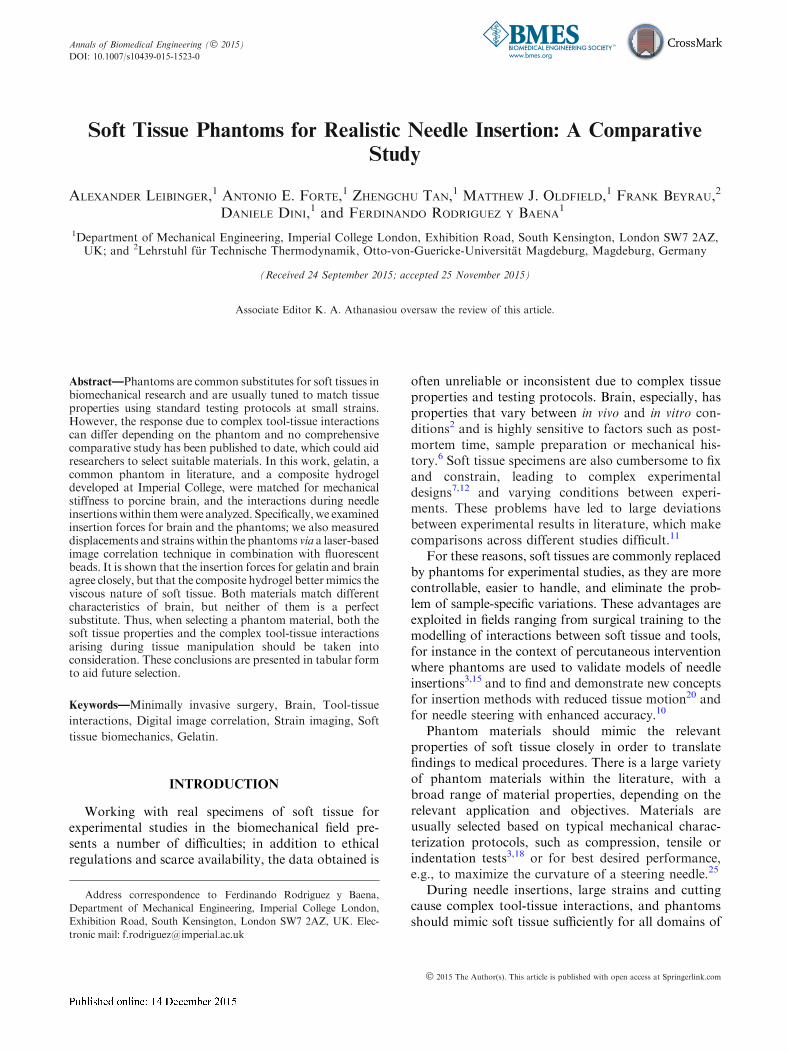
The stiffness of the CH, MCH and gelatin wastuned to match the stiffness of the porcine brain tissueby varying the concentration of the powders dissolvedin deionized water. An indentation test protocol,carried out with a mechanical testing system (Mach-1,Biomomentum Inc., CA) with a 150 g load cell(7.5 mg load resolution) and a spherical indenter(3.175 mm radius), was performed to compare sam-ples that filled the same boxes as used for the needleinsertions. Gefen et al.7 suggested that the indenter tipradius should be no more than 25% the thickness ofthe tested sample and they used an indentation depthequal to the diameter of the indenter. Therefore, theindentation depth was set to 6 mm. The test wasperformed at a displacement rate of 1 mm/s. Afterreaching the maximum depth, the indenter was keptin position for 500 s to record the relaxation behaviorof each material. Five repetitions were carried out foreach material. Figure 1a shows the indentation curvesfor the brain samples and the tuned phantom mate-rials of gelatin (3.4%), MCH (3% PVA + 0.75%PHY), CH (PVA 5% + PHY 0.59%), respectively.The indentation curves for porcine brain tissue andthe phantoms show very good agreement and wereconsidered sufficiently close to each other to assumesimilar stiffness properties. Because of their viscousnature, the relaxation behavior of the compositehydrogels (CH and MCH) is closer to that of braintissue, whereas gelatin, as expected, behaved as anearly elastic material (Fig. 1b). The MCH relaxesmore quickly than the original CH due to its weaklybonded network (‘‘Sample Preparation’’ section).Knowledge of the difference in viscous properties ofthe phantom materials will allow us to draw certain
conclusions from the measured interactions with theinserted needle.
Fracture Behavior
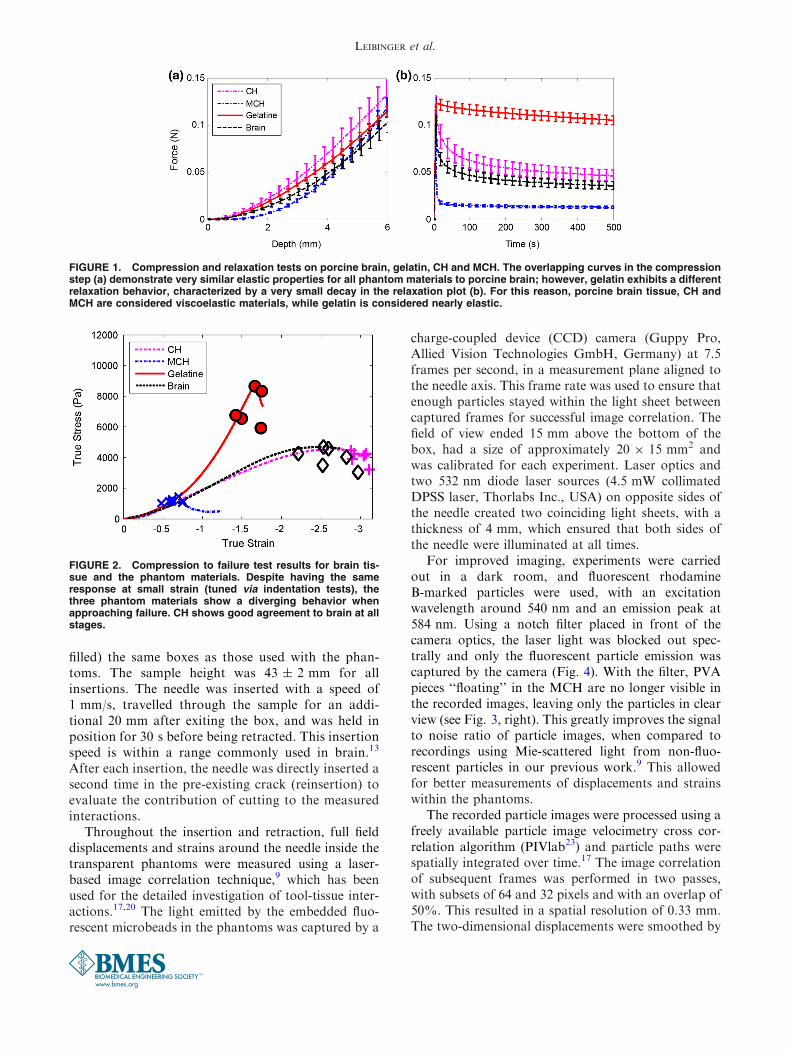
Fracture tests were performed on brain and thephantom materials. Cylindrical samples (diameter14 ± 1 mm, height 9 ± 1 mm) were tested in com-pression, using the same mechanical testing system andwith the same displacement rate as for the needleinsertions. Each sample was compressed between twoplates until failure was detected. Silicon oil was appliedat the interface between the sample and the compres-sion plates in order to minimize friction effects.5 Uni-form expansion of the sample in the radial directionwas monitored. True strains and stresses were com-puted from recorded forces and displacements.
In Fig. 2, the true stresses and strains are shown forthe point of material failure during compression test-ing, for each material tested. Fracture becomes typi-cally visible by a sudden drop in the stress–straincurve. Typically, the brain and the CH samples did notbreak during testing and the values presented in Fig. 2represent the point when the two compression platescame almost into contact, compressing to 95% of thesample’s height.
All four materials show a similar response at thesmall strains for which they were matched, but adiverging behavior at the point of failure. The braintissue and CH show true stresses at failure in the samerange of approximately 3–4.5 kPa. The CH shows aductile and stretchable behavior, which is very similarto brain tissue. MCH shows the lowest final stressesand strains, as it fails early on in the tests. Comparedto brain, gelatin shows more brittle fracture behavior,with high stresses and low strains, whereas thebehavior of brain and CH can be described as ductile,with larger deformations.
METHODS
The needle insertions were performed using thesame mechanical testing system. A straight needle,with a conical tip with a 40� included tip angle and anouter diameter (OD) of 4 mm, was manufactured outof a rigid rapid prototyped material (Elastic Modulus1.7–2.1 GPa, Endur, Stratasys Ltd., USA), connecteddirectly to the load cell.
The needle was inserted from the top and exitedthrough a hole in the bottom of the box (Fig. 3). Eightinsertions were performed for each phantom material,and four insertions were performed on porcine brain.In order to produce equivalent boundary conditionsbetween materials, the brains were placed in (and fully
Soft Tissue Phantoms for Realistic Needle Insertion
filled) the same boxes as those used with the phan-toms. The sample height was 43 ± 2 mm for allinsertions. The needle was inserted with a speed of1 mm/s, travelled through the sample for an addi-tional 20 mm after exiting the box, and was held inposition for 30 s before being retracted. This insertionspeed is within a range commonly used in brain.13
After each insertion, the needle was directly inserted asecond time in the pre-existing crack (reinsertion) toevaluate the contribution of cutting to the measuredinteractions.
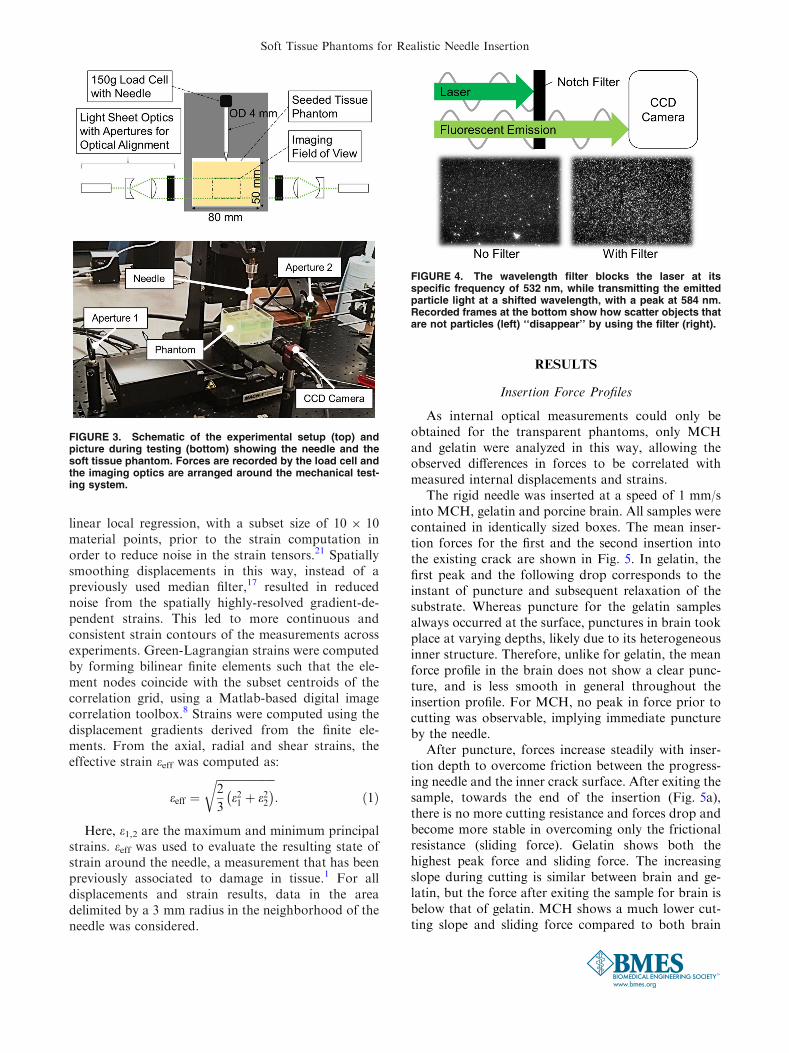
Throughout the insertion and retraction, full fielddisplacements and strains around the needle inside thetransparent phantoms were measured using a laser-based image correlation technique,9 which has beenused for the detailed investigation of tool-tissue inter-actions.17,20 The light emitted by the embedded fluo-rescent microbeads in the phantoms was captured by a
charge-coupled device (CCD) camera (Guppy Pro,Allied Vision Technologies GmbH, Germany) at 7.5frames per second, in a measurement plane aligned tothe needle axis. This frame rate was used to ensure thatenough particles stayed within the light sheet betweencaptured frames for successful image correlation. Thefield of view ended 15 mm above the bottom of thebox, had a size of approximately 20 9 15 mm2 andwas calibrated for each experiment. Laser optics andtwo 532 nm diode laser sources (4.5 mW collimatedDPSS laser, Thorlabs Inc., USA) on opposite sides ofthe needle created two coinciding light sheets, with athickness of 4 mm, which ensured that both sides ofthe needle were illuminated at all times.
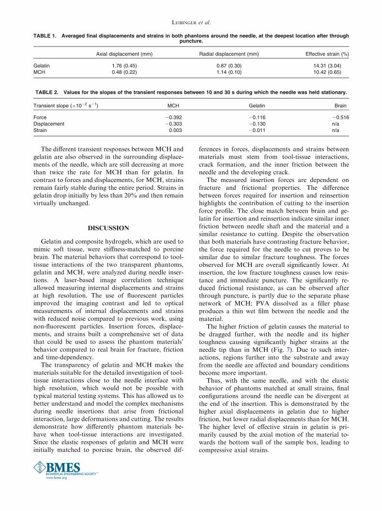
For improved imaging, experiments were carriedout in a dark room, and fluorescent rhodamineB-marked particles were used, with an excitationwavelength around 540 nm and an emission peak at584 nm. Using a notch filter placed in front of thecamera optics, the laser light was blocked out spec-trally and only the fluorescent particle emission wascaptured by the camera (Fig. 4). With the filter, PVApieces ‘‘floating’’ in the MCH are no longer visible inthe recorded images, leaving only the particles in clearview (see Fig. 3, right). This greatly improves the signalto noise ratio of particle images, when compared torecordings using Mie-scattered light from non-fluo-rescent particles in our previous work.9 This allowedfor better measurements of displacements and strainswithin the phantoms.
The recorded particle images were processed using afreely available particle image velocimetry cross cor-relation algorithm (PIVlab23) and particle paths werespatially integrated over time.17 The image correlationof subsequent frames was performed in two passes,with subsets of 64 and 32 pixels and with an overlap of50%. This resulted in a spatial resolution of 0.33 mm.The two-dimensional displacements were smoothed by
FIGURE 1. Compression and relaxation tests on porcine brain, gelatin, CH and MCH. The overlapping curves in the compressionstep (a) demonstrate very similar elastic properties for all phantom materials to porcine brain; however, gelatin exhibits a differentrelaxation behavior, characterized by a very small decay in the relaxation plot (b). For this reason, porcine brain tissue, CH andMCH are considered viscoelastic materials, while gelatin is considered nearly elastic.
FIGURE 2. Compression to failure test results for brain tis-sue and the phantom materials. Despite having the sameresponse at small strain (tuned via indentation tests), thethree phantom materials show a diverging behavior whenapproaching failure. CH shows good agreement to brain at allstages.
LEIBINGER et al.
linear local regression, with a subset size of 10 9 10material points, prior to the strain computation inorder to reduce noise in the strain tensors.21 Spatiallysmoothing displacements in this way, instead of apreviously used median filter,17 resulted in reducednoise from the spatially highly-resolved gradient-de-pendent strains. This led to more continuous andconsistent strain contours of the measurements acrossexperiments. Green-Lagrangian strains were computedby forming bilinear finite elements such that the ele-ment nodes coincide with the subset centroids of thecorrelation grid, using a Matlab-based digital imagecorrelation toolbox.8 Strains were computed using thedisplacement gradients derived from the finite ele-ments. From the axial, radial and shear strains, theeffective strain eeff was computed as:
eeff ¼ffiffiffiffiffiffiffiffiffiffiffiffiffiffiffiffiffiffiffiffiffi
2
3e21 þ e22� �
r
: ð1Þ
Here, e1,2 are the maximum and minimum principalstrains. eeff was used to evaluate the resulting state ofstrain around the needle, a measurement that has beenpreviously associated to damage in tissue.1 For alldisplacements and strain results, data in the areadelimited by a 3 mm radius in the neighborhood of theneedle was considered.
RESULTS
Insertion Force Profiles
As internal optical measurements could only beobtained for the transparent phantoms, only MCHand gelatin were analyzed in this way, allowing theobserved differences in forces to be correlated withmeasured internal displacements and strains.
The rigid needle was inserted at a speed of 1 mm/sinto MCH, gelatin and porcine brain. All samples werecontained in identically sized boxes. The mean inser-tion forces for the first and the second insertion intothe existing crack are shown in Fig. 5. In gelatin, thefirst peak and the following drop corresponds to theinstant of puncture and subsequent relaxation of thesubstrate. Whereas puncture for the gelatin samplesalways occurred at the surface, punctures in brain tookplace at varying depths, likely due to its heterogeneousinner structure. Therefore, unlike for gelatin, the meanforce profile in the brain does not show a clear punc-ture, and is less smooth in general throughout theinsertion profile. For MCH, no peak in force prior tocutting was observable, implying immediate punctureby the needle.
After puncture, forces increase steadily with inser-tion depth to overcome friction between the progress-ing needle and the inner crack surface. After exiting thesample, towards the end of the insertion (Fig. 5a),there is no more cutting resistance and forces drop andbecome more stable in overcoming only the frictionalresistance (sliding force). Gelatin shows both thehighest peak force and sliding force. The increasingslope during cutting is similar between brain and ge-latin, but the force after exiting the sample for brain isbelow that of gelatin. MCH shows a much lower cut-ting slope and sliding force compared to both brain
FIGURE 3. Schematic of the experimental setup (top) andpicture during testing (bottom) showing the needle and thesoft tissue phantom. Forces are recorded by the load cell andthe imaging optics are arranged around the mechanical test-ing system.
FIGURE 4. The wavelength filter blocks the laser at itsspecific frequency of 532 nm, while transmitting the emittedparticle light at a shifted wavelength, with a peak at 584 nm.Recorded frames at the bottom show how scatter objects thatare not particles (left) ‘‘disappear’’ by using the filter (right).
Soft Tissue Phantoms for Realistic Needle Insertion

and gelatin. Overall, the insertion force profiles ofbrain and gelatin are well matched and closer thanthose of brain and MCH.
Expectedly, the reinsertion forces (Fig. 5b) are gen-erally lower than for the first insertions, because theneedle does not need to cut as it travels through the pre-formed crack. This makes the force profiles insensitiveto cutting-related variations, such as those caused byinner membranes in the brain, and reduces the spreadbetween trials. Measured forces for gelatin and brainagree even more closely than for first insertions in both
slope and sliding force, although the forces for gelatinare slightly higher throughout the reinsertion. As be-fore, the profile for MCH is much lower overall and thedifference between first insertion and reinsertion issmaller than for the other two materials.
Interactions During Needle Cutting
Additionally to the measured insertion forces,optical measurements of displacements and strainswithin the transparent phantoms provides a compre-
FIGURE 6. Contour plots of the imaging results during a needle insertion for both phantom materials, gelatin (left) and MCH(right). The first row shows displacement magnitude calculated from axial and radial displacements. The second row shows theeffective strain obtained from the Green-Lagrangian strains (see Eq. 1).
FIGURE 5. Mean forces of the needle for insertions (a) and reinsertions (b) into MCH, gelatin and porcine brain. Left are the forcesof the first insertions and right are the reinsertions into the existing crack, which were performed directly after the first one.
LEIBINGER et al.

hensive understanding of interactions between the nee-dle and the substrates. Figure 6 shows the displacementmagnitude and effective strain that develop around theneedle, in both gelatin and MCH, during cutting, at aninsertion depth of 20 ± 2 mm. Displacements andstrains become higher towards the needle, as the sur-rounding material is deformed during crack formation.Strains in front of the needle are highest, as this is thearea where material is closest to failure. Figure 6 alsoillustrates how, in gelatin, the top interface of thematerial is dragged further in the direction of the needlemotion, resulting in the overall displacements andstrains being distinctly higher than for MCH. Regionsfurther away from the needle surface are more affectedin gelatin. Whereas displacements in MCH becomenegligible (smaller than 0.5 mm) at a distance ofapproximately 3 mm from to the needle surface, thesame region in gelatin shows much higher values, ofaround 1.5 mm. Strain in MCH is also more concen-trated, with local peaks ahead of the needle and at thetip-to-shaft transition, but with strain values of onlyabout 5% on the bevel tip edges. Conversely, strains ingelatin are above 25% around the entire needle tip.
For a more detailed analysis of interactions duringcutting, Fig. 7 shows the mean axial displacements andstrains along the needle axis during cutting, at a depthof 20 ± 2 mm. The needle tip is indicated at x = 0,with negative x-values being positions along the shaftand positive ones being ahead of the needle. This fig-ure shows that the material is dragged along and pu-shed ahead of the needle, leading to a peak incompressive strain in front of the needle. Displace-ments and strains are significantly higher in gelatinalong the needle shaft and ahead of the tip.
Displacements and Strains in Needle Surroundings
The large difference in displacement and strainpatterns observed near the needle is also seen in the
material surrounding the needle shaft, when it is at itsdeepest point after puncturing and travelling throughthe sample for an additional 20 mm. Table 1 showsthe measured mean displacements for gelatin andMCH axially and radially to the needle, and theeffective strain. For gelatin, the axial displacement isnearly four times higher than MCH, indicating thatthe material is dragged further with the needle. Radialdisplacements are almost half of the axial displace-ments. Conversely, MCH shows a reversed pattern ofdisplacements, with significantly higher radial dis-placements than axial ones. The effective strain in theneedle surroundings during sliding is lower for MCHthan for gelatin.
Transient Response
During the experiments, the needle was held in placefor 30 s, after having travelled through the samples for20 mm. Figure 8 shows the averaged transientresponse of brain, MCH and gelatin relative to theinitial state, during the period for which the needleremained stationary. Measured forces and responses ofthe mean axial displacements and effective strains inthe needle surroundings are shown, with displacementand strain data only being available for the twotransparent phantom materials. The approximateslope at 10 and 30 s was computed for forces, dis-placements and strains (Table 2).
Forces for brain and gelatin initially drop at similarrates, but, whereas gelatin forces reach a stable levelaround 50% after approximately 10 s, the forces forbrain continue to decrease throughout the 30 s dura-tion. While for MCH the initial drop of forces is morepronounced than that for brain and gelatin, MCHshows an on-going decrease and a negative slope after10 s, which is 75% of the slope observed for brain.Gelatin shows a slope of only 23% compared to brainfor the same period.
FIGURE 7. Mean axial displacements (a) and strains (b) in the needle surroundings for all experiments, with standard deviations,along the needle axis whilst cutting. The x-coordinate represents the position along the needle axis and the needle tip is marked atx 5 0.
Soft Tissue Phantoms for Realistic Needle Insertion
The different transient responses between MCH andgelatin are also observed in the surrounding displace-ments of the needle, which are still decreasing at morethan twice the rate for MCH than for gelatin. Incontrast to forces and displacements, for MCH, strainsremain fairly stable during the entire period. Strains ingelatin drop initially by less than 20% and then remainvirtually unchanged.
DISCUSSION
Gelatin and composite hydrogels, which are used tomimic soft tissue, were stiffness-matched to porcinebrain. The material behaviors that correspond to tool-tissue interactions of the two transparent phantoms,gelatin and MCH, were analyzed during needle inser-tions. A laser-based image correlation techniqueallowed measuring internal displacements and strainsat high resolution. The use of fluorescent particlesimproved the imaging contrast and led to opticalmeasurements of internal displacements and strainswith reduced noise compared to previous work, usingnon-fluorescent particles. Insertion forces, displace-ments, and strains built a comprehensive set of datathat could be used to assess the phantom materials’behavior compared to real brain for fracture, frictionand time-dependency.
The transparency of gelatin and MCH makes thematerials suitable for the detailed investigation of tool-tissue interactions close to the needle interface withhigh resolution, which would not be possible withtypical material testing systems. This has allowed us tobetter understand and model the complex mechanismsduring needle insertions that arise from frictionalinteraction, large deformations and cutting. The resultsdemonstrate how differently phantom materials be-have when tool-tissue interactions are investigated.Since the elastic responses of gelatin and MCH wereinitially matched to porcine brain, the observed dif-
ferences in forces, displacements and strains betweenmaterials must stem from tool-tissue interactions,crack formation, and the inner friction between theneedle and the developing crack.
The measured insertion forces are dependent onfracture and frictional properties. The differencebetween forces required for insertion and reinsertionhighlights the contribution of cutting to the insertionforce profile. The close match between brain and ge-latin for insertion and reinsertion indicate similar innerfriction between needle shaft and the material and asimilar resistance to cutting. Despite the observationthat both materials have contrasting fracture behavior,the force required for the needle to cut proves to besimilar due to similar fracture toughness. The forcesobserved for MCH are overall significantly lower. Atinsertion, the low fracture toughness causes low resis-tance and immediate puncture. The significantly re-duced frictional resistance, as can be observed afterthrough puncture, is partly due to the separate phasenetwork of MCH; PVA dissolved as a filler phaseproduces a thin wet film between the needle and thematerial.
The higher friction of gelatin causes the material tobe dragged further, with the needle and its highertoughness causing significantly higher strains at theneedle tip than in MCH (Fig. 7). Due to such inter-actions, regions further into the substrate and awayfrom the needle are affected and boundary conditionsbecome more important.
Thus, with the same needle, and with the elasticbehavior of phantoms matched at small strains, finalconfigurations around the needle can be divergent atthe end of the insertion. This is demonstrated by thehigher axial displacements in gelatin due to higherfriction, but lower radial displacements than for MCH.The higher level of effective strain in gelatin is pri-marily caused by the axial motion of the material to-wards the bottom wall of the sample box, leading tocompressive axial strains.
TABLE 2. Values for the slopes of the transient responses between 10 and 30 s during which the needle was held stationary.
Transient slope (91022 s21) MCH Gelatin Brain
Force 20.392 20.116 20.516
Displacement 20.303 20.130 n/a
Strain 0.003 20.011 n/a
TABLE 1. Averaged final displacements and strains in both phantoms around the needle, at the deepest location after throughpuncture.
Axial displacement (mm) Radial displacement (mm) Effective strain (%)
Gelatin 1.76 (0.45) 0.87 (0.30) 14.31 (3.04)
MCH 0.48 (0.22) 1.14 (0.10) 10.42 (0.65)
LEIBINGER et al.
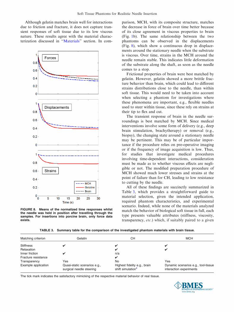
Although gelatin matches brain well for interactionsdue to friction and fracture, it does not capture tran-sient responses of soft tissue due to its low viscousnature. These results agree with the material charac-terization discussed in ‘‘Materials’’ section. In com-
parison, MCH, with its composite structure, matchesthe decrease in force of brain over time better becauseof its close agreement in viscous properties to brain(Fig. 1b). The same relationship between the twophantoms can be observed in the displacements(Fig. 8), which show a continuous drop in displace-ments around the stationary needle when the substrateis viscous. Over time, strains in the MCH around theneedle remain stable. This indicates little deformationof the substrate along the shaft, as soon as the needlecomes to a stop.
Frictional properties of brain were best matched bygelatin. However, gelatin showed a more brittle frac-ture behavior than brain, which could lead to differentstrains distributions close to the needle, than withinsoft tissue. This would need to be taken into accountwhen selecting a phantom for investigations wherethese phenomena are important, e.g., flexible needlesused to steer within tissue, since these rely on strains attheir tip to flex and cut.
The transient response of brain in the needle sur-roundings is best matched by MCH. Since medicalinterventions involve some form of delivery (e.g., deepbrain stimulation, brachytherapy) or removal (e.g.,biopsy), the changing state around a stationary needlemay be pertinent. This may be of particular impor-tance if the procedure relies on pre-operative imagingor if the frequency of image acquisition is low. Thus,for studies that investigate medical proceduresinvolving time-dependent interactions, considerationmust be made as to whether viscous effects are negli-gible or not. The modified preparation procedure ofMCH showed much lower stresses and strains at thepoint of failure than for CH, leading to low resistanceto cutting by the needle.
All of these findings are succinctly summarized inTable 3, which provides a straightforward guide tomaterial selection, given the intended application,required phantom characteristics, and experimentalscenario. Indeed, while none of the materials analyzedmatch the behavior of biological soft tissue in full, eachtype presents valuable attributes (stiffness, viscosity,transparency, etc.) which, if suitably paired to a given
TABLE 3. Summary table for the comparison of the investigated phantom materials with brain tissue.
Matching criterion Gelatin CH MCH
Stiffness 4 4 4
Relaxation 4 4
Inner friction 4 n/a
Fracture resistance 4
Transparency Yes No Yes
Example application Quasi-static scenarios e.g.,
surgical needle steering
Highest fidelity e.g., brain
shift simulation4Dynamic scenarios e.g., tool-tissue
interaction experiments
The tick mark indicates the satisfactory mimicking of the respective material behavior of real tissue.
FIGURE 8. Means of the normalized time responses whilstthe needle was held in position after travelling through thesamples. For insertions into porcine brain, only force dataexists.
Soft Tissue Phantoms for Realistic Needle Insertion

problem, should provide the degree of fidelity requiredto reduce our reliance on ex vivo testing.
CONCLUSIONS AND OUTLOOK
Gelatin and composite hydrogels are often used in avariety of experiments, which aim to assess the per-formance of instruments within biological soft tissue.However, works in the literature capture a wide spreadin the results based on these synthetic media, likelybecause of the necessity to tune their performance tomatch tissue characteristics. In this work, we havestrived to evaluate the performance of compositehydrogels and gelatin against a number of materialproperties, with the aim to provide the reader with akey to the selection process, based on their intendedexperimental setup and end goal.
The investigated materials were shown to performdifferently in terms of several material parameters thatinfluence tool-tissue interactions. Gelatin, which is themost commonly used phantom material in the litera-ture, showed similar behavior to brain during cutting,but low viscoelasticity. The composition of CH canpotentially be tuned to match any of the tissue char-acteristics, but is opaque and thus of limited value inexperimental scenarios where line of site is important.MCH, with its transparency and better viscous prop-erties compared to gelatin, can be used for detailedinteraction studies, but showed low cutting resistance.
In summary, gelatin and composite hydrogels matchdifferent characteristics of brain during needle insertions,but neither is a perfect substitute for mimicking itscomplete behavior. Thus, the selection of phantommaterials should be informedby both the specialmaterialproperties of the soft tissue reference and the complexneedle-tissue interactions needing to be replicated. Thisensures that findings generated with these substitutematerials better reflect real life conditions. New devel-opments, such as those in the areas of tool design orneedle control, must be extensively investigated usingmethodologies that are independent of tissue-specificvariations. A better understanding of brain-mimickingphantoms can potentially accelerate this process.
ACKNOWLEDGEMENTS
This work received funding from the EuropeanResearch Council under the European Union’s Se-venth Framework Programme (FP7/2007-2013), ERCGrant Agreement Number: 258642-STING and by theACTIVE Project (FP7-ICT-2009-6-270460).
CONFLICT OF INTEREST
The authors of this paper have no conflicts ofinterest to report.
OPEN ACCESS
This article is distributed under the terms of theCreative Commons Attribution 4.0 International Li-cense (http://creativecommons.org/licenses/by/4.0/),which permits unrestricted use, distribution, and re-production in any medium, provided you give appro-priate credit to the original author(s) and the source,provide a link to the Creative Commons license, andindicate if changes were made.
REFERENCES
1Bjornsson, C. S., S. J. Oh, Y. A. Al-Kofahi, Y. J. Lim, K.L. Smith, J. N. Turner, S. De, B. Roysam, W. Shain, and S.J. Kim. Effects of insertion conditions on tissue strain andvascular damage during neuroprosthetic device insertion.J. Neural Eng. 3:196–207, 2006.2Chatelin, S., A. Constantinesco, and R. Willinger. Fiftyyears of brain tissue mechanical testing: From in vitro toin vivo investigations. Biorheology 47:255–276, 2010.3DiMaio, S. P., and S. E. Salcudean. Needle insertionmodeling and simulation. IEEE Trans. Robot. Autom.19:864–875, 2003.4Forte, A. E., S. Galvan, F. Manieri, F. Rodriguez y Baena,and D. Dini. A composite hydrogel for brain tissuephantoms. Sci. Rep. 2015 (under review).5Forte, A. E., F. D’Amico, M. N. Charalambides, D. Dini,and J. G. Williams. Modelling and experimental charac-terisation of the rate dependent fracture properties of ge-latine gels. Food Hydrocoll. 46:180–190, 2015.6Garo, A., M. Hrapko, J. A. W. van Dommelen, and G. W.M. Peters. Towards a reliable characterisation of themechanical behaviour of brain tissue: the effects of post-mortem time and sample preparation. Biorheology 44:51–58, 2007.7Gefen, A., and S. S. Margulies. Are in vivo and in situbrain tissues mechanically similar? J. Biomech. 37:1339–1352, 2004.8Jones, E. M. C., M. N. Silberstein, S. R. White, and N. R.Sottos. In situ measurements of strains in composite bat-tery electrodes during electrochemical cycling. Exp. Mech.54:971–985, 2014.9Kerl, J., T. Parittotokkaporn, L. Frasson, M. Oldfield, F.Rodriguez y Baena, and F. Beyrau. Tissue deformationanalysis using a laser based digital image correlationtechnique. J. Mech. Behav. Biomed. Mater. 6:159–165,2012.
10Ko, S. Y., F. Rodriguez, and Y. Baena. Toward a minia-turized needle steering system with path planning forobstacle avoidance. IEEE Trans. Biomed. Eng. 60:910–917,2013.
11Kruse, S., G. Rose, and K. Glaser. Magnetic resonanceelastography of the brain. Neuroimage 39:231–237, 2008.
LEIBINGER et al.

12Miller, K., and K. Chinzei. Constitutive modelling of braintissue: experiment and theory. J. Biomech. 30:1115–1121,1997.
13Miller, K., K. Chinzei, G. Orssengo, and P. Bednarz.Mechanical properties of brain tissue in vivo: experimentand computer simulation. J. Biomech. 33:1369–1376, 2000.
14Misra, S., K. T. Ramesh, and A. M. Okamura. Modelingof tool-tissue interactions for computer-based surgicalsimulation: a literature review. Presence (Camb) 17:463,2008.
15Misra, S., K. B. Reed, B. W. Schafer, K. T. Ramesh, andA. M. Okamura. Mechanics of flexible needles roboticallysteered through soft tissue. Int. J. Robot. Res. 29:1640–1660, 2010.
16Nicolle, S., M. Lounis, and R. Willinger. Shear propertiesof brain tissue over a frequency range relevant for auto-motive impact situations: new experimental results. StappCar Crash J. 48:239–258, 2004.
17Oldfield, M. J. J., C. Burrows, J. Kerl, L. Frasson, T.Parittotokkaporn, F. Beyrau, and F. Rodriguez y Baena.Highly resolved strain imaging during needle insertion:results with a novel biologically inspired device. J. Mech.Behav. Biomed. Mater. 30:50–60, 2013.
18Oldfield, M., D. Dini, G. Giordano, and F. Rodriguez YBaena. Detailed finite element modelling of deep needleinsertions into a soft tissue phantom using a cohesiveapproach. Comput. Methods Biomech. Biomed. Eng. 2012.doi:10.1080/10255842.2011.628448.
19Oldfield, M., A. Leibinger, P.-A. Kaufmann, M. Bertucchi,F. Beyrau, and F. Rodriguez y Baena. Needle geometry,target migration and substrate interactions in high resolu-tion. 2014 36th Annu. Int. Conf. IEEE Eng. Med. Biol. Soc.2014. doi:10.1109/EMBC.2014.6943725.
20Oldfield, M. J., A. Leibinger, T. E. T. Seah, and F. Ro-driguez y Baena. Method to reduce target motion throughneedle–tissue interactions. Ann. Biomed. Eng. 2015. doi:10.1007/s10439-015-1329-0.
21Pan, B., H. Xie, Z. Guo, and T. Hua. Full-field strainmeasurement using a two-dimensional Savitzky-Golaydigital differentiator in digital image correlation. Opt. Eng.46:033601, 2007.
22Swaney, P., J. Burgner, H. Gilbert, and R. Webster. Aflexure-based steerable needle: high curvature with reducedtissue damage. IEEE Trans. Biomed. Eng. 2012. doi:10.1109/TBME.2012.2230001.
23Thielicke, W., and E. J. Stamhuis. PIVlab—towards user-friendly, affordable and accurate digital particle imagevelocimetry in MATLAB. J. Open Res. Softw. 2:e30, 2014.
24van Veen, Y. R., A. Jahya, and S. Misra. Macroscopic andmicroscopic observations of needle insertion into gels.Proc. Inst. Mech. Eng. Part H J. Eng. Med. 226:441–449,2012.
25Webster, R. J., J. Memisevic, and A. M. Okamura. Designconsiderations for robotic needle steering. Proc. 2005 IEEEInt. Conf. Robot. Autom. 2005. doi:10.1109/ROBOT.2005.1570666.
Soft Tissue Phantoms for Realistic Needle Insertion