soft anatomy chapter 4. fish muscle muscle types fish have the same three basic types of muscles as...
TRANSCRIPT
Soft AnatomySoft Anatomy
Chapter 4Chapter 4

Fish MuscleFish Muscle

Muscle typesMuscle types
• Fish have the same three basic types of muscles as other Fish have the same three basic types of muscles as other vertebrates: skeletal, smooth, and cardiac. vertebrates: skeletal, smooth, and cardiac.
• SkeletalSkeletal: Voluntary, used for locomotion, comprises the : Voluntary, used for locomotion, comprises the majority of the fish’s muscle mass.majority of the fish’s muscle mass.
• SmoothSmooth: Involuntary such as intestine, many organs, and : Involuntary such as intestine, many organs, and the circulatory system.the circulatory system.
• CardiacCardiac: Heart: Heart

Skeletal Fish Muscle:Skeletal Fish Muscle:
•Essentially three types of fish muscle: Essentially three types of fish muscle: redred, white, , white, pinkpink..
•Red muscleRed muscle (oxidative): Highly vascularized, myoglobin (oxidative): Highly vascularized, myoglobin containing tissue used during sustained swimming. Small containing tissue used during sustained swimming. Small diameter and high blood volume = rich Odiameter and high blood volume = rich O22 supply! supply!
Presence leads to strong flavor in some fishes (tuna). Presence leads to strong flavor in some fishes (tuna).
•White muscleWhite muscle (glycolytic): Little vascularization. Used (glycolytic): Little vascularization. Used during “burst” swimming. Large diameter fibers. during “burst” swimming. Large diameter fibers.
•Pink musclePink muscle: This one is sort of in between red and : This one is sort of in between red and white. Serves in sustained swimming, but not to the extent white. Serves in sustained swimming, but not to the extent that red muscle is used. that red muscle is used.


Red vs. White Muscle FibersRed vs. White Muscle Fibers
RedRed WhiteWhite
Capilary bedsCapilary beds ExtensiveExtensive SparseSparse
Muscle fiber densityMuscle fiber density LowLow HighHigh
Myoglobin contentMyoglobin content HighHigh LowLow
Glycogen contentGlycogen content LowLow HighHigh
Muscle massMuscle mass LowLow HighHigh

Alimentary CanalAlimentary Canal• Essentially the gastrointestinal tract (GI). Essentially the gastrointestinal tract (GI).
• Two parts: (Antierior) mouth, buccal cavity, pharynxTwo parts: (Antierior) mouth, buccal cavity, pharynx (Posterior) esophagus, stomach, intestine, rectum.(Posterior) esophagus, stomach, intestine, rectum.
• Mouth to esophagus & rectum is comprised of Mouth to esophagus & rectum is comprised of voluntaryvoluntary muscle while the stomach to posterior is muscle while the stomach to posterior is involuntaryinvoluntary. .
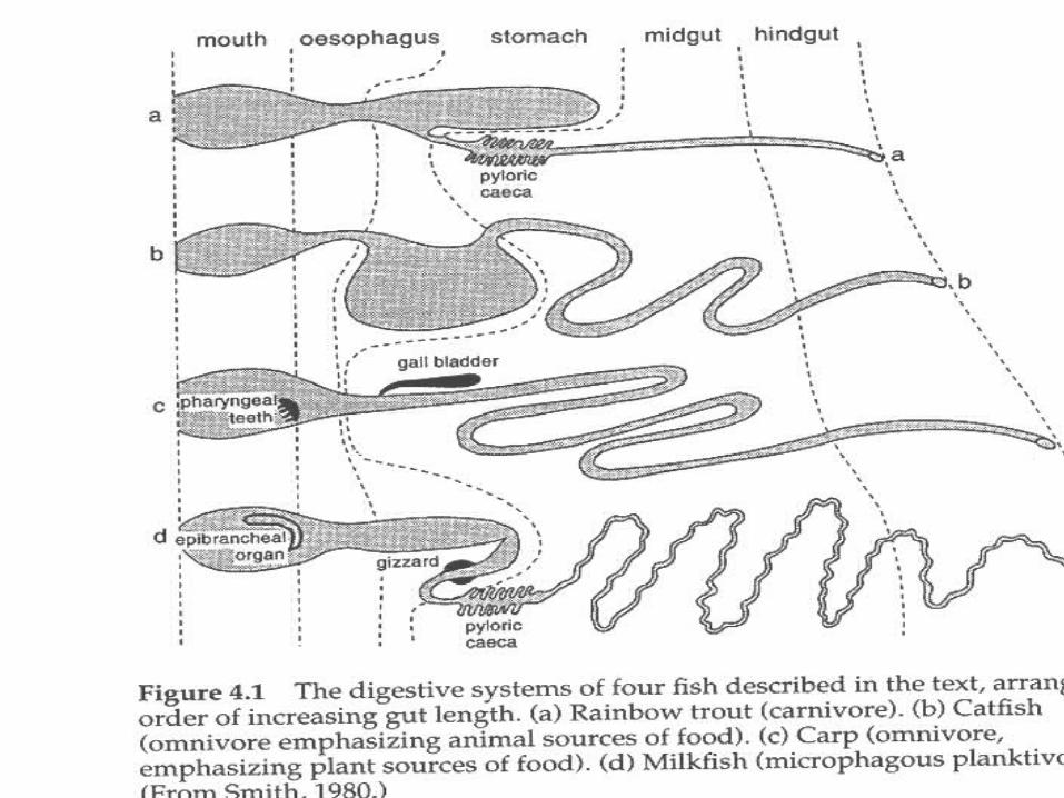
• GI tract varying considerably from spp. to spp. group. GI tract varying considerably from spp. to spp. group.
• Nutrient absorption increased by folding and increase of surface Nutrient absorption increased by folding and increase of surface area (typhlosole, sprial valve).area (typhlosole, sprial valve).

Alimentary Canal Oddities...Alimentary Canal Oddities...
• EsophagusEsophagus (Peristalsis) (Peristalsis)
-one-way trip for food! -one-way trip for food!
• StomachStomach (Killer pH!) (Killer pH!)
-Some tilipia <2! Can -Some tilipia <2! Can actually break down plant cell actually break down plant cell walls in absence of appropriate walls in absence of appropriate gastric enzymes.gastric enzymes.
• No stomach (lungfish)! If no stomach, then no HCl = no shell orNo stomach (lungfish)! If no stomach, then no HCl = no shell or bone digestion.bone digestion.
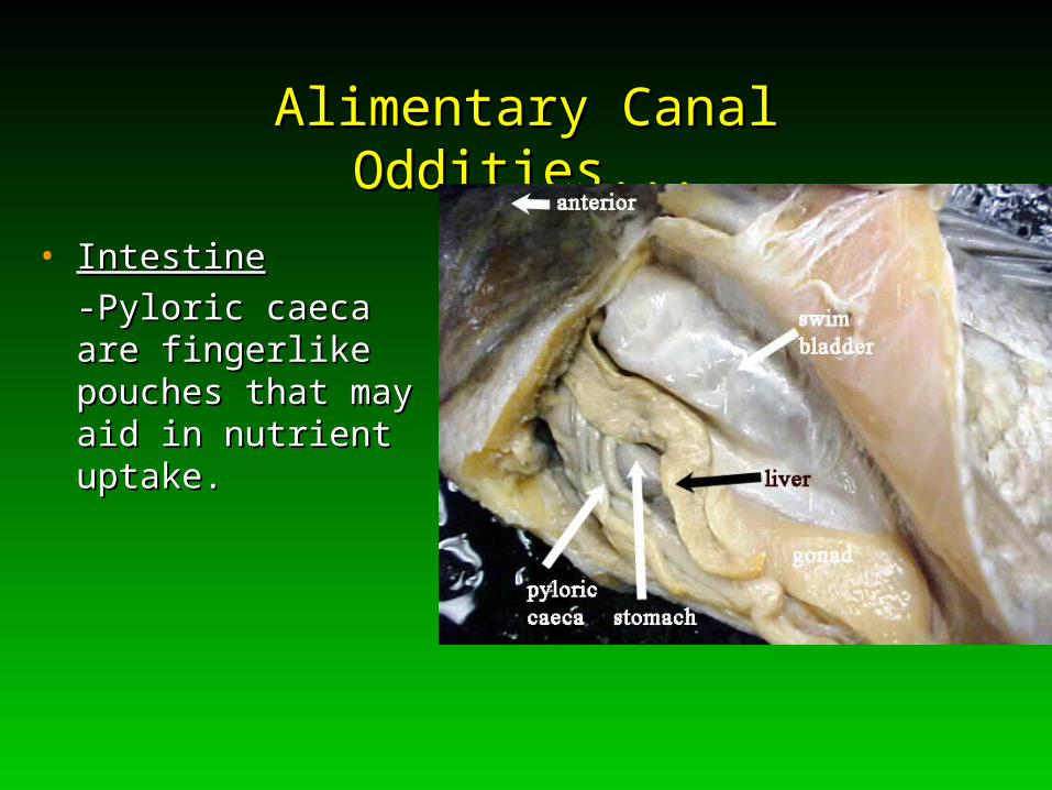
Alimentary Canal Oddities...Alimentary Canal Oddities...
• IntestineIntestine
-Pyloric caeca are -Pyloric caeca are fingerlike pouches that fingerlike pouches that may aid in nutrient may aid in nutrient uptake.uptake.

GI (cont.)GI (cont.)
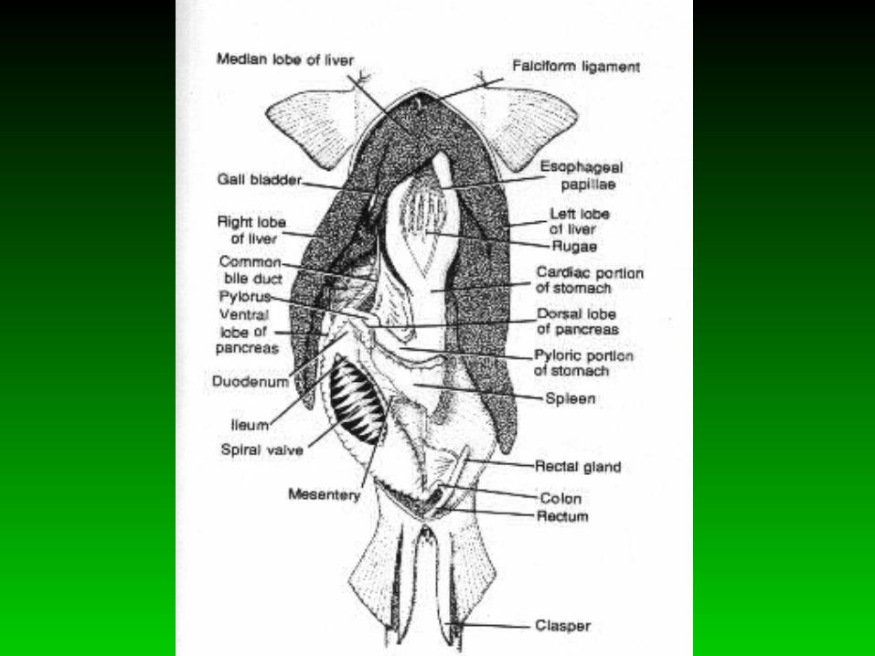
• LiverLiver-fat storage, detox., oil source.-fat storage, detox., oil source.
Sharks—huge liver ~50% of gut space, but they don’t Sharks—huge liver ~50% of gut space, but they don’t get cancer?? get cancer??
• GallbladderGallbladder—source of bile (fat emulsification).—source of bile (fat emulsification).
• PancreasPancreas—source of digestive enzymes —source of digestive enzymes part of liver in some fish and crustaceans (hepatopancreas)part of liver in some fish and crustaceans (hepatopancreas)

BuoyancyBuoyancy
• Fish regulate buoyancy several ways:Fish regulate buoyancy several ways:
(1) Low density tissue (liver in sharks)—increase fat(1) Low density tissue (liver in sharks)—increase fat(2) “Lift” from fin movement or hydrodynamics(2) “Lift” from fin movement or hydrodynamics(3) Reduced heavy tissue (bones and muscle)(3) Reduced heavy tissue (bones and muscle)(4) Gas (swim) bladder(4) Gas (swim) bladder
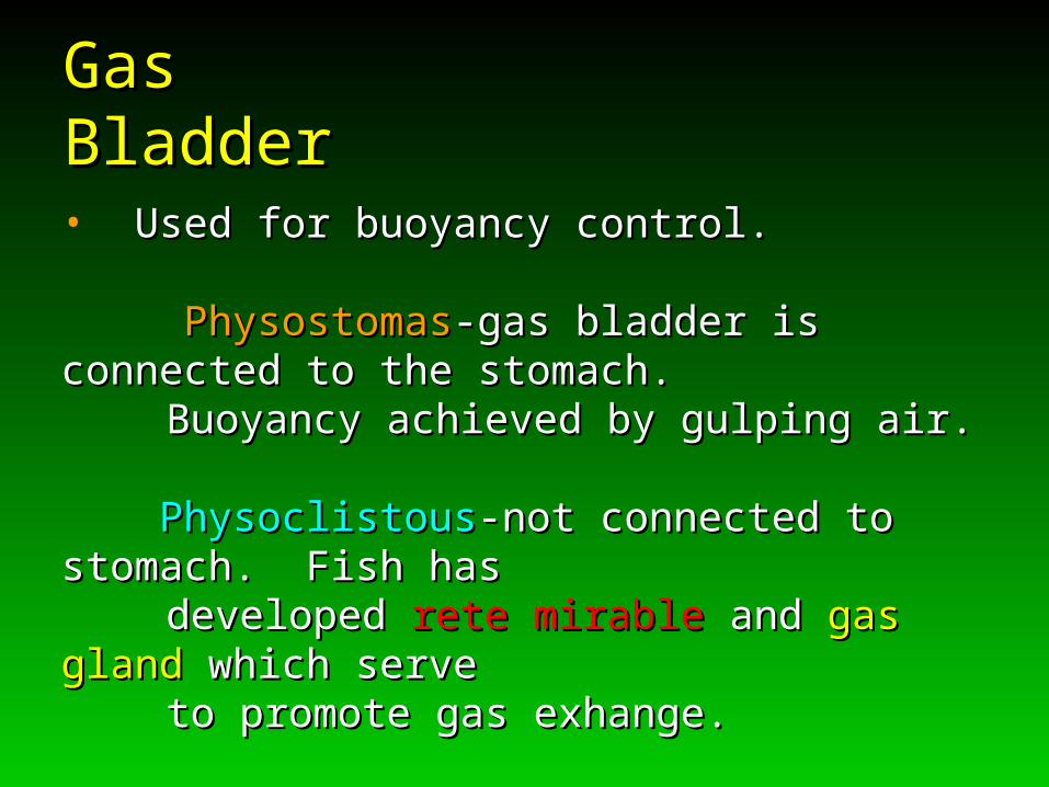
Gas BladderGas Bladder
• Used for buoyancy control.Used for buoyancy control.
PhysostomasPhysostomas-gas bladder is connected to the stomach. -gas bladder is connected to the stomach. Buoyancy achieved by gulping air. Buoyancy achieved by gulping air.
PhysoclistousPhysoclistous-not connected to stomach. Fish has-not connected to stomach. Fish has
developed developed rete mirablerete mirable and and gas glandgas gland which serve which serveto promote gas exhange. to promote gas exhange.


Smooth & Cardiac Muscles:Smooth & Cardiac Muscles:Circulation and the HeartCirculation and the Heart
Smooth & Cardiac Muscles:Smooth & Cardiac Muscles:Circulation and the HeartCirculation and the Heart
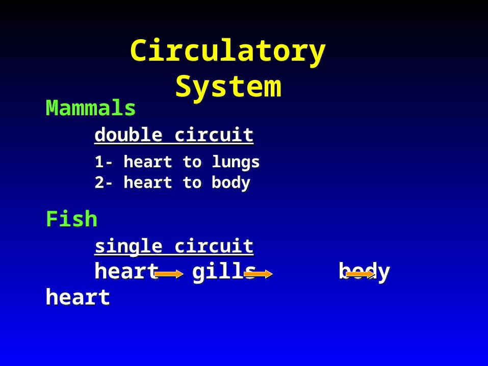
Circulatory SystemCirculatory System
Mammalsdouble circuit1- heart to lungs2- heart to body
Fishsingle circuitheart gills body heart
Mammalsdouble circuit1- heart to lungs2- heart to body
Fishsingle circuitheart gills body heart
Special conditions for fish circulation
Special conditions for fish circulation
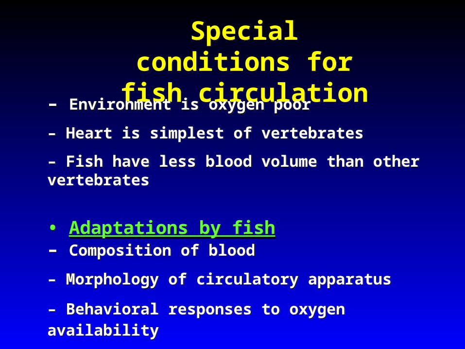
– Environment is oxygen poor
– Heart is simplest of vertebrates
– Fish have less blood volume than other vertebrates
• Adaptations by fish – Composition of blood
– Morphology of circulatory apparatus
– Behavioral responses to oxygen availability
– Environment is oxygen poor
– Heart is simplest of vertebrates
– Fish have less blood volume than other vertebrates
• Adaptations by fish – Composition of blood
– Morphology of circulatory apparatus
– Behavioral responses to oxygen availability

• Delivers oxygen
• Delivers nutrients
• Removes metabolic waste
• Fights pathogens
• Delivers oxygen
• Delivers nutrients
• Removes metabolic waste
• Fights pathogens
Functions of the Circulatory System
Functions of the Circulatory System

Components of the Circulatory System to Study
Components of the Circulatory System to Study
• Blood – Erythrocytes – Leukocytes – Structure of Hemoglobin
• Vascular system – Heart – Vessels
• Blood – Erythrocytes – Leukocytes – Structure of Hemoglobin
• Vascular system – Heart – Vessels
Blood Oxygen Affinity Blood Oxygen Affinity
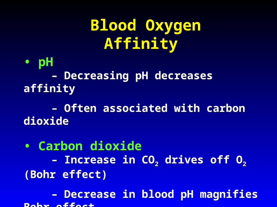
• pH
– Decreasing pH decreases affinity
– Often associated with carbon dioxide
• Carbon dioxide
– Increase in CO2 drives off O2 (Bohr effect)
– Decrease in blood pH magnifies Bohr effect
• pH
– Decreasing pH decreases affinity
– Often associated with carbon dioxide
• Carbon dioxide
– Increase in CO2 drives off O2 (Bohr effect)
– Decrease in blood pH magnifies Bohr effect

Blood Oxygen Affinity Blood Oxygen Affinity
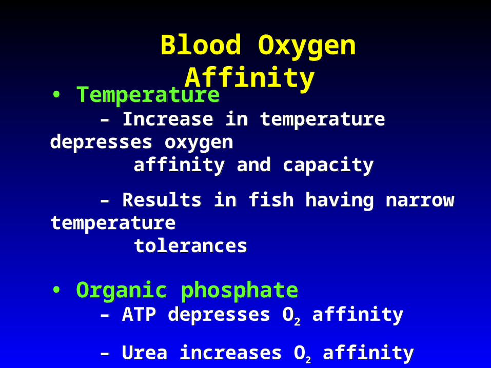
• Temperature
– Increase in temperature depresses oxygen affinity and capacity
– Results in fish having narrow temperature tolerances
• Organic phosphate
– ATP depresses O2 affinity
– Urea increases O2 affinity
• Temperature
– Increase in temperature depresses oxygen affinity and capacity
– Results in fish having narrow temperature tolerances
• Organic phosphate
– ATP depresses O2 affinity
– Urea increases O2 affinity
Fish Circulatory System Fish Circulatory System
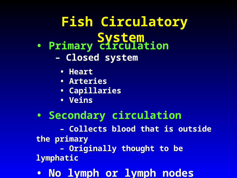
• Primary circulation
– Closed system
• Heart • Arteries • Capillaries • Veins
• Secondary circulation – Collects blood that is outside the primary – Originally thought to be lymphatic
• No lymph or lymph nodes
• Primary circulation
– Closed system
• Heart • Arteries • Capillaries • Veins
• Secondary circulation – Collects blood that is outside the primary – Originally thought to be lymphatic
• No lymph or lymph nodes

Divisions of Primary CirculationDivisions of Primary Circulation
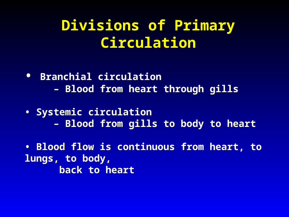
• Branchial circulation – Blood from heart through gills
• Systemic circulation
– Blood from gills to body to heart • Blood flow is continuous from heart, to lungs, to body,
back to heart
• Branchial circulation – Blood from heart through gills
• Systemic circulation
– Blood from gills to body to heart • Blood flow is continuous from heart, to lungs, to body,
back to heart
Proximity of Heart & Gills
Proximity of Heart & Gills
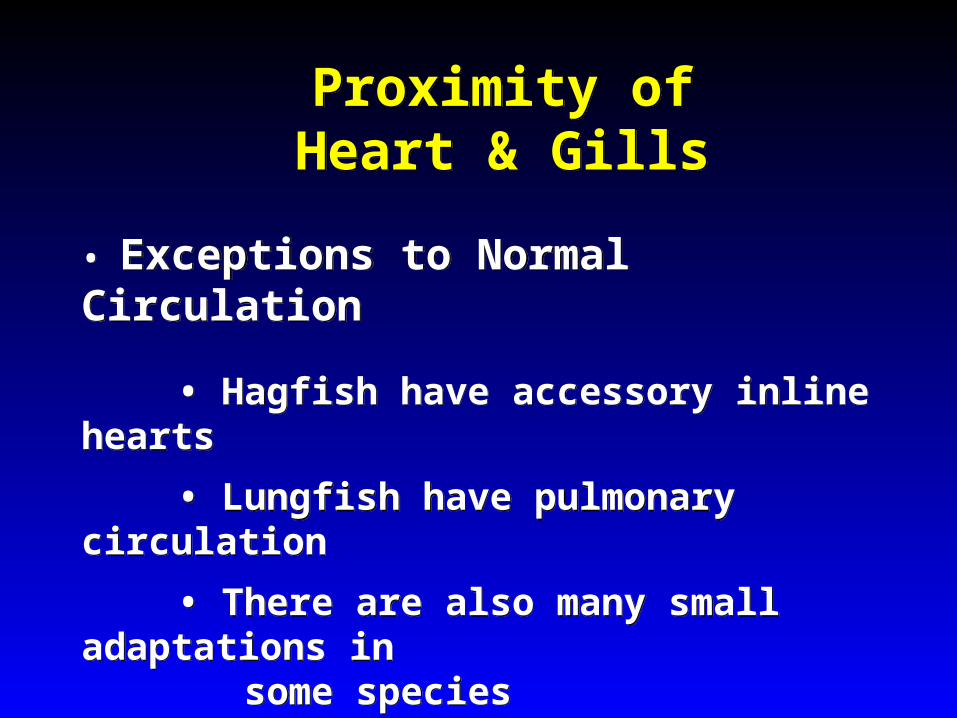
• Exceptions to Normal Circulation
• Hagfish have accessory inline hearts
• Lungfish have pulmonary circulation
• There are also many small adaptations in some species
• Exceptions to Normal Circulation
• Hagfish have accessory inline hearts
• Lungfish have pulmonary circulation
• There are also many small adaptations in some species

(Vascular circulation in lungfish.)
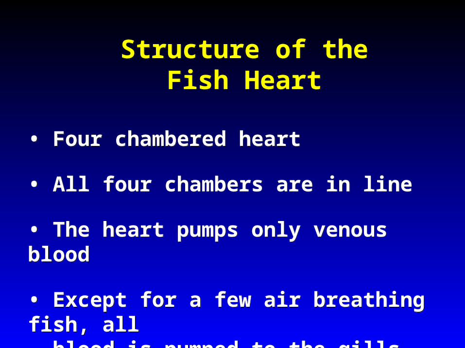
Structure of the Fish HeartStructure of the Fish Heart
• Four chambered heart
• All four chambers are in line
• The heart pumps only venous blood
• Except for a few air breathing fish, all blood is pumped to the gills
• Four chambered heart
• All four chambers are in line
• The heart pumps only venous blood
• Except for a few air breathing fish, all blood is pumped to the gills
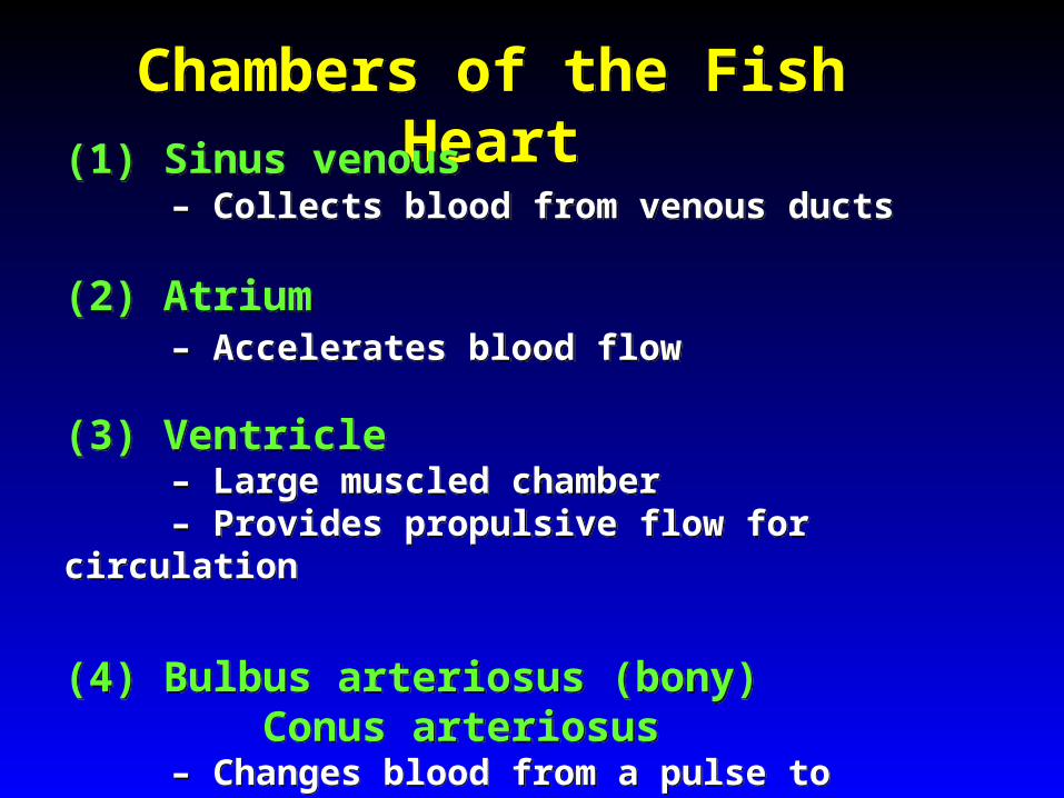
Chambers of the Fish HeartChambers of the Fish Heart
(1) Sinus venous – Collects blood from venous ducts
(2) Atrium
– Accelerates blood flow
(3) Ventricle – Large muscled chamber – Provides propulsive flow for circulation
(4) Bulbus arteriosus (bony) Conus arteriosus
– Changes blood from a pulse to continuous flow
(1) Sinus venous – Collects blood from venous ducts
(2) Atrium
– Accelerates blood flow
(3) Ventricle – Large muscled chamber – Provides propulsive flow for circulation
(4) Bulbus arteriosus (bony) Conus arteriosus
– Changes blood from a pulse to continuous flow

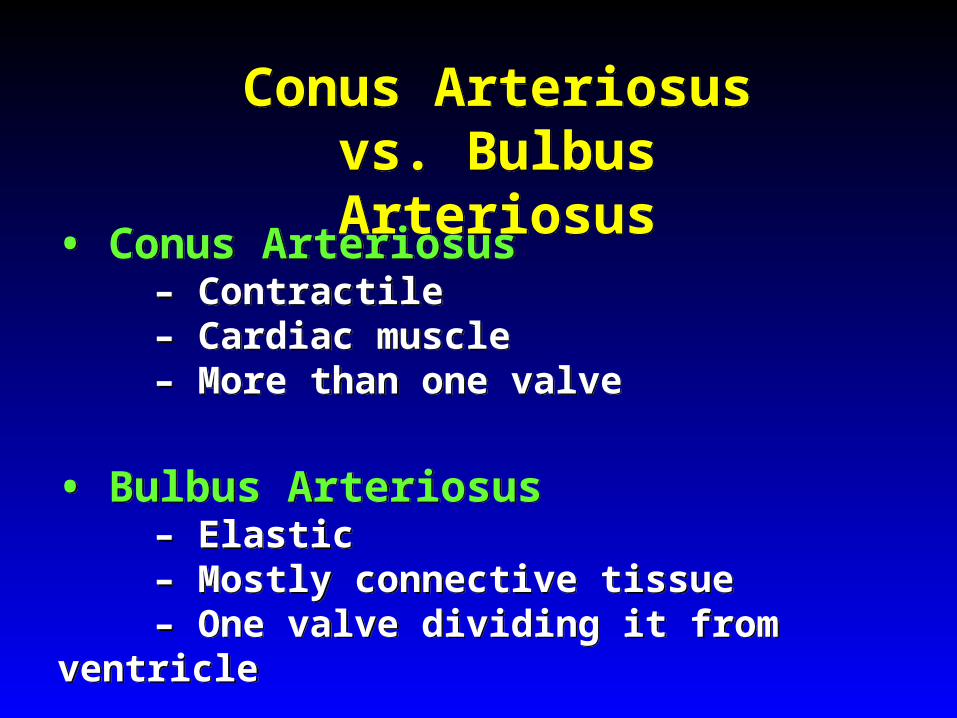
Conus Arteriosus vs. Bulbus Arteriosus
Conus Arteriosus vs. Bulbus Arteriosus
• Conus Arteriosus
– Contractile – Cardiac muscle – More than one valve
• Bulbus Arteriosus
– Elastic – Mostly connective tissue – One valve dividing it from ventricle
• Conus Arteriosus
– Contractile – Cardiac muscle – More than one valve
• Bulbus Arteriosus
– Elastic – Mostly connective tissue – One valve dividing it from ventricle

Regulation of the Fish HeartRegulation of the Fish Heart
• Self-regulating
• Timing can be modified by brain (influence on the autonomic nervous system)
• Pace is set by pacemaker cells
• Many areas show pacemaker activity
• Self-regulating
• Timing can be modified by brain (influence on the autonomic nervous system)
• Pace is set by pacemaker cells
• Many areas show pacemaker activity

The Hagfish Heart The Hagfish Heart
• Most primitive
• Sinus venous well developed
– Divided into two parts to receive different veins
• Bulbus arteriosus
• Have 3 additional hearts
– Cardinal heart in head – Caudal heart near end of tail – Portal heart – pumps blood through liver
• Most primitive
• Sinus venous well developed
– Divided into two parts to receive different veins
• Bulbus arteriosus
• Have 3 additional hearts
– Cardinal heart in head – Caudal heart near end of tail – Portal heart – pumps blood through liver
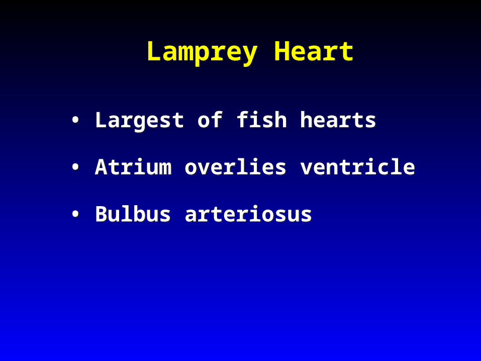
Lamprey HeartLamprey Heart
• Largest of fish hearts
• Atrium overlies ventricle
• Bulbus arteriosus
• Largest of fish hearts
• Atrium overlies ventricle
• Bulbus arteriosus

Elasmobranch HeartElasmobranch Heart
• Conus arteriosus
• Sinus venosus with almost no cardiac muscle
• Ventricle has two muscle layers – Compacta = compact outer layer – Spongiosa = inner layer
• Conus arteriosus
• Sinus venosus with almost no cardiac muscle
• Ventricle has two muscle layers – Compacta = compact outer layer – Spongiosa = inner layer
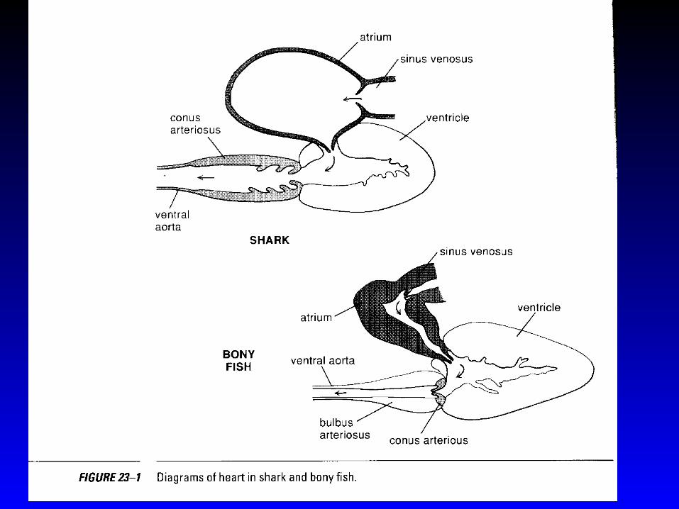
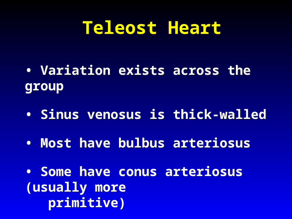
Teleost HeartTeleost Heart
• Variation exists across the group
• Sinus venosus is thick-walled
• Most have bulbus arteriosus
• Some have conus arteriosus (usually more primitive)
• Variation exists across the group
• Sinus venosus is thick-walled
• Most have bulbus arteriosus
• Some have conus arteriosus (usually more primitive)

Lungfish HeartLungfish Heart
• Atrium is divided into two parts by an incomplete septum
– Functional 3 chamber heart – Like amphibians – Right atrium larger than left – Right = deoxygenated from sinus venosus – Left = oxygenated from pulmonary vein
• Atrium is divided into two parts by an incomplete septum
– Functional 3 chamber heart – Like amphibians – Right atrium larger than left – Right = deoxygenated from sinus venosus – Left = oxygenated from pulmonary vein
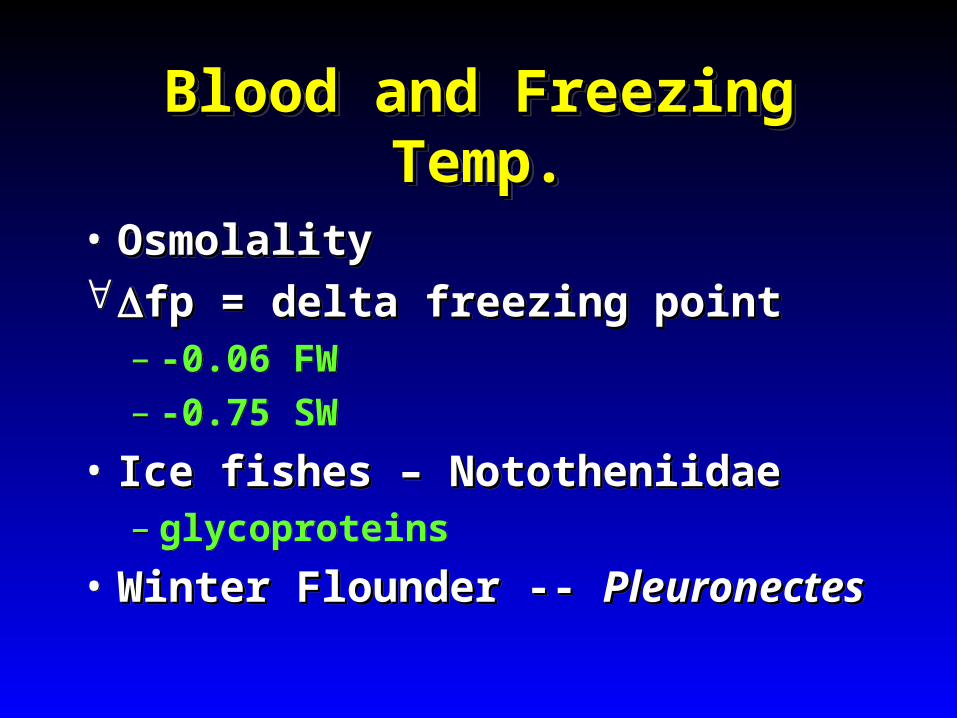
Blood and Freezing Temp.Blood and Freezing Temp.Blood and Freezing Temp.Blood and Freezing Temp.
• OsmolalityOsmolalityfp = delta freezing pointfp = delta freezing point
– -0.06 FW– -0.75 SW
• Ice fishes – NototheniidaeIce fishes – Nototheniidae– glycoproteins
• Winter Flounder -- Winter Flounder -- PleuronectesPleuronectes
• OsmolalityOsmolalityfp = delta freezing pointfp = delta freezing point
– -0.06 FW– -0.75 SW
• Ice fishes – NototheniidaeIce fishes – Nototheniidae– glycoproteins
• Winter Flounder -- Winter Flounder -- PleuronectesPleuronectes
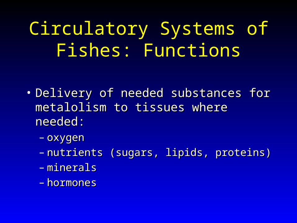
Circulatory Systems of Fishes: Functions
• Delivery of needed substances for Delivery of needed substances for metalolism to tissues where needed:metalolism to tissues where needed:– oxygenoxygen– nutrients (sugars, lipids, proteins)nutrients (sugars, lipids, proteins)– mineralsminerals– hormoneshormones
Functions of Circulatory SystemFunctions of Circulatory System
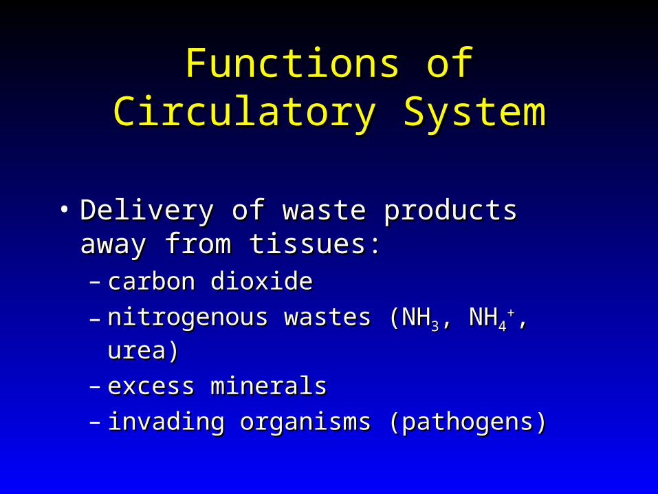
• Delivery of waste products away from Delivery of waste products away from tissues:tissues:– carbon dioxidecarbon dioxide
– nitrogenous wastes (NHnitrogenous wastes (NH33, NH, NH44++, urea), urea)
– excess mineralsexcess minerals– invading organisms (pathogens)invading organisms (pathogens)
Functions of Circulatory SystemFunctions of Circulatory System
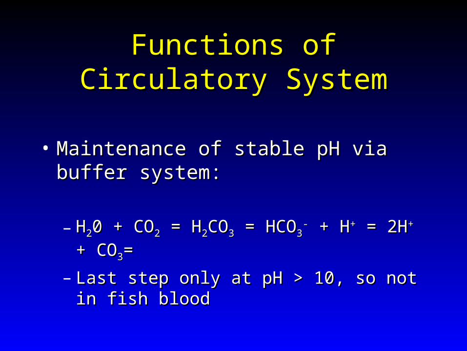
• Maintenance of stable pH via buffer system:Maintenance of stable pH via buffer system:
– HH220 + CO0 + CO22 = H = H22COCO33 = HCO = HCO33-- + H + H++ = 2H = 2H++ + +
COCO33==
– Last step only at pH > 10, so not in fish bloodLast step only at pH > 10, so not in fish blood
Components of Fish Circulatory Components of Fish Circulatory SystemsSystems
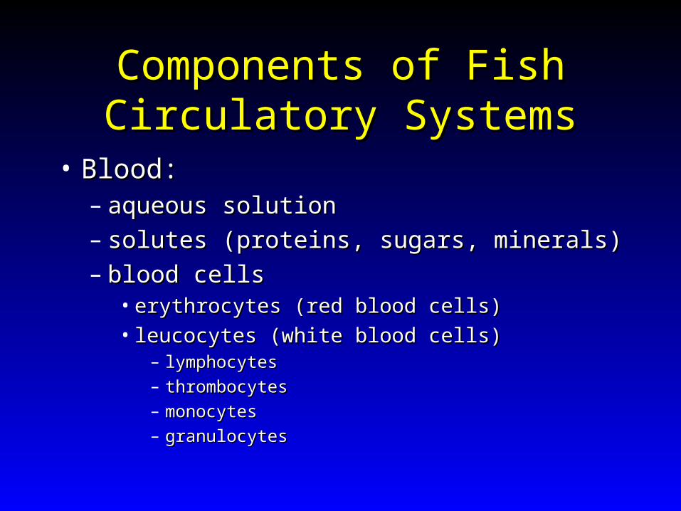
• Blood:Blood:– aqueous solutionaqueous solution– solutes (proteins, sugars, minerals)solutes (proteins, sugars, minerals)– blood cellsblood cells
• erythrocytes (red blood cells)erythrocytes (red blood cells)• leucocytes (white blood cells)leucocytes (white blood cells)
– lymphocyteslymphocytes
– thrombocytesthrombocytes
– monocytesmonocytes
– granulocytesgranulocytes
Components of Fish Circulatory Components of Fish Circulatory SystemsSystems
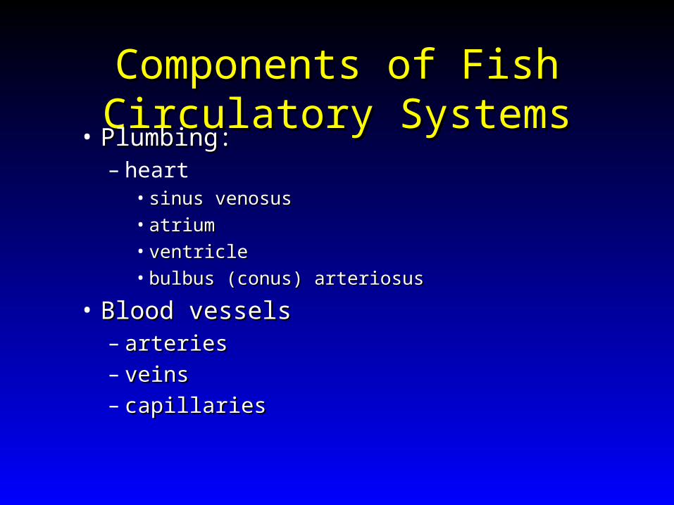
• Plumbing:Plumbing:– heart
• sinus venosussinus venosus• atriumatrium• ventricleventricle• bulbus (conus) arteriosusbulbus (conus) arteriosus
• Blood vesselsBlood vessels– arteriesarteries
– veinsveins
– capillariescapillaries
Nervous Systems of FishesNervous Systems of Fishes
Sensory, Motor and Integrative Sensory, Motor and Integrative FunctionsFunctions
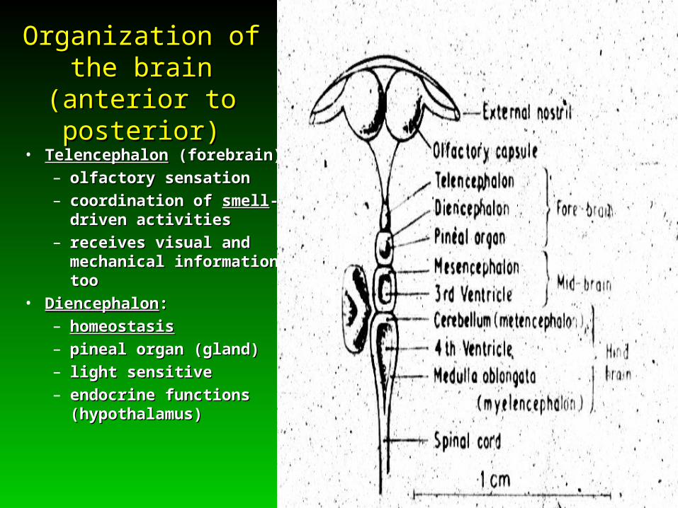
Organization of the brainOrganization of the brain(anterior to posterior)(anterior to posterior)
• TelencephalonTelencephalon (forebrain): (forebrain):
– olfactory sensationolfactory sensation
– coordination of coordination of smellsmell--driven activitiesdriven activities
– receives visual and receives visual and mechanical information, toomechanical information, too
• DiencephalonDiencephalon: :
– homeostasishomeostasis
– pineal organ (gland)pineal organ (gland)
– light sensitivelight sensitive
– endocrine functions endocrine functions (hypothalamus)(hypothalamus)
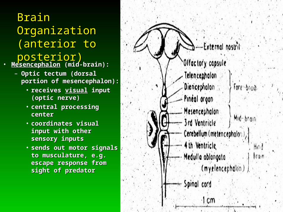
Brain Organization Brain Organization (anterior to posterior)(anterior to posterior)
• MesencephalonMesencephalon (mid-brain): (mid-brain):
– Optic tectum (dorsal portion Optic tectum (dorsal portion of mesencephalon):of mesencephalon):
• receives receives visualvisual input input (optic nerve)(optic nerve)
• central processing centercentral processing center
• coordinates visual input coordinates visual input with other sensory inputswith other sensory inputs
• sends out motor signals sends out motor signals to musculature, e.g. to musculature, e.g. escape response from escape response from sight of predatorsight of predator

Brain (cont.)Brain (cont.)
• MetencephalonMetencephalon (cerebellum):(cerebellum):
– coordinatescoordinates
swimming activityswimming activity
– coordinates:coordinates:
• balancebalance input input with motor with motor responseresponse
• electrical senseelectrical sense input with input with motor motor responseresponse
Organization of the brainOrganization of the brain(anterior to posterior)(anterior to posterior)
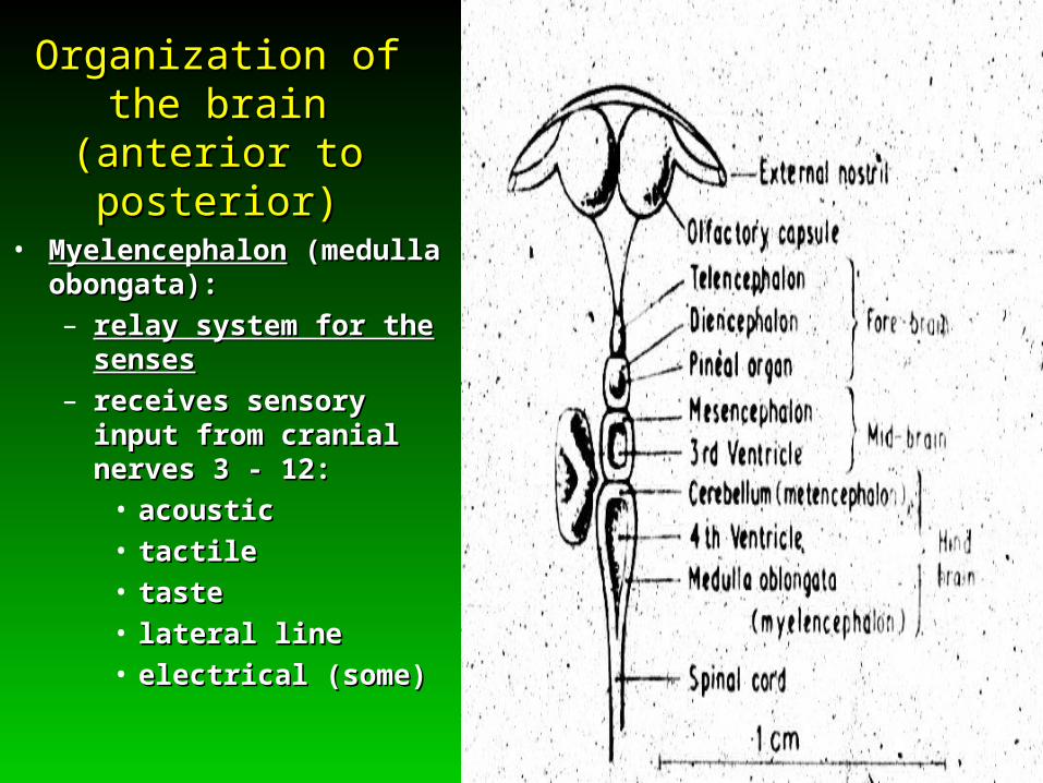
• MyelencephalonMyelencephalon (medulla (medulla obongata):obongata):
– relay system for the sensesrelay system for the senses
– receives sensory input from receives sensory input from cranial nerves 3 - 12:cranial nerves 3 - 12:
• acousticacoustic
• tactiletactile
• tastetaste
• lateral linelateral line
• electrical (some)electrical (some)