social cognition in mesial temporal lobe epilepsy (mtle) · of prof. dr. hennric jokeit and prof....
TRANSCRIPT

Zurich Open Repository andArchiveUniversity of ZurichMain LibraryStrickhofstrasse 39CH-8057 Zurichwww.zora.uzh.ch
Year: 2012
Social cognition in mesial temporal lobe epilepsy (MTLE)
Broicher, Sarah Dinah
Posted at the Zurich Open Repository and Archive, University of ZurichZORA URL: https://doi.org/10.5167/uzh-93509Dissertation
Originally published at:Broicher, Sarah Dinah. Social cognition in mesial temporal lobe epilepsy (MTLE). 2012, University ofZurich, Faculty of Arts.

SOCIAL COGNITION
IN MESIAL TEMPORAL LOBE EPILEPSY (MTLE)
Thesis
Presented to the Faculty of Arts
of
the University of Zurich
for the degree of Doctor of Philosophy
by
Sarah Dinah Broicher
of Germany
Accepted in the spring/autumn semester 2012 on the recommendation
of Prof. Dr. Hennric Jokeit and Prof. Dr. Lutz Jäncke and Prof. Dr.
Martin Meyer
(Zurich, 2012)
ACKNOWLEDGMENT
2
dedicated to
my family and friends
ACKNOWLEDGMENT
3
ACKNOWLEDGEMENT
This doctoral thesis was carried out at the department of neuropsychology at the Swiss
Epilepsy Centre in Zurich under supervision of Prof. Dr. rer. nat. Hennric Jokeit, head of
department of neuropsychology. Functional MRI measurements were performed at the MR
Institute of Radiology of Dominik Huber at Schulthess Clinic.
I would like to express my gratitude to the following:
Prof. Dr. rer. nat. Hennric Jokeit for his supervision during the three years of my
thesis, for sharing his rich experience and immense knowledge in all research and
clinical related questions, for his inspiration and support throughout my work, for his
critical reviews, his prompt answers to any of my questions, and for guiding me
throughout the three years of study
Prof. Dr. Boris B. Quednow for providing me the possibility to take part in numerous
scientific meetings at the Psychiatric University Hospital Zurich and his support and
motivation in many ways
Prof. Dr. Katharina Henke for inspiring me to write a thesis and thus layed the
foundation for this work
Each of my three advisors contributed in their own way to the success of this
dissertation and to my personal development.
In addition, I am particularly indepted to:
All patients who participated in this projects. Without their help and cooperation, this
work would not have been realised
Dr. Giorgi Kuchukhidze from the Medical University of Innsbruck and Dr. Lars
Frings from the Center of Geriatrics and Gerontology of Freiburg (Germany) for their
kind support, encouragement and fruitful cooperation
Dr. Dominik Huber, Gesa Osterkamp and Theresa Biesold from the Schulthess Clinic
in Zurich for the technical support during fMRI measurements and beyond that Gesa
Osterkamp for the joy she has given me with her witty sense of humour
Victoria Reed for meticulously correcting the English in the manuscript

ACKNOWLEDGMENT
4
Many thanks are also due to my colleagues at the Department of Neuropsychology for
their kind support and encouragement and numerous cosy and delicious lunch and
coffee breaks which were always empowering
My research colleagues for sharing both success and setbacks and for always being
there whenever I needed ready listeners
Finally, I would like to give a special thanks to:
My friends for their loyal support and encouragement. Without them, I could not have
accomplished nearly as much. They all stood by my side from year to year and
inspired me in silent ways that resound in endless echoes
My parents, sisters and my little niece and nephew for their endless love and support
help me through any of life’s challenges

CONTENT
5
CONTENT
1 ZUSAMMENFASSUNG ............................................................................................. 7
2 SUMMARY ................................................................................................................. 10
3 GENERAL INTRODUCTION AND OUTLINE OF THE THESIS..................... 13
3.1 DEFINITION EPILEPSY AND CLASSIFICATION ....................................................................................14
3.1.1 Mesial temporal lobe epilepsy (MTLE) .....................................................................................15
3.2 SOCIAL COGNITION .........................................................................................................................16
3.2.1 Basal social cognitive processes ...............................................................................................17
3.2.2 Theory of Mind (ToM) ...............................................................................................................18
3.3 TESTING SOCIAL COGNITION ...........................................................................................................19
3.3.1 Selected tests of basal processes of social cognition .................................................................19
3.3.2 Selected tests of theory of mind .................................................................................................20
3.3.3 Self-report questionnaires of psychopathology and quality of life ............................................23
3.4 IMAGING OF SOCIAL COGNITION ......................................................................................................25
3.5 SOCIAL COGNITION IN TEMPORAL LOBE EPILEPSY ...........................................................................27
4 RESEARCH OBJECTIVES ..................................................................................... 40
4.1 FIRST STUDY: EMOTION RECOGNITION AND THEORY OF MIND IN SYMPTOMATIC MESIAL TEMPORAL
LOBE EPILEPSY PATIENTS (MTLE) .....................................................................................................................41
4.2 SECOND STUDY: ASSOCIATION BETWEEN IPSILATERAL AMYGDALAR DYSFUNCTION IN MESIAL
TEMPORAL LOBE EPILEPSY (MTLE), PERFORMANCE IN THEORY OF MIND (TOM) ABILITIES, AND
LATERALIZATION OF SEIZURE-ONSET SIDE .........................................................................................................41
4.3 THIRD STUDY: ASSOCIATION BETWEEN IPSILATERAL AMYGDALAR DYSFUNCTION IN MTLE AND
FUNCTIONAL CONNECTIVITY OF THE AMYGDALE WITH REMOTE TEMPORAL AND FRONTAL BRAIN AREAS .........42
4.4 FOURTH STUDY: ASSOCIATION BETWEEN STRUCTURAL ABNORMALITIES AND FMRI RESPONSE IN
THE AMYGDALA IN PATIENTS WITH TEMPORAL LOBE EPILEPSY ..........................................................................42
5 ORIGINAL ARTICLES ............................................................................................ 45
5.1 TELL ME HOW DO I FEEL – EMOTION RECOGNITION AND THEORY OF MIND IN SYMPTOMATIC MESIAL
TEMPORAL LOBE EPILEPSY ................................................................. FEHLER! TEXTMARKE NICHT DEFINIERT.
5.2 ADVANCED SOCIAL COGNITION AND AMYGDALAR FMRI RESPONSE IN PATIENTS WITH UNILATERAL
MESIAL TEMPORAL LOBE EPILEPSY .....................................................................................................................88
5.3 ALTERATIONS IN FUNCTIONAL CONNECTIVITY OF THE AMYGDALE IN UNILATERAL MESIAL
TEMPORAL LOBE EPILEPSY ...............................................................................................................................112
5.4 FMRI RESPONSE OF DYSPLASTIC AMYGDALA IN PATIENTS WITH MESIAL TEMPORAL LOBE EPILEPSY .
......................................................................................................................................................135

CONTENT
6
6 SUMMARY AND GENERAL DISCUSSION ....................................................... 150
6.1 SUMMARY .....................................................................................................................................150
6.2 METHODOLOGICAL CONSIDERATIONS ...........................................................................................154
6.3 STRENGTHS AND LIMITATIONS OF THE PRESENT STUDIES ..............................................................156
6.4 CLINICAL IMPLICATIONS ...............................................................................................................157
6.5 SUGGESTIONS FOR FUTURE STUDIES .............................................................................................158
8 PUBLICATIONS ..................................................................................................... 160
8.1 PAPERS .........................................................................................................................................160
8.2 BOOK CHAPTER .............................................................................................................................160
8.3 POSTERS........................................................................................................................................160
8.4 TALKS ...........................................................................................................................................161
9 CURRICULUM VITAE .......................................................................................... 162
ZUSAMMENFASSUNG
7
1 ZUSAMMENFASSUNG
Zahlreiche klinische Studien deuten darauf hin, dass Patienten mit chronischer Epilepsie
häufig fehlangepasstes Verhalten und psychiatrische Komorbidiäten aufweisen (Hermann,
Seidenberg, & Bell, 2000). Inwiefern diese Fehlanpassungen durch Ängste und Sorgen
hinsichtlich der epileptischen Anfälle, die Wahrnehmung von Stigmatisierung und
Diskriminierung (z.B. im Arbeitsumfeld) sowie fehlende soziale Unterstützung begründet
sind oder auf mögliche Beeinträchtigungen im Bereich sozialer Kognition hindeuten, ist
Gegenstand kontroverser Diskussionen (Devinsky & Najjar, 1999; Shackleton, Kasteleijn-
Nolst Trenite, de Craen, Vandenbroucke, & Westendorp, 2003).
Aufgaben, welche sozial-kognitive Fähigkeiten verlangen scheinen ein weit distribuiertes
neuronales Netzwerk zu aktivieren. Experimente mit bildgebenden Verfahren haben zu
Grunde liegende neuronale Prozesse in verschiedenen frontal und temporal gelagerten
Hirnregionen lokalisiert (Adolphs, 2003; Gallagher & Frith, 2003; Vollm et al., 2006). Doch
obwohl diese Hirnregionen auch bei einer mesialen Temporallappenepilepsie (MTLE)
betroffen sind, wurden sozial kognitive Fähigkeiten bei dieser Patientengruppe bisher kaum
untersucht (Kirsch, 2006).
Die vorliegende Doktorarbeit hatte zum Ziel, die Frage zu beantworten, inwiefern
Patienten mit MTLE gegenüber Patienten mit einer Epilepsie ausserhalb der mesiotemporalen
und frontalen Strukturen in ihren sozial-kognitiven Fähigkeiten beeinträchtigt sind. Dabei
sollte auch der Frage nachgegangen werden, welche Bedeutung spezifisch der Amygdala
zukommt und welche affektiven Funktionen durch diese Struktur und die mit ihr verknüpften
Hirnregionen vermittelt werden.
Zu diesem Zweck wurden in einer ersten Studie die Emotionserkennung, das Erkennen von
Intentionen und Ansichten anderer (Theory of Mind) sowie die emotional-basierte
Entscheidungsfähigkeit mittels einer umfangreichen Testbatterie gut etablierter und empirisch
erprobter Testparametern untersucht. MTLE Patienten zeigten gegenüber gesunden
Kontrollen sowohl Beeinträchtigungen in der Emotionserkennung in Gesichtern als auch der
gesprochenen Sprache, in zahlreichen ToM Tests sowie der Entscheidungsfähigkeit unter
Ambiguität und Unsicherheit. MTLE Patienten wiesen gegenüber der Epilepsiekontrollgruppe
insbesondere Beeinträchtigungen in der allgemeinen Emotionserkennung auf und zeigten
zudem keine adäquate Anpassung des Verhaltens an Rückmeldungen in einem
Entscheidungstest. Darüber hinaus unterschied sich die Leistung der Epilepsiekontrollgruppe

ZUSAMMENFASSUNG
8
weder signifikant von der Leistung der MTLE Patienten noch von jener der gesunden
Kontrollen und lag zwischen diesen beiden Gruppen.
Eine Untersuchung von Schacher et al. (2006) deutet darauf hin, dass bei Patienten mit
unilateraler MTLE häufig die ipsilaterale Funktion der Amygdala beeinträchtigt ist. Die
Bedeutung der Amygdala für die Theory of Mind (ToM) Fähigkeit ist aufgrund der
uneindeutigen Befundlage bisher noch nicht geklärt. Somit untersuchten wir in einer zweiten
Studie, ob pathologisch asymmetrische amygdaläre fMRI Aktivierungen, die mit einem
Lateralisationsindex operationalisiert wurden, mit der Leistung im Recognition of Faux Pas
Test in einem Zusammenhang stehen. Patienten mit einer rechtsseitigen MTLE zeigten
beeinträchtigte Leistungen im Vergleich zu linksseitigen MTLE Patienten. In Patienten mit
rechtsseitiger MTLE korrelierte der fMRT-Lateralisationsindex der ipsilateralen amygdalären
Aktivierung signifikant mit der Leistung im Faux Pas Test. Mittels schrittweiser multipler
Regressionsanalyse konnte jedoch gezeigt werden, dass der fMRT-Lateralisationsindex, nebst
der Seite der Epilepsie, nicht signifikant zur Erklärung der Variation der Leistung im Faux
Pas Test beitragen konnte.
Die Amygdala weist Verbindungen mit zahlreichen kortikalen und subkortikalen Arealen
auf, einschliesslich Rückprojektionen zum frontalen Kortex. Aufgrund dieser weitreichenden
Verknüpfungen, gingen wir in einer dritten Studie der Frage nach, inwiefern sich bei
Patienten mit einer Signalminderung in der ipsilateralen Amygdala, funktionelle,
modulatorische Einflüsse auf weiter entfernt liegende, aber mit der Amygdala verknüpfte
Hirnregionen zeigen. Darüber hinaus untersuchten wir, welche Strukturen innerhalb des
Amygdala-Netzwerkes für ToM Leistungen relevant sind. Zur Erfassung funktioneller
Konnektivität wurde die nabhängige Komponentenanalyse (Independent Component
Analysis, ICA) angewendet, bei der es sich um eine relativ neue, hypothesenfreie,
datengetriebene Analysemethode handelt, die dazu dient, unabhängige Signalquellen aus
einem Gesamtsignal zu extrahieren und die Konnektivität signifikanter Aktivitätsknoten
abzubilden (Calhoun, Adali, Pearlson, & Pekar, 2001). Mittels funktioneller
Konnektivitätsanalyse wurde ein Netzwerk identifiziert, welches bei gesunden
Kontrollpersonen asymmetrisch organisiert war. Bei MTLE Patienten war diese beobachtete
Asymmetrie der Amygdalakonnektivität durch die Lateralisation der Seite des Anfallsbeginns
moduliert. Es zeigten sich sowohl bei links- als auch bei rechtsseitigen MTLE Patienten eine
verminderte Koaktivierung in linkshemisphärischen temporalen und frontalen Strukturen.
Zudem konnten wir zeigen, dass die kontralaterale Integrität eine grössere Rolle für ToM
Leistungen zu spielen scheint als die verbleibende ipsilaterale Aktivität. Diese Ergebnisse

ZUSAMMENFASSUNG
9
wurden als Hinweis auf kompensatorische Aktivierungen kontralateraler mesiotemporaler
Strukturen gedeutet.
Eine vierte Studie ging schliesslich der Frage nach, inwiefern die amygdaläre Reaktion im
fMRT bei einer dysplastischen Amygdala beeinträchtigt ist. Es zeigte sich, dass eine
amygdaläre Läsion (Dysplasie) nicht notwendigerweise mit funktionellen Beeinträchtigungen
assoziiert ist, was anhand dreier Patienten mit linksseitiger Amygdaladysplasie gezeigt
werden konnte.
Zusammenfassend lassen die vier Studien dieser Dissertation darauf schliessen, dass
sozial-kognitive Fähigkeiten bei Patienten mit einer chronischen Epilepsie gegenüber
Gesunden beeinträchtigt sind. Auch Patienten mit nicht fokalen Epilepsien, die
gleichermassen wie die MTLE Patienten antiepileptische Medikation erhalten, können
Schwierigkeiten in der sozialen Kognition aufweisen, dies jedoch deutlich seltener als
Patienten mit MTLE. Allein die Chronizität der Erkrankung oder Medikation lassen sich
somit für die auftretenden Defizite nicht verantwortlich machen. Die MTLE scheint einen
spezifischen Risikofaktor für Defizite in der sozialen Kognition darzustellen, wobei dies
vermutlich auf die Beteiligung des fronto-limbischen Systems, einschliesslich der Amygdala,
am Krankheitsgeschehen zurückzuführen ist.

SUMMARY
10
2 SUMMARY
Numerous clinical studies have revealed that psychosocial maladjustment is a serious issue
for many patients with chronic epilepsies (Hermann et al., 2000). To what extent these
maladjustments are caused by social burdens, stigma, and the risk factors of an active
epilepsy, and to what extent they are due to dysfunctional social cognition, is still a matter of
controversy (Devinsky & Najjar, 1999; Shackleton et al., 2003).
Tasks that demand social cognitive abilities appear to activate a consistent set of brain
regions. Experiments using imaging techniques have found underlying neural processes in
different frontal and temporal localized brain regions (Adolphs, 2003; Gallagher & Frith,
2003; Vollm et al., 2006). Despite knowledge that these brain regions are frequently affected
in patients with mesial temporal lobe epilepsy (MTLE), social-cognitive abilities, such as
theory of mind (ToM), have received little attention in this patient group (Kirsch, 2006).
In the present thesis, we conducted four experimental studies to answer the question
whether patients with MTLE compared to patients with epilepsies outside the mesiotemporal
and frontal structures were specifically impaired in their social-cognitive abilities. Thereby,
the questions should be answered which role the amygdala plays in emotional and social
function and which affective functions were mediated by the amygdala and its connected
brain regions.
Our first study was aimed at investigating emotion recognition, theory of mind, and
decision making using a set of tasks that combine behavioural and psychological measures of
social and emotional variables. MTLE patients were significantly impaired relative to healthy
controls (HC) in all measures of social perception affecting the ability to interpret emotional
expressions and feelings from faces and voices and, with one exception, on all advanced tests
of reasoning about the mental states of others. MTLE patients were predominantely impaired
in their general emotion recognition abilities compared to extra-MTLE patients. In contrast,
subjects with extra-MTLE showed no significant impairment in tests of social cognition
relative to HC. Performance in the epileptic control group, while not significantly differing
from performance in either the MTLE or healthy control group, lay between these two groups
on nearly all measures.
Secondly, we wanted to investigate possible effects of functional and structural amygdalar
abnormalities on higher-order social behaviour. Thus, patients with mesial temporal lobe
epilepsy (MTLE) frequently show ipsilateral amygdalar dysfunctions (Schacher, Haemmerle
et al., 2006). However, the role of the amygdala in higher-order social cognition such as ToM

SUMMARY
11
is still under debate. Therefore, our second study aimed at investigating whether the
asymmetry ratio of functional magnetic resonance imaging (fMRI) amygdalar activity is
related to performance in an advanced ToM task, the recognition of faux pas test. We found
lower performances in patients with right-sided MTLE compared to patients with left-sided
MTLE in the recognition of faux pas test. In patients with right-sided MTLE, the recognition
of faux pas scores correlated with the degree of asymmetry of fMRI activation in the
ipsilateral amygdala. However, stepwise multiple regression analysis revealed that the
amygdalar asymmetry ratio did not significantly contribute to the explanation of faux pas test
performance variability beyond that explained by side of MTLE.
Since the amygdala has extensive connections with many cortical and subcortical areas,
including backprojections to the frontal cortex, we further wanted to evaluate wheter
ipsilateral amygdalar dysfunction has functional modulatory influences on these remote brain
structures. To pursue functional connectivity, we used independent component analysis (ICA)
of fMRI data to characterize possible amygdala network alterations that may be caused by
ipsilateral amygdala dysfunction. ICA is a relatively new, hypothesis-free, data-driven method
which is used to extract independent source signals from an overall signal and that allows one
to image connectivity of significant nodes of activity (Calhoun et al., 2001). We furthermore
investigated the relationship between activation within the amygdala network and behavioural
performance in a ToM task. In healthy controls a hemispheric asymmetry of the amygdala
network was present with amygdala co-activation in predominantely left temporolateral and
frontal brain strucutes. In MTLE patients the side of pathology modulated the observed
asymmetry of amygdala connectivity. In MTLE patients the extent of amygdalar connectivity
to the prahippocampal gyrus and insula was related to ToM test performance.
The fourth study was aimed at investigating, whether amygdalar response in fMRI is
impaired if it is affected by dysplasia. We found that amygdala lesion (dysplasia) is not
necessarily associated with the functional impairment, which was demonstrated in three
patients with left-sided dysplasia.
In conclusion, we take our data to suggest that MTLE can be considered as a specific risk
factor for the development of social-cognitive deficits. It can be concluded that the chronicity
of epilepsy is an important factor in the predisposition of these patients to social-cognitive
deficits, but that brain dysfunction can pose an additional hazard, probably related to the
involvement of the fronto-limbic system.
ABBREVIATIONS
12
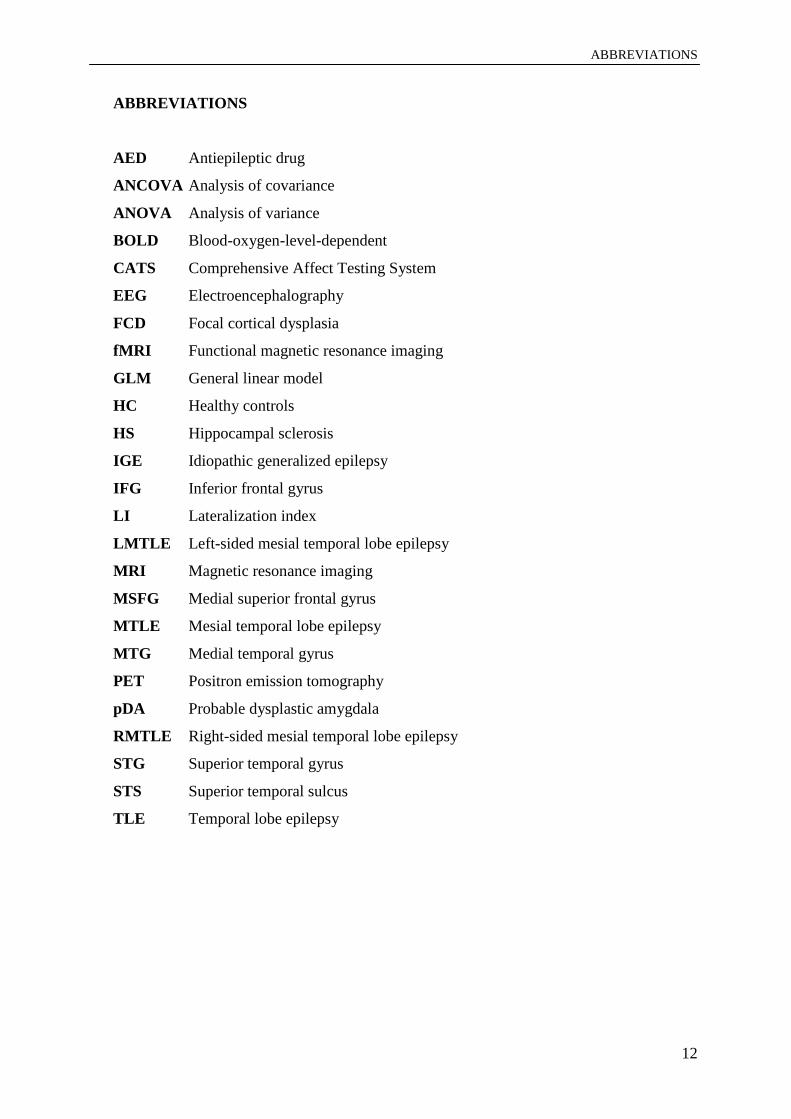
ABBREVIATIONS
AED Antiepileptic drug
ANCOVA Analysis of covariance
ANOVA Analysis of variance
BOLD Blood-oxygen-level-dependent
CATS Comprehensive Affect Testing System
EEG Electroencephalography
FCD Focal cortical dysplasia
fMRI Functional magnetic resonance imaging
GLM General linear model
HC Healthy controls
HS Hippocampal sclerosis
IGE Idiopathic generalized epilepsy
IFG Inferior frontal gyrus
LI Lateralization index
LMTLE Left-sided mesial temporal lobe epilepsy
MRI Magnetic resonance imaging
MSFG Medial superior frontal gyrus
MTLE Mesial temporal lobe epilepsy
MTG Medial temporal gyrus
PET Positron emission tomography
pDA Probable dysplastic amygdala
RMTLE Right-sided mesial temporal lobe epilepsy
STG Superior temporal gyrus
STS Superior temporal sulcus
TLE Temporal lobe epilepsy

GENERAL INTRODUCTION AND OUTLINE OF THESIS
13
3 GENERAL INTRODUCTION AND OUTLINE OF THE THESIS
This dissertation contains six sections. Section 1 and 2 give a short summary of the thesis.
Section 3 is the general introduction to the subject of this dissertation. Section 4 concerns the
aims and hypotheses of all four studies. In section 5.1 emotion recognition and ToM in
symptomatic MTLE patients are investigated. Patients with MTLE are compared with patients
with extra-MTLE and healthy controls (HC). Also, various aspects of psychopathology and
quality of life are assessed to study their contribution to deficits in social cognition.
Additionally, other epilepsy related variables are evaluated as potential risk factors for social-
cognitive deficits. In section 5.2 the relationship between the asymmetry ratio of fMRI
amygdalar activity and performance in an advanced ToM task, the recognition of faux pas
test, is investigated. In section 5.3 patients with MTLE are compared with HC in functional
connectivity of the amygdala. In section 5.4 the association between structural abnormalities
and functional magnetic resonance imaging (fMRI) bold oxygen level dependent (BOLD)
response in the amygdala in patients with temporal lobe epilepsy is investigated. In the final
section, section 6, the results of the studies are summarized and methodological issues and
implications for clinical practice and suggestions for future research are discussed.
Social neuroscience is an emerging interdisciplinary field aimed at investigating the
fundamentals of human social and emotional behaviour, the quintessence of which is the
relationship between brain processes and social interaction. Studies on the impact of
neurological, psychiatric, and psychological conditions on human social behaviour contribute
to our understanding of the complexity of social interactions and highlight important social
and affective symptoms in brain disorders such as epilepsies which continue to be overlooked
in clinical practice.
In patients with epilepsy non-social cognitive functions including memory, language and
executive functions have been studied for many decades, whereas social cognitive abilities
have received little attention (Kirsch, 2006). This is quite astonishing in light of what we
know about the remarkable overlap between structures associated with social cognition and
anterior brain structures which are frequently affected in patients with epilepsy. The paucity
of research becomes more understandable when one considers the lack of readily apparent
social deficits in the majority of patients with epilepsies (Phelps & LeDoux, 2005).
Nevertheless, comprehensive clinical studies have revealed that psychosocial
maladjustment is a serious problem in many patients with chronic epilepsies (Hermann et al.,

GENERAL INTRODUCTION AND OUTLINE OF THESIS
14
2000). To what extent these maladjustments are caused by social burdens, stigma, and risk
factors of active epilepsy, and to what extent they are due to dysfunctional social cognition,
remains an open question (Devinsky & Najjar, 1999; Shackleton et al., 2003). However, the
fact that psychosocial maladjustment and psychiatric comorbidity are more frequent in certain
focal epilepsies compared with other epilepsy syndromes may reflect a specific pathological
association (Perini et al., 1996).
In the past, psychiatry and neurology have used different terms and concepts and differed
in their diagnostic approaches, research and treatment methods. Their focus converges to
some degree within the framework of the modern neurosciences. As such, social and affective
neuroscience provides insight into behavioural disorders in patients with epilepsy via new
unifying concepts that can be investigated by means of behavioural tests, structural and
functional imaging as well as by neuropsychopharmacological interventions. These
opportunities allow us to advance our understanding of brain diseases, how they affect
behaviour and raise the hope of new and more efficient therapeutic interventions.
3.1 Definition epilepsy and classification
Epilepsy is a worldwide common brain disorder. According to the world health
organization (WHO) up to 10% of the general population will suffer from cerebral seizures at
least once in their lifetime. Epilepsy is defined as a brain disorder characterised
predominantly by recurrent and unpredictable interruptions of normal brain function, called
epileptic seizures and by the neurobiologic, cognitive, psychologic, and social consequences
of this condition (Fisher et al., 2005). An epileptic seizure is a transient occurrence of signs
and/or symptoms due to abnormal hypersynchronous discharges of cortical neurons. The
clinical signs and symptoms of seizures depend on the localization of foci and the extent of
propagation of the epileptiform discharge. Seizures can affect consciousness, memory,
emotional state, cognition, behaviour and sensory-, motor-, and autonomic functions.
The International League Against Epilepsy (ILAE) proposed an international classification
of epilepsy, for both seizure types and epilepsy syndromes to achieve uniform of terminology
(1981; 1989). Based on whether the source of the seizure within the brain is localized or
distributed, seizures are classified as partial (or focal) or generalized. A seizure is classified as
partial when there is evidence of a clinical partial onset, regardless of whether the seizure
secondarily generalizes. Partial seizures are further devided on the basis of whether or not
consciousness is affected (simple partial seizures vs. complex partial seizues). When a partial
seizure spreads within the brain, it is classified as partial seizure with secondarily
GENERAL INTRODUCTION AND OUTLINE OF THESIS
15
generalization. A seizure is considered primary generalized when clinical symptomatology
provides no indication of an anatomic localisation and no clinical evidence of focal onset.
They can be divided according to the effect on the body into absence, myoclonic, clonic,
tonic, tonic-clonic, and atonic seizures. The ictal EEG patterns are usually bilateral and reflect
neuronal discharge that is widespread in both hemispheres.
Epilepsy syndromes can be further classified by presumptive cause into idiopathic,
symptomatic and cryptogenic. In general, idiopathic (or primary) generalized epilepsy (IGE)
is virtually synonymous with genetic epilepsy and means that no underlying cause is apparent
other than a possible hereditary predisposition to seizures. When the epilepsy arises from the
effects of an epileptic lesion (e.g. metabolic disturbances, cerebral malformations, head
injury, infections, brain tumours), the epilepsy syndrome is called symptomatic (or secondary
epilepsy). Cryptogenic epilepsy is considered as secondary epilepsy, but the underlying cause
has not been identified.
3.1.1 Mesial temporal lobe epilepsy (MTLE)
Mesial temporal lobe epilepsy (MTLE) is the most prevalent focal epilepsy. It is
characterized by recurrent seizures that originate from mesial temporal structures, most
frequently within the hippocampus. Therefore, hippocampal sclerosis represents the most
common pathological substrate in MTLE (Elger, Helmstaedter, & Kurthen, 2004).
Neuropsychological examinations often uncover memory impairments which are usually
material-specific to the side of ictal onset (Rausch, 1987).
Resective surgery can be highly effective in obtaining seizure freedom in medically
intractable patients with MTLE, but bears a significant risk of memory and language
impairments. Most candidates for epilepsy surgery are patients with partial epilepsy
syndromes refractory to medical treatment. Anterior temporal lobectomy and selective
amygdalohippocampectomy are the most common epilepsy surgery and is associated with
high success rates (70-80% of patients becoming free of seizures) and low complication rates
(Blume, Holloway, & Wiebe, 2001; Clusmann et al., 2002; Siegel, 2004), but bears the risk of
side effects such as loss of memory, language disturbances, and emotional alterations,
associated with the removal of brain tissue. Such side effects are minimized by careful patient
selection, as well as the use of structural and functional imaging techniques (Bonelli et al.,
2010; Janszky et al., 2005; Powell et al., 2008) and a battery of neurological and
neuropsychological tests that indicate where resection can be made to minimize effects on
neurological and cognitive function. Accordingly, a considerable amount of research has been
carried out on pre- and postoperative performances on measures of memory, language, and
GENERAL INTRODUCTION AND OUTLINE OF THESIS
16
executive functions (Baxendale, Thompson, Harkness, & Duncan, 2006; Chelune, Naugle,
Luders, & Awad, 1991; Helmstaedter & Elger, 1996; Majdan, Sziklas, & Jones-Gotman,
1996).
Moreover, data have been presented in the literature that show an increased rate of
psychiatric disturbances in patients with MTLE compared to patients with other types of
epilepsy (Hermann et al., 2000; Perini et al., 1996; Quiske, Helmstaedter, Lux, & Elger,
2000). However, it is not clear to what extent psychosocial difficulties are caused by
medication (e.g. number and types of medication), psychological and social factors (e.g. fear
of seizures, perceived stigma, discrimination, lack of social support) and to what extend they
are related to deficits in social cognitive functions.
3.2 Social cognition
Many patients with epilepsy suffer from communication problems and interpersonal
difficulties that have a significant bearing on their quality of life. Imaging and lesion studies
have identified cerebral networks associated with social cognitive functions which are
frequently affected in patients with temporal or frontal lobe epilepsies. Accordingly, recent
studies have demonstrated impairments in social cognition in these patient groups using
specific tasks involving emotional recognition and theory of mind (Benuzzi et al., 2004;
Fowler et al., 2006; Meletti et al., 2003; Schacher, Winkler et al., 2006; Walpole, Isaac, &
Reynders, 2008).
Social cognition is a complex and extensive concept that comprises a wide spectrum of
sub-processes at different levels of brain functioning (Adolphs, 2006). It includes the
perception, encoding, organising and accessing of a variety of relevant social information.
Social cognition is based upon the exchange of signals, whereby the processing of these
signals can take place at the automatic and controlled level and is influenced by motivational
aspects (Beer & Ochsner, 2006). It is noteworthy that these processes rapidly act in different
modalities in parallel and draw on implicit as well as explicit memories. Therefore, it is
reasonable to assume that lesions in one or more widely distributed independent components
may lead to greater or less severe impairments in social cognition.
Adequate social interactions are a prerequisite for normal human development from an
anthropgenetical as well as ontogenetical point of view. Social cognition encompasses any
cognitive process that involves conspecifics, either as a group or an individual. It
encompasses the ability to build representations about others, oneself, and the relationships
between oneself and others, and to apply them flexibly to execute social behaviour (Beer &

GENERAL INTRODUCTION AND OUTLINE OF THESIS
17
Ochsner, 2006). Therefore, the success of social interactions depends upon the ability to
understand the cognitive and emotional processes of others (Vollm et al., 2006).
3.2.1 Basal social cognitive processes
Within social cognition one can differentiate between more advanced social cognitive
abilities, which require the understanding of complex mental conditions, and more basal
processes such as the perception and expression of emotional information.
Processing of emotional information plays an important role in many aspects of cognition
(Cacioppo & Gardner, 1999), including decision-making (Damasio, Grabowski, Frank,
Galaburda, & Damasio, 1994), memory, and attention (Christianson, 1992). Furthermore,
understanding other people requires relevant information from different modalities which may
provide social information about others including speech, facial expression, prosody, lexical
information, gaze direction, gestures and posture. Besides the predominant meaning gleaned
from visual information, olfactory, auditory and tactile sensations can also influence
processing of social signals (Adolphs, 2006). However, the majority of studies have explored
the processing of facial expressions because of longstanding research traditions and well
established test materials (Ekman & Friesen, 1976).
Brain damaged patients who exhibit impaired emotional processing, but who are otherwise
neuropsychologically intact, show marked deficits in social behaviour and in their
interpersonal relationships (Damasio et al., 1994). Emotional agnosia, also called expressive
or social emotional agnosia, can be seen as an emotion perception deficit and refers to a form
of agnosia in which individuals are unable to perceive facial expressions, body language and
intonation, thus making it impossible for them non-verbally to perceive people’s emotions
and limiting their social interactions. Social-emotional agnosias are commonly observed
following amygdala and right cerebral lesions, particularly those involving the temporal lobe
(Joseph, 1988).
Although not a form of agnosia in the narrow sense of the word, alexithymia may be
difficult to distinguish from, or even co-occur with, emotional agnosia. Whereas emotional
agnosia refers to the inability to recognize affect in others (oriented towards others),
alexithymia refers to the inability to recognize affect in oneself (oriented towards oneself).
Peter Sifneos introduced the term to describe people who appeared to have impairments in
understanding, processing, or describing their own emotions (Taylor, 2000).

GENERAL INTRODUCTION AND OUTLINE OF THESIS
18
Despite the importance of emotional expression and processing of emotional information,
there are only a few measures available to assess these functions, most of which are not
standardised (Borod, 2000) or cross-culturally validated.
More detailed information about measures of basal social cognitive functions is provided
in the following sections covering methodological issues and imaging.
3.2.2 Theory of Mind (ToM)
Humans are by far the most talented species in reading the minds of others. This implies
that we constantly make assumptions about the intentions and beliefs of others which form the
framework of our complex interpretations of human behaviour in daily life. These mentalistic
interpretations often seem trivial to us to the point that we fail to perceive them as meaningful,
not to mention consider them part of an intuitive psychological theory. Nevertheless they
represent a fundamental aspect of social cognition which has been coined theory of mind
(ToM) (Premack & Woodruff, 1978). ToM is thought to be the proximate mechanism
enabling humans to find their way in complex, collaborative social networks.
The terms empathy, social intelligence, and perspective taking are, along with ToM,
related abilities and concepts and were often used as equivalents in the literature as well as in
everyday speech. Therefore, social cognition is not equivalent to ToM since there are a
number of cognitive abilities which fall within the realm of social cognition which do not
involve ToM operations in the narrow sense of the word, e.g. social reasoning and decision
making, the recall of knowledge regarding social schemata and moral judgment (Greene &
Haidt, 2002).
According to numerous findings, ToM is considered a specific cognitive domain that needs
to be delineated from general intelligence and from executive functions. There are many
studies in which social cognition has been shown to be dissociable from general intelligence.
For example, Baron-Cohen et al. (1997) showed that very high functioning adults (HFA) with
autism or Asperger Syndrome (AS), despite being of normal or above average IQ, were
nevertheless impaired on a subtle theory of mind test. A further example of this dissociation is
seen in Down`s syndrome where intellectual function is impaired, but individuals perform
well on theory of mind tasks (Karmiloff-Smith, Klima, Bellugi, Grant, & Baron-Cohen,
1995).
In another study, Baron-Cohen et al. (2001) used a revised version of the “Reading the
mind in the eyes Test” (Eyes Test) and administered this test to a group of adults with AS or
HFA. Again, there was no significant correlation between IQ and the performance in the Eyes
GENERAL INTRODUCTION AND OUTLINE OF THESIS
19
Test, confirming that this is independent of general (non-social) intelligence. Using the “Mind
in the Voice” Task, which extends the aforementioned test into the auditory domain,
Rutherford et al. (2002) found that individuals with AS/HFA have difficulty extracting mental
state information from vocalizations. Here, too, no significant correlation was found between
verbal IQ and performance on the voice task for either the AS/HFA group or the noncollege
control group.
Apart from theory of mind, memory, attention, executive functions (including planning of
action), motivation and decision making equally contribute to the cognitive and behavioural
outputs in social interactions. ToM should be considered a complex neuropsychological
function that can be selectively disturbed, but which is correlated with distinct cognitive
abilities, in particular executive functions (Rowe, Bullock, Polkey, & Morris, 2001).
Tests which go above and beyond simple attribution performances are also called „higher-
order“ or „advanced“ ToM tests and require the understanding of complex mental states (what
does x think or feel?) or also the comprehension of mental states in role-taking activities (e.g.
does X also really mean what X says? Why does X behave thus?). The inferences one makes
regarding others` mental states include knowledge regarding their thoughts and beliefs
(“cognitive ToM component”) as well as knowledge and empathic understanding of their
emotional states and feelings (“affective ToM component”).
3.3 Testing social cognition
The perception and expression of emotional information and ToM abilities have been
investigated in numerous studies in a variety of patient groups and healthy persons using a
number of experimental paradigms and tests. The following list of selected tests is not
intended to be exhaustive, but broadly to cover the most commonly used or representative
tests. Short descriptions and behavioural data from a variety of tests are presented below in
order to reveal their differences and to highlight recent developments and research
perspectives.
3.3.1 Selected tests of basal processes of social cognition
Comprehensive Affect Testing System (CATS)
Test description. Most studies on social cognition have used visual stimuli, but it is clear
that real-life social interactions necessarily draw on additional modalities. Audition provides
important social signals in addition to language. Accordingly, the intonation of speech –
GENERAL INTRODUCTION AND OUTLINE OF THESIS
20
prosody – can signal various emotions, and is recognised using some of the same structures
that we use for recognising facial expressions (Adolphs, Damasio, & Tranel, 2002). Froming
et al. (2006) took this issue into account and developed a computerised measurement of visual
and auditory emotional processing of the six basic emotions (Comprehensive Affect Testing
System, CATS). The CATS consists of thirteen subtests assessing facial identification,
emotion matching with and without verbal denotation, emotional tone or prosodic processing
with and without verbal denotation, and with conflicting or congruent semantic content.
Behavioural data. The CATS has been administered to patients with Asperger`s syndrome
(AS) and comparisons between these patients and healthy controls on CATS subtest results
revealed general impairments in the comprehension of facial and prosodic information in the
AS group (Froming et al., 2006). Recently, Rocca et al. (2009) applied the CATS to a group
of patients with schizophrenia and healthy controls and found that controls performed better
on all subtests, the only exception being an affect discrimination task. Data collection is in
progress with different groups of patients with brain damage.
3.3.2 Selected tests of theory of mind
Various experimental paradigms exist for evaluating ToM-skills. However a truly
theoretically based differentiation of relevant aspects and dimensions of the ToM-construct
and its test psychological considerations remain absent.
According to the conceptual classification of a “cognitive” and an “affective” ToM
component (with overlaps with empathy), some tests require the attribution of epistemic
mental conditions such as knowledge, attention or beliefs while other tests investigate the
attribution of affective mental conditions e.g. “feel happy” or “want something” (Stone,
Baron-Cohen, Calder, Keane, & Young, 2003). According to Shamhay-Tsoory and Aharon-
Peretz (2007), performance on second-order false belief tasks requires cognitive components
of ToM while “higher-order” or “advanced ToM tests” such as the faux-pas test (Stone,
Baron-Cohen, & Knight, 1998) require both components. The attribution of intention assumes
the recognition of whether an action was executed intentionally or accidentally and can be
considered as a further type of attribution, although its inclusion under the attribution of
epistemic mental conditions seems to be reasonable as well.
Apart from the classification of ToM tests according to their type of attribution, they also
differ with regard to the stimulus modality they employ. While some contain verbal material
such as stories and subsequently demand adequate language comprehension, complex visual

GENERAL INTRODUCTION AND OUTLINE OF THESIS
21
stimuli are applied in other tests (dynamic and non-dynamic); rarely have verbal and visual
material been combined.
Moving Triangles
Test description. Heider and Simmel (1944) conducted an experimental study over 65
years ago that can be seen as the starting point of attribution theory research. In their
experiment healthy subjects were asked to interpret a short film sequence (2.5 min) in which
three geometric shapes (a big and a small triangle and a circle) move around at different
speeds. Another shape in the field is a rectangle which also acts as door that can be opened
and closed. All in all, Heider and Simmel’s (1944) study contained three experiments. In the
first experiment subjects freely described what they saw after watching film sequences twice.
In a second experiment, subjects were asked to interpret the movements of the figures as
human actions and to answer structured interview questions after presentation of the film. In
the third experiment the video was shown in reverse and subjects took part in a short,
structured interview. The authors observed that people attributed intentions and desires to
moving geometric shapes if these actions are of adequate complexity.
Behavioural data. Klin (2000) developed the Social Attribution Task (SAT), a new
cognitive procedure based on Heider and Simmels’s cartoon animation and applied it to a
group of individuals with autism, with Asperger syndrome (AS), and normally developing
adolescents and adults. The SAT is adapted for presentation to developmentally disabled
individuals by minimising factors thought to promote ToM task performance but that are
absent in real-life social situations. Furthermore, it includes a coding system to examine and
quantify different aspects of the subject’s social cognitive responses. Both clinical groups
showed significant deficits in making social attributions.
Based on the classic Heider and Simmel (1944) paradigm, Abell et al. (2000) aimed to
design novel stimuli whose properties of motion would evoke mental state attributions.
Protagonists of the new test were two shapes (a big red and a small blue triangle) moving
around the screen, which on most trials contained an enclosure. Mental state attributions were
restricted to pure movement and interaction in the absence of vocal or facial expression. In
their study they presented three different types of animation sequences: random movement in
which no interaction occurs (e.g. bouncing), goal-directed (G-D) interactions that elicit
attributions of simple actions (e.g. fighting) and ToM interactions that elicit attributions of
mental states to the agents (e.g. tricking). The G-D and ToM condition consisted of four
animations each, while the random condition had two animations. The computerised

GENERAL INTRODUCTION AND OUTLINE OF THESIS
22
animations were presented to high-functioning children with autism, children with general
intellectual impairment, normally developing 8-year olds and adults. The authors found that
high-functioning children with autism frequently used inappropriate descriptions when
characterising the ToM animations. Castelli et al. (2002) used twelve silent animations, four
of each of the three types of animations, and here as well the autism group gave fewer and
less accurate descriptions of the ToM animations.
Finally, Heberlein and Adolphs (2004a) used a video of the original Heider and Simmel
(1944) film in a single case study and found that a patient who acquired bilateral focal
damage during childhood failed to attribute social intent to the moving geometrical objects in
the normative manner.
Reading the mind in the Eyes Test
Test description. There are only a few tests which examine ToM skills in adults. So-called
“higher-order” or “advanced ToM tests” go far beyond simple attributions and can only be
used to study adolescents and adults of normal intelligence, e.g. “Reading the mind in the
Eyes test” (“Eyes test”) (Baron-Cohen et al., 1997; Baron-Cohen et al., 2001). The subject’s
task is to choose which of four words best describes what the person in the picture, that shows
only a pair of eyes, is thinking or feeling (e.g. terrified, upset, arrogant, annoyed) (Baron-
Cohen et al., 2001).
Behavioural data. The Eyes test has enjoyed wide use and has demonstrated reduced test
performance in patients with psychiatric diagnoses including autism and AS (Baron-Cohen et
al., 1997; Baron-Cohen, O'Riordan, Stone, Jones, & Plaisted, 1999; Baron-Cohen et al., 2001)
and in patients with schizophrenia (Craig, Hatton, Craig, & Bentall, 2004).
Further, patients with unilateral or bilateral amygdalar lesions (Adolphs, Baron-Cohen, &
Tranel, 2002; Stone et al., 2003), with frontotemporal dementia (Gregory et al., 2002) as well
as with frontal lobe epilepsy (Farrant et al., 2005) have been found to have impaired
performance in the Eyes test. Farrant et al. (2005), however, presume that the discovered
deficits in the fvFTD and FLE group are in fact caused by the emotional component rather
than ToM itself.
All in all, findings from this widely used test show it to be sensitive for detecting specific
ToM impairments in populations that have been found to have deficits in other ToM tests.

GENERAL INTRODUCTION AND OUTLINE OF THESIS
23
Faux Pas Test
Test description. The Recognition of Faux pas Test (Baron-Cohen et al., 1999; Stone et al.,
1998) is another ToM test for adults and estimates the ability to recognise and understand a
social faux pas. It was designed to evaluate metalizing abilities in individuals with high
functioning autism who are able to pass second-order false belief tests. A faux-pas is
understood as a statement in which the speaker accidentally offends or insults another person.
For example, person “A” complains to person “B” about a wedding present without realising
that he is talking to the person from whom he received it. The faux pas test measures several
ToM components by including deductions concerning epistemic mental conditions as well as
affective mental conditions (Stone et al., 2003; 1998). As verbal materials, in the form of
rather complex stories, are used in this task, it makes fairly high verbal demands of the
individual.
Behavioural data. Baron-Cohen et al. (1999) administered an age-adapted version of the
faux pas test to a group of younger subjects (mean age = 12 years-old) with HFA/AS and
found that they had difficulties using mental state knowledge and had difficulties in detecting
the faux pas. Unlike the children with HFA/AS in the Baron-Cohen et al. study (1999), adults
with AS in Zalla et al.’s study (2009) and the two adolescents with AS in Shamay-Tsoory et
al.’s case-study (2002) reported that something awkward or wrong was perpetrated in the faux
pas stories; they were generally unable to provide correct justifications in terms of reasons
and intentions and failed to attribute emotions to others.
The adult version of this test has also been applied to patients with orbitofrontal and
amygdalar lesions (Stone et al., 2003; Stone et al., 1998), TBI (Milders, Fuchs, & Crawford,
2003), patients with mesial temporal lobe epilepsy (Schacher, Winkler et al., 2006), patients
with Parkinson disease (Peron et al., 2009), patients with fronto-temporal dementia and
patients with Alzheimer disease (Gregory et al., 2002); all of whom had difficulties
recognising that a faux pas had been committed.
3.3.3 Self-report questionnaires of psychopathology and quality of life
Saarbrueck Personality Questionnaire (SPF)
The “Saarbrueck Personality Questionnaire” (SPF) is designed to assess empathy. It is
translated and validated from the Interpersonal Reactivity Index (IRI) of Davis (1983). We
used a 16-item, shortened version of the SPF (Paulus, 2009).
The SPF is a 37-item, 5 point, self-reporting questionnaire. It combines affective and
cognitive aspects of empathic reactions and therefore includes the subscales perspective

GENERAL INTRODUCTION AND OUTLINE OF THESIS
24
taking, fantasy, empathic concern, and personal distress. Perspective taking is pointing to the
ability to see things from another person’s perspective spontaneously; fantasy measures the
tendency, to put oneself in the position or role of characters in motion pictures or novels. The
subscale empathic concern is constructed to display emotions directed to others as remorse or
care of people in trouble, the personal distress subscale though is related to emotions oriented
to oneself as concern or indisposition in close interpersonal relationships.
Toronto-Alexithymia Scale (TAS-26)
The Toronto-Alexithymia Scale (TAS-26) assesses various facets of the alexithymia
construct. Alexithymia, meaning literally no words for moods, is basically a communication
disorder. The construct has been introduced in psychiatry and medical psychology because of
a consistent body of clinical and phenomenological observations relating to a particular way
of interacting emotionally. The TAS-26 has become one of the widely used measure of the
construct and was constructed after a literature review revealed five main content areas
thought to reflect the construct (Taylor, 2000; Taylor, Ryan, & Bagby, 1985). We used the
German version of the 26-item TAS-26 (Kupfer, 2001).
The TAS-26 uses a 5-point Likert type rating scale from 1 (strongly disagree) to 5
(strongly agree). Apart from a total score, the TAS-26 has three sub-scale scores including:
difficulty identifying feelings and distinguishing them from bodily sensations, difficulty
describing feelings to others, and externally oriented thinking. According to Taylor et al.
(1992), a score equal to or greater than 54 is regarded as indicative for alexithymia.
Beck Depression Inventory (BDI)
Depressive mood was assessed with the Beck Depression Inventory (BDI), a reliably and
widely used self-rating questionnaire (Beck, 1984).
The BDI consists of 21 items, with scores that range from 0 to3. A score larger than 12 is
considered to indicate a mild form of depressive mood, and a score above 18 is regarded as
indicative of clinical depression. Scores were analysed as a continuous variable.
Eysenck Personality Questionnaire (EPQ-RK)
Personality traits were assessed with the German short version of the Eysenck Personality
Questionnaire Revised (EPQ-R) (Eysenck & Eysenck, 1976; Ruch, 1999)..
GENERAL INTRODUCTION AND OUTLINE OF THESIS
25
The EPQ-RK comprises of 50 items to be answered in a binary mode ‘yes’/ ‘no’ (Ruch,
1999). The responses define the three personality dimensions of psychoticism, extraversion
and neuroticism, complemented by a social desirability scale.
Quality of Life with Epilepsy-31 (QOLIE-31)
The Quality of Life with Epilepsy-31 (QOLIE-31) is the short form of the QOLIE-89, an
international, widely used, epilepsy specific questionnaire of quality of life. We applied the
German translation of QOLIE-31 which was confirmed as a reliable and valid instrument for
assessing aspects of quality of life in patients with epilepsy by Cramer et al. (1998).
The 31 items of the QUOLIE-31 comprised seven subscales (Seizure Worry, Overall
Quality of Life, Emotional Well-Being, Energy-Fatigue, Cognitive Functioning, Medication
Effects, Social Functioning) and an overall item (Health Status). The raw values were
converted to 0–100 scores, whereby higher values reflect better QOL. The total score and the
scores for the subscales were calculated according to the QOLIE-31 Scoring Form.
3.4 Imaging of social cognition
Tasks that demand social cognitive abilities appear to activate a consistent set of brain
regions. Experiments using imaging techniques have found underlying neural processes in
different frontal and temporal localised brain regions (Amodio & Frith, 2006; Stone et al.,
1998) including particularly the medial frontal cortex (MFC), inclusive the anterior cingulare
cortex (ACC), the superior temporal sulcus (STS) at the temporal parietal junction (TPJ), the
temporal poles (TP) and the amygdala.
Medial Frontal Cortex (MFC) and Anterior Cingulate Cortex (ACC). For a better
understanding of its role in social cognition, one can functionally divide the MFC into a
posterior rostral region (prMFC, associated with cognitive processes) and an anterior rostral
region (arMFC, associated with emotional processes), as well as into an orbital region
(oMFC, associated with the monitoring of task outcomes). While the prMFC is thought to be
engaged in monitoring the value of possible future actions, the oMFC guides behaviour
regarding the evaluation of possible consequences. The arMFC appears to be activated by a
wide range of social cognition tasks that involve thinking about the psychological attributes of
people regardless of whether the person was the self, another person, or whether judgments
pertained to dispositions or mental states (Amodio & Frith, 2006). Thus, activations of the
arMFC and ACC were found for the perception of oneself as well as one’s own mental
conditions (Lane, Reiman, Ahern, Schwartz, & Davidson, 1997; Vogeley et al., 2001) and for
the thinking about the mental states of others (Rilling, Sanfey, Aronson, Nystrom, & Cohen,
GENERAL INTRODUCTION AND OUTLINE OF THESIS
26
2004). Based on this knowledge and results which have revealed involvement of the ACC in
the control of the attention (Bush, Luu, & Posner, 2000), Gallagher and Frith (Gallagher &
Frith, 2003) proposed that the activated parts of the ACC could govern the attention allocated
to mental conditions. Thus, the ACC could correspond to the „decoupling“ mechanism which
was suggested by Leslie (1994) and which differentiates hypothetical conditions from reality
(Frith & Frith, 2003).
Superior Temporal Sulcus (STS). Activation in the area of the STS has consistently and
robustly been reported in many studies. It is assumed that the STS represents rather
elementary processes involved in a variety of ToM tasks and that the posterior STS is
particularly sensitive to biological motion (Allison, Puce, & McCarthy, 2000). Overall, the
results point to the participation of the STS in the perception of purposeful actions and their
attribution as self-caused or other-caused (Brunet, Sarfati, Hardy-Bayle, & Decety, 2000;
Castelli, Happe, Frith, & Frith, 2000).
Temporal Parietal Junction (TPJ). The TPJ appears to be involved in reasoning about the
contents of another person’s mind (Saxe & Kanwisher, 2003). In particular, it has been
proposed that the right TPJ is selectively involved in representing the beliefs of others (Saxe,
Jamal, & Powell, 2006). However, this remains a controversial issue as this region has also
consistently been activated during spatial reorienting of visual attention (Mitchell, 2008).
Temporal Pole (TP). The TP may be involved with the retrieval of memory contents,
especially autobiographical memories and memories for faces (Gallagher & Frith, 2003).
Accordingly, the studies which presumably made only negligible demands on the memory or
imagination of the test participant were unable to find any activation in the temporal pole
(Gallagher, Cole, & McNeill, 2002; Rilling et al., 2004). Olsen et al. (2007) reviewed the
literature in both non-human primates and humans and their findings indicated that the TP has
some role in both social and emotional processes including face recognition and ToM.
Amygdala. The amygdala-complex is considered to have a central role in the perception
and processing of socially relevant information (Adolphs, 2003; Spezio, Huang, Castelli, &
Adolphs, 2007), emotional learning (Phelps et al., 2001) and memory (McGaugh, 2004). The
amygdala was shown to react to angry and fearful faces (Adams, Gordon, Baird, Ambady, &
Kleck, 2003), be involved in gaze monitoring (Kawashima et al., 1999), and is crucial for the
recognition of social emotions . Furthermore, there is converging evidence that amygdala
structures and their connecting complex of neural systems are at the core of the ability to
interpret the mental states of others (Baron-Cohen et al., 2000; Stone et al., 2003). In their
current overview of results from different functional imaging studies of the brain basis of

GENERAL INTRODUCTION AND OUTLINE OF THESIS
27
ToM skills, Carrington and Bailey (Carrington & Bailey, 2009) found the amygdala to be less
consistently activated. However, its influence on social and emotional reactions (Adolphs,
Schul, & Tranel, 1998) clearly indicates involvement of the amygdala in certain ToM
functions.
3.5 Social cognition in temporal lobe epilepsy
Despite knowledge that cerebral networks associated with social cognitive functions are
frequently affected in patients suffering from temporal lobe epilepsies, investigations into
social cognitive abilities have been scarce (Kirsch, 2006). This paucity of research could be
due to the lack of readily apparent social deficits in temporal lobe epilepsy patients (Phelps &
LeDoux, 2005).
At the same time, TLE is often associated with behavioural disturbances such as psycho-
social maladjustments and psychiatric co-morbidities including depression and social anxiety
(Hermann et al., 2000). However, since anxiety and distress related to epileptic seizures and
their consequences, stigmatisation and discrimination as well as a lack of social support can
be seen as causative variables in the development of psychiatric afflictions (Devinsky &
Najjar, 1999; Shackleton et al., 2003), it remains unclear to what extent psychosocial
difficulties are caused by these factors and to what extent they are related to deficits in social
cognitive functions and, accordingly, to lesions in structures associated with social cognition.
The fact that psychosocial difficulties and psychiatric symptoms appear more often in MTLE
compared to other chronic epilepsy syndromes (Perini et al., 1996) supports the assumption of
an association between MTLE and impairments in social cognition and offers an indication of
a possible specific pathology associated with this epilepsy syndrome. Of course, there are
other epilepsy syndromes, such as frontal lobe (Farrant et al., 2005) or juvenile myoclonic
epilepsy (Piazzini, Turner, Vignoli, Canger, & Canevini, 2008), which may also be at risk of
social cognitive impairments, but these have only rarely been investigated and we therefore
focus below on TLE.
Basal social cognitive processes
Several studies of basal aspects of social cognition suggest that the recognition of basic
emotions in facial expressions is frequently impaired in TLE-patients (Benuzzi et al., 2004;
Fowler et al., 2006; Meletti et al., 2003; Shaw et al., 2007; Walpole et al., 2008). In particular,
patients with early seizure onset within the right, non-speech dominant, hemisphere showed
pronounced difficulties in the recognition of fearful faces (Benuzzi et al., 2004; Meletti et al.,

GENERAL INTRODUCTION AND OUTLINE OF THESIS
28
2003). Also, the early-onset right MTLE-HS patients in Hlobil et al.’s (2008) study were
impaired in their ability to recognize fear when compared to other MTLE patients and control
subjects, indicating that age of damage is an important factor determining this ability.
Moreover, impairments in the recognition of basic emotions with negative valence have
also been reported in temporal lobectomy patients with amygdala damage on the basis of
facial- and vocal expressions (Brierley, Medford, Shaw, & David, 2004). The patients in
Shaw et al.’s (2007) study who underwent a left anterior temporal lobectomy for medically
intractable epilepsy which incorporated the entire amygdala, evaluated fearful facial
expressions in a more normative manner. By contrast, in right-sided MTLE patients the
operation did not have any effect on the level of impairment.
Theory of mind
Apart from impairments in the recognition of basic emotions (considered to be a
prerequisite for a ToM), deficits in emotional memory (Boucsein, Weniger, Mursch,
Steinhoff, & Irle, 2001) and in ToM abilities (Schacher, Winkler et al., 2006) have been
associated with MTLE.
Abnormalities in higher-order social cognition were directly attributed to MTLE in a study
by Schacher et al. (2006). The authors compared patients with MTLE to patients with
epilepsy not originating within the MTL and healthy controls in their ability to detect a social
faux pas. They used a shortened version of the faux pas test (Stone et al., 2003), consisting of
three short prose passages, and found that MTLE patients performed significantly worse in
this test than patients with epilepsy other than MTLE (extra MTLE) and healthy controls.
This finding was not accounted for by variables such as age, age at seizure onset, duration of
epilepsy, text comprehension or IQ and, thus, corroborate earlier findings that ToM abilities
are mainly independent of other cognitive functions (Frith & Frith, 2003). Considering that
the epilepsy control group exhibited no impairments in the ToM task, the authors concluded
that the observed deficit comprised a specific impairment in focal epilepsies with lesions in
the ToM-network.
The role of the amygdala in social cognition
The question of the role of the amygdala and the affective functions which it mediates is
still under debate (Shaw et al., 2004). The amygdala has been associated with ToM processes
in numerous studies (Adolphs, 2003), whereby it appears to be of particular importance in the
attribution of affective mental states (Vollm et al., 2006). To detect a social faux pas, as

GENERAL INTRODUCTION AND OUTLINE OF THESIS
29
required in Schacher et al.’s (2006) study, one has to be able to understand the emotional
condition of another person. In patients with MTLE, the amygdala are often part of the
epileptogenic zone and in about a quarter of patients with hippocampal sclerosis (HS), the
ipsilateral amygdala shows volume reduction or even atrophy (Goncalves Pereira et al., 2005;
Salmenpera, Kalviainen, Partanen, & Pitkanen, 2001). Furthermore, neuropathological
findings in temporal lobe epilepsy patients point to variable degrees of neuronal cell loss and
astrogliosis in the amygdala (Aliashkevich et al., 2003; Yilmazer-Hanke et al., 2000).
Disagreement remains as to what degree the amygdala merely supports the development of
ToM abilities (Frith & Frith, 2003; Shaw et al., 2004; Tager-Flusberg & Sullivan, 2000) or
whether it additionally represents an important part of the neural network which underlies
ToM processing abilities (Channon & Crawford, 2000; Happe, Brownell, & Winner, 1999;
Sommer, Dohnel, Meinhardt, & Hajak, 2008; Stone et al., 2003). The majority of authors
agree with the latter supposition, which receives support in particular from lesion studies that
indicate a clear connection between uni- and bilateral lesions of the amygdala and deficits in
ToM (Heberlein & Adolphs, 2004b; Stone et al., 2003).
Amygdala fMRI
Apart from these behavioural studies, imaging studies have also been conducted that have
detected amygdalar dysfunctions. Using an animated fearful face-paradigm in their fMRI
study, Schacher et al. (2006) showed that ipsilateral amygdala functioning is impaired in the
majority of patients with mTLE. In contrast, the paradigm resulted in symmetrical bilateral
amygdala activation in healthy volunteers.
Bonelli et al. (2009) used a fearful face paradigm to study the role of the amygdala in the
processing of emotions in patients with mTLE and to examine whether this may be a potential
preoperative predictive marker for emotional disturbances following surgery. Healthy control
subjects looking at photographs of fearful faces demonstrated left lateralised amgydala
activation, while right-sided TLE patients showed bilateral amygdala activations. Left-sided
TLE-patients, however, had significantly reduced activations of either the left or right
amygdala in comparison to the control group and the right-sided TLE-patients. During
scanning, subjects in Bonelli et al.’s (2009) study were instructed to make judgments of
whether photographs of faces were pleasant or unpleasant, a task in which patients with right-
sided MTLE were previously shown to have impairments as compared to left-sided MTLE
patients and healthy controls (Benuzzi et al., 2004; Meletti et al., 2003). In Bonelli et al.’s
(2009) study, the left-sided MTLE patients displayed on average bilaterally reduced fMRI
GENERAL INTRODUCTION AND OUTLINE OF THESIS
30
amygdala reactivity. Inspections of scatter plots revealed, however, considerable
interindividual variability in the asymmetry of amygdalar responses, even in patients with
left-sided MTLE.
Structure-function analyses have also shown an association between impairments in the
recognition of facial expressions, especially of fear (Meletti et al., 2003), and reduced fMRI
activity in patients with early onset right-sided TLE (Benuzzi et al., 2004). In addition, an
association has been observed between fear recognition deficits and the duration of epilepsy
as well as the amount of decrease in amygdalar volume (Houghton et al., 2000; Reynders,
Broks, Dickson, Lee, & Turpin, 2005).
In sum, the majority of studies suggest that the degree of impairment and which aspects of
social cognition are impaired is influenced by amygdalar pathology in addition to mediating
factors such as the age at which and side of which a lesion was acquired, age at seizure onset,
the expansion of the symptomatogenic zone as well as the functional deficit zone.
GENERAL INTRODUCTION AND OUTLINE OF THESIS
31
REFERENCES
Commission on Classification and Terminology of the ILAE: Proposal for revised clinical and
electroencephalographic classification of epileptic seizures. Epilepsia 1981: 489-501.
Commission on Classification and Terminology of the ILAE: Proposal for revised clinical and
electroencephalographic classification of epileptic seizures. Epilepsia 1989: 389-399.
Abell F, Happé F, Frith U. Do triangles play tricks? Attribution of mental states to animated
shapes in normal and abnormal development. Cognitive Development 2000; 15: 1-16.
Adams RB, Jr., Gordon HL, Baird AA, Ambady N, Kleck RE. Effects of gaze on amygdala
sensitivity to anger and fear faces. Science 2003; 300: 1536.
Adolphs R. Is the human amygdala specialized for processing social information? Ann N Y
Acad Sci 2003; 985: 326-40.
Adolphs R. How do we know the minds of others? Domain-specificity, simulation, and
enactive social cognition. Brain Research 2006; 1079: 25-35.
Adolphs R, Baron-Cohen S, Tranel D. Impaired recognition of social emotions following
amygdala damage. J Cogn Neurosci 2002a; 14: 1264-74.
Adolphs R, Damasio H, Tranel D. Neural systems for recognition of emotional prosody: a 3-
D lesion study. Emotion 2002b; 2: 23-51.
Adolphs R, Schul R, Tranel D. Intact recognition of facial emotion in Parkinson's disease.
Neuropsychology 1998; 12: 253-8.
Aliashkevich AF, Yilmazer-Hanke D, Van Roost D, Mundhenk B, Schramm J, Blumcke I.
Cellular pathology of amygdala neurons in human temporal lobe epilepsy. Acta
Neuropathol 2003; 106: 99-106.
Allison T, Puce A, McCarthy G. Social perception from visual cues: role of the STS region.
Trends Cogn Sci 2000; 4: 267-278.
Amodio DM, Frith CD. Meeting of minds: the medial frontal cortex and social cognition. Nat
Rev Neurosci 2006; 7: 268-77.
Baron-Cohen S, Jolliffe T, Mortimore C, Robertson M. Another advanced test of theory of
mind: evidence from very high functioning adults with autism or asperger syndrome. J
Child Psychol Psychiatry 1997; 38: 813-22.
Baron-Cohen S, O'Riordan M, Stone V, Jones R, Plaisted K. Recognition of faux pas by
normally developing children and children with Asperger syndrome or high-
functioning autism. J Autism Dev Disord 1999; 29: 407-18.
GENERAL INTRODUCTION AND OUTLINE OF THESIS
32
Baron-Cohen S, Ring HA, Bullmore ET, Wheelwright S, Ashwin C, Williams SC. The
amygdala theory of autism. Neurosci Biobehav Rev 2000; 24: 355-64.
Baron-Cohen S, Wheelwright S, Hill J, Raste Y, Plumb I. The "Reading the Mind in the
Eyes" Test revised version: a study with normal adults, and adults with Asperger
syndrome or high-functioning autism. J Child Psychol Psychiatry 2001; 42: 241-51.
Baxendale S, Thompson P, Harkness W, Duncan J. Predicting memory decline following
epilepsy surgery: a multivariate approach. Epilepsia 2006; 47: 1887-94.
Beck AT, Guthy, D., Steer, R. A., Ball, R. Internal consistencies of the original and revised
Beck Depression Inventory. Journal of Clinical Psychology 1984; 40: 1365-1367.
Beer JS, Ochsner KN. Social cognition: A multi level analysis. Brain Research
Multiple Perspectives on the Psychological and Neural Bases of Understanding Other
People's Behavior 2006; 1079: 98-105.
Benuzzi F, Meletti S, Zamboni G, Calandra-Buonaura G, Serafini M, Lui F, et al. Impaired
fear processing in right mesial temporal sclerosis: a fMRI study. Brain Res Bull 2004;
63: 269-81.
Blume WT, Holloway GM, Wiebe S. Temporal epileptogenesis: localizing value of scalp and
subdural interictal and ictal EEG data. Epilepsia 2001; 42: 508-14.
Bonelli SB, Powell R, Yogarajah M, Thompson PJ, Symms MR, Koepp MJ, et al.
Preoperative amygdala fMRI in temporal lobe epilepsy. Epilepsia 2009; 50: 217-27.
Bonelli SB, Powell RH, Yogarajah M, Samson RS, Symms MR, Thompson PJ, et al. Imaging
memory in temporal lobe epilepsy: predicting the effects of temporal lobe resection.
Brain 2010; 133: 1186-99.
Borod JC. The neuropsychology of emotion. New York: Oxford University Press, 2000.
Boucsein K, Weniger G, Mursch K, Steinhoff BJ, Irle E. Amygdala lesion in temporal lobe
epilepsy subjects impairs associative learning of emotional facial expressions.
Neuropsychologia 2001; 39: 231-6.
Brierley B, Medford N, Shaw P, David AS. Emotional memory and perception in temporal
lobectomy patients with amygdala damage. J Neurol Neurosurg Psychiatry 2004; 75:
593-9.
Brunet E, Sarfati Y, Hardy-Bayle MC, Decety J. A PET investigation of the attribution of
intentions with a nonverbal task. Neuroimage 2000; 11: 157-66.
Bush G, Luu P, Posner MI. Cognitive and emotional influences in anterior cingulate cortex.
Trends Cogn Sci 2000; 4: 215-222.
Cacioppo JT, Gardner WL. Emotion. Annu Rev Psychol 1999; 50: 191-214.

GENERAL INTRODUCTION AND OUTLINE OF THESIS
33
Carrington SJ, Bailey AJ. Are there theory of mind regions in the brain? A review of the
neuroimaging literature. Hum Brain Mapp 2009; 30: 2313-35.
Castelli F, Frith C, Happe F, Frith U. Autism, Asperger syndrome and brain mechanisms for
the attribution of mental states to animated shapes. Brain 2002; 125: 1839-49.
Castelli F, Happe F, Frith U, Frith C. Movement and mind: a functional imaging study of
perception and interpretation of complex intentional movement patterns. Neuroimage
2000; 12: 314-25.
Channon S, Crawford S. The effects of anterior lesions on performance on a story
comprehension test: left anterior impairment on a theory of mind-type task.
Neuropsychologia 2000; 38: 1006-17.
Chelune GJ, Naugle RI, Luders H, Awad IA. Prediction of cognitive change as a function of
preoperative ability status among temporal lobectomy patients seen at 6-month follow-
up. Neurology 1991; 41: 399-404.
Christianson SA. Emotional stress and eyewitness memory: a critical review. Psychol Bull
1992; 112: 284-309.
Clusmann H, Schramm J, Kral T, Helmstaedter C, Ostertun B, Fimmers R, et al. Prognostic
factors and outcome after different types of resection for temporal lobe epilepsy. J
Neurosurg 2002; 97: 1131-41.
Craig JS, Hatton C, Craig FB, Bentall RP. Persecutory beliefs, attributions and theory of
mind: comparison of patients with paranoid delusions, Asperger's syndrome and
healthy controls. Schizophr Res 2004; 69: 29-33.
Cramer JA, Perrine K, Devinsky O, Bryant-Comstock L, Meador K, Hermann B.
Development and cross-cultural translations of a 31-item quality of life in epilepsy
inventory. Epilepsia 1998; 39: 81-8.
Damasio H, Grabowski T, Frank R, Galaburda AM, Damasio AR. The return of Phineas
Gage: clues about the brain from the skull of a famous patient. Science 1994; 264:
1102-5.
Davis MH. Measuring individual differences in empathy: Evidence for a multidimensional
approach. Journal of Personality and Social Psychology 1983; 44: 113-126.
Devinsky O, Najjar S. Evidence against the existence of a temporal lobe epilepsy personality
syndrome. Neurology 1999; 53: S13-25.
Ekman P, Friesen WV. Pictures of facial affect. Palo Alto, CA: Consulting Psychologist’s
Press, 1976.
GENERAL INTRODUCTION AND OUTLINE OF THESIS
34
Elger CE, Helmstaedter C, Kurthen M. Chronic epilepsy and cognition. Lancet Neurol 2004;
3: 663-72.
Eysenck HJ, Eysenck SBG. Manual of the Eysenck Personality Scales (EPS Adult). London,
1976.
Farrant A, Morris RG, Russell T, Elwes R, Akanuma N, Alarcon G, et al. Social cognition in
frontal lobe epilepsy. Epilepsy Behav 2005; 7: 506-16.
Fisher RS, van Emde Boas W, Blume W, Elger C, Genton P, Lee P, et al. Epileptic seizures
and epilepsy: definitions proposed by the International League Against Epilepsy
(ILAE) and the International Bureau for Epilepsy (IBE). Epilepsia 2005; 46: 470-2.
Fowler HL, Baker GA, Tipples J, Hare DJ, Keller S, Chadwick DW, et al. Recognition of
emotion with temporal lobe epilepsy and asymmetrical amygdala damage. Epilepsy
Behav 2006; 9: 164-72.
Frith U, Frith CD. Development and neurophysiology of mentalizing. Philos Trans R Soc
Lond B Biol Sci 2003; 358: 459-73.
Froming KB, Levy CM, Schaffer SG, Ekman P. Comprehensive Affect Testing System
(CATS): http://www.psychologysoftware.com/testing_instruments.htm, © 2000-2006.
Gallagher HL, Frith CD. Functional imaging of 'theory of mind'. Trends Cogn Sci 2003; 7:
77-83.
Gallagher S, Cole J, McNeill D. Social cognition and primacy of movement revisited. Trends
Cogn Sci 2002; 6: 155-156.
Goncalves Pereira PM, Insausti R, Artacho-Perula E, Salmenpera T, Kalviainen R, Pitkanen
A. MR volumetric analysis of the piriform cortex and cortical amygdala in drug-
refractory temporal lobe epilepsy. AJNR Am J Neuroradiol 2005; 26: 319-32.
Greene J, Haidt J. How (and where) does moral judgment work? Trends Cogn Sci 2002; 6:
517-523.
Gregory C, Lough S, Stone V, Erzinclioglu S, Martin L, Baron-Cohen S, et al. Theory of
mind in patients with frontal variant frontotemporal dementia and Alzheimer's disease:
theoretical and practical implications. Brain 2002; 125: 752-64.
Happe F, Brownell H, Winner E. Acquired 'theory of mind' impairments following stroke.
Cognition 1999; 70: 211-40.
Heberlein AS, Adolphs R. Impaired spontaneous anthropomorphizing despite intact
perception and social knowledge. Proc Natl Acad Sci U S A 2004a; 101: 7487-7491.
Heberlein AS, Adolphs R. Impaired spontaneous anthropomorphizing despite intact
perception and social knowledge. Proc Natl Acad Sci U S A 2004b; 101: 7487-91.

GENERAL INTRODUCTION AND OUTLINE OF THESIS
35
Heider F, Simmel M. An experimental study of apparent behavior. The American Journal of
Psychology 1944; 57: 243-259.
Helmstaedter C, Elger CE. Cognitive consequences of two-thirds anterior temporal lobectomy
on verbal memory in 144 patients: a three-month follow-up study. Epilepsia 1996; 37:
171-80.
Hermann BP, Seidenberg M, Bell B. Psychiatric comorbidity in chronic epilepsy:
identification, consequences, and treatment of major depression. Epilepsia 2000; 41
Suppl 2: S31-41.
Hlobil U, Rathore C, Alexander A, Sarma S, Radhakrishnan K. Impaired facial emotion
recognition in patients with mesial temporal lobe epilepsy associated with
hippocampal sclerosis (MTLE-HS): Side and age at onset matters. Epilepsy Res 2008;
80: 150-7.
Houghton JM, Broks P, Wing A, Eldridge P, Walsh R, Davies P, et al. Does TLE impair the
ability to recognise cues to the emotional state of others? Epilepsia 2000; 41: 249.
Janszky J, Jokeit H, Kontopoulou K, Mertens M, Ebner A, Pohlmann-Eden B, et al.
Functional MRI predicts memory performance after right mesiotemporal epilepsy
surgery. Epilepsia 2005; 46: 244-50.
Jokeit H, Seitz RJ, Markowitsch HJ, Neumann N, Witte OW, Ebner A. Prefrontal asymmetric
interictal glucose hypometabolism and cognitive impairment in patients with temporal
lobe epilepsy. Brain 1997; 120 ( Pt 12): 2283-94.
Joseph R. The right cerebral hemisphere: emotion, music, visual-spatial skills, body-image,
dreams, and awareness. J Clin Psychol 1988; 44: 630-73.
Karmiloff-Smith A, Klima A, Bellugi U, Grant J, Baron-Cohen S. Is there a social module?
Language, face processing and theory of mind in individuals with Williams syndrome.
Journal of Cognitive Neuroscience 1995; 7: 169-208.
Kawashima R, Sugiura M, Kato T, Nakamura A, Hatano K, Ito K, et al. The human amygdala
plays an important role in gaze monitoring. A PET study. Brain 1999; 122 ( Pt 4):
779-83.
Kirsch HE. Social cognition and epilepsy surgery. Epilepsy Behav 2006; 8: 71-80.
Klin A. Attributing social meaning to ambiguous visual stimuli in higher-functioning autism
and Asperger syndrome: The Social Attribution Task. J Child Psychol Psychiatry
2000; 41: 831-46.
Kupfer J, Brosig, B., Brähler, E. TAS-26: Toronto-Alexithymie-Skala-26 (deutsche Version).
Göttingen: Hogrefe, 2001.

GENERAL INTRODUCTION AND OUTLINE OF THESIS
36
Lane RD, Reiman EM, Ahern GL, Schwartz GE, Davidson RJ. Neuroanatomical correlates of
happiness, sadness, and disgust. Am J Psychiatry 1997; 154: 926-33.
Leslie AM. Pretending and believing: issues in the theory of ToMM. Cognition 1994; 50:
211-38.
Majdan A, Sziklas V, Jones-Gotman M. Performance of healthy subjects and patients with
resection from the anterior temporal lobe on matched tests of verbal and
visuoperceptual learning. J Clin Exp Neuropsychol 1996; 18: 416-30.
McGaugh JL. The amygdala modulates the consolidation of memories of emotionally
arousing experiences. Annu Rev Neurosci 2004; 27: 1-28.
Meletti S, Benuzzi F, Rubboli G, Cantalupo G, Stanzani Maserati M, Nichelli P, et al.
Impaired facial emotion recognition in early-onset right mesial temporal lobe epilepsy.
Neurology 2003; 60: 426-31.
Milders M, Fuchs S, Crawford JR. Neuropsychological impairments and changes in
emotional and social behaviour following severe traumatic brain injury. J Clin Exp
Neuropsychol 2003; 25: 157-72.
Mitchell D. Activity in Right Temporo-Parietal Junction is Not Selective for Theory-of-Mind.
Cereb Cortex 2008; 18: 262-271.
Olson IR, Plotzker A, Ezzyat Y. The Enigmatic temporal pole: a review of findings on social
and emotional processing. Brain 2007; 130: 1718-31.
Paulus C. Der Saarbrücker Persönlichkeitsfragebogen SPF (IRI) zur Messung von Empathie:
Psychometrische Evaluation der deutschen Version des Interpersonal Reactivity
Index: http://psydok.sulb.uni-saarland.de/volltexte/2009/2363/, 2009.
Perini GI, Tosin C, Carraro C, Bernasconi G, Canevini MP, Canger R, et al. Interictal mood
and personality disorders in temporal lobe epilepsy and juvenile myoclonic epilepsy. J
Neurol Neurosurg Psychiatry 1996; 61: 601-5.
Perner J, Wimmer H. Misinformation and unexpected change: testing the development of
epistemic-state attribution. Psychol Res 1988; 50: 191-7.
Peron J, Vicente S, Leray E, Drapier S, Drapier D, Cohen R, et al. Are dopaminergic
pathways involved in theory of mind? A study in Parkinson's disease.
Neuropsychologia 2009; 47: 406-14.
Phelps EA, LeDoux JE. Contributions of the amygdala to emotion processing: from animal
models to human behavior. Neuron 2005; 48: 175-87.
Phelps EA, O'Connor KJ, Gatenby JC, Gore JC, Grillon C, Davis M. Activation of the left
amygdala to a cognitive representation of fear. Nat Neurosci 2001; 4: 437-41.

GENERAL INTRODUCTION AND OUTLINE OF THESIS
37
Piazzini A, Turner K, Vignoli A, Canger R, Canevini MP. Frontal cognitive dysfunction in
juvenile myoclonic epilepsy. Epilepsia 2008; 49: 657-62.
Powell HW, Richardson MP, Symms MR, Boulby PA, Thompson PJ, Duncan JS, et al.
Preoperative fMRI predicts memory decline following anterior temporal lobe
resection. J Neurol Neurosurg Psychiatry 2008; 79: 686-93.
Premack DG, Woodruff G. Does the chimpanzee have a theory of mind? Behavioral and
Brain Sciences 1978; 1: 515-526.
Quiske A, Helmstaedter C, Lux S, Elger CE. Depression in patients with temporal lobe
epilepsy is related to mesial temporal sclerosis. Epilepsy Res 2000; 39: 121-5.
Rausch R. Anatomical substrates of interictal memory deficits in temporal lobe epileptics. Int
J Neurol 1987; 21-22: 17-32.
Reynders HJ, Broks P, Dickson JM, Lee CE, Turpin G. Investigation of social and emotion
information processing in temporal lobe epilepsy with ictal fear. Epilepsy Behav 2005;
7: 419-29.
Rilling JK, Sanfey AG, Aronson JA, Nystrom LE, Cohen JD. The neural correlates of theory
of mind within interpersonal interactions. Neuroimage 2004; 22: 1694-703.
Rocca P, Castagna TM, Montemagni C, Rassetti R, Rocca G, Bogetto F. Exploring the role of
face processing in facial emotion recognition in schizophrenia. Acta
Neuropsychiatrica 2009; 21: 292-300.
Rowe AD, Bullock PR, Polkey CE, Morris RG. "Theory of mind" impairments and their
relationship to executive functioning following frontal lobe excisions. Brain 2001;
124: 600-16.
Ruch W. Die revidierte Fassung des Eysenck personality questionnaire und die Konstruktion
des deutschen EPQ-R bzw. EPQ-RK. Zeitschrift für Differentielle und Diagnostische
Psychologie 1999; 20: 1-14.
Rutherford MD, Baron-Cohen S, Wheelwright S. Reading the mind in the voice: a study with
normal adults and adults with Asperger syndrome and high functioning autism. J
Autism Dev Disord 2002; 32: 189-94.
Salmenpera T, Kalviainen R, Partanen K, Pitkanen A. Hippocampal and amygdaloid damage
in partial epilepsy: a cross-sectional MRI study of 241 patients. Epilepsy Res 2001;
46: 69-82.
Saxe R, Jamal N, Powell L. My body or yours? The effect of visual perspective on cortical
body representations. Cereb Cortex 2006; 16: 178-82.

GENERAL INTRODUCTION AND OUTLINE OF THESIS
38
Saxe R, Kanwisher N. "People thinking about thinking people: The role of the temporo-
parietal junction in 'theory of mind',". NeuroImage 2003; 19: 1835-1842.
Schacher M, Haemmerle B, Woermann FG, Okujava M, Huber D, Grunwald T, et al.
Amygdala fMRI lateralizes temporal lobe epilepsy. Neurology 2006a; 66: 81-7.
Schacher M, Winkler R, Grunwald T, Kraemer G, Kurthen M, Reed V, et al. Mesial temporal
lobe epilepsy impairs advanced social cognition. Epilepsia 2006b; 47: 2141-6.
Shackleton DP, Kasteleijn-Nolst Trenite DG, de Craen AJ, Vandenbroucke JP, Westendorp
RG. Living with epilepsy: long-term prognosis and psychosocial outcomes. Neurology
2003; 61: 64-70.
Shamay-Tsoory SG, Aharon-Peretz J. Dissociable prefrontal networks for cognitive and
affective theory of mind: a lesion study. Neuropsychologia 2007; 45: 3054-67.
Shamay-Tsoory SG, Tomer R, Yaniv S, Aharon-Peretz J. Empathy deficits in Asperger
syndrome: a cognitive profile. Neurocase 2002; 8: 245-52.
Shaw P, Lawrence E, Bramham J, Brierley B, Radbourne C, David AS. A prospective study
of the effects of anterior temporal lobectomy on emotion recognition and theory of
mind. Neuropsychologia 2007; 45: 2783-90.
Shaw P, Lawrence EJ, Radbourne C, Bramham J, Polkey CE, David AS. The impact of early
and late damage to the human amygdala on 'theory of mind' reasoning. Brain 2004;
127: 1535-48.
Siegel AM. Presurgical evaluation and surgical treatment of medically refractory epilepsy.
Neurosurg Rev 2004; 27: 1-18; discussion 19-21.
Sommer M, Dohnel K, Meinhardt J, Hajak G. Decoding of affective facial expressions in the
context of emotional situations. Neuropsychologia 2008; 46: 2615-21.
Spezio ML, Huang PY, Castelli F, Adolphs R. Amygdala damage impairs eye contact during
conversations with real people. J Neurosci 2007; 27: 3994-7.
Stone VE, Baron-Cohen S, Calder A, Keane J, Young A. Acquired theory of mind
impairments in individuals with bilateral amygdala lesions. Neuropsychologia 2003;
41: 209-20.
Stone VE, Baron-Cohen S, Knight RT. Frontal lobe contributions to theory of mind. J Cogn
Neurosci 1998; 10: 640-56.
Tager-Flusberg H, Sullivan K. A componential view of theory of mind: evidence from
Williams syndrome. Cognition 2000; 76: 59-90.
Taylor GJ. Recent developments in alexithymia theory and research. Can J Psychiatry 2000;
45: 134-42.
GENERAL INTRODUCTION AND OUTLINE OF THESIS
39
Taylor GJ, Bagby RM, Parker JD. The Revised Toronto Alexithymia Scale: some reliability,
validity, and normative data. Psychother Psychosom 1992; 57: 34-41.
Taylor GJ, Ryan D, Bagby RM. Toward the development of a new self-report alexithymia
scale. Psychother Psychosom 1985; 44: 191-9.
Vogeley K, Bussfeld P, Newen A, Herrmann S, Happe F, Falkai P, et al. Mind reading: neural
mechanisms of theory of mind and self-perspective. Neuroimage 2001; 14: 170-81.
Vollm BA, Taylor AN, Richardson P, Corcoran R, Stirling J, McKie S, et al. Neuronal
correlates of theory of mind and empathy: a functional magnetic resonance imaging
study in a nonverbal task. Neuroimage 2006; 29: 90-8.
Walpole P, Isaac CL, Reynders HJ. A comparison of emotional and cognitive intelligence in
people with and without temporal lobe epilepsy. Epilepsia 2008; 49: 1470-4.
Yilmazer-Hanke DM, Wolf HK, Schramm J, Elger CE, Wiestler OD, Blumcke I. Subregional
pathology of the amygdala complex and entorhinal region in surgical specimens from
patients with pharmacoresistant temporal lobe epilepsy. J Neuropathol Exp Neurol
2000; 59: 907-20.
Zalla T, Sav AM, Stopin A, Ahade S, Leboyer M. Faux pas detection and intentional action in
Asperger Syndrome. A replication on a French sample. J Autism Dev Disord 2009;
39: 373-82.

RESEARCH OBJECTIVES
40
4 RESEARCH OBJECTIVES
On the basis of the preceding observations, we remain unsure as to whether we should
consider deficits in social cognition as defining symptoms of the MTLE syndrome. Since
social-cognitive impairments may have a devastating impact on interpersonal relationships,
social functioning and quality of life and may promote the occurrence of the frequently
reported comorbid symptoms of depression and anxiety, we need to improve and expand our
understanding of the relationship between epilepsy and deficits in social cognition. Yet,
aspects of social cognition are not often part of the psychiatric or neuropsychological
assessment of patients with epilepsies and no comprehensive study exists that investigated
emotion recognition, theory of mind, decision-making, psychopathology and quality of life in
two well-defined groups of epilepsy patients.
Based on recent findings suggesting that ipsilateral amygdala functioning is impaired in the
majority of MTLE patients (Schacher, Haemmerle et al., 2006), a further purpose of our
experiments was to investigate possible effects of functional and structural amygdalar
abnormalities on higher-order social behaviour.
Therefore, the main objective of this thesis was to study the relationship between MTLE,
amygdala functioning and social cognition by investigating the following key objectives:
Emotion recognition, ToM, decision-making, psychopathology and quality of life
in MTLE patients
Association between ipsilateral amygdalar dysfunction in MTLE, performance in
ToM abilities, and lateralization of seizure-onset side
Association between ipsilateral amygdalar dysfunction in MTLE and functional
connectivity of the amygdala with remote temporal and frontal brain areas
Association between congenital structural abnormalities and fMRI response in the
amygdala in patients with TLE
All patients that participate in these studies are recruited in the Swiss Epilepsy Centre in
Zurich and the Department of Neurology of Innsbruck Medical University (for the first
study). Both clinics are dedicated to the diagnosis, care, counselling and support of patients
with epilepsy and related disorders. The referral of patients often entails complex questions
concerning the diagnosis, therapy, and psychosocial issues that demand a multidisciplinary

RESEARCH OBJECTIVES
41
approach. All patients received a thorough neurological, psychiatric and neuropsychological
assessment.
4.1 First study: Emotion recognition and theory of mind in symptomatic mesial
temporal lobe epilepsy patients (MTLE)
In patients with mesial temporal lobe epilepsy (MTLE), it is common clinical practice to
apply memory-, language-, attention- and executive function tests, pointing out cognitive
deficits as well as resources, enabling the optimization of the patients’ psychosocial and
socioeconomic situation. However, none of these tests take account of the fact that patients
with MTLE often suffer from communication problems and interpersonal difficulties.
The purpose of this first study was to answer the question whether patients with MTLE are
impaired in emotion recognition, theory of mind (ToM), and decicion-making. To address this
question we used a set of tasks that combine behavioural and psychological measures of
social and emotional variables.
Hypotheses and explorations of the first study:
(1) We hypothesize that patients with MTLE compared to an epilepsy control group and HC
are at increased risk for developing social cognitive deficits.
(2) We will explore psychological states and traits, and quality of life of patients with
epilepsy.
(3) We will further explore the association of social cognitive performance with epilepsy-
related as well as demographic variables.
4.2 Second study: Association between ipsilateral amygdalar dysfunction in mesial
temporal lobe epilepsy (MTLE), performance in theory of mind (ToM) abilities,
and lateralization of seizure-onset side
Patients with mesial temporal lobe epilepsy (MTLE) frequently show ipsilateral amygdalar
dysfunctions as well as deficits in “Theory of Mind” (ToM) tasks. However, the role of the
amygdala in higher-order social cognition such as ToM is still under debate. The question was
what the amygdala contributes to social cognition.
Therefore, the present study was aimed at investigating whether the asymmetry ratio of
functional magnetic resonance imaging (fMRI) amygdalar activity is related to performance
in an advanced ToM task, the recognition of faux pas test.

RESEARCH OBJECTIVES
42
Hypotheses and explorations of the third study:
(1) We hypothesize that amygdala activity induced by fearful face processing is associated
with performance in a ToM task.
(2) We will explore the relationship between ToM abilities and the laterality of the
epileptogenic zone and amygdala BOLD reactivity.
4.3 Third study: Association between ipsilateral amygdalar dysfunction in MTLE and
functional connectivity of the amygdale with remote temporal and frontal brain
areas
The amygdala plays a central role in emotion processing and has extensive connections
with many cortical and subcortical areas, including backprojections to the frontal cortex. In
mesial temporal lobe epilepsy (MTLE) patients with hippocampal sclerosis, reductions in
both functional and structural connectivity between hippocampal structures and adjacent brain
regions have been reported, whereas little is known about the functional connectivity of the
amygdala in unilateral MTLE.
The aim of this study was to evaluate whether ipsilateral amygdala dysfunction has
functional modulatory influences on remote temporal, frontal, and parietal brain structures,
known to be closely interconnected with the amygdala and to identify their association to
theory of mind (ToM) abilities.
Hypotheses of the fourth study:
(1) We hypothesize that MTLE patients show an altered amygdala network in relation to the
lateralization of seizure-onset side.
(2) We further hypothesize that left- and right-sided MTLE patients show primarily impaired
ipsilateral amygdala connectivity whereas contralateral amygdala connectivity should be less
affected.
4.4 Fourth study: Association between structural abnormalities and fMRI response in
the amygdala in patients with temporal lobe epilepsy
Functional MRI (fMRI) studies, have demonstrated a major role of amygdalae in
processing of emotions by responsivity to fearful facial expressions (Bonelli et al., 2009;
Schacher, Haemmerle et al., 2006). In the majority of patients with unilateral temporal lobe
epilepsy (TLE) and hippocampal sclerosis (HS), amygdala as well as parahippocampal fMRI

RESEARCH OBJECTIVES
43
activations were observed contralaterally to the seizure onset side. However, in some patients,
dissociation between amygdalar activations and epileptogenic lesion were found. In two
patients ipsilateral amygdala activity and reversed asymmetries in parahippocampal
activations were observed (Schacher, Haemmerle et al., 2006).
HS is frequently associated with extrahippocampal lesions like focal cortical dysplasia
(FCD) Palmini type I, which affects temporal lobe in the majority of cases (Fauser et al.,
2006). Few studies addressing the function of FCD demonstrated either its integration in
function or a functional “shift” from FCD, which was largely determined by cellular and
histochemical properties of FCD. This has been shown mainly in invasive studies related to
FCD located in convexital cortical regions (Marusic et al., 2002). This study represents a first
effort to investigate non-invasively the functional integrity of amygdala affected by FCD.
The aim of this study was to investigate whether dysplastic amygdalae show an impaired
response by functional MRI (fMRI).
Explorations of the second study:
(1) We will explore whether amygdalar response in fMRI is impaired in patients with TLE if it
is affected by dysplasia.
RESEARCH OBJECTIVES
44
REFERENCES
Bonelli SB, Powell R, Yogarajah M, Thompson PJ, Symms MR, Koepp MJ, et al.
Preoperative amygdala fMRI in temporal lobe epilepsy. Epilepsia 2009; 50: 217-27.
Fauser S, Huppertz HJ, Bast T, Strobl K, Pantazis G, Altenmueller DM, et al. Clinical
characteristics in focal cortical dysplasia: a retrospective evaluation in a series of 120
patients. Brain 2006; 129: 1907-16.
Marusic P, Najm IM, Ying Z, Prayson R, Rona S, Nair D, et al. Focal cortical dysplasias in
eloquent cortex: functional characteristics and correlation with MRI and
histopathologic changes. Epilepsia 2002; 43: 27-32.
Schacher M, Haemmerle B, Woermann FG, Okujava M, Huber D, Grunwald T, et al.
Amygdala fMRI lateralizes temporal lobe epilepsy. Neurology 2006; 66: 81-7.

ORIGINAL ARTICLES
45
5 ORIGINAL ARTICLES
This doctoral thesis is based on four first authorship articles:
5.1 Broicher, S.D., Kuchukhidze, G., Grunwald, T., Krämer, G., Kurthen, M. and Jokeit,
H. (2011). “Tell me how do I feel”a - Emotion recognition and theory of mind in
symptomatic mesial temporal lobe epilepsy. Neuropsychologia,50,118-128.
5.2 Broicher, S.D., Arter, F., Grunwald, T., Huber, D., Kurthen, M., Krämer, G. and
Jokeit, H. Amygdalar fMRI response is related to advanced social cognition in patients
with unilateral mesial temporal lobecepilepsy. Manuscript submitted to Epilepsy and
Behavior.
5.3 Broicher, S.D., Frings, L., Huppertz, H.-J., Grunwald, T., Kurthen, M., Krämer, G.
and Jokeit, H. Alterations in functional connectivity of the amygdala in unilateral
mesial temporal lobe epilepsy. Journal of Neurology,DOI10.1007/s00415-012-6533-3.
5.4 Broicher, S., Kuchukhidze, G., Grunwald, T., Krämer, G., Kurthen, M., Trinka, E.
and Jokeit, H. (2010). Association between structural abnormalities and fMRI
response in the amygdala in patients with temporal lobe epilepsy. Seizure,19,426-431.

ORIGINAL ARTICLES
46
5.1 “Tell me how do I feel”a - Emotion recognition and theory of mind in symptomatic
mesial temporal lobe epilepsy
Sarah D. Broicher1, Giorgi Kuchukhidze
2, Thomas Grunwald
1, Günter Krämer
1, Martin
Kurthen1, Hennric Jokeit
1
1 Swiss Epilepsy Centre, Zürich, Switzerland
2 Department of Neurology, Innsbruck Medical University, Innsbruck, Austria
Corresponding author:
Sarah D. Broicher, MSc
Swiss Epilepsy Centre
Bleulerstrasse 60, 8008 Zürich, Switzerland
Tel.: ++41/44/387-6348
Fax: ++41/44/387-6134
e-mail: [email protected]
Key words: social cognition, temporal lobe epilepsy, emotion recognition, theory of mind
Word count: 8272
a „Blue Monday“, New Order, 1983
ORIGINAL ARTICLES
47
ABSTRACT
Specific interictal personality characteristics in epilepsy, sometimes referred to as
“Waxmann–Geschwind Syndrome”, have been recognised for centuries and extensively
described. Despite the persevering clinical impression that patients with mesial temporal lobe
epilepsies (MTLE) suffer from problems in communication and interpersonal relations,
uncertainties and controversies remain as to the precise origin of these psychosocial
difficulties. Here, we investigated social-cognitive and decision-making abilities using a set of
tasks that combine behavioural and psychological measures of social and emotional variables
to answer the question of whether patients with MTLE are specifically impaired in social
cognition compared to both an epilepsy and a healthy control group.
MTLE patients, an epilepsy control group (extra-MTLE; patients with epilepsy, not
originating within the frontal or mesial temporal lobe) and healthy controls (HC) were
assessed according to their general cognitive status as well as with our Social Cognition
Battery, which included tests of basic processes of social cognition, theory of mind, decision
making, and various aspects of psychopathology and quality of life.
MTLE patients were significantly impaired compared to HC on most measures of the
Social Cognition Battery. MTLE patients were predominantly impaired in general emotion
recognition compared to extra-MTLE patients. Performance in the epilepsy control group,
although not significantly differing from performance in either the MTLE or healthy control
group, lay between these two groups.
MTLE can be considered a significant risk factor for the development of deficits in social
cognition beyond weaknesses that might be associated with epilepsy as a stigmatized chronic
neurological disorder. The presence of deficits in social cognition may explain various
behavioural symptoms that have historically driven concepts such as “epileptic personality” or
“interictal personality disorder” and may indicate new routes for therapeutic interventions.

ORIGINAL ARTICLES
48
1. Introduction
Standards in neuropsychological diagnostics stem from the pre-imaging period when
neuropsychology contributed to the determination of side and location of lesions as well as to
the evaluation of functional deficits. For this reason, it was very effective to focus clinical
diagnostics on deficits in language and material-specific memory. Nevertheless, problems
with social interactions that cannot be explained by deficits related to language, memory or
perception had been noticed long ago by epileptologists who coined the terms “interictal
personality syndrome” and “Waxmann–Geschwind Syndrome” (Bear & Fedio, 1977;
Waxman & Geschwind, 1975). According to this view, a “social abscess” develops more
frequently in patients with epilepsy than in patients with other neurological conditions
(Guerrant et al., 1962). However, at this time there were neither scientific tools nor concepts
to study deficits in social interactions using the paradigms of neuropsychological diagnostics.
The very recent development of social neuroscience within the last decade has provided tests
and paradigms that may help reveal the underlying deficits responsible for social mal-adaptive
behaviour in clinical populations.
Social cognition refers to the capacity to interpret and predict others’ behaviour in terms
of their beliefs, intentions, feelings, attitudes and perspectives. It enables us to interact in
complex social environments and to engage in the activities that we value most, such as
family, friendship, love, and cooperation. Accordingly, impairments in social cognition can
have a devastating impact on social interactions, interpersonal relationships, employment and
experiential activities which are identified as key factors for subjective well being and
happiness (Helliwell & Putnam, 2004; Sherman et al., 2008).
Several psychiatric disorders, including autism, Asperger syndrome (Castelli, Frith,
Happe, & Frith, 2002; Lind & Bowler, 2009), schizophrenia (Brune, 2005), depression (Zobel
et al., 2010), and neurological conditions including uni- or bilateral amygdala lesions (Stone,
Baron-Cohen, Calder, Keane, & Young, 2003), traumatic brain injury (McDonald, Flanagan,
Rollins, & Kinch, 2003), fronto-temporal dementia, Alzheimer (Gregory et al., 2002), and
Parkinson’s disease (Peron et al., 2009; Yoshimura, 2007) are accompanied by deficits in
social cognition. They all may affect the same brain network by impacting on different nodes
of the network that may support the processing of fast automatic, and controlled multimodal
information (Lieberman, 2007). Whether social-cognitive deficits also constitute a specific
pathophysiological core symptom within epilepsy is still inconclusive, as is the relationship of
these deficits to psychopathology.
ORIGINAL ARTICLES
49
Mesial temporal lobe epilepsy (MTLE) is the most prevalent focal epilepsy in adults, a
chronic neurological condition characterized by clinical manifestations that are a direct
reflection of the pathological changes that occur in the inner parts of the temporal cortices
(Benbadis, Heriaud, Tatum, & Vale, 2003). Moreover, data have been presented in the
literature which show an increased rate of psychiatric disturbances in patients with MTLE
compared to patients with other types of epilepsy (Hermann, Seidenberg, & Bell, 2000; Perini
et al., 1996; Quiske, Helmstaedter, Lux, & Elger, 2000). However, it is not clear to what
extent psychosocial difficulties are caused by medication, psychological and social factors
(e.g. fear of seizures, perceived stigma, discrimination, lack of social support) and to what
extend they are related to underlying deficits in social cognitive functions caused by brain
lesion.
Despite the major involvement of the fronto-limbic system in seizure generation in
temporal lobe epilepsy and the recognition of emotions, theory of mind (ToM), and decision-
making, few researchers have studied social-cognitive abilities in this patient group (Benuzzi
et al., 2004; Bonora et al., 2011; Fowler et al., 2006; Meletti et al., 2009; 2003; Reynders,
Broks, Dickson, Lee, & Turpin, 2005; Schacher et al., 2006; Walpole, Isaac, & Reynders,
2008). Research has tended to focus on more basal processes such as the perception and
expression of emotional information rather than more advanced social cognitive abilities that
require the understanding of complex mental states. One of the first reported studies of
higher-order social cognition in epilepsy comes from our laboratory and suggests that patients
with MTLE, as compared to patients with epilepsy not originating in the mesio-temporal or
frontal lobe and healthy controls, were impaired in their ability to recognize a faux pas, a
typical ToM skill (Schacher et al., 2006). Furthermore, few studies have assessed decision-
making abilities in MTLE patients (Bonatti et al., 2009; Butman et al., 2007; Delazer et al.,
2010a; Delazer et al., 2010b; Labudda et al., 2009). Decision making is a cognitive ability that
falls within the realm of social cognition. Patients with MTLE were shown to have difficulties
learning from feedback and making decisions under conditions of uncertainty and ambiguity.
However, most of these studies failed to include a clinical control group of epilepsy patients
who, like patients with MTLE, suffer from a chronic medical condition and receive
antiepileptic drug treatment that may influence their psycho-affective states (Schmitz, 2011).
Hence, additional studies of the relationship between type of epilepsy and social-cognitive
deficits are needed. However, social cognition is a complex and extensive concept that
comprises a wide spectrum of sub-processes at different levels of brain functioning (Adolphs,
2006) and that involve different sensory modalities (verbal-nonverbal, auditory, visual). At a
ORIGINAL ARTICLES
50
basic level, the assessment of emotion recognition provides an avenue for examining the
ability to interpret simple social signals that are assessed quickly and effortlessly. At a more
sophisticated level, an assessment of the capacity to understand conversational inference will
reveal difficulties in making abstract judgments about the meanings, beliefs, desires and
intentions of a speaker. Therefore, we devised a test battery of social cognition that examines
both the ability to interpret emotional expressions from basic paralinguistic and nonverbal
information as well as the ability to interpret higher order social cognition from verbal and
visual material.
The aim of the present study was to investigate social cognitive functions in patients with
MTLE using a variety of tests involving emotion recognition and theory of mind (ToM) in
different modalities as well as decision making. As patients with epilepsy often experience
psychiatric disturbances, various aspects of psychopathology and quality of life were assessed
by means of self-report questionnaires and inventories. The MTLE patients’ performance was
compared to responses from patients with epilepsy not originating from the frontal or mesial
temporal lobes and healthy controls. Furthermore, the association of social cognitive
performance with various epilepsy-related variables was studied, as well as the association
between social cognitive performance and demographic variables. Finally, the relationship
between general psychopathology, quality of life and social cognitive abilities was determined
in order to investigate the contribution of psychopathology to deficits in social cognition.
2. Material and methods
2.1 Participants
All patients admitted to the epilepsy monitoring units of the Swiss Epilepsy Centre in
Zurich and the Department of Neurology of Innsbruck Medical University were considered
for participation in the present study. A total of 42 right-handed adult patients suffering from
epilepsies (Innbruck: n=13; Zurich: n=29) qualified and agreed to participate in the study. To
be included in this study, a good knowledge of the German language and a confirmed
diagnosis of epileptic seizures was required. Patients with a diagnosis of frontal lobe epilepsy
were excluded from the study because of the assumed role of frontal lobe function in social
cognition. Additional exclusion criteria were an IQ below 75 (representing intellectual
impairment) and psychiatric disorders. Adjustment disorders and mild to moderate depression
constituted an exception to this rule since it was assumed that they are comorbid symptoms of
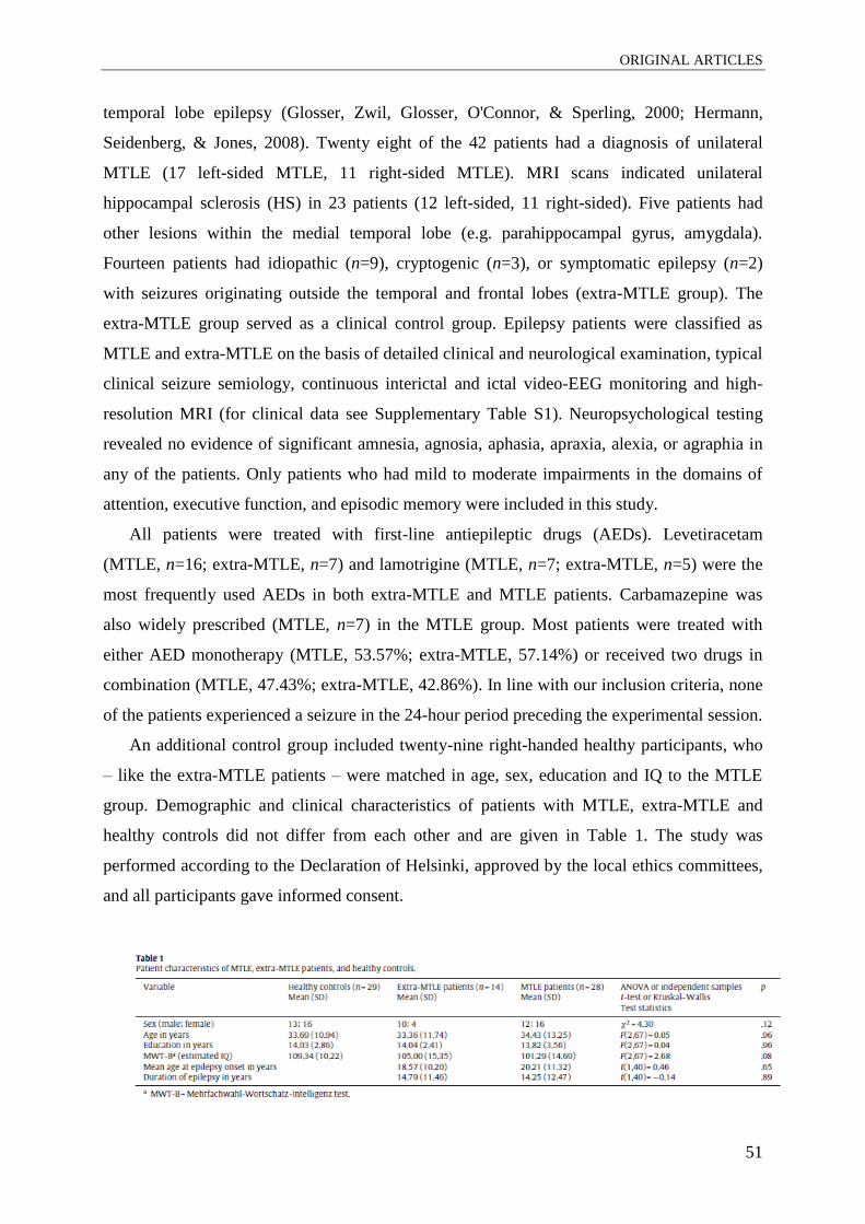
ORIGINAL ARTICLES
51
temporal lobe epilepsy (Glosser, Zwil, Glosser, O'Connor, & Sperling, 2000; Hermann,
Seidenberg, & Jones, 2008). Twenty eight of the 42 patients had a diagnosis of unilateral
MTLE (17 left-sided MTLE, 11 right-sided MTLE). MRI scans indicated unilateral
hippocampal sclerosis (HS) in 23 patients (12 left-sided, 11 right-sided). Five patients had
other lesions within the medial temporal lobe (e.g. parahippocampal gyrus, amygdala).
Fourteen patients had idiopathic (n=9), cryptogenic (n=3), or symptomatic epilepsy (n=2)
with seizures originating outside the temporal and frontal lobes (extra-MTLE group). The
extra-MTLE group served as a clinical control group. Epilepsy patients were classified as
MTLE and extra-MTLE on the basis of detailed clinical and neurological examination, typical
clinical seizure semiology, continuous interictal and ictal video-EEG monitoring and high-
resolution MRI (for clinical data see Supplementary Table S1). Neuropsychological testing
revealed no evidence of significant amnesia, agnosia, aphasia, apraxia, alexia, or agraphia in
any of the patients. Only patients who had mild to moderate impairments in the domains of
attention, executive function, and episodic memory were included in this study.
All patients were treated with first-line antiepileptic drugs (AEDs). Levetiracetam
(MTLE, n=16; extra-MTLE, n=7) and lamotrigine (MTLE, n=7; extra-MTLE, n=5) were the
most frequently used AEDs in both extra-MTLE and MTLE patients. Carbamazepine was
also widely prescribed (MTLE, n=7) in the MTLE group. Most patients were treated with
either AED monotherapy (MTLE, 53.57%; extra-MTLE, 57.14%) or received two drugs in
combination (MTLE, 47.43%; extra-MTLE, 42.86%). In line with our inclusion criteria, none
of the patients experienced a seizure in the 24-hour period preceding the experimental session.
An additional control group included twenty-nine right-handed healthy participants, who
– like the extra-MTLE patients – were matched in age, sex, education and IQ to the MTLE
group. Demographic and clinical characteristics of patients with MTLE, extra-MTLE and
healthy controls did not differ from each other and are given in Table 1. The study was
performed according to the Declaration of Helsinki, approved by the local ethics committees,
and all participants gave informed consent.

ORIGINAL ARTICLES
52
2.2 Procedure
Neuropsychological and psychiatric assessments were carried out after all participants
gave written informed consent. All participants performed the multiple-choice vocabulary test
(MWT-B) which served an estimate of crystalline verbal intelligence. (Lehrl, Triebig, &
Fischer, 1995). The test battery comprised emotion recognition, theory of mind, decision-
making, and self-report measures (see below). The entire battery took about 3.5 to 4 hours to
complete. Subjects were allowed to request a break at any time during the psychiatric and
neuropsychological assessment.
2.3 Emotion Recognition
2.3.1 Comprehensive Affect Testing System (CATS)
CATS aims to measure the perception of facial expressions, prosody, and linguistically
presented emotional material and therefore employs the visual and auditory modality of
communication (Froming et al., 2006). CATS consists of 13 subtests: 11 emotion tasks and
two control tasks assessing facial identification, emotion matching with and without verbal
denotation (e.g. in some tasks both emotional faces and the name of the target emotion is
displayed on the screen, whereas in other tasks no additional verbal cues are given), emotional
tone or prosodic processing with and without verbal denotation (e.g. in some tasks both
emotional prosody and the name of the target emotion is displayed on the screen, whereas in
other tasks no additional verbal cues are given), and with conflicting or congruent semantic
content. Emotional stimuli covered happy, sad, angry, surprised, disgusted, fearful or neutral
mood. Instructions and verbal and auditory stimuli for the CATS task were translated by our
group into German.
Composite Scales. Each item within a subtest is scored as either correct or incorrect, and
items are summed to obtain a raw score for each of the 13 subtests. Data from the 11 emotion
related subtests were combined and reduced to five different composite scales: the Simple
Facial Scale (Subtests 2 & 5), Complex Facial Scale (Subtests 7, 8, 13), Prosody Scale
(Subtests 4, 6, 9), Lexical Scale (Subtests 10), and Cross-Modal Scale (Subtest 11 & 12).
Quotient Scales. Broader scales, based on mode of communication (facial affect and
prosody) and emotion per se, are also generated. The Affect Recognition Quotient is obtained
by combining the two facial scales; the Prosody Recognition Quotient is identical to the
ORIGINAL ARTICLES
53
Prosody Scale, and the Emotion Recognition Quotient is an overarching scale and includes all
11 emotional subtests.
Discrete emotion scales. There are additional scales for each of the six basic emotions that
provide information about performance based on type of emotion. Items in the discrete
emotion scales test facial affect recognition and are taken from subtests five, 7, 8, and 13.
2.4 Selected tests of theory of mind
2.4.1 Moving Triangles
The Moving Triangles Test aims to measure the extent to which subjects make mental
state attributions to dynamic visual stimuli (Abell, Happé, & Frith, 2000; Castelli et al., 2002;
Heider & Simmel, 1944). The protagonists of this test were two shapes, a big red triangle and
a small blue triangle, that move around the screen. Mental state attributions were restricted to
pure movement and interaction in the absence of vocal or facial expression. There were three
different types of animation sequences: random movement (RD) in which no interaction
occurred (e.g. bouncing), goal-directed (G-D) interactions that elicited attributions of simple
actions (e.g. fighting) and theory of mind (ToM) interactions that elicited attributions of
mental states to the agents (e.g. tricking). Each sequence lasted between 34 and 45 seconds,
and the three types of animations were matched for length.
Verbal descriptions for each of the four examples of the RD, G-D and ToM animation
sequences were recorded and coded with respect to three dimensions: ‘intentionality’ (degree
of mental state attribution, range 0-5, with absence of mental state language at one extreme
and elaborate use of mental state language at the other); ‘appropriateness’ (0-3, with incorrect
at one extreme and highly appropriate at the other); and ‘length’ (0-4, ranging from no
response to more than four clauses). For more details of materials and scoring procedure see
Castelli et al. (2002).
2.4.2 Reading the Mind in the Eyes Test
The “Reading the mind in the Eyes Test” (“Eyes Test”) (Baron-Cohen, Jolliffe,
Mortimore, & Robertson, 1997; Baron-Cohen, Wheelwright, Hill, Raste, & Plumb, 2001)
aims to measure higher-level facial emotion perception where the subject has to infer mental
and affective states from social cues of the eye region and thus employ both the visual and
verbal modalities. In the Eyes Test the subject’s task is to choose which of four words best
describes what the person in the picture is thinking or feeling (e.g. terrified, amused, regretful,
ORIGINAL ARTICLES
54
flirtatious). Each picture shows only a pair of eyes from ten different faces (male and female).
All faces were standardized to one size (15x10cm), all were black and white, with the same
region of the face selected for each photo. All 73 subjects performed a shortened version of
the Eyes Test (Baron-Cohen et al., 2001) that comprised ten selected pair of eyes (item
numbers 5, 6, 9, 11, 16, 19, 23, 26, 30, and 36). The short version of the Eyes Test was
assembled from an independent sample of 20 healthy subjects who completed the full version
(unpublished data). In the short version we eliminated those pair of eyes that did not show a
sufficient amount of interindividual variability. Reliability analysis between the long and the
short version in this sample revealed a sufficient correlation between the two versions (r=.78;
p<.001).
The maximum score on this test is 10.
2.4.3 Faux Pas Test
The Recognition of Faux Pas Test (Baron-Cohen, O'Riordan, Stone, Jones, & Plaisted,
1999; Stein, Corte, Colling, & Whall, 1998; Stone, Baron-Cohen, & Knight, 1998) is an
advanced ToM test because it aims to measure mental and affective state attributions from
verbal material. This test estimates the ability to recognise and understand a social faux-pas,
which is understood as a statement in which the speaker accidentally offends or insults
another person. All subjects performed a shortened version of a Faux Pas Test (Stone et al.,
2003) that comprised five selected stories. Patients were asked to read each story silently,
knowing that they would then be asked questions about the story. The first question
concerned the detection and comprehension of the faux pas, while question two, three, and
four required the patient to impute the mental state of and attribute emotions to another.
Further information related to the test can be found in Schacher et al. (2006).
Data analysis involved the sum of the first four questions, where each question was scored
with one point, resulting in a maximum of twenty points for all five stories. Points for the
control question were excluded from the total score, but served as an indicator of text
comprehension.
2.5 Decision Making
2.5.1 Iowa Gambling Task (IGT)
The Iowa Gambling Task (IGT) aims to measure decision making under initial ambiguity
(Bechara, Damasio, Damasio, & Anderson, 1994) and is widely used in research of cognition

ORIGINAL ARTICLES
55
and emotion. It tests the ability to choose between favourable card decks with lower gains but
also lower risk for losses and unfavourable card decks with higher gains but also higher
losses. Participants are presented four decks of cards on a computer screen and are instructed
to use the mouse to select cards (overall 100 trials). They were told that each time they choose
a card they would win some game money. Occasionally, a card will also have a penalty
function and they lose money. Participants were instructed that they could choose cards from
any deck and that the goal of the game is to win as much money as possible. Participants were
also informed that some decks are better than others and that to win they had to avoid the
disadvantageous decks which would lead to losses over the long run and keep selecting from
the advantageous decks which would lead to gains. Participants learn the nature of the decks
through trial and error.
For each block the number of selections from blocks A and B (disadvantageous) and the
number of selections from blocks C and D (advantageous) were counted. Performance on the
IGT is scored by a global outcome score (net score) and a score for each of the five
consecutive blocks of 20 cards. These scores correspond to the number of cards chosen from
the advantageous decks (C+D) minus the number of cards chosen from the disadvantageous
decks (A+B). The analysis of the IGT performance by blocks of 20 cards provides
information about the learning capacity and strategy used by participants (Bechara, 2001).
2.6 Self-Report-Questionnaires
We further investigated empathy, psychological distress, personality dimensions, empathy
and quality of life using the following reliably and widely used self-report measures: (1) the
Saarbruecken Personality Questionnaire (SPF) to assess empathy (Davis, 1983); (Paulus,
2009), (2) the Toronto-Alexithymia Scale (TAS-26) to assess various facets of the
alexithymia construct (Kupfer, 2001; Taylor, 2000; Taylor, Ryan, & Bagby, 1985), (3) the
Beck Depression Inventory (BDI) to assess depressive mood (Beck, 1984), (4) the Eysenck
Personality Questionnaire (EPQ-RK) to assess personality traits (Eysenck & Eysenck, 1976;
Ruch, 1999), and (5) the Quality of Life with Epilepsy-31 (QOLIE-31) to assess aspects of
quality of life in patients with epilepsy (Cramer et al., 1998).
2.7 Statistical analysis
ORIGINAL ARTICLES
56
Analyses were carried out using SPSS Version 15. Besides for the overall score of the
Beck Depression Inventory and the Moving Triangles Test, we conducted a multivariate
analysis of variance (MANOVA) to test the effect of group (MTLE, extra-MTLE, HC) and
sex on the test scores of the Social Cognition Test Battery. A separate MANOVA was
performed to determine significant differences between groups and sex on the self-report
questionnaires. We then applied Bonferroni corrected post-hoc tests.
Performance on the Iowa Gambling Task was separately analysed to explore changes in
performance over time. Therefore, IGT data were analysed by a repeated-measures analysis of
variance (ANOVA) with blocks (1-5) as the within-subjects factor and group (MTLE patients,
extra-MTLE patients, healthy controls) as the between subjects factor. Bonferroni corrected
post-hoc tests were then applied. Paired-sample t-tests were carried out to inspect differences
within groups.
Differences between patient groups on the QOLIE-31 were examined using unpaired t-
tests. For the BDI and Moving Triangles Test, groups (MTLE, extra-MTLE, healthy controls)
were compared by means of Kruskal-Wallis tests. Significant results were further analysed
using Mann-Whitney tests.
Additionally, separate MANOVA’s for test scores in the Social Cognition Test Battery,
and the self-report questionnaires as dependent variables and lateralization (right- vs. left-
sided MTLE patients) as an independent variable, were calculated. The confirmatory
statistical comparisons of all data across groups were carried out at a significance level set at
p<.05 (two-tailed).
As the various tests of social cognition and the self-report questionnaires used different
response scales, we expressed all data as Z scores in order to accurately compare subscale
responses with each other.
2.7.1 Correlation Analysis
Correlations of dependent variables with each other and with demographic variables were
tested using Pearson’s product-moment correlation.
Within MTLE and extra-MTLE patient groups, we used additional correlation analysis
(Pearson Correlation Coefficient) to investigate the relation between age at seizure onset,
duration of epilepsy and each test of the Social Cognition Test Battery, the self-report
questionnaires and the decision making task.

ORIGINAL ARTICLES
57
3. Results
3.1 Demographic and neuropsychological background tasks
Means and standard deviation of socio-demographic, clinical and cognitive variables of
healthy controls and patient groups are shown in Table 1. Demographic variables did not
differ between the three groups. Therefore, a MANOVA was conducted to investigate group
differences on measures of the Social Cognition Test Battery, the self-report questionnaires
and the IGT.
3.2 Selected tests of emotion recognition and theory of mind
A MANOVA comparing the three groups on all tests of the Social Cognition Test Battery
as a whole revealed neither a significant main effect for group, based on Pillai’s Trace,
F(32,88)=1.13, p=.32, nor a significant main effect for gender, F(16,43)=.54, p=.91, nor for
group x gender interaction, F(32,88)=, p=.76. We report effects of group for single dependent
variables below. Sample sizes vary slightly by subgroup due to missing values.
3.2.1 CATS
All patients (MTLE: n=24, extra-MTLE: n=14) and HC (n=28) showed normal levels of
effort, indicated by validity scores within the normal range. Less than 10% of our sample
obtained combined scores of less than 31, paralleling the results of Froming et al. (Froming et
al., 2006).
Quotient Scales. Comparing the three groups on the quotient scales revealed a significant
main effect of group for all three quotients, namely the Affect, Prosody, and Emotion
Recognition Quotient. Post hoc analysis revealed that MTLE patients performed significantly
worse compared to HC on all quotient scales (ARQ, p=.006; PRQ, p<.001; ERQ, p<.001) as
well as compared to extra-MTLE patients on the ERQ (p=.02). Extra-MTLE patients and HC
did not differ in terms of the quotient scales (all p>.05). (see Table 2 and Fig. 1).
Composite Scales. Comparing the three groups on the composite scales showed a
significant main effect of group for the complex facial and prosody scale (see Table 2). Post
hoc tests showed that MTLE patients were impaired compared to HC (p=.01), but not
compared to extra-MTLE patients (p=.07), in complex facial emotion recognition. Also
prosody recognition was impaired in MTLE patients compared to HC (Prosody Scale,

ORIGINAL ARTICLES
58
p=.003), but not compared to extra-MTLE patients (p=.15). Extra-MTLE patients and HC did
not differ in terms of the composite scales (all p>.05). There were no significant differences
between groups with regard to the simple facial, lexical, and cross modal scale (see Table 2
and Fig. 1).
Discrete emotion scales. With regard to the specific type of emotion, there was a
significant main effect of group for recognition of fear, sadness, and disgust (see Table 2),
with post hoc tests showing that MTLE patients compared to HC were specifically impaired
on the emotion type fear (p=.005), and disgust (p=.04) (see Table 2 and Fig. 1)
Correlation Analysis. With regard to CATS quotients, ARQ (r=−.287, p<.05), PRQ (r
=−.449, p<.001), and ERQ (r=−.410, p<.001) correlated with age, PRQ (r=.266, p<.05) and
ERQ (r=.298, p=.02) with verbal IQ. These results indicate a decline in visual and auditory
emotion recognition performance with the person’s age. Additionally, lower performance in
general emotion recognition performance, particularly in auditory emotion recognition
abilities, were associated with reduced verbal IQ. ERQ (r=−.316, p<.01) correlated with years
of education, indicating that poorer general emotion recognition performance is associated
with fewer years of education.
Within MTLE and extra-MTLE patients, there were no significant correlations between
quotients and clinical variables including duration of epilepsy and age at epilepsy onset as
well as between composite scales and the same clinical variables.
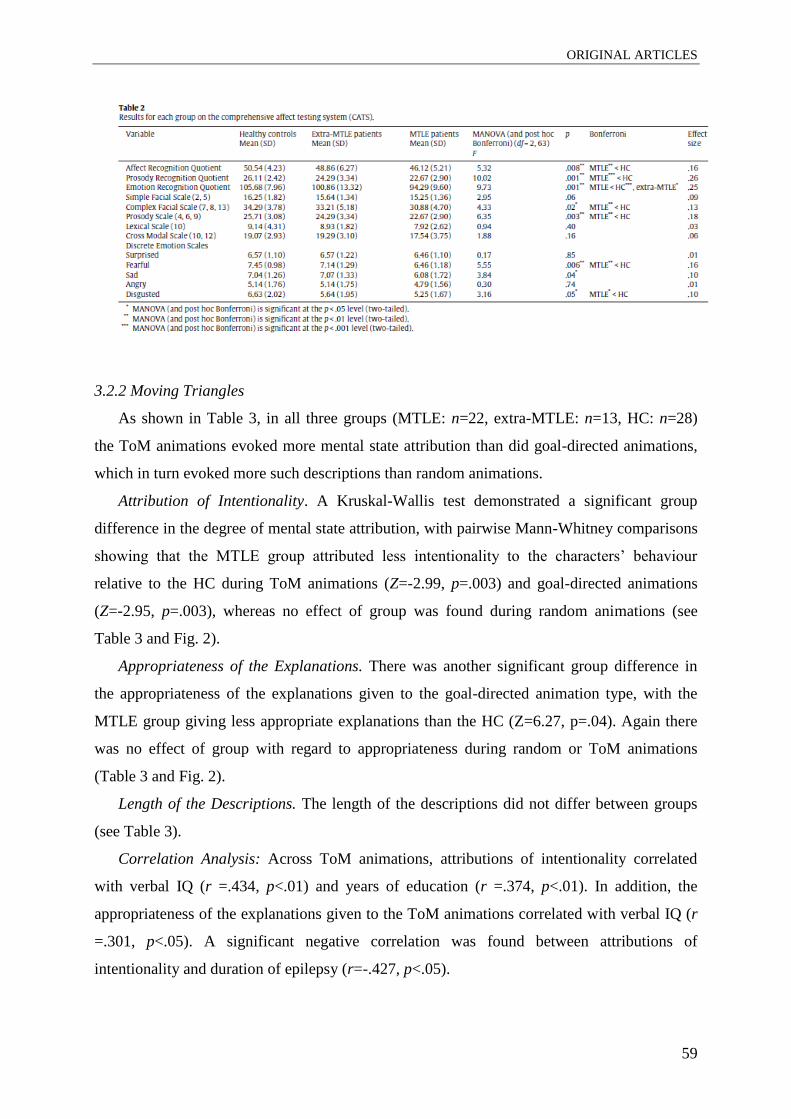
ORIGINAL ARTICLES
59
3.2.2 Moving Triangles
As shown in Table 3, in all three groups (MTLE: n=22, extra-MTLE: n=13, HC: n=28)
the ToM animations evoked more mental state attribution than did goal-directed animations,
which in turn evoked more such descriptions than random animations.
Attribution of Intentionality. A Kruskal-Wallis test demonstrated a significant group
difference in the degree of mental state attribution, with pairwise Mann-Whitney comparisons
showing that the MTLE group attributed less intentionality to the characters’ behaviour
relative to the HC during ToM animations (Z=-2.99, p=.003) and goal-directed animations
(Z=-2.95, p=.003), whereas no effect of group was found during random animations (see
Table 3 and Fig. 2).
Appropriateness of the Explanations. There was another significant group difference in
the appropriateness of the explanations given to the goal-directed animation type, with the
MTLE group giving less appropriate explanations than the HC (Z=6.27, p=.04). Again there
was no effect of group with regard to appropriateness during random or ToM animations
(Table 3 and Fig. 2).
Length of the Descriptions. The length of the descriptions did not differ between groups
(see Table 3).
Correlation Analysis: Across ToM animations, attributions of intentionality correlated
with verbal IQ (r =.434, p<.01) and years of education (r =.374, p<.01). In addition, the
appropriateness of the explanations given to the ToM animations correlated with verbal IQ (r
=.301, p<.05). A significant negative correlation was found between attributions of
intentionality and duration of epilepsy (r=-.427, p<.05).
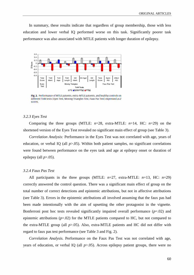
ORIGINAL ARTICLES
60
In summary, these results indicate that regardless of group membership, those with less
education and lower verbal IQ performed worse on this task. Significantly poorer task
performance was also associated with MTLE patients with longer duration of epilepsy.
3.2.3 Eyes Test
Comparing the three groups (MTLE: n=28, extra-MTLE: n=14, HC: n=29) on the
shortened version of the Eyes Test revealed no significant main effect of group (see Table 3).
Correlation Analysis: Performance in the Eyes Test was not correlated with age, years of
education, or verbal IQ (all p>.05). Within both patient samples, no significant correlations
were found between performance on the eyes task and age at epilepsy onset or duration of
epilepsy (all p>.05).
3.2.4 Faux Pas Test
All participants in the three groups (MTLE: n=27, extra-MTLE: n=13, HC: n=29)
correctly answered the control question. There was a significant main effect of group on the
total number of correct detections and epistemic attributions, but not in affective attributions
(see Table 3). Errors in the epistemic attributions all involved assuming that the faux pas had
been made intentionally with the aim of upsetting the other protagonist in the vignette.
Bonferroni post hoc tests revealed significantly impaired overall performance (p=.02) and
epistemic attributions (p=.02) for the MTLE patients compared to HC, but not compared to
the extra-MTLE group (all p>.05). Also, extra-MTLE patients and HC did not differ with
regard to faux pas test performance (see Table 3 and Fig. 2).
Correlation Analysis. Performance on the Faux Pas Test was not correlated with age,
years of education, or verbal IQ (all p>.05). Across epilepsy patient groups, there were no

ORIGINAL ARTICLES
61
significant correlations between Faux Pas Test performance and the clinical variables age at
epilepsy onset and duration of epilepsy (all p>.05).
3.3 Decision making
3.3.1 Iowa Gambling Task (IGT)
Data for 25 HC, 14 extra-MTLE patients, and 19 MTLE patients were available for
analysis. Performance of the experimental groups on the IGT is shown in Fig. C.1.
An ANOVA of the IGT global outcome score across all groups showed a significant main
effect of the factor group, F(2,50)=5.74, p=.006. Post-hoc Bonferroni tests revealed that the
MTLE patients performed significantly worse compared to HC (p=.02), but not compared to
extra-MTLE patients (p=1.00). Extra-MTLE patients and HC also differed in terms of the
global outcome score (p=.03).
A repeated-measures ANOVA with blocks (1–5) as the within-subjects factor and group
(MTLE, extra-MTLE, healthy controls) as the between-subjects factor was carried out on the
IGT net scores. The main effect of block, F(4,220)=13.92, p<.001, as well as the main effect
of group, F(2,55)=6.67, p=.003 was significant, reflecting the strategy shift across blocks and
indicating different preferences of decks in total between the groups. The interaction of both
factors was not significant, F(8,220)=1.14, p=.34. Post-hoc Bonferroni tests showed that
MTLE patients performed significantly worse compared to HC (p=.007), but not compared to
extra-MTLE patients (p=1.00). Extra-MTLE patients and HC also differed in terms of the net
score (p=.02).
When only comparing performance of the first to the fifth block, paired samples t-tests
revealed that the frequency of advantageous selections markedly increased over the task for

ORIGINAL ARTICLES
62
HC (block 1 vs. block 5: t(24)=−6.80, p<.001) and extra-MTLE patients, t(13)=−3.23, p=.007,
but did not significantly increase for MTLE patients, t(18)=−1.65, p=.12.
Correlation Analysis. Performance on the IGT was not correlated with age, years of
education, or verbal IQ (all p>.05). Within the MTLE group, the global outcome score of the
IGT was correlated with duration of epilepsy (r=-.53, p<.05), indicating that a longer duration
of epilepsy was associated with impaired decision-making performance.
3.4 Self-Report Questionnaires
We used MANOVA to investigate group x gender differences on the self-report
questionnaires. A significant main effect for group, based on Pillai’s Trace, F(24,102)=1.82,
p=.02, but no significant main effect for gender, F(12,50)=1.64, p=.12, or for gender x group
interaction, F(12,50)=1.28, p=.27, was observed.
Across patient groups, no significant correlations between any self-report questionnaire
and the clinical variables age at epilepsy onset, or duration of epilepsy were found (all p>.05).
Below we report the effects of group on single dependent variables. Sample sizes vary slightly
by subgroup due to missing values.
3.4.1 Saarbruecken Personality Questionnaire (SPF)
Groups (MTLE: n=27, extra-MTLE: n=13, HC: n=29) differed neither on the total score
of the SPF, nor on the subscales fantasy, empathic concern and perspective taking (see
Supplementary Table S2).
ORIGINAL ARTICLES
63
3.4.2 Toronto-Alexithymia Scale (TAS-26)
The majority of MTLE (22/28; 79%) and extra-MTLE patients (13/14; 93%) and all
subjects in the healthy control group (28/28) had TAS scores below the cut-off of 54, which
would be indicative for alexithymia. No significant group effect emerged for the subscales
difficulty communicating emotions and externally oriented thinking (Table S2). A significant
effect of group was found for the mean TAS score and the difficulty identifying emotions
subscale of the TAS, with post hoc comparisons revealing that the MTLE group was overall
more alexithymic (p=.005), and had significantly more difficulty in identifying (p=.001) and
communicating emotions (p=.05) compared to HC (see Supplementary Table S2).
3.4.3 Beck Depression Inventory (BDI)
In all three groups the mean score for the BDI was within the normal range of HC (Beck,
1979) (for mean depression scores on the BDI see Supplementary Table S2). No subjects
within the healthy control group had a BDI score above the cut-off of 12 (indicative for a mild
form of depressive mood). The majority of MTLE (16/23; 70%) and extra-MTLE patients
(10/13; 80%) had BDI scores lying under the cut-off of 12. Seven out of 23 (30%) in the
MTLE group and three out of 13 (25%) in the extra-MTLE group had a score above 12 and
below 19, indicating mild to moderate depressive mood, and none had a score above 18,
indicating a clinical depression.
Kruskal-Wallis test revealed significant group difference in the overall score, with
pairwise Mann-Whitney comparisons showing that the MTLE group (Z=-5.11, p<.001) and
the extra-MTLE group (Z=-2.99, p=.003) had more depressive symptoms relative to HC (see
Supplementary Table S2).
3.4.4 Eysenck Personality Questionnaire (EPQ-RK)
Comparisons of the three groups (MTLE: n=28, extra-MTLE: n=12, HC: n=28) on the
EPQ-RK revealed a significant main effect of group for the subscale Neuroticism, whereas
groups did not differ in the other personality domains Extraversion and Psychoticism, or with
regard to Social Conformity. Post hoc analysis revealed that MTLE patients compared to HC
(p=.01), but not compared to extra-MTLE patients (p=.09), described themselves as distinctly
more neurotic (see Supplementary Table S2).
ORIGINAL ARTICLES
64
3.4.5 Quality of Life with Epilepsy-31 (QOLIE-31)
T-Tests that examined mean differences between MTLE (MTLE: n=28) and extra-MTLE
patients (MTLE: n=14) in quality of life indicated that MTLE patients had a lower subjective
quality of life compared to extra-MTLE patients as indicated by a lower Overall Score as well
as lower scores on the subscales Overall Quality of Life and Social Functioning. With respect
to the QOLIE Emotional Well Being subscale score, no significant differences were found
between the two groups (see Supplementary Table S2).
3.5 Correlations between behavioural measures of social cognition
Pearson’s product-moment correlations between the emotion recognition, the theory of
mind, and the decision-making task revealed that facial emotion recognition (ARQ) was
highly correlated with emotion recognition from prosody (PRQ) as well as with general
emotion recognition (ERQ), which in turn was also highly correlated with PRQ. Emotion
recognition from a face and prosody were also highly correlated with the extent of intentional
attributions that had been made as well as with the appropriateness of the explanations that
had been given for ToM animations. It also appears that, independent of the channel of
communication, emotion recognition was related to the IGT global outcome score. Whereas
the Eyes and Faux Pas Test were not correlated with emotion recognition, they were found to
correlate with each other.
3.6 Correlations between behavioural measures of social cognition and subjective measures of
psychological distress, personality dimensions, empathy and quality of life
Across both epilepsy groups, Beck Depression Inventory (BDI) scores did not correlate
with emotion recognition, neither from face nor from prosody, nor with any other measures of
theory of mind or the decision-making task (all Spearman’s p>.05). Within all groups, a lack
of correlation between behavioural and subjective measures was also observed (all p>.05).
Only when both epilepsy patient groups were combined was QOLIE Social Functioning
correlated with emotion recognition from prosody (r=.43, p=.008), whereas the lack of
association between all other measures remained (all p>.05).
ORIGINAL ARTICLES
65
3.7 Effect of Side of MTLE
Right- and left-sided MTLE did not differ with regard to demographic variables such as
sex, age, verbal IQ, years of education, age at epilepsy onset and duration of epilepsy (all
p>.05).
We analyzed the MTLE group in more detail by first analyzing the influence of the side of
epilepsy within the MTLE group on tests from the Social Cognition Test Battery as well as
the self-report questionnaires by applying separate MANOVA’s with the fixed factor side of
epilepsy.
On the Social Cognition Test Battery, a significant effect of the factor group was found
only for the CATS emotion type fear. Patients with right-sided MTLE performed worse on
CATS emotion type fear compared to left-sided MTLE patients, F(1, 20)=5.42; p=.03, eta2
=0.21. No effect of the factor side of epilepsy was found on any of the tests of the self-report
questionnaires (all p>.05).
For the IGT, repeated-measures ANOVA’s were performed for right- and left-sided
MTLE groups as well as for men and women on all relevant parameters. No significant group
effects were found in these analyses.
4. Discussion
4.1 Main findings
In the present study we assessed emotion recognition, theory of mind, decision making,
psychological states and traits, and quality of life in a group of patients with MTLE.
The study demonstrated deficits in subjects with MTLE compared to HC in all measures
of social perception affecting the ability to interpret emotional expressions and feelings from
faces and voices and, with one exception, on all advanced tests of reasoning about the mental
states of others. MTLE patients were predominantly impaired in their general emotion
recognition abilities compared to extra-MTLE patients. In contrast, subjects with extra-MTLE
showed no significant impairment in tests of social cognition relative to HC. Their
performance, however, also did not differ from the performance of the MTLE group and lay
between these two groups on nearly all measures. It is reasonable to assume that, independent
of the site of seizure onset in the extra-MTLE group, fronto-limbic structures might be
affected due to frequent propagation of epileptic brain electric activity from posterior to
ORIGINAL ARTICLES
66
anterior areas. This also implies that patients with other epilepsy syndromes, who equally
were treated with antiepileptic drugs (AEDs), might be at risk of suffering from difficulties in
social-cognitive abilities, albeit to a lesser degree than MTLE patients. Another possible
explanation for the absence of significant differences between the two patient groups is the
heterogenous composition of the extra-MTLE group with patients affected by different
epilepsy syndromes which emphasizes the need for more homogenous control groups. Overall
our results support the claim that MTLE is a risk factor for deficits in social cognition beyond
weaknesses that are associated with an epileptic disorder in general.
4.2 Emotion recognition from faces and prosody
We revealed that patients with MTLE, as compared to HC, are impaired in complex facial
and auditory emotion processing, but not in lexical emotion recognition. This is in accordance
with a recent finding suggesting that the basic emotion recognition impairment in MTLE
patients is not dependent on the sensory modality through which emotional material is
perceived (Bonora et al., 2011). Moreover, impairments in the recognition of basic emotions
with negative valence have also been reported in temporal lobectomy patients with amygdala
damage on the basis of facial and vocal expressions (Brierley, Medford, Shaw, & David,
2004). By contrast, two other studies failed to find differences in recognizing emotional
prosody in a small group of patients after temporal lobectomy (Adolphs, Tranel, & Damasio,
2001) and in a group of patients with MTLE and asymmetrical amygdala damage (Fowler et
al., 2006). However, these negative results could be due to small sample sizes, item and task
characteristics and different clinical patient characteristics. Rocca et al. (2009), who applied
the CATS to a group of patients with schizophrenia and healthy controls, found that controls
performed better on all subtests with the exception of an affect discrimination task. By
contrast, analysis of component scales revealed that MTLE patients as compared to HC
performed worse on the complex facial and prosody scale, whereas performance on the
simple facial, lexical and cross modal scale was comparable between the three groups,
indicating more circumscribed deficits in emotion recognition in MTLE compared to
schizophrenia patients.
In our study, analysis of emotion scales revealed that patients with MTLE were
significantly impaired compared to HC in recognizing fear and disgust, while performance on
all other basic emotions did indeed lie below the performance of the two other groups (HC
and extra-MTLE patients). However, these differences failed to reach significance. This

ORIGINAL ARTICLES
67
observation is in line with findings of previous studies investigating facial emotion
recognition abilities in MTLE patients (Benuzzi et al., 2004; Bonora et al., 2011; Meletti et
al., 2009; 2003). Furthermore, our findings are in accordance with a recent meta-analysis
showing that a set of interacting fronto-temporal brain regions are active during the
experiencing and perception of emotion across a range of emotion categories (Lindquist,
2011). Moreover, we did not find statistically significant correlations between epilepsy-
related variables such as age at seizure onset and duration of epilepsy with CATS quotients
and scales.
4.3 Theory of Mind Tests
We further investigated ToM performances in MTLE patients using other experimental
paradigms that allow differentiation between different types of attribution as well as with
regard to the stimulus modality they employ.
The result of the present study supports our previous finding that patients with MTLE
show impairments in theory of mind abilities (Schacher et al., 2006). Extending findings of
this earlier study, we could show here that these deficits are not limited to the verbal modality.
Accordingly, MTLE patients were impaired in faux pas detection, made significantly worse
epistemic attributions and gave fewer and less accurate interpretations of animations that
elicited mentalizing compared to HC, but not compared to extra-MLTE patients (whose
performance again lay between that of HC and MTLE patients). The Eyes Test was the only
ToM test that failed to find significant differences between groups. While HC were highly
accurate in inferring the mental states of triangles without facial expressions or other human
cues, i.e., from movement cues alone, our data parallel those found in patients with autism
spectrum disorders who, regardless of general intelligence, were also shown to be impaired in
making mental state attributions about animated shapes (Abell et al., 2000; Castelli et al.,
2002; Klin, 2000). If we directly compare the results of the MTLE patients with data from
patients with autism spectrum disorder, the mean scores for intentionality attributions within
ToM animations for the epilepsy patients are comparable to those of the autism groups
(Castelli et al., 2002). These results suggest that MTLE patients show impairments in making
mental state attributions that are comparable to individuals with autism which is characterized
by impaired social functioning, communication, and interaction (Wing, 1986). Furthermore,
performance on ToM animations of the Moving Triangles Task was positively correlated with
emotion recognition from face and prosody, indicating that both tasks seem to measure facets

ORIGINAL ARTICLES
68
of the same construct. It is conceivable that the patients' poor understanding of others' minds
is associated with impairment in verbal and auditory emotion recognition. Interestingly, as
opposed to most measures of ToM, MTLE and extra-MTLE patients self-rated their empathic
skills as comparable to those of HC. This fact emphasizes the importance of objective social-
cognitive testing in epilepsy patients as patients may be insufficiently aware of their social-
cognitive deficits.
Additionally, we explored the association between ToM performance and epilepsy-related
variables and found several modest positive associations between the degree of mental state
attributions with age at epilepsy onset and duration of epilepsy. From these results we can
conclude that patients with earlier epilepsy onset and longer duration of epilepsy are more
prone to have deficits in ToM.
4.4 Decision Making
Our results confirm previous findings (Bonatti et al., 2009; Butman et al., 2007; Delazer,
Zamarian, Bonatti, Kuchukhidze et al., 2010; Delazer, Zamarian, Bonatti, Walser et al., 2010;
Labudda et al., 2009) that MTLE patients have deficits in decision making under conditions
of ambiguity. MTLE patients showed a significantly lower decision-making performance than
HC, but not compared to extra-MTLE patients, who also obtained lower global outcome and
net scores compared to HC. However, the frequency of advantageous selections markedly
increased over the task for HC and extra-MTLE patients, but did not significantly increase for
MTLE patients, indicating impaired learning via feedback. In addition, emotion recognition,
particularly from the face and decision-making performance were correlated. A possible
explanation for this association is the shared structural involvement of parts of the limbic
system in decision making under ambiguity and to emotion perception (Hsu et al., 2005).
Moreover, the decision-making performance was correlated with duration of epilepsy within
MTLE patients.
4.5 Psychic states and traits and quality of life
In our group of clinically distinct epilepsy patients, MTLE patients did not show more
psychiatric symptoms compared to extra-MTLE patients. Patients with MTLE are thought to
have a particular specific risk for affective disorders because of the major involvement of the
limbic system in both seizure generation in TLE and in the regulation of mood and affect. Our

ORIGINAL ARTICLES
69
study assessed psychological distress (BDI, TAS-26), personality dimensions (EPQ-RK), and
quality of life (QOLIE-31) in patients with MTLE, extra-MTLE and HC.
We did not find an overrepresentation of depressive symptoms in epilepsy patients.
However, as expected, both patient groups had more depressive symptoms compared to HC.
Furthermore, MTLE patients reported more neurotic complaints (emotional instability,
nervous tendency towards negative emotionality, and inability to cope) and were more
alexithymic compared to HC. Taylor et al. (2000) examined the relationship between the
alexithymia construct and emotional intelligence (represented by EQ-i scores) and found a
strong negative correlation between the TAS and the EQ-i, indicating a considerable overlap
of the two constructs. The higher dimensional scores for the MTLE patients on the personality
dimension neuroticism corresponds with the clinical impression that patients with MTLE are
often characterized by high levels of negative affect such as depression and anxiety (Bonora
et al., 2011). MTLE patients rated their quality of life lower than extra-MTLE patients. This
may also explain why patients with MTLE had higher neuroticism scores, because it can be
assumed that psychological and social problems cause complaints of distress. The lower
quality of life experienced by MTLE patients compared to extra-MTLE patients suggests that
this difference is not the consequence of a chronic medical condition or AED medication per
se.
4.6 Associations between social cognition and psychic states and traits, empathy, and quality
of life
Furthermore, we investigated the correlations between behavioural measures of social
cognition and subjective measures of psychological distress, personality dimensions, empathy
and quality of life. No correlations emerged between any of these measures, indicating that
social-cognitive abilities were not related to subjective measures of psychiatric symptoms,
personality dimensions, self-reported empathic abilities, or quality of life. In line with these
findings, Bonara et al. (2011) and Reynders et al. (2005) found that MTLE patients’ ability to
recognize emotions was not related to subjective measures of well-being assessed with
QOLIE-31 and disease disability awareness assessed with the BDI. Interestingly, when we
investigated the correlations between emotion recognition ability and subjective quality of life
within the whole group of epilepsy patients, a significant correlation emerged between
QOLIE Social Functioning and emotion recognition from prosody. If one considers how
central emotion recognition and ToM abilities are to everyday life, one can assume that any

ORIGINAL ARTICLES
70
functional impairment due to structural and functional disconnections of the underlying neural
substrates of social cognition could have a devastating impact on interpersonal relationships,
social functioning and quality of life. If a person can understand semantic content but is
unable to detect prosody, he or she may take a sarcastic comment quite literally. When, in
addition to the missing prosodic information, changes in facial expression cannot be
recognized, significant information within conversations and interactions with others may go
undetected, and as a result, the individual may react inappropriately. This may create
misunderstandings and disagreements with others within the private or non-private sector that
can be source of social problems. It becomes clear with such examples that in order to address
such issues in diagnosis and rehabilitation, the exact nature and severity of the impairment
must be accurately assessed.
4.7 Side of epilepsy
In general, we found no differences between patients with seizures originating within the
left versus right mesial temporal lobe with regard to their performance in emotion recognition,
ToM, or decision making. Notably, the comparable performance in emotion recognition from
face and prosody of both left and right MTLE patients was surprising because patients with
right MTLE were expected to be more impaired (Meletti et al., 2009; 2003). However, in a
recent study of this group (Bonora et al., 2011) the authors also failed to find significant
differences between right- and left-sided MTLE patients in both visual and auditory emotion
recognition abilities. Evidence of an effect of the side of epilepsy onset was only found for the
specific emotion type fear, indicating that right-sided MTLE patients were more severely
impaired. In accordance with our findings, patients with seizure onset within the right, non-
language dominant hemisphere were reported to show pronounced difficulties in the
recognition of fearful faces (Benuzzi et al., 2004; Hlobil, Rathore, Alexander, Sarma, &
Radhakrishnan, 2008; Meletti et al., 2003).
4.8 Demographic variables and verbal intelligence quotient
Demographic variables such as gender, age, verbal intelligence, and years of education
were also evaluated with regard to their possible effects on emotion recognition and theory of
mind.

ORIGINAL ARTICLES
71
There was no effect of gender on any performance in the test battery. Age negatively
correlated with the ability to recognize emotions from both face and prosody, to a particular
degree from prosody, indicating a decline in performance with the person’s age. These results
are consistent with a previous finding of robust relationships between advancing age and
decline in performance on prosody tasks (Kiss & Ennis, 2001). In contrast, age had no effect
on ToM performance or self-reported psychological distress, personality dimensions, or
empathy.
Our study replicated previous findings of significant correlations between verbal IQ and
auditory emotion recognition abilities as well as a lack of correlations between verbal IQ and
facial emotion recognition abilities (Bonora et al., 2011; Reynders et al., 2005). Bonora et al.
(2011) ascribed the reported correlation between IQ and emotional prosody recognition to the
fact that emotional prosody recognition, as opposed to facial emotion recognition, might
impose more cognitive demands on the examinee. It may be assumed that prosodic stimuli
require greater working memory demands than facial stimuli. This assumption is consistent
with results from imaging studies that have shown the greatest activation in the right frontal
regions during prosody tasks (George et al., 1996). Another significant correlation was found
between verbal IQ and attributions of intentionality and the appropriateness of explanations
given during ToM animations of the Moving Triangles task. Effects of education were also
found on this task which is not surprising when one considers the comparatively high verbal
demands. By contrast, there were no significant correlations between verbal IQ and any other
ToM task. Such tasks also require attributions of intentions; however, the examinee is given
more structured guidance through the answering of questions (e.g. Faux Pas Test) or receives
additional input (e.g. Eyes Test). Overall, with a few exceptions, social-cognitive abilities
were shown to be independent of verbal intelligence and supported numerous findings that
assumed that emotion recognition and ToM are specific cognitive domains that should be
delineated from general intelligence (Baron-Cohen et al., 1997; Karmiloff-Smith, Klima,
Bellugi, Grant, & Baron-Cohen, 1995). Although only patients who had mild to moderate
impairments in the domains of attention, executive function, and episodic memory were
included in this study, we cannot completely rule out effects of cognitive deficits, especially
in executive functions, on emotional processing and ToM.

ORIGINAL ARTICLES
72
5. Conclusions
Our study demonstrated that patients with MTLE are at risk of impairments in various
aspects of social cognition. Patients with epilepsies not originating from the medial temporal
lobes may also be at risk, albeit to a much lesser extent. Therefore, the specific type of
epilepsy, particularly MTLE, and not epilepsy per se as a chronic disorder or its treatment
with AED can be considered as significant risk factor in developing social-cognitive deficits.
It is reasonable to argue that the frequent presence of deficits in social cognition in MTLE
patients contributed to the formulation of the concept of an “interictal personality syndrome”
or a “Waxman-Geschwind Syndrome”. Further neurobehavioural research in this field is
necessary to reveal the causes and demystify the concept of the “epileptic personality” and
hopefully provide new approaches for treatment.
ORIGINAL ARTICLES
73
Acknowledgments
This work was supported by the Swiss Epilepsy Foundation.
We thank Prof. Dr. Eugen Trinka, Prof. Dr. Gerhard Luef and Dr. Iris Unterberger for
enabling us to recruit patients from the Epilepsy Unit of the Department of Neurology,
Innsbruck Medical University.

References
Abell, F., Happé, F., & Frith, U. (2000). Do triangles play tricks? Attribution of mental states
to animated shapes in normal and abnormal development. Cognitive Development, 15,
1-16.
Adolphs, R. (2006). How do we know the minds of others? Domain-specificity, simulation,
and enactive social cognition. Brain Research, 1079(1), 25-35.
Adolphs, R., Tranel, D., & Damasio, H. (2001). Emotion recognition from faces and prosody
following temporal lobectomy. Neuropsychology, 15(3), 396-404.
Baron-Cohen, S., Jolliffe, T., Mortimore, C., & Robertson, M. (1997). Another advanced test
of theory of mind: evidence from very high functioning adults with autism or asperger
syndrome. Journal of Child Psychology and Psychiatry, 38(7), 813-822.
Baron-Cohen, S., O'Riordan, M., Stone, V., Jones, R., & Plaisted, K. (1999). Recognition of
faux pas by normally developing children and children with Asperger syndrome or
high-functioning autism. Journal of Autism and Developmental Disorders, 29(5), 407-
418.
Baron-Cohen, S., Wheelwright, S., Hill, J., Raste, Y., & Plumb, I. (2001). The "Reading the
Mind in the Eyes" Test revised version: a study with normal adults, and adults with
Asperger syndrome or high-functioning autism. Journal of Child Psychology and
Psychiatry, 42(2), 241-251.
Bear, D. M., & Fedio, P. (1977). Quantitative analysis of interictal behavior in temporal lobe
epilepsy. Archives of Neurology, 34(8), 454-467.
Bechara, A. (2001). Neurobiology of decision-making: risk and reward. Seminars in Clinical
Neuropsychiatry, 6(3), 205-216.
Bechara, A., Damasio, A. R., Damasio, H., & Anderson, S. W. (1994). Insensitivity to future
consequences following damage to human prefrontal cortex. Cognition, 50(1-3), 7-15.
Beck, A. T., Guthy, D., Steer, R. A., Ball, R. (1984). Internal consistencies of the original and
revised Beck Depression Inventory. Journal of Clinical Psychology, 40, 1365-1367.
Benbadis, S. R., Heriaud, L., Tatum, W. O., & Vale, F. L. (2003). Epilepsy surgery, delays
and referral patterns-are all your epilepsy patients controlled? Seizure, 12(3), 167-170.
Benuzzi, F., Meletti, S., Zamboni, G., Calandra-Buonaura, G., Serafini, M., Lui, F., et al.
(2004). Impaired fear processing in right mesial temporal sclerosis: a fMRI study.
Brain Research Bulletin, 63(4), 269-281.

ORIGINAL ARTICLES
75
Bonatti, E., Kuchukhidze, G., Zamarian, L., Trinka, E., Bodner, T., Benke, T., et al. (2009).
Decision making in ambiguous and risky situations after unilateral temporal lobe
epilepsy surgery. Epilepsy and Behavior, 14(4), 665-673.
Bonora, A., Benuzzi, F., Monti, G., Mirandola, L., Pugnaghi, M., Nichelli, P., et al. (2011).
Recognition of emotions from faces and voices in medial temporal lobe epilepsy.
Epilepsy and Behavior, 20(4), 648-654.
Brierley, B., Medford, N., Shaw, P., & David, A. S. (2004). Emotional memory and
perception in temporal lobectomy patients with amygdala damage. Journal of
Neurology, Neurosurgery, and Psychiatry, 75(4), 593-599.
Brune, M. (2005). "Theory of mind" in schizophrenia: a review of the literature.
Schizophrenia Bulletin, 31(1), 21-42.
Butman, J., Allegri, R. F., Thomson, A., Fontela, E., Abel, C., Viaggio, B., et al. (2007).
Behavioral flexibility impairment with negative feedback in refractory temporal lobe
epileptic patients with unilateral amygdala and hippocampal resection. Actas
Espanolas Psiquiatria, 35(1), 8-14.
Castelli, F., Frith, C., Happe, F., & Frith, U. (2002). Autism, Asperger syndrome and brain
mechanisms for the attribution of mental states to animated shapes. Brain, 125(Pt 8),
1839-1849.
Cramer, J. A., Perrine, K., Devinsky, O., Bryant-Comstock, L., Meador, K., & Hermann, B.
(1998). Development and cross-cultural translations of a 31-item quality of life in
epilepsy inventory. Epilepsia, 39(1), 81-88.
Davis, M. H. (1983). Measuring individual differences in empathy: Evidence for a
multidimensional approach. Journal of Personality and Social Psychology, 44, 113-
126.
Delazer, M., Zamarian, L., Bonatti, E., Kuchukhidze, G., Koppelstatter, F., Bodner, T., et al.
(2010a). Decision making under ambiguity and under risk in mesial temporal lobe
epilepsy. Neuropsychologia, 48(1), 194-200.
Delazer, M., Zamarian, L., Bonatti, E., Walser, N., Kuchukhidze, G., Bodner, T., et al.
(2010b). Decision making under ambiguity in temporal lobe epilepsy: does the
location of the underlying structural abnormality matter? Epilepsy and Behavior,
20(1), 34-37.
Eysenck, H. J., & Eysenck, S. B. G. (1976). Manual of the Eysenck Personality Scales (EPS
Adult). London.

ORIGINAL ARTICLES
76
Fowler, H. L., Baker, G. A., Tipples, J., Hare, D. J., Keller, S., Chadwick, D. W., et al.
(2006). Recognition of emotion with temporal lobe epilepsy and asymmetrical
amygdala damage. Epilepsy and Behavior, 9(1), 164-172.
Froming, K. B., Levy, C. M., Schaffer, S. G., & Ekman, P. (2006). Comprehensive Affect
Testing System (CATS) (Publication., from
http://www.psychologysoftware.com/testing_instruments.htm:
George, M. S., Parekh, P. I., Rosinsky, N., Ketter, T. A., Kimbrell, T. A., Heilman, K. M., et
al. (1996). Understanding emotional prosody activates right hemisphere regions.
Archives of Neurology, 53(7), 665-670.
Glosser, G., Zwil, A. S., Glosser, D. S., O'Connor, M. J., & Sperling, M. R. (2000).
Psychiatric aspects of temporal lobe epilepsy before and after anterior temporal
lobectomy. Journal of Neurology, Neurosurgery, and Psychiatry, 68(1), 53-58.
Gregory, C., Lough, S., Stone, V., Erzinclioglu, S., Martin, L., Baron-Cohen, S., et al. (2002).
Theory of mind in patients with frontal variant frontotemporal dementia and
Alzheimer's disease: theoretical and practical implications. Brain, 125(Pt 4), 752-764.
Guerrant, J., Anderson, W. W., Fischer, A., Weinstein, M. R., Jaros, R. M., & Deskins, A.
(1962). Personality in Epilepsy. Springfield, Illinois: Thomas, C. C.
Heider, F., & Simmel, M. (1944). An experimental study of apparent behavior. The American
Journal of Psychology, 57, 243-259.
Helliwell, J. F., & Putnam, R. D. (2004). The social context of well-being. Philosophical
Transactions of the Royal Society London B Biological Sciences, 359(1449), 1435-
1446.
Hermann, B., Seidenberg, M., & Jones, J. (2008). The neurobehavioural comorbidities of
epilepsy: can a natural history be developed? Lancet Neurology, 7(2), 151-160.
Hermann, B. P., Seidenberg, M., & Bell, B. (2000). Psychiatric comorbidity in chronic
epilepsy: identification, consequences, and treatment of major depression. Epilepsia,
41 Suppl 2, S31-41.
Hlobil, U., Rathore, C., Alexander, A., Sarma, S., & Radhakrishnan, K. (2008). Impaired
facial emotion recognition in patients with mesial temporal lobe epilepsy associated
with hippocampal sclerosis (MTLE-HS): Side and age at onset matters. Epilepsy
Research, 80(2-3), 150-157.
Hsu, M., Bhatt, M., Adolphs, R., Tranel, D., & Camerer, C. F. (2005). Neural systems
responding to degrees of uncertainty in human decision-making. Science, 310(5754),
1680-1683.

ORIGINAL ARTICLES
77
Karmiloff-Smith, A., Klima, A., Bellugi, U., Grant, J., & Baron-Cohen, S. (1995). Is there a
social module? Language, face processing and theory of mind in individuals with
Williams syndrome. Journal of Cognitive Neuroscience, 7, 169-208.
Kiss, I., & Ennis, T. (2001). Age-related decline in perception of prosodic affect. Applied
Neuropsychology, 8(4), 251-254.
Klin, A. (2000). Attributing social meaning to ambiguous visual stimuli in higher-functioning
autism and Asperger syndrome: The Social Attribution Task. Journal of Child
Psychology and Psychiatry, 41(7), 831-846.
Kupfer, J., Brosig, B., Brähler, E. (2001). TAS-26: Toronto-Alexithymie-Skala-26 (deutsche
Version). Göttingen: Hogrefe.
Labudda, K., Frigge, K., Horstmann, S., Aengenendt, J., Woermann, F. G., Ebner, A., et al.
(2009). Decision making in patients with temporal lobe epilepsy. Neuropsychologia,
47(1), 50-58.
Lehrl, S., Triebig, G., & Fischer, B. (1995). Multiple choice vocabulary test MWT as a valid
and short test to estimate premorbid intelligence. Acta Neurol Scand, 91(5), 335-345.
Lieberman, M. D. (2007). Social cognitive neuroscience: a review of core processes. Annual
Review of Psychology, 58, 259-289.
Lind, S. E., & Bowler, D. M. (2009). Recognition memory, self-other source memory, and
theory-of-mind in children with autism spectrum disorder. Journal of Autism and
Developmental Disorders, 39(9), 1231-1239.
Lindquist, K. A., Wager, T. D., Kober, H., Bliss-Moreau, E., Feldmann Barrett, L. (in press).
The brain basis of emotion: A meta-analytic review. Behavioral and Brain Sciences.
McDonald, S., Flanagan, S., Rollins, J., & Kinch, J. (2003). TASIT: A new clinical tool for
assessing social perception after traumatic brain injury. Journal of Head Trauma
Rehabilitation, 18(3), 219-238.
Meletti, S., Benuzzi, F., Cantalupo, G., Rubboli, G., Tassinari, C. A., & Nichelli, P. (2009).
Facial emotion recognition impairment in chronic temporal lobe epilepsy. Epilepsia,
50(6), 1547-1559.
Meletti, S., Benuzzi, F., Rubboli, G., Cantalupo, G., Stanzani Maserati, M., Nichelli, P., et al.
(2003). Impaired facial emotion recognition in early-onset right mesial temporal lobe
epilepsy. Neurology, 60(3), 426-431.
Paulus, C. (2009). Der Saarbrücker Persönlichkeitsfragebogen SPF (IRI) zur Messung von
Empathie: Psychometrische Evaluation der deutschen Version des Interpersonal

ORIGINAL ARTICLES
78
Reactivity Index (Publication., from http://psydok.sulb.uni-
saarland.de/volltexte/2009/2363/:
Perini, G. I., Tosin, C., Carraro, C., Bernasconi, G., Canevini, M. P., Canger, R., et al. (1996).
Interictal mood and personality disorders in temporal lobe epilepsy and juvenile
myoclonic epilepsy. Journal of Neurology, Neurosurgery, and Psychiatry, 61(6), 601-
605.
Peron, J., Vicente, S., Leray, E., Drapier, S., Drapier, D., Cohen, R., et al. (2009). Are
dopaminergic pathways involved in theory of mind? A study in Parkinson's disease.
Neuropsychologia, 47(2), 406-414.
Quiske, A., Helmstaedter, C., Lux, S., & Elger, C. E. (2000). Depression in patients with
temporal lobe epilepsy is related to mesial temporal sclerosis. Epilepsy Research,
39(2), 121-125.
Reynders, H. J., Broks, P., Dickson, J. M., Lee, C. E., & Turpin, G. (2005). Investigation of
social and emotion information processing in temporal lobe epilepsy with ictal fear.
Epilepsy and Behavior, 7(3), 419-429.
Rocca, P., Castagna, T. M., Montemagni, C., Rassetti, R., Rocca, G., & Bogetto, F. (2009).
Exploring the role of face processing in facial emotion recognition in schizophrenia.
Acta Neuropsychiatrica, 21, 292-300.
Ruch, W. (1999). Die revidierte Fassung des Eysenck personality questionnaire und die
Konstruktion des deutschen EPQ-R bzw. EPQ-RK. Zeitschrift für Differentielle und
Diagnostische Psychologie, 20, 1-14.
Schacher, M., Winkler, R., Grunwald, T., Kraemer, G., Kurthen, M., Reed, V., et al. (2006).
Mesial temporal lobe epilepsy impairs advanced social cognition. Epilepsia, 47(12),
2141-2146.
Schmitz, B. (2011). The effects of antiepileptic drugs on behavior. In M. R. Trimble & B.
Schmitz (Eds.), The Neuropsychiatry of Epilepsy (2nd ed. ed., pp. 133-142).
Cambridge: Cambridge University Press.
Sherman, E. M., Griffiths, S. Y., Akdag, S., Connolly, M. B., Slick, D. J., & Wiebe, S.
(2008). Sociodemographic correlates of health-related quality of life in pediatric
epilepsy. Epilepsy and Behavior, 12(1), 96-101.
Stein, K. F., Corte, C., Colling, K. B., & Whall, A. (1998). A theoretical analysis of Carper's
ways of knowing using a model of social cognition. Scholarly Inquiry for Nursing
Practice, 12(1), 43-60; discussion 61-44.

ORIGINAL ARTICLES
79
Stone, V. E., Baron-Cohen, S., Calder, A., Keane, J., & Young, A. (2003). Acquired theory of
mind impairments in individuals with bilateral amygdala lesions. Neuropsychologia,
41(2), 209-220.
Stone, V. E., Baron-Cohen, S., & Knight, R. T. (1998). Frontal lobe contributions to theory of
mind. Journal of Cognitive Neuroscience, 10(5), 640-656.
Taylor, G. J. (2000). Recent developments in alexithymia theory and research. Canadian
Journal of Psychiatry, 45(2), 134-142.
Taylor, G. J., Ryan, D., & Bagby, R. M. (1985). Toward the development of a new self-report
alexithymia scale. Psychotherapy and Psychosomatics, 44(4), 191-199.
Walpole, P., Isaac, C. L., & Reynders, H. J. (2008). A comparison of emotional and cognitive
intelligence in people with and without temporal lobe epilepsy. Epilepsia, 49(8), 1470-
1474.
Waxman, S. G., & Geschwind, N. (1975). The interictal behavior syndrome of temporal lobe
epilepsy. Archives of General Psychiatry, 32(12), 1580-1586.
Wing, L. (1986). Clarification on Asperger's syndrome. Journal of Autism and Developmental
Disorders, 16(4), 513-515.
Yoshimura, N. (2007). [Impairment of social cognition in Parkinson disease--comparison of
idiopathic Parkinson disease with juvenile parkinsonism]. Brain Nerve, 59(9), 917-
922.
Zobel, I., Werden, D., Linster, H., Dykierek, P., Drieling, T., Berger, M., et al. (2010). Theory
of mind deficits in chronically depressed patients. Depression and Anxiety, 27(9), 821-
828.

ORIGINAL ARTICLES
80
Supplementary Table S1 Clinical data from MTLE and extra-MTLE patients.
Patient Type of
Epilepsy Sex Handedness Age
Mean Age
at
Epilepsy
Onset in
Years
Duration
of
Epilepsy
in Years
Side of
Seizure
Onset
MRI Findings
A1 MTLE w RH 58 27 31 Right HS
A3 MTLE w RH 27 3 24 Right HS
A3 MTLE w RH 21 1 20 Right HS
A4 MTLE m RH 26 23 3 Right HS
A5 MTLE w RH 27 6 21 Right HS
A6 MTLE m RH 53 23 30 Right HS
A7 MTLE m RH 45 36 9 Right HS
A8 MTLE m RH 53 3 50 Right HS
A9 MTLE w RH 42 30 12 Right HS
A10 MTLE w RH 32 31 1 Right HS
A11 MTLE m RH 47 32 15 Right HS
A12 MTLE w RH 30 29 1 Left
Space-consuming cystic lesion in
the parahippocampal gyrus
A13 MTLE m RH 65 35 30 Left HS
A14 MTLE w RH 42 34 8 Left HS

ORIGINAL ARTICLES
81
Patient Type of
Epilepsy Sex Handedness Age
Mean Age
at
Epilepsy
Onset in
Years
Duration
of
Epilepsy
in Years
Side of
Seizure
Onset
MRI Findings
A15 MTLE w RH 26 23 3 Left Dysplasia of amygdala
A16 MTLE w RH 26 23 3 Left Dysplasia of amygdala
A17 MTLE m RH 18 17 1 Left HS
A18 MTLE w RH 44 9 35 Left HS
A19 MTLE w RH 46 37 9 Left HS
A20 MTLE w RH 20 4 16 Left HS
A21 MTLE w RH 15 10 5 Left HS
A22 MTLE m RH 33 18 15 Left HS
A23 MTLE w RH 35 22 13 Left HS
A24 MTLE m RH 19 17 3 Left
Space-consuming lesion in the
fusiform gyrus
A25 MTLE m RH 33 17 16 Left HS
A26 MTLE m RH 34 33 1 Left Dysplasia of amygdala
A27 MTLE m RH 30 10 20 Left HS
A28 MTLE w RH 17 13 4 Left HS
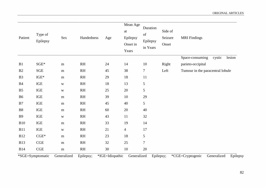
ORIGINAL ARTICLES
82
Patient Type of
Epilepsy Sex Handedness Age
Mean Age
at
Epilepsy
Onset in
Years
Duration
of
Epilepsy
in Years
Side of
Seizure
Onset
MRI Findings
B1 SGE* m RH 24 14 10 Right
Space-consuming cystic lesion
parieto-occipital
B2 SGE m RH 45 38 7 Left Tumour in the paracentral lobule
B3 IGE* m RH 29 18 11
B4 IGE w RH 18 13 5
B5 IGE w RH 25 20 5
B6 IGE m RH 39 10 29
B7 IGE m RH 45 40 5
B8 IGE m RH 60 20 40
B9 IGE w RH 43 11 32
B10 IGE m RH 33 19 14
B11 IGE w RH 21 4 17
B12 CGE* m RH 23 18 5
B13 CGE m RH 32 25 7
B14 CGE m RH 30 10 20
*SGE=Symptomatic Generalized Epilepsy; *IGE=Idiopathic Generalized Epilepsy; *CGE=Cryptogenic Generalized Epilepsy
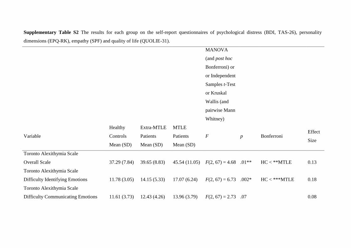
Supplementary Table S2 The results for each group on the self-report questionnaires of psychological distress (BDI, TAS-26), personality
dimensions (EPQ-RK), empathy (SPF) and quality of life (QUOLIE-31).
MANOVA
(and post hoc
Bonferroni) or
or Independent
Samples t-Test
or Kruskal
Wallis (and
pairwise Mann
Whitney)
Variable
Healthy
Controls
Mean (SD)
Extra-MTLE
Patients
Mean (SD)
MTLE
Patients
Mean (SD)
F p Bonferroni Effect
Size
Toronto Alexithymia Scale
Overall Scale 37.29 (7.84) 39.65 (8.83) 45.54 (11.05) F(2, 67) = 4.68 .01** HC < **MTLE 0.13
Toronto Alexithymia Scale
Difficulty Identifying Emotions 11.78 (3.05) 14.15 (5.33) 17.07 (6.24) F(2, 67) = 6.73 .002* HC < ***MTLE 0.18
Toronto Alexithymia Scale
Difficulty Communicating Emotions 11.61 (3.73) 12.43 (4.26) 13.96 (3.79) F(2, 67) = 2.73 .07 0.08

ORIGINAL ARTICLES
84
Toronto Alexithymia Scale
Externally Oriented Thinking 13.85 (3.11) 13.16 (4.29) 14.53 (4.17) F(2, 67) = 0.58 .5 0.02
Beck Depression Inventory
Overall Score 1.31 (2.46) 6.77 (8.39) 10.43 (9.19) x2 = 27.78 .001**
HC < ***MTLE,
**extra-MTLE
Eysenck Personality Questionnaire
Neuroticism 3.61 (2.71) 3.75 (3.08) 5.75 (2.95) F(2,65) = 4.02 .02* HC < **MTLE 0.12
Eysenck Personality Questionnaire
Psychoticism 7.11 (3.40) 7.71 (3.07) 6.75 (3.62) F(2, 65) = 2.36 .1 0.07
Eysenck Personality Questionnaire
Extraversion 6.89 (2.78) 7.42 (2.23) 7.39 (2.32) F(2, 66) = 0.80 .45 0.03
Eysenck Personality Questionnaire Lie
Scale 6.54 (3.57) 8.33 (1.50) 6.64 (2.23) F(2, 66) = 2.06 .14 0.06
Saarbruecken Personality Questionnaire
Fantasy 14.38 (2.46) 12.38 (3.80) 13.33 (2.88) F(2, 66) = 1.74 .19 0.05
Saarbruecken Personality Questionnaire
Empathic Concern 15.10 (2.26) 15.54 (3.50) 15.11 (1.78) F(2, 66) = 0.17 .84 0.01
Saarbruecken Personality Questionnaire
Perspective Taking 14.59 (2.81) 14.31 (4.23) 14.26 (2.58) F(2, 66) = 0.14 .87 0.01
Saarbruecken Personality Questionnaire
Personal Distress 10.93 (3.21) 10.54 (4.27) 11.41 (3.63) F(2, 66) = 0.19 .83 0.01

ORIGINAL ARTICLES
85
Quality of Life in Epilepsy-31
Overall Score 72.13 (12.80) 56.01 (15.65) t(1, 40) = -3.33 .002**
MTLE < **Extra-
MTLE
Quality of Life in Epilepsy-31
Overall QOL 74.82 (15.39) 58.73 (18.55) t(1, 40) = -2.80 .008**
MTLE < **Extra-
MTLE
Quality of Life in Epilepsy-31
Emotional Well Being 68.57 (19.40) 58.61 (16.68) t(1, 40) = -1.73 .09
Quality of Life in Epilepsy-31
Social Functioning 82.86 (13.85) 59.68 (22.04) t(1, 40) = -3.58 .001**
MTLE < **Extra-
MTLE
* MANOVA (and post hoc Bonferroni) or Independent Samples t-Test is significant at the p<.05 level (two-tailed).
** MANOVA (and post hoc Bonferroni) or Independent Samples t-Test is significant at the p<.01 level (two-tailed).
*** Kruskal Wallis (and pairwise Mann Whitney) is significant at the p<.001 level (two-tailed).
Sample sizes vary slightly by subgroup due to missing values.

ORIGINAL ARTICLES
86
Supplementary Table S3 Pearson’s product-moment correlations between Emotion Recognition measures (CATS Quotients), Theory of Mind
measures (Eyes Test, Moving Triangles, Faux Pas Test), and the Decision-Making Task (Iowa Gambling Task).
n=66 a
CATS,
ARQ
CATS,
PRQ
CATS,
ERQ Eyes Test
MT,
ToM,
I.
MT,
ToM,
A.
MT,
ToM,
L.
Faux Pas
Test
IGT
CATS
Affect Recognition Quotient (ARQ) 1
CATS
Prosody Recognition Quotient (PRQ) .49** 1
CATS
Emotion Recognition Quotient (ERQ) .84** .83** 1
Eyes Test
Total Score .16 .05 .12 1
Moving Triangles, ToM
Intentionality (I) .34** .45** .50** .12 1
Moving Triangles, ToM
Appropriateness (A) .19 .43** .36** .02 .70** 1
Moving Triangles, ToM
Length (L) .21 .15 .24 .13 .57** .52** 1

ORIGINAL ARTICLES
87
Faux Pas Test
Total Score .01 .23 .15 .25* .22 .06 .18 1
Iowa Gambling Task (IGT)
Global Outcome Score .43** .29* .41** .15 .25 .13 .20 .20 1
* Correlation is significant at the p<.05 level (two-tailed).
** Correlation is significant at the p<.01 level (two-tailed).
aCATS n=66, Eyes Test n=71, Moving Triangles n=63, Faux Pas Test n=69, Iowa Gambling Task n=53
Sample sizes vary slightly by subgroup due to missing values.
5.2 Advanced social cognition and amygdalar fMRI response in patients with
unilateral mesial temporal lobe epilepsy
Sarah D. Broicher, Franziska Arter, Thomas Grunwald, Dominik Huber, Martin Kurthen,
Günter Krämer, Hennric Jokeit
Swiss Epilepsy Centre, Zürich, Switzerland
Corresponding author:
Sarah Dinah Broicher, MSc
Swiss Epilepsy Center
Bleulerstrasse 60, 8008 Zürich, Switzerland
Tel.: ++41/44/387-6348
Fax: ++41/44/387-6134
e-mail: [email protected]
Key words: amygdala, fMRI, social cognition, temporal lobe epilepsy, theory of mind
Number of text pages: 14
Word count: 4 572
Number and proposed size of figures: 3; 147 KB
Number and proposed size of tables: 1; 88 KB
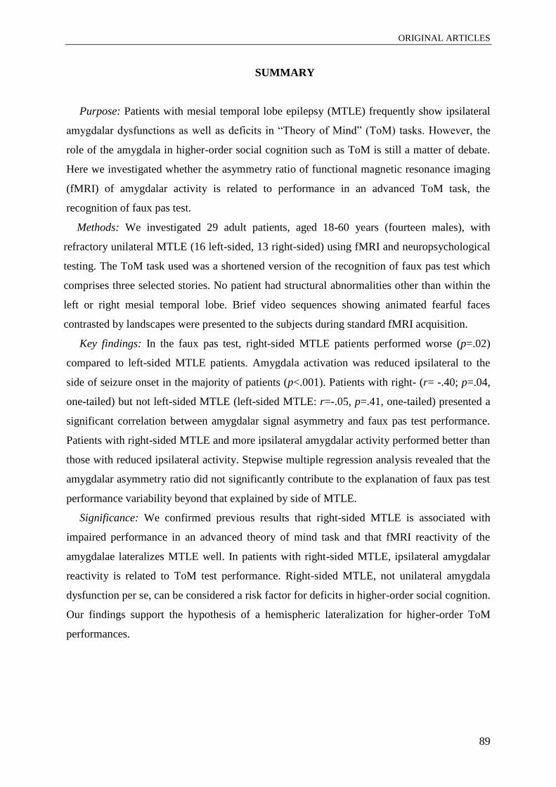
ORIGINAL ARTICLES
89
SUMMARY
Purpose: Patients with mesial temporal lobe epilepsy (MTLE) frequently show ipsilateral
amygdalar dysfunctions as well as deficits in “Theory of Mind” (ToM) tasks. However, the
role of the amygdala in higher-order social cognition such as ToM is still a matter of debate.
Here we investigated whether the asymmetry ratio of functional magnetic resonance imaging
(fMRI) of amygdalar activity is related to performance in an advanced ToM task, the
recognition of faux pas test.
Methods: We investigated 29 adult patients, aged 18-60 years (fourteen males), with
refractory unilateral MTLE (16 left-sided, 13 right-sided) using fMRI and neuropsychological
testing. The ToM task used was a shortened version of the recognition of faux pas test which
comprises three selected stories. No patient had structural abnormalities other than within the
left or right mesial temporal lobe. Brief video sequences showing animated fearful faces
contrasted by landscapes were presented to the subjects during standard fMRI acquisition.
Key findings: In the faux pas test, right-sided MTLE patients performed worse (p=.02)
compared to left-sided MTLE patients. Amygdala activation was reduced ipsilateral to the
side of seizure onset in the majority of patients (p<.001). Patients with right- (r= -.40; p=.04,
one-tailed) but not left-sided MTLE (left-sided MTLE: r=-.05, p=.41, one-tailed) presented a
significant correlation between amygdalar signal asymmetry and faux pas test performance.
Patients with right-sided MTLE and more ipsilateral amygdalar activity performed better than
those with reduced ipsilateral activity. Stepwise multiple regression analysis revealed that the
amygdalar asymmetry ratio did not significantly contribute to the explanation of faux pas test
performance variability beyond that explained by side of MTLE.
Significance: We confirmed previous results that right-sided MTLE is associated with
impaired performance in an advanced theory of mind task and that fMRI reactivity of the
amygdalae lateralizes MTLE well. In patients with right-sided MTLE, ipsilateral amygdalar
reactivity is related to ToM test performance. Right-sided MTLE, not unilateral amygdala
dysfunction per se, can be considered a risk factor for deficits in higher-order social cognition.
Our findings support the hypothesis of a hemispheric lateralization for higher-order ToM
performances.

ORIGINAL ARTICLES
90
INTRODUCTION
Mesial temporal lobe epilepsy (MTLE) is typically associated with lesions involving
temporal limbic structures such as the hippocampus and amygdala. The role of the
hippocampus and adjacent cortical regions in memory functions is well established and is
taken into account in the diagnostic workup in candidates for epilepsy surgery. In contrast, the
role the amygdala plays in clinical and neuropsychological symptoms and in the context of
epilepsy surgery is less well understood.
Recently, we showed that ipsilateral amygdala reactivity in fMRI is reduced in response to
fearful faces in the majority of patients with unilateral MTLE (Schacher, Haemmerle et al.,
2006). Moreover, we found that MTLE patients, as compared to patients with epilepsy not
originating within the MTL and healthy controls, were impaired in their ability to recognise
social faux pas (Schacher, Winkler et al., 2006). The faux pas test can be seen as a “higher-
order” or “advanced” theory of mind (ToM) task as it goes beyond simple attributions and
requires inferences regarding others’ mental states with respect to their thoughts and beliefs
(“cognitive ToM component”) as well as with respect to their emotional states and feelings
(“affective ToM component”) (Shamay-Tsoory & Aharon-Peretz, 2007; Stone et al., 2003;
Stone et al., 1998). Particularly patients with right-sided MTLE performed worse on this test
than patients with left-sided MTLE.
In epilepsy, the laterality of the seizure focus has especially been considered as a potential
risk factor for disturbances in basal aspects of social cognition such as the processing of
emotional stimuli. TLE-patients, in particular patients with early seizure onset and right or
bilateral medial temporal dysfunction, were found to have pronounced difficulties in the
recognition of fearful faces (Benuzzi et al., 2004; Bonora et al., 2011; Meletti et al., 2009;
2003). Lesion studies have also found impairments in the recognition of fear after right, rather
than left, amygdala damage (Adolphs et al., 2001; Anderson, Spencer, Fulbright, & Phelps,
2000). However, since little research has been conducted on higher-order social cognition in
MTLE, evidence on the question of whether patients with right-sided MTLE were at
increased risk for disturbances of higher-order social cognition is still lacking.
ToM tasks are thought to activate a consistent set of frontal and temporal brain regions
(Amodio & Frith, 2006; Stone et al., 1998). In their current overview of functional imaging
studies investigating the neural basis of ToM, Carrington and Bailey (Carrington & Bailey,
2009) found the amygdala to be less consistently activated. However, there is converging
evidence that the amygdala, besides its central role in the perception and processing of
ORIGINAL ARTICLES
91
socially relevant information (Adolphs, 2003; Spezio et al., 2007), is at the core of the ability
to interpret the mental states of others (Baron-Cohen et al., 2000; Castelli et al., 2000;
Gallagher & Frith, 2004; Stone et al., 2003).
Disagreement remains as to what degree the amygdala merely supports the development
of ToM abilities (Frith & Frith, 2003; Shaw et al., 2004; Tager-Flusberg & Sullivan, 2000) or
whether it represents an essential part of the ToM network (Channon & Crawford, 2000;
Happe et al., 1999; Sommer et al., 2008; Stone et al., 2003). The majority of authors agree
with the latter supposition, which receives support in particular from lesion studies that
indicate a clear connection between uni- and bilateral lesions of the amygdala and deficits in
ToM (Heberlein & Adolphs, 2004a; Stone et al., 2003). A recent meta-analytic review of the
literature on human neuroimaging of emotion showed that a set of interacting fronto-temporal
brain regions, including the amygdala, are active during the experiencing and perception of
emotion across a range of emotion categories (Lindquist, 2011). The amygdala was found to
be more active during fear perception, but also during all other emotion categories. Whereas a
considerable number of studies support the close relationship between emotion recognition
and ToM using behavioural data (Bora et al., 2005; Brune, 2005a; Buitelaar & van der Wees,
1997; Dyck, Piek, Hay, Smith, & Hallmayer, 2006; Henry, Phillips, Crawford, Ietswaart, &
Summers, 2006), but see also (Langdon, Coltheart, & Ward, 2006; Phillips, MacLean, &
Allen, 2002 for divergent results), the only study reported to date which has investigated its
relationship on both behavioural and neuronal levels was a recent event-related fMRI study by
Mier et al. (2010). The authors reported a positive correlation between the number of
correctly recognized emotions and correctly recognized intentions as well as overlapping
patterns of activation during performance of both tasks. Areas conjointly activated by both
tasks included, among others, the amygdala.
The purpose here is to evaluate whether functional impairments of the amygdalae as
established by fMRI are associated with degraded performance in a recognition of faux pas
test in patients with MTLE. Our hypothesis was that amygdala activity induced by fearful face
processing is associated with performances in higher-order ToM tasks.
Following the finding of our previous investigation (Schacher, Winkler et al., 2006) which
suggested that the right hemisphere could be more strongly associated with theory of mind
abilities in epilepsy patients, we further wanted to assess the relationship between ToM
abilities and the laterality of the epileptogenic zone and amygdala BOLD reactivity within the
same group of patients.

ORIGINAL ARTICLES
92
PATIENTS AND METHODS
Subjects
Thirty-one patients aged 18 to 60 (mean 35.97 years, SD 13.09; 14 men, 17 women; 29
right handed, 1 left handed and 1 ambidexter) with medically refractory focal epilepsies
(mean age at seizure onset 13.32 years, SD 10.65 and epilepsy duration 22.32 years, SD
14.83) were investigated (see Table 1). Patients were recruited from consecutive inpatient
admissions to the Swiss Epilepsy Center between 2006 and 2010.
Patients underwent neurological examination and routine EEG recordings using the 10-20
system. Seizure types and epilepsy syndromes were diagnosed according to the classification
of the International League Against Epilepsy (1989; 1981).
All patients had MTLE with a clear unilateral seizure onset of MTL origin, as shown by
continuous interictal and ictal video/EEG monitoring with scalp and sphenoidal electrodes
and a clear diagnosis of MTL pathology, as shown by high-resolution routine MRI. Of the
thirty-one patients, thirty had hippocampal sclerosis (HS) (twelve right- and eighteen left-
sided). The MRI of one patient revealed a space-consuming lesion of unclear pathology at the
medial border of the amygdala (Table 1). All patients were treated with first line antiepileptic
drugs (AEDs).
Language laterality was determined either by use of an fMRI paradigm employing a
verbal fluency task (n=29) or by Wada test if the fMRI was inconclusive (n=2) (Woermann et
al., 2003). The majority of patients (29/31) had left-sided language dominance; two had
bilateral language representations in fMRI. Handedness was determined by the Edinburgh
Handedness Inventory (Table 1).
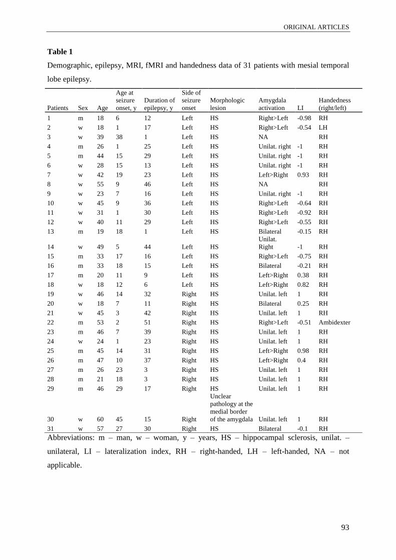
ORIGINAL ARTICLES
93
Table 1
Demographic, epilepsy, MRI, fMRI and handedness data of 31 patients with mesial temporal
lobe epilepsy.
Patients Sex Age
Age at
seizure
onset, y
Duration of
epilepsy, y
Side of
seizure
onset
Morphologic
lesion
Amygdala
activation LI
Handedness
(right/left)
1 m 18 6 12 Left HS Right>Left -0.98 RH
2 w 18 1 17 Left HS Right>Left -0.54 LH
3 w 39 38 1 Left HS NA RH
4 m 26 1 25 Left HS Unilat. right -1 RH
5 m 44 15 29 Left HS Unilat. right -1 RH
6 w 28 15 13 Left HS Unilat. right -1 RH
7 w 42 19 23 Left HS Left>Right 0.93 RH
8 w 55 9 46 Left HS NA RH
9 w 23 7 16 Left HS Unilat. right -1 RH
10 w 45 9 36 Left HS Right>Left -0.64 RH
11 w 31 1 30 Left HS Right>Left -0.92 RH
12 w 40 11 29 Left HS Right>Left -0.55 RH
13 m 19 18 1 Left HS Bilateral -0.15 RH
14 w 49 5 44 Left HS
Unilat.
Right -1 RH
15 m 33 17 16 Left HS Right>Left -0.75 RH
16 m 33 18 15 Left HS Bilateral -0.21 RH
17 m 20 11 9 Left HS Left>Right 0.38 RH
18 w 18 12 6 Left HS Left>Right 0.82 RH
19 w 46 14 32 Right HS Unilat. left 1 RH
20 w 18 7 11 Right HS Bilateral 0.25 RH
21 w 45 3 42 Right HS Unilat. left 1 RH
22 m 53 2 51 Right HS Right>Left -0.51 Ambidexter
23 m 46 7 39 Right HS Unilat. left 1 RH
24 w 24 1 23 Right HS Unilat. left 1 RH
25 m 45 14 31 Right HS Left>Right 0.98 RH
26 m 47 10 37 Right HS Left>Right 0.4 RH
27 m 26 23 3 Right HS Unilat. left 1 RH
28 m 21 18 3 Right HS Unilat. left 1 RH
29 m 46 29 17 Right HS Unilat. left 1 RH
30 w 60 45 15 Right
Unclear
pathology at the
medial border
of the amygdala Unilat. left 1 RH
31 w 57 27 30 Right HS Bilateral -0.1 RH
Abbreviations: m – man, w – woman, y – years, HS – hippocampal sclerosis, unilat. –
unilateral, LI – lateralization index, RH – right-handed, LH – left-handed, NA – not
applicable.

ORIGINAL ARTICLES
94
Mentally handicapped patients were excluded (IQ<70). All patients had an IQ equal to or
greater than 75. Further exclusion criteria were psychiatric disorders, whereby adjustment
disorders and mild to moderate depression constituted an exception to this rule since it was
assumed that they are comorbid symptoms of temporal lobe epilepsy (Glosser et al., 2000; B.
Hermann et al., 2008). Adjustment disorders and depressive mood were diagnosed by a highly
experienced clinical psychologist using ICD-10 criteria.
Amygdala activation was measured in all patients with a fearful face paradigm (Schacher,
Haemmerle et al., 2006). In addition to the fearful face paradigm, subjects underwent
neuropsychological investigation and performed a typical advanced ToM task in which they
had to detect a faux pas in a brief prose passage (Stone et al., 2003). fMRI and
neuropsychological testing took place either on the same (n=19) or different days (n=12), but
did not take place more than three months apart, except in one patient in whom this interval
was seven months.
All patients gave written informed consent following a thorough explanation of the study.
The present study was performed in adherence of the Declaration of Helsinki and was also
approved by the local medical ethics committee.
Recognition of faux pas test
All 31 subjects performed a shortened version of a faux pas test (Stone et al., 2003) that
comprised three selected stories. The shortened version of the faux pas test was previously
investigated in healthy controls and patients with TLE by Schacher et al. (Schacher, Winkler
et al., 2006). Further information related to the selection procedure of the stories has been
described by Schacher et al. (2006). The faux pas test is a ToM test for adults and estimates
the ability to recognize and understand a social faux pas.
A faux-pas is understood as a statement in which the speaker unintentionally offends or
insults another person. For example, person “A” complains to person “B” about a wedding
present without realising that he is talking to the person from whom he received it. The faux
pas test measures several ToM components by including deductions concerning epistemic
mental states as well as affective mental states (Schacher, Winkler et al., 2006; Stone et al.,
2003; 1998). Patients were asked to read each story silently, knowing that they would then be
asked questions about the story. There were three questions regarding interpersonal relations
and emotional states, and the last one was a control question to control for text comprehension
(see Appendix). The first question concerned the detection and comprehension of the faux
pas, while question two and three required the patient to impute the mental state of

ORIGINAL ARTICLES
95
andattribute emotions to another. The data analysis involved the sum of correct answers to the
first three questions, where each question was scored with one point, resulting in maximum
nine points for all three stories. Points for the control question were excluded from the total
score but served as an indicator of text comprehension.
Additionally, to control for language comprehension, data from the Chapman-Cook test
were used (Chapman, 1923). This test comprises 12 short text passages in which a single
word does not fit with the overall context of the text. All patients included in the study had
intact language-comprehension abilities (i.e., all answered every faux pas control question
correctly and correctly identified at least 11 of the 12 words in the Chapman Cook test).
Intelligence quotients (IQs) were estimated using the full-scale IQ derived from the
German version of the Wechsler Adult Intelligence Scale, Revised (HAWIE-R) (Tewes,
1994) and a vocabulary-based intelligence test (MWT-B). In this test, patients were required
to identify a real word from a choice of four pseudo-words with increasing test difficulty
(Lehrl et al., 1995). The mean score for Full-Scale, Verbal, and Performance IQ was within
the normal range (Full-Scale IQ: M=98.48; SD=14.10; Verbal IQ: M=94.93; SD=15.18;
Performance IQ: M=103.17; SD=16.65). The majority of patients (80.65%; 25 out of 31)
tested had a Full-Scale, Verbal and Performance IQ within the norm range (i.e., above 85).
fMRI task design
The fMRI paradigm used here has been extensively described by Schacher et al. (2006).
The applied block design paradigm consisted of eight activation and eight baseline blocks
each lasting 24 seconds. The activation condition comprised 75 brief episodes (two to three
seconds) from thriller and horror films. All episodes showed close-ups of the faces of actors
who were expressing fear with high intensity. None of the episodes showed violence or
aggression. During baseline blocks 72 short episodes of similar length with dynamic
landscape video recordings were presented. Video clips of dull domestic landscapes were
used owing to their stable low emotional content while their general visual stimulus properties
were comparable with the movie clips. Frequency and duration of the sequences were
matched in the activation and control conditions. Stimuli were presented via a back-projection
screen and viewed through a tilted overhead mirror. Prior to beginning subjects were told that
they would see rapid presentations of film sequences depicting fearful faces intermixed with
landscape film sequences. They were instructed to relax while watching the film and to focus
on the eyes of the actors during the activation blocks.

ORIGINAL ARTICLES
96
MRI acquisition
fMRI data were recorded using a 1.5 T system (Magneton Sonata, Siemens, Erlangen,
Germany) and a 3.0 T system (Achieva Philips Medical Systems, Best, The Netherlands). A
comparison between data which were collected on different systems with different MRI-
parameters is likely unproblematic as images were considered and evaluated intra-
individually and only the activity ratios of different patients were compared. Routine skull
base magnetic resonance imaging (MRI) protocols were performed for each patient. MRI
sequences included T1-weighted spin echo and gradient echo three-dimensional multiplanar
reconstruction images (MPRAGE), coronal T2-weighted turbo spin echo, T2-weighted fast
fluid attenuated inversion recovery (FLAIR) and diffusion weighted sequences. Coronal T2-
weighted and FLAIR slices were 1-3 mm thick and were acquired at 90° perpendicular to the
long axis of the hippocampi. Subjects were sited in the head coil with ear pads and foam
padding to minimize head motion and noise exposition. T1-weighted (T1W) acquisition
parameters, according to the pre-existing protocols, were 176 axial slices with 1-mm single-
slice thickness, repetition time (TR) 8.2 ms, echo time (TE) 3.93 ms, 8° flip angle, field of
view (FOV) 250 mm, and 288 x 288 matrix on the 3.0 T system, and TR 1.900 ms, TE 3.93
ms, 15° flip angle, FOV 250 mm, and 256 x 256 matrix on the 1.5 T system.
Functional data were acquired using echo planar imaging (EPI) T2*-weighted sequence.
The following parameters were applied to measure amygdalar activation: 18 coronal slices on
the 3.0 T vs. 12 coronal slices on the 1.5 T, 4-mm slice thickness and 0-mm interslice gap vs.
5-mm slice thickness and 1-mm interslice gap, TR 1500 ms vs. TR 1490 ms, TE 35 ms vs. TE
60, 75° flip angle vs. 90° flip angle, FOV 220 mm vs. FOV 250 mm, matrix size 64 x 64,
voxel size 2.75 x 2.75 x 4 mm vs. voxel size 3.9 x 3.9 x 5 mm, reconstructed into an image
matrix of 128 x 128 on both systems. Coronal slices were geared orthogonally to the
hippocampal formation and were spread over the anterior temporal lobe.
Data analyses
Functional MRI single subject data analysis was performed with BrainVoyager QX
software (BrainInnovation, Maastricht, the Netherlands). In the primary analyses the data
were preprocessed with (1) three-dimensional motion correction and (2) trend removal by
temporal fast Fourier transform-based high-pass filtering and transformed into Talairach co-
ordinate space. Images of the fearful face task were additionally spatially smoothed with a full
width at half-maximum of 4 mm.
ORIGINAL ARTICLES
97
For multiple regression analysis a general linear model (GLM) with the predictor for the
activation condition was computed. The time courses of the predictor were obtained by using
a linear model of the hemodynamic response. The overall model fit was assessed using F
statistics. Significant differences between the experimental conditions were assessed using
contrast (t) maps.
For images in the fearful face task, individual volumes of interests (VOIs) were defined
for the amygdalar region. VOIs were specified functionally for each patient separately using a
predefined statistical threshold of p<.001 (see Table 1). Anatomic borders of the functional
clusters were the uncal recess caudally and the optical chiasm rostrally and white matter
superiorly. For each VOI the number of activated voxels was counted in the left and right
hemisphere on a predefined threshold of p<.001. Asymmetry ratios (AR) were defined for the
number of significantly activated voxels in the amygdalar/periamygdalar area using the
formula: AR = (left - right)/(left + right). If the AR was greater than 0.25 and less than -0.25
these cases were categorized as asymmetric.
Statistical Analysis
Statistical evaluations were performed with the SPSS 15 software package (SPSS Inc,
Chicago, IL, U.S.A).
We applied the non-parametric Mann-Whitney U test to examine whether right- vs. left-
sided MTLE patients and men vs. women differed in their overall score in the faux pas test.
We additionally used nonparametric correlation analysis (Kendall Rank Correlation
Coefficient) to investigate the relation between age at seizure onset, duration of epilepsy, age,
Full-Scale, Verbal and Performance IQ, and the ability to detect faux pas.
Since data are not measured on an interval scale and deviate from a normal distribution,
we computed the Kendall Rank Correlation Coefficient of the “performance in the faux pas
test” and “Asymmetry Ratio (AR)”. Because of the directional hypothesis of a better
performance in the faux pas test and increased activation of the right amygdala in right-sided
MTLE patients, single correlations between fMRI asymmetry ratio and performance in the
faux pas test for subgroups of MTLE patients were carried out at a significance level set at
p<.05 (one-tailed).
A stepwise multiple regression analysis was performed to examine the possible predicting
abilities of side of MTLE and AR for faux pas test performance. The level of significance was
set to p=.05 (two-tailed tests).
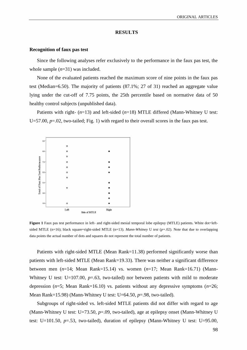
ORIGINAL ARTICLES
98
RESULTS
Recognition of faux pas test
Since the following analyses refer exclusively to the performance in the faux pas test, the
whole sample (n=31) was included.
None of the evaluated patients reached the maximum score of nine points in the faux pas
test (Median=6.50). The majority of patients (87.1%; 27 of 31) reached an aggregate value
lying under the cut-off of 7.75 points, the 25th percentile based on normative data of 50
healthy control subjects (unpublished data).
Patients with right- (n=13) and left-sided (n=18) MTLE differed (Mann-Whitney U test:
U=57.00, p=.02, two-tailed; Fig. 1) with regard to their overall scores in the faux pas test.
Figure 1 Faux pas test performance in left- and right-sided mesial temporal lobe epilepsy (MTLE) patients. White dot=left-
sided MTLE (n=16); black square=right-sided MTLE (n=13). Mann-Whitney U test (p=.02). Note that due to overlapping
data points the actual number of dots and squares do not represent the total number of patients.
Patients with right-sided MTLE (Mean Rank=11.38) performed significantly worse than
patients with left-sided MTLE (Mean Rank=19.33). There was neither a significant difference
between men (n=14; Mean Rank=15.14) vs. women (n=17; Mean Rank=16.71) (Mann-
Whitney U test: U=107.00, p=.63, two-tailed) nor between patients with mild to moderate
depression (n=5; Mean Rank=16.10) vs. patients without any depressive symptoms (n=26;
Mean Rank=15.98) (Mann-Whitney U test: U=64.50, p=.98, two-tailed).
Subgroups of right-sided vs. left-sided MTLE patients did not differ with regard to age
(Mann-Whitney U test: U=73.50, p=.09, two-tailed), age at epilepsy onset (Mann-Whitney U
test: U=101.50, p=.53, two-tailed), duration of epilepsy (Mann-Whitney U test: U=95.00,
ORIGINAL ARTICLES
99
p=.40, two-tailed), and Full-Scale, Verbal and Performance IQ (Mann-Whitney U test; Full-
Scale IQ: U=96.00, p=.40, two-tailed; Verbal IQ: U=93.50, p=.65, two-tailed; Performance
IQ: U=90.50, p=.55, two-tailed).
Non-parametric correlational analysis revealed a negative association between the MTLE
patient’s age and deficits in faux pas test performance (Kendall r=-.32; p=.02, two-tailed).
Age at seizure onset (Kendall r=-.06; p=.67, two-tailed), duration of epilepsy (Kendall r=-.23;
p=.08, two-tailed) and Full-Scale, Verbal and Performance IQ (Kendall; Full-Scale IQ: r=.12;
p=.36, two-tailed; Verbal IQ: r=.11; p=.43, two-tailed; Performance IQ: r=.15; p=.27, two-
tailed) were not related to faux pas test performance.
fMRI asymmetry ratio (AR)
T2*-weighted contrast differences were found within the amygdalae in all but two
subjects in response to watching video sequences with fearful faces compared to watching
landscape scenes (p<.01). Due to the nonquantifiable AR of two patients, they were excluded
from further analysis. In the following we refer to the sample excluding these two patients
(n=29). The activation focus was located in the superior portion of the amygdala, as found by
Schacher et al. (Schacher, Haemmerle et al., 2006). The mean AR of the amygdala was .05
(SD=.83).
In patients with right-sided MTLE (n=13) the mean of the AR was .69 (SD=.52), which
reflects predominant contralateral left-sided activation of the amygdala. Patients with left-
sided MTLE (n=16) showed the opposite activity pattern with predominant contralateral
right-sided amygdala activation (AR=-.47, SD=.66). The AR of patients with right-sided
MTLE significantly differed from patients with left-sided MTLE (Mann-Whitney U test:
U=13.00, p<.001; Fig. 2).

ORIGINAL ARTICLES
100
Figure 2 Asymmetry ratio of fMRI amygdalar activity in left- and right-sided mesial temporal lobe epilepsy (MTLE)
patients. White dot=left-sided MTLE (n=16); black square=right-sided MTLE (n=13). Mann-Whitney U test (p<.001). Note
that due to overlapping data points the actual number of dots and squares do not represent the total number of patients.
In patient seven, 17 and 18 with left-sided MTLE and patient 22 with right-sided MTLE
we found an asymmetric activation of the amygdala ipsilateral to the seizure onset side.
Of the 29 patients, bilateral amygdalae activation without appreciable asymmetry was
observed in four patients (patients 13, 16, 20 and 31), unilateral right-sided activation in five,
unilateral left-sided activation in eight, lateralized right-sided activation in seven and
lateralized left-sided activation in five. Altogether, 11 out of 13 patients with right-sided
MTLE showed a positive AR and 13 out of 16 patients with left-sided MTLE showed a
negative AR.
In relation to the side of seizure onset, amygdala activation was contralateral in 21 out of
29 (72 %) patients, and ipsilateral in four (14%) patients. In two patients with left-sided
MTLE as well as two patients with right-sided MTLE, amygdala activation was bilateral
(14%).
Correlation between fMRI asymmetry ratio and performance in the faux pas test
The bivariate non-parametric correlation coefficient between the AR and the total score in
the faux pas test was r=-.31, p=.03, two-tailed (n=29) (Fig. 3). Patients with predominantly
right-sided amygdalar signal tended to perform better in the faux pas test.
Post hoc, a better performance in the faux pas test with increased activation of the right
amygdala was found within the subgroup of right-sided MTLE patients (r=-.40; p=.04, one-
tailed), while this was not the case in the subgroup of left-sided MTLE patients (left-sided
MTLE: r=-.05, p=.41, one-tailed; Fig. 3).

ORIGINAL ARTICLES
101
Figure 3 Correlations between the total score in the faux pas test and the asymmetry ratio of fMRI amygdalar activity in
mesial temporal lobe epilepsy (MTLE). White dot=left-sided MTLE (n=16); black square=right-sided MTLE (n=13); solid
line=correlation for left- and right-sided MTLE patients (r=-.31, p=.03, two-tailed); line with three dots=correlation for left-
sided MTLE patients (r=-.05, p=.41, one-tailed); line with one dot=correlation for right-sided MTLE patients (r= -.40; p=.04,
one-tailed). Note that due to overlapping data points the actual number of dots and squares do not represent the total number
of patients.
Intercorrelation between fMRI asymmetry ratio, side of MTLE and performance in the
faux pas test
A stepwise multiple regression analysis was conducted using performance in the faux pas
test as a dependent variable and the side of MTLE and the fMRI asymmetry ratio as the
predictor variables. The results of the stepwise analysis revealed that the side of MTLE, not
the AR, was a significant predictor (r=.43; F=6.30, p=.02, two-tailed) of faux pas task
performance.
DISCUSSION
We reproduced data from two previous independent former studies (Schacher, Haemmerle
et al., 2006; Schacher, Winkler et al., 2006) using a single sample of 29 consecutive
pharmacoresistant MTLE patients.
Patients with MTLE were tested using a shortened version of the faux pas test in which
patients with MTLE were previously shown to be specifically impaired (Schacher, Winkler et
al., 2006). In concordance with our previous study, we found that patients with right-sided
MTLE performed worse in identifying a faux pas than patients with left-sided MTLE
(Schacher, Winkler et al., 2006).
ORIGINAL ARTICLES
102
Furthermore, we used an animated fearful faces paradigm that reliably elucidates
amygdalar BOLD activation in fMRI studies. Schacher et al. (2006) have previously shown
that this paradigm results in bilateral amygdalar activation in the majority of healthy controls,
whereas the activation is mostly contralateral to the side of seizure onset in patients with
unilateral TLE (Schacher, Haemmerle et al., 2006). Once again we were able to demonstrate
that the fearful faces fMRI paradigm is a reliable method for visualizing amygdala activation
and lateralizing temporal lobe epilepsy. However, we found considerable variability of
amygdalar activity. This is in accordance with T2 measurements in the amygdala
demonstrating bilateral pathological changes even in patients with unilateral hippocampal
sclerosis (Bartlett, Richardson, & Duncan, 2002).
Extending the findings of earlier studies, we aimed to investigate whether the asymmetry
ratio of fMRI amygdalar activity was related to performance in the recognition of faux pas
test. We found a weak but significant correlation between the asymmetry ratio of amygdalar
activity and faux-pas test performance. A predominantly right-sided BOLD-signal of the
amygdala during an animated fearful face paradigm was associated with better performance in
the faux pas test.
Stepwise multiple regression analysis of our data revealed that the side of pathology,
however, explained a more considerable part of the variance of faux pas test performance than
the lateralization of the amygdala BOLD signal. Thus, patients with right-sided MTLE may
be at increased risk for deficits in higher-order social cognition. Our findings are in line with
previous studies of the effect of cerebral damage on ToM skills that support the view that
ToM relies, at least in part, on the right hemisphere (Brunet et al., 2000; Calarge, Andreasen,
& O'Leary, 2003; H. L. Gallagher et al., 2000; Griffin et al., 2006; Shamay-Tsoory, Tomer, &
Aharon-Peretz, 2005; Siegal, Carrington, & Radel, 1996; Winner, Brownell, Happe, Blum, &
Pincus, 1998), whereas unilateral amygdalar dysfunction per se is not sufficient to explain the
variability of ToM task performance in our sample. Especially within the prefrontal
structures, there seems to be a hemispheric lateralization, indicating that the right frontal lobe
is more strongly associated with ToM abilities (Shamay-Tsoory, Tomer, Berger, Goldsher, &
Aharon-Peretz, 2005; Stuss, Gallup, & Alexander, 2001; Vollm et al., 2006). Seizures which
do have their onset in the temporal lobe will often invade extra-temporal structures in the
course of the seizure (Jokeit et al., 1997; Oyegbile et al., 2004). Therefore, impairment of
frontal lobe function may also be considered as a risk factor for deficits in higher-order ToM
tasks in epilepsy (Farrant et al., 2005). However, other studies have found contradictory
results with predominantly left hemisphere activation of the frontal lobe (Calarge et al., 2003;
ORIGINAL ARTICLES
103
Fletcher et al., 1995; Goel, Grafman, Sadato, & Hallett, 1995) or no effect of a hemispheric
lateralization for ToM performances at all (Weed, 2008).
Post hoc analyses of separate subject groups showed that patients with right- but not left-
sided MTLE presented a significant correlation between amygdalar signal asymmetry and
faux pas test performance. Our results suggest that patients with right-sided MTLE, but more
right-sided amygdalar reactivity tend to perform better than patients with right-sided MTLE
and reduced amygdalar reactivity. Whereas the side of lesion explains the majority of
variance in our sample, this specific correlation may suggest that ipsilateral amygdalar
reactivity in patients with right-sided MTLE indicates the extension of epilepsy-related
structural and functional pathology within a right hemisphere fronto-limbic network that
governs affective and social processing.
Furthermore, the impaired performance on the advanced ToM task was not accounted for
by variables such as age at seizure onset, duration of epilepsy or cognitive ability (as
measured by IQ) and, thus, corroborate earlier findings that ToM abilities are mainly
independent of other cognitive functions (Frith & Frith, 2003). However, we are aware that
the present study did not specifically address the relationship between ToM and cognitive
functions in TLE, elucidation of which requires more focused and exhaustive investigation.
By contrast, age is supposed to have an influence upon faux pas test performance. We found a
negative association with age. The effects of aging on ToM and their links with other
cognitive processes, such as executive functions and memory, are still inconclusive. However,
our finding is in accordance with recent studies, indicating that theory of mind abilities may
decline with adult age (Duval, Piolino, Bejanin, Eustache, & Desgranges, ; Slessor, Phillips,
& Bull, 2007).
There are some limitations of the study that need to be addressed.
A first remark concerns the weakness of the correlation between faux pas test performance
and fMRI reactivity. Reasons include methodological limitations due to temporarily
independent measurements of amygdalar activity and behavioral test performance as well as
the imperfect reliability of both measures.
Second, due to the relatively small sample size, caution should be used, as the findings
might not be transferable to the entire MTLE group. Therefore, it would be worthwhile to
further study and isolate clinical characteristics within our subgroups of right- and left-sided
MTLE patients.
In conclusion, right-sided MTLE, not unilateral amygdala dysfunction, can be considered
as a primary risk factor for deficits in higher-order social cognition. At least in patients with
ORIGINAL ARTICLES
104
right-sided MTLE, ipsilateral amygdalar reactivity is related to social behavioural measures.
Whether amygdala activation acts an unspecific indicator of the integrity of fronto-limbic
circuitry or whether it is indeed involved in ToM processing needs to be investigated in
patients who undergo amygdala resection for surgical treatment of epilepsy. Overall, our
results support the hypothesis of a hemispheric lateralization for higher-order ToM
performances.

ORIGINAL ARTICLES
105
ACKNOWLEDGMENTS
We would like to thank the Swiss Epilepsy Foundation for financial support and Gesa
Osterkamp and Theresa Biesold for technical support. We also would like to thank Victoria
Reed for valuable comments regarding the improvement of the manuscript.
We confirm that we have read the Journal’s position on issues involved in ethical
publication and affirm that this report is consistent with those guidelines.
DISCLOSURE
None of the authors has any conflict of interest to disclose.

ORIGINAL ARTICLES
106
REFERENCES
Proposal for revised clinical and electroencephalographic classification of epileptic seizures.
(1981) From the Commission on Classification and Terminology of the International
League Against Epilepsy. Epilepsia 22:489-501.
Proposal for revised classification of epilepsies and epileptic syndromes. (1989) Commission
on Classification and Terminology of the International League Against Epilepsy.
Epilepsia 30:389-399.
Adolphs R (2003) Is the human amygdala specialized for processing social information? Ann
N Y Acad Sci 985:326-340.
Adolphs R, Tranel D, Damasio H (2001) Emotion recognition from faces and prosody
following temporal lobectomy. Neuropsychology 15:396-404.
Amodio DM, Frith CD (2006) Meeting of minds: the medial frontal cortex and social
cognition. Nat Rev Neurosci 7:268-277.
Anderson AK, Spencer DD, Fulbright RK, Phelps EA (2000) Contribution of the
anteromedial temporal lobes to the evaluation of facial emotion. Neuropsychology
14:526-536.
Baron-Cohen S, Ring HA, Bullmore ET, Wheelwright S, Ashwin C, Williams SC (2000) The
amygdala theory of autism. Neurosci Biobehav Rev 24:355-364.
Bartlett PA, Richardson MP, Duncan JS (2002) Measurement of amygdala T2 relaxation time
in temporal lobe epilepsy. J Neurol Neurosurg Psychiatry 73:753-755.
Benuzzi F, Meletti S, Zamboni G, Calandra-Buonaura G, Serafini M, Lui F, Baraldi P,
Rubboli G, Tassinari CA, Nichelli P (2004) Impaired fear processing in right mesial
temporal sclerosis: a fMRI study. Brain Res Bull 63:269-281.
Bonora A, Benuzzi F, Monti G, Mirandola L, Pugnaghi M, Nichelli P, Meletti S (2011)
Recognition of emotions from faces and voices in medial temporal lobe epilepsy. Epilepsy
Behav 20:648-654.
Bora E, Vahip S, Gonul AS, Akdeniz F, Alkan M, Ogut M, Eryavuz A (2005) Evidence for
theory of mind deficits in euthymic patients with bipolar disorder. Acta Psychiatr Scand
112:110-116.
Brune M (2005) Emotion recognition, 'theory of mind,' and social behavior in schizophrenia.
Psychiatry Res 133:135-147.
Brunet E, Sarfati Y, Hardy-Bayle MC, Decety J (2000) A PET investigation of the attribution
of intentions with a nonverbal task. Neuroimage 11:157-166.

ORIGINAL ARTICLES
107
Buitelaar JK, van der Wees M (1997) Are deficits in the decoding of affective cues and in
mentalizing abilities independent? J Autism Dev Disord 27:539-556.
Calarge C, Andreasen NC, O'Leary DS (2003) Visualizing how one brain understands
another: a PET study of theory of mind. Am J Psychiatry 160:1954-1964.
Carrington SJ, Bailey AJ (2009) Are there theory of mind regions in the brain? A review of
the neuroimaging literature. Hum Brain Mapp 30:2313-2335.
Castelli F, Happe F, Frith U, Frith C (2000) Movement and mind: a functional imaging study
of perception and interpretation of complex intentional movement patterns. Neuroimage
12:314-325.
Channon S, Crawford S (2000) The effects of anterior lesions on performance on a story
comprehension test: left anterior impairment on a theory of mind-type task.
Neuropsychologia 38:1006-1017.
Chapman F (1923) The Community Chest. Cal State J Med 21:317-318.
Duval C, Piolino P, Bejanin A, Eustache F, Desgranges B (2010) Age effects on different
components of theory of mind. Conscious Cogn.
Dyck MJ, Piek JP, Hay D, Smith L, Hallmayer J (2006) Are abilities abnormally
interdependent in children with autism? J Clin Child Adolesc Psychol 35:20-33.
Farrant A, Morris RG, Russell T, Elwes R, Akanuma N, Alarcon G, Koutroumanidis M
(2005) Social cognition in frontal lobe epilepsy. Epilepsy Behav 7:506-516.
Fletcher PC, Happe F, Frith U, Baker SC, Dolan RJ, Frackowiak RS, Frith CD (1995) Other
minds in the brain: a functional imaging study of "theory of mind" in story
comprehension. Cognition 57:109-128.
Frith U, Frith CD (2003) Development and neurophysiology of mentalizing. Philos Trans R
Soc Lond B Biol Sci 358:459-473.
Gallagher HL, Frith CD (2004) Dissociable neural pathways for the perception and
recognition of expressive and instrumental gestures. Neuropsychologia 42:1725-1736.
Gallagher HL, Happe F, Brunswick N, Fletcher PC, Frith U, Frith CD (2000) Reading the
mind in cartoons and stories: an fMRI study of 'theory of mind' in verbal and nonverbal
tasks. Neuropsychologia 38:11-21.
Glosser G, Zwil AS, Glosser DS, O'Connor MJ, Sperling MR (2000) Psychiatric aspects of
temporal lobe epilepsy before and after anterior temporal lobectomy. J Neurol Neurosurg
Psychiatry 68:53-58.
Goel V, Grafman J, Sadato N, Hallett M (1995) Modeling other minds. Neuroreport 6:1741-
1746.

ORIGINAL ARTICLES
108
Griffin R, Friedman O, Ween J, Winner E, Happe F, Brownell H (2006) Theory of mind and
the right cerebral hemisphere: refining the scope of impairment. Laterality 11:195-225.
Happe F, Brownell H, Winner E (1999) Acquired 'theory of mind' impairments following
stroke. Cognition 70:211-240.
Heberlein AS, Adolphs R (2004) Impaired spontaneous anthropomorphizing despite intact
perception and social knowledge. Proc Natl Acad Sci U S A 101:7487-7491.
Henry JD, Phillips LH, Crawford JR, Ietswaart M, Summers F (2006) Theory of mind
following traumatic brain injury: the role of emotion recognition and executive
dysfunction. Neuropsychologia 44:1623-1628.
Hermann B, Seidenberg M, Jones J (2008) The neurobehavioural comorbidities of epilepsy:
can a natural history be developed? Lancet Neurol 7:151-160.
Jokeit H, Seitz RJ, Markowitsch HJ, Neumann N, Witte OW, Ebner A (1997) Prefrontal
asymmetric interictal glucose hypometabolism and cognitive impairment in patients with
temporal lobe epilepsy. Brain 120 ( Pt 12):2283-2294.
Langdon R, Coltheart M, Ward PB (2006) Empathetic perspective-taking is impaired in
schizophrenia: evidence from a study of emotion attribution and theory of mind. Cogn
Neuropsychiatry 11:133-155.
Lehrl S, Triebig G, Fischer B (1995) Multiple choice vocabulary test MWT as a valid and
short test to estimate premorbid intelligence. Acta Neurol Scand 91:335-345.
Lindquist KA, Wager TD, Kober H, Bliss-Moreau E, Feldmann Barrett L (in press) The brain
basis of emotion: A meta-analytic review. Behavioral and Brain Sciences.
Meletti S, Benuzzi F, Cantalupo G, Rubboli G, Tassinari CA, Nichelli P (2009) Facial
emotion recognition impairment in chronic temporal lobe epilepsy. Epilepsia 50:1547-
1559.
Meletti S, Benuzzi F, Rubboli G, Cantalupo G, Stanzani Maserati M, Nichelli P, Tassinari CA
(2003) Impaired facial emotion recognition in early-onset right mesial temporal lobe
epilepsy. Neurology 60:426-431.
Mier D, Lis S, Neuthe K, Sauer C, Esslinger C, Gallhofer B, Kirsch P (2010) The
involvement of emotion recognition in affective theory of mind. Psychophysiology
47:1028-1039.
Oyegbile TO, Dow C, Jones J, Bell B, Rutecki P, Sheth R, Seidenberg M, Hermann BP
(2004) The nature and course of neuropsychological morbidity in chronic temporal lobe
epilepsy. Neurology 62:1736-1742.
ORIGINAL ARTICLES
109
Phillips LH, MacLean RD, Allen R (2002) Age and the understanding of emotions:
neuropsychological and sociocognitive perspectives. J Gerontol B Psychol Sci Soc Sci
57:P526-530.
Schacher M, Haemmerle B, Woermann FG, Okujava M, Huber D, Grunwald T, Kramer G,
Jokeit H (2006a) Amygdala fMRI lateralizes temporal lobe epilepsy. Neurology 66:81-87.
Schacher M, Winkler R, Grunwald T, Kraemer G, Kurthen M, Reed V, Jokeit H (2006b)
Mesial temporal lobe epilepsy impairs advanced social cognition. Epilepsia 47:2141-
2146.
Shamay-Tsoory SG, Aharon-Peretz J (2007) Dissociable prefrontal networks for cognitive
and affective theory of mind: a lesion study. Neuropsychologia 45:3054-3067.
Shamay-Tsoory SG, Tomer R, Aharon-Peretz J (2005a) The neuroanatomical basis of
understanding sarcasm and its relationship to social cognition. Neuropsychology 19:288-
300.
Shamay-Tsoory SG, Tomer R, Berger BD, Goldsher D, Aharon-Peretz J (2005b) Impaired
"affective theory of mind" is associated with right ventromedial prefrontal damage. Cogn
Behav Neurol 18:55-67.
Shaw P, Lawrence EJ, Radbourne C, Bramham J, Polkey CE, David AS (2004) The impact of
early and late damage to the human amygdala on 'theory of mind' reasoning. Brain
127:1535-1548.
Siegal M, Carrington J, Radel M (1996) Theory of mind and pragmatic understanding
following right hemisphere damage. Brain Lang 53:40-50.
Slessor G, Phillips LH, Bull R (2007) Exploring the specificity of age-related differences in
theory of mind tasks. Psychol Aging 22:639-643.
Sommer M, Dohnel K, Meinhardt J, Hajak G (2008) Decoding of affective facial expressions
in the context of emotional situations. Neuropsychologia 46:2615-2621.
Spezio ML, Huang PY, Castelli F, Adolphs R (2007) Amygdala damage impairs eye contact
during conversations with real people. J Neurosci 27:3994-3997.
Stone VE, Baron-Cohen S, Calder A, Keane J, Young A (2003) Acquired theory of mind
impairments in individuals with bilateral amygdala lesions. Neuropsychologia 41:209-
220.
Stone VE, Baron-Cohen S, Knight RT (1998) Frontal lobe contributions to theory of mind. J
Cogn Neurosci 10:640-656.
Stuss DT, Gallup GG, Jr., Alexander MP (2001) The frontal lobes are necessary for 'theory of
mind'. Brain 124:279-286.

ORIGINAL ARTICLES
110
Tager-Flusberg H, Sullivan K (2000) A componential view of theory of mind: evidence from
Williams syndrome. Cognition 76:59-90.
Tewes U (1994) HAWIE R Hamburg-Wechsler Intelligenztest für Erwachsene. Verlag Hans
Huber, Bern.
Vollm BA, Taylor AN, Richardson P, Corcoran R, Stirling J, McKie S, Deakin JF, Elliott R
(2006) Neuronal correlates of theory of mind and empathy: a functional magnetic
resonance imaging study in a nonverbal task. Neuroimage 29:90-98.
Weed E (2008) Theory of mind impairment in right hemisphere damage: A review of the
evidence. International Journal of Speech-Language Pathology 10:414-424.
Winner E, Brownell H, Happe F, Blum A, Pincus D (1998) Distinguishing lies from jokes:
theory of mind deficits and discourse interpretation in right hemisphere brain-damaged
patients. Brain Lang 62:89-106.
Woermann FG, Jokeit H, Luerding R, Freitag H, Schulz R, Guertler S, Okujava M, Wolf P,
Tuxhorn I, Ebner A (2003) Language lateralization by Wada test and fMRI in 100 patients
with epilepsy. Neurology 61:699-701.

ORIGINAL ARTICLES
111
APPENDIX
At Fernhaven Elementary School, there was a story competition. Everyone was invited to
enter. Several of the fifth graders did so. Christine, a fifth grader, loved the story she had
entered in the competition. A few days later, the results of the competition were announced:
Christine’s story had not won anything, and a classmate, Jake, had won first prize. The
following day, Christine was sitting on a bench with Jake. They were looking at his first prize
trophy. Jake said, “It was so easy to win that contest. All of the other stories in the
competition were terrible.” “Where are you going to put your trophy?” asked Christine.”
Questions:
1. “Did anyone say something they shouldn’t have said or something awkward? If yes, who
said something they shouldn’t have said or something awkward? and why shouldn’t he/she
have said it or why was it awkward?”
2. “Did Jake know that Christine had entered a story in the contest?”
3. “How do you think Christine felt?”
4. “(Control question): in the story, who won the contest?”
ORIGINAL ARTICLES
112
5.3 Alterations in functional connectivity of the amygdale in unilateral mesial
temporal lobe epilepsy
Sarah D. Broicher1, Lars Frings
2, Hans-Jürgen Huppertz
1, Thomas Grunwald
1, Martin
Kurthen1, Günter Krämer
1, and Hennric Jokeit
1
1 Swiss Epilepsy Centre, Zürich, Switzerland
2 Center of Geriatrics and Gerontology, Freiburg, Germany
Corresponding author:
Sarah D. Broicher, MSc
Swiss Epilepsy Centre
Bleulerstrasse 60, 8008 Zürich, Switzerland
Tel.: ++41/44/387-6348
Fax: ++41/44/387-6134
e-mail: [email protected]
Key words: mesial temporal lobe epilepsy, amygdala, fMRI, functional connectivity, theory
of mind

ORIGINAL ARTICLES
113
ABSTRACT
The aim of this work was to evaluate the relationship between ipsilateral amygdala
dysfunction in uni- lateral mesial temporal lobe epilepsy (MTLE) and remote temporal,
frontal, and parietal brain structures and to identify their association with theory of mind
(ToM) abilities. Functional magnetic resonance imaging (fMRI) data were acquired from
MTLE patients with unilateral hippocampal sclerosis (n = 28; 16 left-sided) and healthy
controls (HC, n = 18) watching an animated fearful face paradigm. To explore functional
connectivity, we used independent component analysis (ICA) of fMRI data to characterize
possible amygdala network alterations that may be caused by lateralized amygdala
dysfunction. We furthermore investigated the relationship between activation within the
amygdala network and ToM task performance. The pattern of amygdalar BOLD activation
observed in response to an animated fearful face paradigm was bilateral amygdalar activation
in HC and amygdala activation lateralized to the contralateral side in MTLE patients. In HC, a
hemispheric asymmetry of the amygdala network was present with amygdala co-activation in
predominantly left temporolateral and frontal brain structures. In MTLE patients, the observed
asymmetry of amygdala connectivity was modulated by the side of pathology and the extent
of amygdalar connectivity to the parahippocampal gyrus and insula was related to ToM test
performance. These findings suggest that ipsilateral amygdalar dysfunction in MTLE is
associated with altera- tions in remote temporal and frontal brain areas. The study of
psychiatric and neurological disorders via network analysis allows for a shift of focus away
from viewing dysfunctions of individual structures to a pathological network that pos- sibly
gives rise to a variety of symptoms.

ORIGINAL ARTICLES
114
INTRODUCTION
Patients with mesial temporal lobe epilepsy (MTLE) are at considerable risk of
developing psychiatric problems such as affective mood and personality disorders [16]. The
extent of structural lesions in the mesiotemporal lobes, epileptogenic activity, age at onset of
seizures, and etiology are among the multiple factors that can influence psychopathology in
MTLE. Furthermore, given the importance of the limbic system in emotion processing and
social cognition, it is not surprising that epileptogenic lesions capable of disrupting limbic
connectivity are associated with a great variety of clinically significant behavioral abnor-
malities [6, 7, 20, 29].
We have recently shown that ipsilateral amygdalar dysfunction is present in the majority
of MTLE patients, illustrating that functional pathology includes the amygdala as revealed by
BOLD functional magnetic resonance imaging (fMRI) [28]. The amygdala has extensive con-
nections with various neocortical areas in the prefrontal cortex and anterior temporal lobe as
well as subcortical structures [1, 3, 14, 27, 30, 31]. Considering these interconnections, the
amygdala emerges as a nodal point in a network that links together cortical and subcortical
brain regions considered to be critically involved in emotion processing [26].
In MTLE patients with hippocampal sclerosis, reductions in both functional and structural
connectivity between hippocampal structures and adjacent brain regions have been reported
[13, 19, 34], whereas little is known about the functional connectivity of the amygdala in
unilateral MTLE. Using a region-of-interest (ROI) approach, Vuilleumier et al. [33] have
shown that structural amygdalar lesions lead to deafferentation in extrastriate visual cortical
areas. This is strong evidence that the amygdala is capable of modulating processes in
functionally and anatomically distant parts of the brain [11].
The purpose of our study is to evaluate the relationship between ipsilateral amygdala
dysfunction and remote temporal, frontal, and parietal brain structures, known to be closely
interconnected with the amygdala. We used independent component analysis (ICA), a new
data-driven method that allows one to image connectivity of significant nodes of activity [10].
We hypothesized that MTLE patients show an altered amygdala network depending on the
lateralization of their epilepsies. Accordingly, left- and right-sided MTLE (LMTLE, RMTLE)
patients were expected to show primarily impaired ipsilateral connectivity, whereas
contralateral connectivity should be less affected.
Moreover, we investigate whether amygdala connectivity is related to theory of mind
(ToM) abilities, which were assessed using the recognition of Faux Pas Test in which MTLE
ORIGINAL ARTICLES
115
patients have previously been shown specifically to be impaired and which, among different
validated tests, approved to be the most sensitive one for ToM deficits in MTLE [9, 29].
MATERIALS AND METHODS
Patients and healthy controls
A total of 28 consecutive patients with a diagnosis of unilateral medically refractory
mesial temporal lobe epilepsy (MTLE) and unilateral hippocampal sclerosis, ipsilateral to
epilepsy side (HS, 16 left-sided; 12 right-sided), admitted to the Swiss Epilepsy Centre
between 2009 and 2010 were enrolled in this study (17 females; mean age 37.4 years+12.6
years; age range 16–60). Twenty-three patients were right-handed (self-reported), and fMRI
indicated left-sided language dominance in all 5 left-handed patients. In all patients, the
diagnosis of unilateral MTLE with partial and/or secondary generalized tonic-clonic seizures
was based on typical clinical seizure semiology, interictal and ictal EEG findings and the
results of MRI scans (see Table 1).
Table 1. Clinical data from MTLE patients.
Patient
Side of
Seizure
Onset Sex Handedness Age
Mean Age at
Epilepsy Onset
in Years
Duration of
Epilepsy in
Years
1 Left w RH 43 19 24
2 Left w RH 17 13 4
3 Left m RH 32 4 28
4 Left m RH 19 17 2
5 Left w RH 45 41 4
6 Left w RH 36 1 35
7 Left w RH 35 22 13
8 Left m RH 30 11 19
9 Left m RH 33 17 16
10 Left m RH 42 24 18
11 Left w RH 45 9 36
12 Left w LH 49 35 14
13 Left w RH 18 4 14

ORIGINAL ARTICLES
116
Patient
Side of
Seizure
Onset Sex Handedness Age
Mean Age at
Epilepsy Onset
in Years
Duration of
Epilepsy in
Years
14 Left m LH 35 4 31
15 Left w LH 44 9 35
16 Left w RH 16 10 5
17 Right w RH 30 7 23
18 Right m RH 60 43 17
19 Right w RH 48 15 33
20 Right m RH 34 8 26
21 Right m RH 48 28 20
22 Right m LH 47 32 15
23 Right m RH 53 3 50
24 Right w RH 44 34 10
25 Right w RH 17 1 16
26 Right w LH 25 22 3
27 Right w RH 45 4 41
28 Right w RH 58 27 31
All patients were therapeutically refractory to various first-line antiepileptic drugs
(AEDs). None of the patients experienced a seizure in the 24-h period preceding the
experimental session.
Eighteen right-handed (as assessed by the Edinburgh Handedness Inventory) [23], left-
hemispheric dominant (as indicated by fMRI), native German-speaking participants with no
history of psychiatric or neurological illness (confirmed by psychiatric clinical assessment)
were enrolled (12 females; mean age 31.2 ± 5.8 years; age range 24–44). No abnormal
findings on conventional brain MRI were observed in these controls.
The mean age of groups (MTLE, HC) did not differ significantly (ANOVA, n.s.), nor did
gender distribution (Chi-square test, n.s.). LMTLE and RMTLE patients did not differ
concerning age at epilepsy onset or duration of epilepsy (t test, n.s.). All subjects gave written
informed consent. The study was approved by the local medical ethics committee and was in
ORIGINAL ARTICLES
117
compliance with the Declaration of Helsinki. Demographics and clinical characteristics are
shown in Table 2.
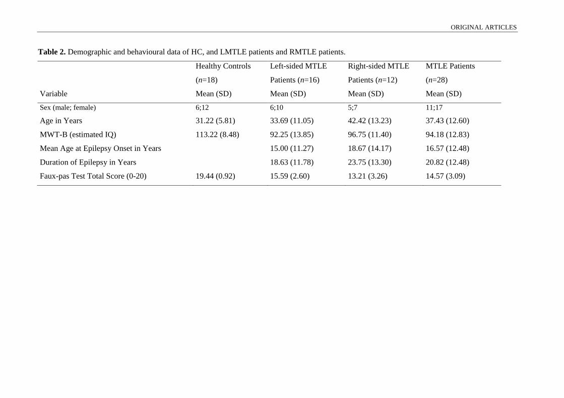
ORIGINAL ARTICLES
Table 2. Demographic and behavioural data of HC, and LMTLE patients and RMTLE patients.
Variable
Healthy Controls
(n=18)
Mean (SD)
Left-sided MTLE
Patients (n=16)
Mean (SD)
Right-sided MTLE
Patients (n=12)
Mean (SD)
MTLE Patients
(n=28)
Mean (SD)
Sex (male; female) 6;12 6;10 5;7 11;17
Age in Years 31.22 (5.81) 33.69 (11.05) 42.42 (13.23) 37.43 (12.60)
MWT-B (estimated IQ) 113.22 (8.48) 92.25 (13.85) 96.75 (11.40) 94.18 (12.83)
Mean Age at Epilepsy Onset in Years 15.00 (11.27) 18.67 (14.17) 16.57 (12.48)
Duration of Epilepsy in Years 18.63 (11.78) 23.75 (13.30) 20.82 (12.48)
Faux-pas Test Total Score (0-20) 19.44 (0.92) 15.59 (2.60) 13.21 (3.26) 14.57 (3.09)
ORIGINAL ARTICLES
fMRI task design
The fMRI paradigm used here was first developed, validated, and applied to HC and
patients with TLE by Schacher et al. [28]. Further information related to the selection
procedure of the stimuli can be found in Schacher et al. [28]. The block-design paradigm
consisted of eight activation and eight baseline blocks each lasting 24 s. The activation
condition comprised 75 brief episodes (2–3 s) from thriller and horror films. All episodes
showed the faces of actors who were expressing fear with high intensity. None of the episodes
showed violence or aggression. During baseline blocks, 72 short episodes of similar length
(2–3 s) with dynamic landscape video recordings were presented. Video clips of dull domestic
landscapes were used owing to their stable low emotional content while their general visual
stimulus properties were comparable to the movie clips. Frequency and duration of the
sequences (2–3 s) were matched in the activation and control conditions. Stimuli were
presented via a back-projection screen and viewed through a tilted overhead mirror. Prior to
beginning, subjects were told that they would see rapid presentations of film sequences
depicting fearful faces intermixed with landscape film sequences. They were instructed to
relax while watching the film and to focus on the eyes of the actors during the activation
blocks.
MRI acquisition
The structural and functional MRI data were recorded using a 3.0-Tesla Achieva scanner
(Philips Medical Systems, Best, The Netherlands) between 2009 and 2010 using a stan-
dardized protocol. MRI sequences included T1-weighted spin-echo and gradient-echo three-
dimensional multiplanar reconstruction images (MPRAGE) with (patients) and without
(healthy controls) intravenous contrast application, coronal T2-weighted turbo spin echo, T2-
weighted fast-fluid attenuated inversion recovery (FLAIR) and diffusion-weighted sequences.
Coronal T2 and FLAIR slices were 2–3 mm thick and were acquired at 90° perpendicular to
the long axis of the hippocampus. Subjects were sited in the head coil with ear pads and foam
padding to minimize head motion. There were successive parameters for the anatomic
sequence: 176 axial slices with 1-mm single-slice thickness, repetition time (TR) 8.2 ms, echo
time (TE) 3.93 ms, 8° flip angle, field of view (FOV) 250 mm, and 288 x 288 matrix.
Functional data were acquired using EPI T2*-weighted sequences. The following
parameters were applied to measure amygdalar activation: 18 coronal slices, 4-mm slice
thickness (interslice gap: 0 mm), TR 1,500 ms, TE 35 ms, 75° flip angle, FOV 220 mm,
matrix size 64 x 64 (voxel size 3 x 3 x 3 mm), reconstructed into an image matrix of 128 x

ORIGINAL ARTICLES
120
128. Coronal slices were geared orthogonally to the hippocampal formation and covered most
of the temporal and frontal lobes. Ten ‘‘dummy’’ scans were first acquired to reach steady-
state magnetization and discarded.
fMRI data analyses
fMRI single subject data analyses were performed using the SPM8 software package
(http://www.fil.ion.ucl.ac.uk) running on MATLAB R2009b (http://www.mathworks.com).
We used two different types of analytical techniques for fMRI data.
We first applied a classical, model-driven approach where specific hypothesis about the
expected blood-oxy- gen-level-dependent (BOLD) response at the individual voxel locations
are tested using a general linear model (GLM). This analysis was used to functionally define
regions-of-interest (ROI) in the amygdala that show the typical activation and deactivation
pattern in response to dynamic fearful faces and reduced ipsilateral amygdala reactivity in the
majority of patients with unilateral MTLE. In a second step, independent component analysis
(ICA) was conducted, which is a hypothesis-free, data-driven approach, aimed at
identification of a network of functionally connected brain regions that included the
amygdala. This network will be referred to as the ‘amygdala network’ for brevity.
Preprocessing
Data were preprocessed with (1) realignment of all images to correct for head movement
during functional MR scanning using six parameter rigid-body transformations; (2)
coregistration to obtain an overlap between functional images and the anatomical image; (3)
normalization of the resulting images to match the Montreal Neurological Institute (MNI)
template brain in Talairach-space and resampling to 3 x 3 x 3 mm3. Images were additionally
spatially smoothed with a Gaussian kernel of 4 x 4 x 4 mm3 full width at half-maximum.
General linear model (GLM) analysis
Single-subject analysis: For each subject, fMRI responses were modeled using a
canonical hemodynamic response function and the general linear model was used to perform a
first level, within-subjects analysis on the functional data from each subject individually for
the primary contrast ‘fearful faces minus landscape’, with spatial realignment parameters
entered as covariates. Before model estimation, data were band-pass filtered (high-pass filter:
128 s, low- pass filter: hemodynamic response function) to remove high- and low-frequency
noise. The overall model fit was assessed using an F statistics. A statistical parametric map of
ORIGINAL ARTICLES
121
brain activation was computed representing the contrast ‘fearful faces minus landscape’.
Results were taken to a second level group analysis.
Group level analysis: To identify the main effect of task for each study group, we used
one-sample t tests. Mean activation and deactivation patterns were obtained to visualize
group-level activation maps that were generated with a global threshold set at p < 0.001
following correction for multiple comparisons on a whole-brain level family-wise error
(FWE) basis, and a cluster extent (kE) threshold set at 5 voxels.
Independent component analysis (ICA)
Functional connectivity analyses were performed using the group ICA of fMRI (GIFT)
toolbox (http://mialab.mrn.org/software/gift/) running on MATLAB R2009b. ICA is a
multivariate data-driven decomposition method whose application on functional imaging data
is relatively new and constitutes an alternative method to classical model-driven analyses of
fMRI data [10]. ICA allows for separation of independent 3D spatial maps based on the
characteristic signal time course of the voxels grouped together to form the respective spatial
map. In that sense, ICA is commonly referred to as decomposing time series data into maps of
voxels which display a mutual functional connectivity; i.e., brain regions whose activity over
time follow an identical, or similar, time course are interpreted to represent a network of
functionally connected brain regions.
Preprocessed fMRI time series data were subjected to a group spatial ICA for the
identification of spatial, independently distributed component networks. Analysis steps were
carried out with default settings and comprised group data reduction by means of a principal
component analysis, ICA calculation using an infomax algorithm, and back-reconstruction in
order to obtain individual independent component maps [10].
The amygdala network was selected from the ICA-calculated, group-averaged
components by two independent, experienced raters (S. B., L. F.). The selected component
was the only component that involved the amygdalae and was selected by visual inspection of
the respective component maps overlayed on the group-averaged T1-weighted image. The
identified component not only included the amygdalae but was centered on the amygdalae,
i.e., highest levels of connectivity were observed within the amygdalae.
Individual amygdala network maps were then taken to second level analyses performed
with SPM8. In order to reveal the amygdala network in each group, separate one-sample t
tests were computed. Group differences within the amygdala network were then assessed in a
subsequent two-way analysis of covariance (ANCOVA). Fixed factors included group

ORIGINAL ARTICLES
122
(LMTLE, RMTLE, HC) and sex. Control variables were age, age at epilepsy onset, duration
of epilepsy, and IQ. Significant differences between groups were assessed using t contrast
maps, restricted to the amygdala network of HC by inclusive masking.
Behavioural data
All participants performed a multiple-choice vocabulary test (MWT-B), which served an
estimate of crystalline verbal intelligence [18] as well as a shortened version of a Faux Pas
Test [32] that comprised five selected stories. Patients were asked to read each story silently,
knowing that they would then be asked questions about the story. The first question
concerned the detection and comprehension of the faux pas, while questions two, three, and
four required the patient to impute the mental state of and attribute emotions to another agent.
Further information related to the test can be found in Schacher et al. [29].
Data analysis involved the sum of the first four questions, where each question was scored
with one point, resulting in a maximum of twenty points for all five stories. Points for the
control question were excluded from the total score, but served as an indicator of text
comprehension.
Relationship between amygdala network and behavioural data
A two-way ANCOVA with the fixed factors group (LMTLE, RMTLE, HC) and sex and
the covariates Faux Pas Test performance, age, age at epilepsy onset, duration of epilepsy,
and IQ was performed to determine significant differences between the groups in the
involvement of amygdala network nodes for Faux Pas Test performance. Again, significant
differences between groups were assessed using t contrasts that were masked with an image
of the amygdala component of HC.
RESULTS
Main BOLD effects (general linear model, GLM)
Task-related activation patterns: mean activation across HC in response to fearful faces
was observed in bilateral hippocampus, amygdala, precentral gyrus, inferior frontal gyrus
(IFG), and insula as well as in the right middle temporal gyrus (MTG) and left thalamus. By
contrast, group-wise activation in LMTLE patients was observed contralateral to the lesion, in
right hippocampus, amygdala, precentral gyrus, IFG, MTG, fusiform gyrus, medial temporal
pole (TP), and pallidum. In contrast, mean activation in RMTLE patients was found

ORIGINAL ARTICLES
123
contralateral to the lesion, in the left amygdala, hippocampus, superior temporal gyrus/ sulcus
(STG/STS), and ipsilateral in the right IFG (see supplementary table and figure).
Connectivity analysis of amygdalar BOLD responses (independent component analysis,
ICA)
Amygdala connectivity: ICA analysis identified a set of six clusters significantly co-
activated with bilateral amygdala BOLD activity in HC (thresholded at p<0.001, uncorrected,
k=5 contiguous voxels) (see Table 3; Fig.1, red). Besides the two larger clusters centered
around the bilateral amygdalo-hippocampal transition area, the amygdalae and hippocampi
were found to be co-activated with the left TP, the left IFG, left superior medial frontal gyrus
(SMFG), left anterior cingulate cortex (ACC), left MTG and right cerebellum. All peak
voxels in this analysis survived a threshold of p<0.05 (FDR corrected).
Group differences in amygdala connectivity: A two-way ANCOVA with the fixed factors
group (LMTLE, RMTLE, HC) and sex and the covariates age, age at epilepsy onset, duration
of epilepsy, and IQ were calculated in order to detect group differences within the amygdala
component. Group contrasts revealed significantly reduced amygdalae co-activation in
RMTLE patients compared to HC on the contralateral side in the left hippocampus, TP, and
ACC (see Table 1; Fig. 1, blue). LMTLE patients also showed reduced amygdala co-
activation in the left hippocampus, the left STG/STS, and the right IFG (see Table 3; Fig. 1,
green). RMTLE patients compared to LMTLE patients showed reduced amygdalae co-
activation within the right medial TP, right MTG and left IFG (see Table 3; Fig. 1, yellow),
whereas LMTLE patients compared to RMTLE patients showed no reduced amygdala
connectivity.

ORIGINAL ARTICLES
Table 3. Brain regions co-activated with the amygdala in HC, LMTLE patients and RMTLE patients.
HC (n=18)
Mean (SD)
LMTLE
Patients
(n=16)
Mean (SD)
RMTLE
Patients
(n=12)
Mean (SD)
Difference between
groups
Region
Peak MNI
coordinates
[x y z] t
Peak MNI
coordinates
[x y z] t
Peak MNI
coordinates
[x y z] t
R amygdalo-hippocampal area (1) +21 -10 -17 22.51
L amygdalo-hippocampal area (2) -24 -13 -17 17.38 -33 -25 -17 3.83 -24 -13 -17 3.79 RMTLE, LMTLE < HC
L temporal pole (3) -57 +6 -6 7.71 -54 +5 -5 3.88 RMTLE < HC
R medial temporal pole +45 +5 -26 3.93 RMTLE < LMTLE
L inferior frontal gyrus (3) -57 +17 +28 7.34 -45 +11 -6 5.04 RMTLE < LMTLE
R inferior frontal gyrus +54 +6 +34 4.20 LMTLE < HC
L middle temporal gyrus (3) -60 -10 -17 4.28
R middle temporal gyrus +48 +2 -29 3.95 RMTLE < LMTLE
L superior temporal gyrus -57 -1 -6 3.90 LMTLE < HC
L anterior cingulate cortex (4) -8 +20 +28 9.29 -3 +17 +31 4.34 RMTLE < HC
L superior medial frontal gyrus (5) -6 +66 +34 6.91
R cerebellum (6) +18 -49 -47 7.44
Coordinates (x y z) are given in MNI space. For ANCOVA, t contrast maps were masked with an image of the amygdala component of healthy
controls, all p<0.001, k=5 contiguous voxels. Number in brackets corresponds to cluster number.

ORIGINAL ARTICLES
Figure 1. Amygdala network in healthy controls (HC) and left- and right-sided mesial temporal lobe epilepsy patients
(LMTLE, RMTLE), overlaid on a mean anatomical scan of all subjects (L=Left side; R=Right side). Amygdalae co-
activation with both hippocampi, left temporal pole, left inferior frontal gyrus, left superior medial frontal gyrus, left anterior
cingulate cortex, left middle temporal gyrus and right cerebellum of the HC group (red) (p<0.001, uncorrected). Significantly
reduced amygdala co-activation in LMTLE (green) and RMTLE patients (blue) compared to HC (red), and in RMTLE
patients compared to LMTLE patients (yellow) (p<0.001, uncorrected; k=5 contiguous voxels).
Behavioural data
To directly compare the Faux Pas Test results of MTLE patients with those of HC, we
analysed the fixed factors group (MTLE, HC) and sex in a two-way ANCOVA with the
covariate age and IQ. Only the factor group influenced the test performance: MTLE patients
performed worse than HC [F(1, 40)=20.51; p<0.001; eta2=0.34; power=0.99]. No significant
influence of the factor sex or age was noted (p>0.05).
In a next step, we analysed the MTLE group in more detail by investigating the influence
of the side of epilepsy using a two-way ANCOVA with the fixed factors side of epilepsy, sex,
and the covariates age, age at seizure onset, duration of epilepsy, and IQ. Patients with
RMTLE performed worse than patients with LMTLE [F(1, 20)=12.30; p<0.002; eta2=0.39;
power=0.93]. Again, no significant influence of the factor sex or any of the covariates was
found (p>0.05).
Red: HC: L+R amygdala & hippocampus, L TP, L IFG, L SMFG, L ACC, L MTG, R Cerebellum
Green: LMTLE<HC: L hippocampus, L STG, R IFG
Blue: RMTLE<HC: L hippocampus, L TP, L ACC;
Yellow: RMTLE<LMTLE: R TP, L IFG, R MTG

ORIGINAL ARTICLES
126
Relationship between amygdala network and behavioural data
Faux Pas Test performance of HC was not included in this analysis due to reduced
variance and a strong ceiling effect in this group.
To investigate the relationship between activation in the amygdala network and Faux Pas
Test performance in LMTLE and RMTLE, we performed a two-way ANCOVA with the
fixed factors group (LMTLE, RMTLE) and sex and overall Faux Pas Test performance, age,
age at epilepsy onset, and duration of epilepsy as covariates. Group contrasts were masked
with an image of the amygdala component of HC and revealed that in LMTLE patients better
Faux Pas Test performance was associated with increased activation in the contralateral insula
(x y z coordinates=+39 -7 -8; see Fig. 2a, green) whereas in RMTLE patients, the
contralateral hippocampal formation including the amygdala (x y z coordinates=-21 -25 -23;
see Fig. 2b, blue) was related to overall Faux Pas Test performance (t=2.53, thresholded at
p<0.01, k=5 contiguous voxels). There was no significant influence of the factor sex or any of
the covariates (p>0.05).
Figure 2. Relationship between amygdala network and ToM task (faux pas test) in left-sided (Fig. 2a; green) and right-sided
mesial temporal lobe epilepsy patients (LMTLE, RMTLE) (Fig. 2b; blue), overlaid on a mean anatomical scan of all subjects
(L=Left side; R=Right side).
DISCUSSION
Main results
We explored connectivity of the amygdala with various temporal, frontal, and parietal
brain areas in patients with unilateral MTLE.
There were three main findings: (1) we found the typical amygdalar BOLD activation in
response to an animated fearful-faces paradigm: bilateral amygdalar activation in HC and
amygdala activation lateralized to the contralateral side in MTLE patients; (2) in HC, a
ORIGINAL ARTICLES
127
hemispheric asymmetry of the amygdala network was present with amygdala co-activation
predominantly in the left temporolateral and frontal brain structures, whereby in MTLE
patients the observed asymmetry of amygdala connectivity was modulated by lateralization of
side of seizure onset; (3) the level of connectivity was related to Faux Pas Test performance.
In previous studies [8, 28] we were able to show that the animated fearful-faces paradigm
results in bilateral amygdalar activation in HC and amygdala activation lateralized to the
contralateral side of seizure onset in the majority of MTLE patients. In the present study, we
replicated this finding identifying symmetrical activation pattern within HC and a
corresponding asymmetrical activation pattern within MTLE patients. Our analysis of
functional connectivity of the amygdala was based on this prior identification of a main effect
of task in each study under investigation.
The amygdala network in HC and MTLE patients
The amygdala network, activated by the applied stimulus material in HC, was found to be
asymmetrically organized within different temporal and frontal lobe brain structures. A left
predominance was observed in the TP, IFG, SMFG, ACC, and MTG. Furthermore, we found
crosswise co- activation within the right cerebellum.
Whether this asymmetry reflects certain task or stimulus properties or brain hemispheric
asymmetry cannot be answered by our study. However, this left predominance of connectivity
is in line with previous structural data showing leftward asymmetry in parietotemporal white
matter as well as the fiber density of the arcuate fasciculus in subjects with left-sided speech
dominance [15, 22, 25]. Moreover, predictions of language lateralization have been
demonstrated from gray matter probabilistic maps of high-resolution structural scans [17] as
well as from white matter by means of diffusion tensor imaging [12]. Additionally, recent
resting state fMRI studies reported a left predominance in basal functional connectivity within
the medial temporal lobe network in HC [5] and more pronounced effects of impaired
functional connectivity ipsilateral to the seizure focus in LMTLE patients compared to
RMTLE patients [24].
We found a clear effect of lesion, whereby this effect of pathology was most prominent in
brain areas showing greater connectivity. In MTLE patients, the observed asymmetry of
amygdala connectivity was modulated by lateralization of side of seizure onset. Compared to
HC, we found reduced amygdala co-activation in the left hippocampus, the left STS, and the
right IFG in LMTLE patients whereas RMTLE patients showed reduced connectivity in the
left hippocampus, the left TP, and the left ACC. Thus, we did not find the expected
ORIGINAL ARTICLES
128
impairment of ipsilateral connectivity in RMTLE patients, which suggests that despite only
unilateral morphological MRI-proven pathology exists, the contralateral hemisphere may also
be involved through transcallosal projections [27].
Which nodes within the amygdala network are associated with ToM performances?
We further investigated ToM performance in MTLE patients using the Faux Pas Test. The
results of the present study support our previous finding that patients with MTLE, especially
those with right-sided seizure onset, show impairments in the recognition of faux pas [29].
This observation is also in accordance with our finding that RMTLE patients, compared to
LMTLE patients, had significantly reduced amygdala-coactivation within the ipsilateral
medial TP and the MTG. Further, compared to HC, the contralateral activation in the
amygdalo-hippocampal area, in addition to other structures, was observed to be significantly
reduced in RMTLE patients. Thus, RMTLE patients were found to have functional
disturbances in both amygdalo-hippocampal areas: the left as revealed by reduced co-
activation as well as the right, due to ipsilateral mesiotemporal pathology. This may be an
explanatory factor underlying their especially poor performance in the ToM test. At the
behavioral level, pronounced difficulties in RMTLE patients have also been reported for
facial emotion recognition abilities [20, 21]. Extending the findings of our earlier studies [29],
we investigated in the present study which structures within the amygdala network are related
to ToM test performance. In patients with LMTLE, connectivity within the contralateral
insula, a limbic-related cortical area known to have widespread reciprocal connections to the
amygdala and hippocampus [2, 31], was related to Faux Pas Test performance. In RMTLE
patients connectivity within the contralateral hippocampal formation, including amygdala,
was related to the behavioral data.
Our results suggest that the integrity of contralateral mesiotemporal lobe structures play a
more important role in MTLE patients for performance on a ToM task than remaining
spatially connected ipsilateral activity. This result may be indicative for compensatory
activation of the contralateral mesiotemporal region [5]. However, since no comparison was
performed with controls, no final conclusion can be drawn regarding the relationship between
ToM and MTLE.
There are some limitations of the study that need to be addressed. First, the degree of
hippocampal atrophy was not controlled in this study. Second, extension of structural mesial
temporal lobe lesions may exceed the ipsilateral hippocampus and may also be found in
ipsilateral or even bilateral amygdala. Accordingly, amygdala T2 measurements demonstrated
ORIGINAL ARTICLES
129
bilateral pathological changes even in patients with unilateral hippocampal sclerosis [4].
Finally, methodological limitations may exist due to temporarily independent measurements
of amygdalar activity and behavioral test performance that can be avoided in forthcoming
studies.
Conclusion and future perspectives
The amygdala network in HC was found to be asymmetrically organized within various
temporal and frontal lobe brain structures, whereby in MTLE patients the observed
asymmetric pattern of amygdala connectivity was strongly modulated by pathology. These
findings suggest that ipsilateral amygdalar dysfunction in MTLE patients may result in loss of
functional connectivity of brain structures remote to but interconnected with the amygdala.
The use of functional connectivity analysis in psychiatric and neurological disorders as
well as in epilepsy surgery patients may allow a broader view, away from the roles and
dysfunctions of individual regions of the brain, towards a perspective in which affective
symptoms and deficits in social cognition may be seen as symptom domains arising from
certain pathological networks.

ORIGINAL ARTICLES
130
Acknowledgements
We would like to thank the Swiss Epilepsy Foundation for financial support and Dr. Dominik
Huber, Gesa Osterkamp, and Theresa Biesold for technical support.

ORIGINAL ARTICLES
131
References
1. Amaral DG, Price JL (1984) Amygdalo-cortical projections in the monkey (Macaca
fascicularis). The Journal of comparative neurology 230:465-496
2. Augustine JR (1996) Circuitry and functional aspects of the insular lobe in primates
including humans. Brain Res Brain Res Rev 22:229-244
3. Barbas H, De Olmos J (1990) Projections from the amygdala to basoventral and
mediodorsal prefrontal regions in the rhesus monkey. The Journal of comparative
neurology 300:549-571
4. Bartlett PA, Richardson MP, Duncan JS (2002) Measurement of amygdala T2
relaxation time in temporal lobe epilepsy. J Neurol Neurosurg Psychiatry 73:753-755
5. Bettus G, Guedj E, Joyeux F, Confort-Gouny S, Soulier E, Laguitton V, Cozzone PJ,
Chauvel P, Ranjeva JP, Bartolomei F, Guye M (2009) Decreased basal fMRI
functional connectivity in epileptogenic networks and contralateral compensatory
mechanisms. Human brain mapping 30:1580-1591
6. Bonora A, Benuzzi F, Monti G, Mirandola L, Pugnaghi M, Nichelli P, Meletti S
(2011) Recognition of emotions from faces and voices in medial temporal lobe
epilepsy. Epilepsy Behav 20:648-654
7. Broicher S, Jokeit H (2011) Emotional agnosis and theory of mind. In: Trimble MR,
Schmitz B (eds) The Neuropsychiatry of Epilepsy. Cambridge University Press,
Cambridge, pp 109-123
8. Broicher S, Kuchukhidze G, Grunwald T, Kramer G, Kurthen M, Trinka E, Jokeit H
(2010) Association between structural abnormalities and fMRI response in the
amygdala in patients with temporal lobe epilepsy. Seizure 19:426-431
9. Broicher SD, Kuchukhidze G, Grunwald T, Kramer G, Kurthen M, Jokeit H (2011)
"Tell me how do I feel" - Emotion recognition and theory of mind in symptomatic
mesial temporal lobe epilepsy. Neuropsychologia
10. Calhoun VD, Adali T, Pearlson GD, Pekar JJ (2001) A method for making group
inferences from functional MRI data using independent component analysis. Human
brain mapping 14:140-151
11. Dolan RJ (2002) Emotion, cognition, and behavior. Science (New York, NY
298:1191-1194
ORIGINAL ARTICLES
132
12. Ellmore TM, Beauchamp MS, Breier JI, Slater JD, Kalamangalam GP, O'Neill TJ,
Disano MA, Tandon N (2010) Temporal lobe white matter asymmetry and language
laterality in epilepsy patients. NeuroImage 49:2033-2044
13. Frings L, Schulze-Bonhage A, Spreer J, Wagner K (2009) Remote effects of
hippocampal damage on default network connectivity in the human brain. Journal of
neurology 256:2021-2029
14. Ghashghaei HT, Barbas H (2002) Pathways for emotion: interactions of prefrontal and
anterior temporal pathways in the amygdala of the rhesus monkey. Neuroscience
115:1261-1279
15. Hagmann P (1972) [Milestones in the development of language in infancy. A
prospective examination of 65 children including sets of twins]. Helvetica paediatrica
acta 21:71-84
16. Hermann B, Seidenberg M, Jones J (2008) The neurobehavioural comorbidities of
epilepsy: can a natural history be developed? Lancet Neurol 7:151-160
17. Josse G, Kherif F, Flandin G, Seghier ML, Price CJ (2009) Predicting language
lateralization from gray matter. J Neurosci 29:13516-13523
18. Lehrl S, Triebig G, Fischer B (1995) Multiple choice vocabulary test MWT as a valid
and short test to estimate premorbid intelligence. Acta Neurol Scand 91:335-345
19. Liao W, Zhang Z, Pan Z, Mantini D, Ding J, Duan X, Luo C, Lu G, Chen H (2010)
Altered functional connectivity and small-world in mesial temporal lobe epilepsy.
PloS one 5:e8525
20. Meletti S, Benuzzi F, Cantalupo G, Rubboli G, Tassinari CA, Nichelli P (2009) Facial
emotion recognition impairment in chronic temporal lobe epilepsy. Epilepsia 50:1547-
1559
21. Meletti S, Benuzzi F, Rubboli G, Cantalupo G, Stanzani Maserati M, Nichelli P,
Tassinari CA (2003) Impaired facial emotion recognition in early-onset right mesial
temporal lobe epilepsy. Neurology 60:426-431
22. Nucifora PG, Verma R, Melhem ER, Gur RE, Gur RC (2005) Leftward asymmetry in
relative fiber density of the arcuate fasciculus. Neuroreport 16:791-794
23. Oldfield RC (1971) The assessment and analysis of handedness: the Edinburgh
inventory. Neuropsychologia 9:97-113
24. Pereira FR, Alessio A, Sercheli MS, Pedro T, Bilevicius E, Rondina JM, Ozelo HF,
Castellano G, Covolan RJ, Damasceno BP, Cendes F (2010) Asymmetrical
ORIGINAL ARTICLES
133
hippocampal connectivity in mesial temporal lobe epilepsy: evidence from resting
state fMRI. BMC neuroscience 11:66
25. Powell HW, Parker GJ, Alexander DC, Symms MR, Boulby PA, Wheeler-Kingshott
CA, Barker GJ, Noppeney U, Koepp MJ, Duncan JS (2006) Hemispheric asymmetries
in language-related pathways: a combined functional MRI and tractography study.
NeuroImage 32:388-399
26. Purves D, Augustine GJ, Fitzpatrick D, Hall WC, LaMantia A-S, McNamara JO,
White LE (2008) Neuroscience. Sinauer Associates, Sunderland: Massachusetts
27. Roy AK, Shehzad Z, Margulies DS, Kelly AM, Uddin LQ, Gotimer K, Biswal BB,
Castellanos FX, Milham MP (2009) Functional connectivity of the human amygdala
using resting state fMRI. Neuroimage 45:614-626
28. Schacher M, Haemmerle B, Woermann FG, Okujava M, Huber D, Grunwald T,
Kramer G, Jokeit H (2006) Amygdala fMRI lateralizes temporal lobe epilepsy.
Neurology 66:81-87
29. Schacher M, Winkler R, Grunwald T, Kraemer G, Kurthen M, Reed V, Jokeit H
(2006) Mesial temporal lobe epilepsy impairs advanced social cognition. Epilepsia
47:2141-2146
30. Stefanacci L, Suzuki WA, Amaral DG (1996) Organization of connections between
the amygdaloid complex and the perirhinal and parahippocampal cortices in macaque
monkeys. The Journal of comparative neurology 375:552-582
31. Stein JL, Wiedholz LM, Bassett DS, Weinberger DR, Zink CF, Mattay VS, Meyer-
Lindenberg A (2007) A validated network of effective amygdala connectivity.
Neuroimage 36:736-745
32. Stone VE, Baron-Cohen S, Calder A, Keane J, Young A (2003) Acquired theory of
mind impairments in individuals with bilateral amygdala lesions. Neuropsychologia
41:209-220
33. Vuilleumier P, Richardson MP, Armony JL, Driver J, Dolan RJ (2004) Distant
influences of amygdala lesion on visual cortical activation during emotional face
processing. Nature neuroscience 7:1271-1278
34. Waites AB, Briellmann RS, Saling MM, Abbott DF, Jackson GD (2006) Functional
connectivity networks are disrupted in left temporal lobe epilepsy. Ann Neurol
59:335-343
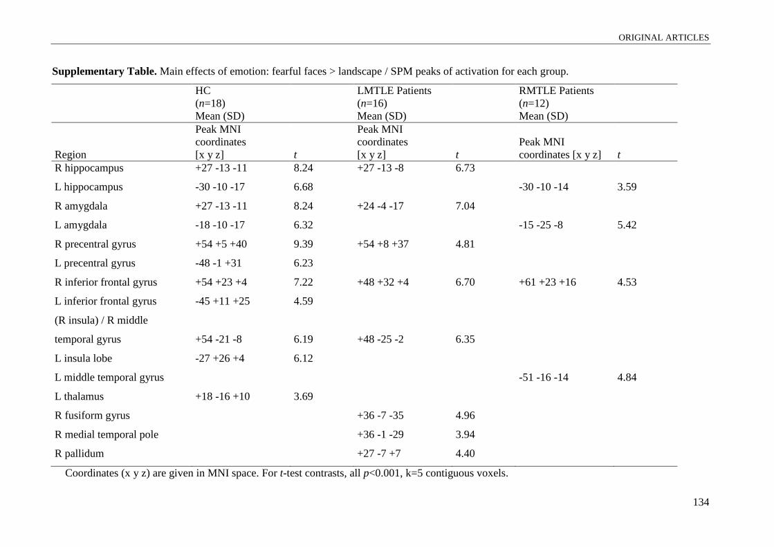
ORIGINAL ARTICLES
134
Supplementary Table. Main effects of emotion: fearful faces > landscape / SPM peaks of activation for each group.
HC
(n=18)
Mean (SD)
LMTLE Patients
(n=16)
Mean (SD)
RMTLE Patients
(n=12)
Mean (SD)
Region
Peak MNI
coordinates
[x y z] t
Peak MNI
coordinates
[x y z] t
Peak MNI
coordinates [x y z] t
R hippocampus +27 -13 -11 8.24 +27 -13 -8 6.73
L hippocampus -30 -10 -17 6.68 -30 -10 -14 3.59
R amygdala +27 -13 -11 8.24 +24 -4 -17 7.04
L amygdala -18 -10 -17 6.32 -15 -25 -8 5.42
R precentral gyrus +54 +5 +40 9.39 +54 +8 +37 4.81
L precentral gyrus -48 -1 +31 6.23
R inferior frontal gyrus +54 +23 +4 7.22 +48 +32 +4 6.70 +61 +23 +16 4.53
L inferior frontal gyrus -45 +11 +25 4.59
(R insula) / R middle
temporal gyrus +54 -21 -8 6.19 +48 -25 -2 6.35
L insula lobe -27 +26 +4 6.12
L middle temporal gyrus -51 -16 -14 4.84
L thalamus +18 -16 +10 3.69
R fusiform gyrus +36 -7 -35 4.96
R medial temporal pole +36 -1 -29 3.94
R pallidum +27 -7 +7 4.40
Coordinates (x y z) are given in MNI space. For t-test contrasts, all p<0.001, k=5 contiguous voxels.
ORIGINAL ARTICLES
135
5.4 fMRI response of dysplastic amygdala in patients with mesial temporal lobe
epilepsy
Broicher S1, Kuchukhidze G
2, Grunwald T
1, Krämer G
1, Kurthen M
1, Trinka E
2, Jokeit H
1
1 Swiss Epilepsy Center, Zürich, Switzerland
2 Department of Neurology, Innsbruck Medical University, Innsbruck, Austria
Corresponding author:
Sarah Broicher, MSc
Swiss Epilepsy Center
Bleulerstrasse 60, 8008 Zürich, Switzerland
Tel.: ++41/44/387-6348
Fax: ++41/44/387-6134
e-mail: [email protected]
Key words: amygdala, cortical dysplasia, epilepsy, fMRI
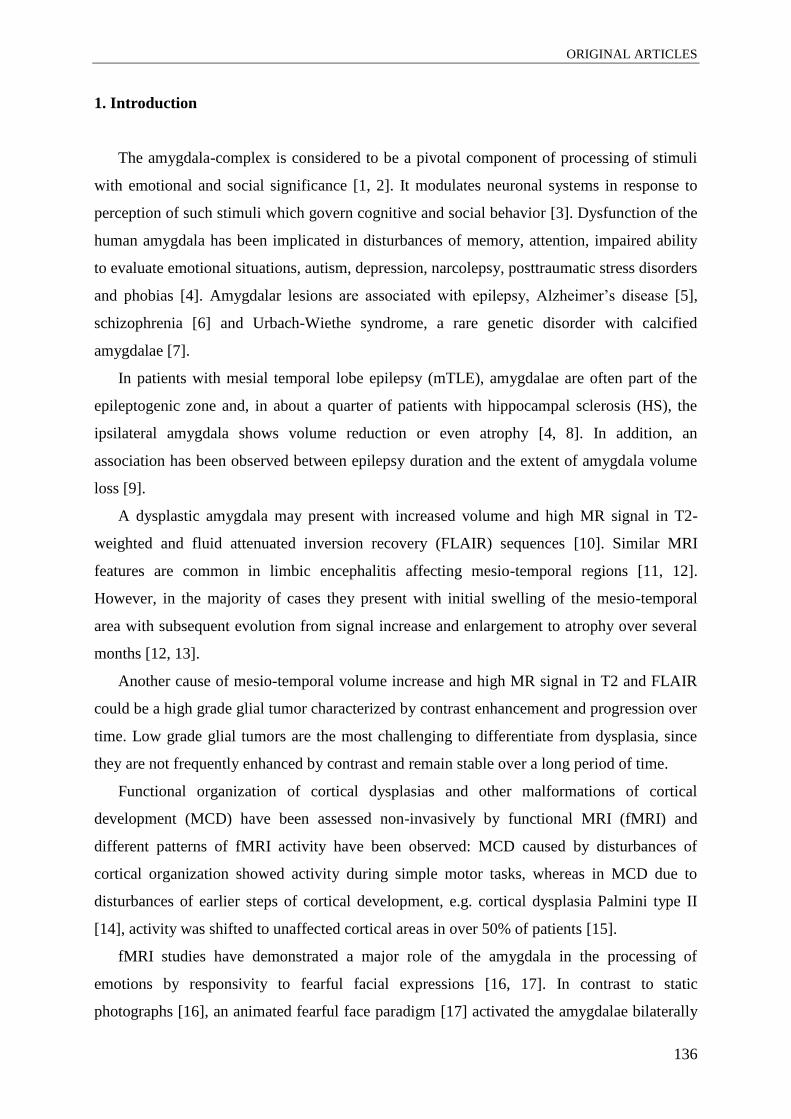
ORIGINAL ARTICLES
136
1. Introduction
The amygdala-complex is considered to be a pivotal component of processing of stimuli
with emotional and social significance [1, 2]. It modulates neuronal systems in response to
perception of such stimuli which govern cognitive and social behavior [3]. Dysfunction of the
human amygdala has been implicated in disturbances of memory, attention, impaired ability
to evaluate emotional situations, autism, depression, narcolepsy, posttraumatic stress disorders
and phobias [4]. Amygdalar lesions are associated with epilepsy, Alzheimer’s disease [5],
schizophrenia [6] and Urbach-Wiethe syndrome, a rare genetic disorder with calcified
amygdalae [7].
In patients with mesial temporal lobe epilepsy (mTLE), amygdalae are often part of the
epileptogenic zone and, in about a quarter of patients with hippocampal sclerosis (HS), the
ipsilateral amygdala shows volume reduction or even atrophy [4, 8]. In addition, an
association has been observed between epilepsy duration and the extent of amygdala volume
loss [9].
A dysplastic amygdala may present with increased volume and high MR signal in T2-
weighted and fluid attenuated inversion recovery (FLAIR) sequences [10]. Similar MRI
features are common in limbic encephalitis affecting mesio-temporal regions [11, 12].
However, in the majority of cases they present with initial swelling of the mesio-temporal
area with subsequent evolution from signal increase and enlargement to atrophy over several
months [12, 13].
Another cause of mesio-temporal volume increase and high MR signal in T2 and FLAIR
could be a high grade glial tumor characterized by contrast enhancement and progression over
time. Low grade glial tumors are the most challenging to differentiate from dysplasia, since
they are not frequently enhanced by contrast and remain stable over a long period of time.
Functional organization of cortical dysplasias and other malformations of cortical
development (MCD) have been assessed non-invasively by functional MRI (fMRI) and
different patterns of fMRI activity have been observed: MCD caused by disturbances of
cortical organization showed activity during simple motor tasks, whereas in MCD due to
disturbances of earlier steps of cortical development, e.g. cortical dysplasia Palmini type II
[14], activity was shifted to unaffected cortical areas in over 50% of patients [15].
fMRI studies have demonstrated a major role of the amygdala in the processing of
emotions by responsivity to fearful facial expressions [16, 17]. In contrast to static
photographs [16], an animated fearful face paradigm [17] activated the amygdalae bilaterally

ORIGINAL ARTICLES
137
in all 17 healthy controls, whereas the majority of patients (11 out of 12) with unilateral
temporal lobe epilepsy (TLE) and HS showed amygdalar activation contralateral to the
seizure onset side.
However, in some patients, dissociation between amygdalar activations and epileptogenic
lesion were found [17]. In two patients ipsilateral amygdala activity and reversed asymmetries
in parahippocampal activations were observed.
Here, we investigate for the first time, whether amygdalar response in fMRI is principally
impaired if it is affected by dysplasia as defined by stationary but increased volume in T1-
and signal enhancement in T2-weighted and FLAIR MRI sequences.
2. Methods
2.1. Participants
Twenty-five patients aged 16 to 60 (mean 38 years, SD 13.2; 13 men, 12 women; 24 right
handed and 1 ambidexter) with TLE (mean age at seizure onset 15.2 years, SD 12.7 and
epilepsy duration 23.1 years, SD 13.7) were investigated (see Table 1). Patients were recruited
from consecutive inpatient admissions to the Swiss Epilepsy Center.
All patients underwent neurological examination and routine EEG recordings using the
10-20 system. Seizure types and epilepsy syndromes were diagnosed according to the
classification of the International League Against Epilepsy [18, 19]. Seizure onset zone was
determined by continuous interictal and ictal video/EEG monitoring with scalp and
sphenoidal electrodes.
Twenty-four patients had mTLE and one had neocortical TLE with cavernoma localized
in the lateral temporal lobe as well as ipsilateral HS and pDA. Eighteen patients had HS (17
unilateral and one bilateral). All patients had pharmacoresistant epilepsy (14/25 patients had
daily or weekly seizures) (see Table 1).
ORIGINAL ARTICLES
138
Language laterality was determined either by use of an fMRI paradigm employing a
verbal fluency task [17] (n=23) or by Wada test if the fMRI was inconclusive (n=2) [20]. The
majority of patients (23/25) had left-sided language dominance, two had bilateral language
representation. Handedness was determined by the Edinburgh Handedness Inventory.
Every patient gave written informed consent after a complete explanation of the study.
The present study was performed in adherence to the Declaration of Helsinki and was also
approved by a local medical ethics committee.
2.2. fMRI task design
The fMRI paradigm used here was first developed, validated and applied to healthy
controls and patients with TLE by Schacher et al. [17]. Further information related to the
selection procedure of the stimuli can be found in Schacher et al. [17]. The block design
paradigm consisted of eight activation and eight baseline blocks each lasting 24 seconds. The
activation condition comprised 75 brief episodes (2 to 3 seconds) from thriller and horror
films. All episodes showed the faces of actors who were expressing fear with high intensity.
None of the episodes showed violence or aggression. During baseline blocks 72 short
episodes of similar length (two to three seconds) with dynamic landscape video recordings
were presented. Video clips of dull domestic landscapes were used owing to their stable low
ORIGINAL ARTICLES
139
emotional content while their general visual stimulus properties were comparable with the
movie clips. Frequency and duration of the sequences (2 to 3 seconds) were matched in the
activation and control conditions. Stimuli were presented via a back-projection screen and
viewed through a tilted overhead mirror. Prior to beginning, subjects were told that they
would see rapid presentations of film sequences depicting fearful faces intermixed with
landscape film sequences. They were instructed to relax while watching the film and to focus
on the eyes of the actors during the activation blocks.
2.3. MRI acquisition
The fMRI-data were recorded using a 3.0 Tesla Achieva scanner (Philips Medical
Systems, Best, The Netherlands) between 2007 and 2009 using a standardized protocol. MRI
sequences included T1-weighted spin echo and gradient echo three-dimensional multiplanar
reconstruction images (MPRAGE) with and without intravenous contrast application, coronal
T2-weighted turbo spin echo, T2-weighted fast fluid attenuated inversion recovery (FLAIR)
and diffusion weighted sequences. Coronal T2 and FLAIR slices were 1-3 mm thick and were
acquired at 90° perpendicular to the long axis of the hippocampus. Subjects were sited in the
head coil with ear pads and foam padding to minimize head motion. There were successive
parameters for the anatomic sequence: 176 axial slices with 1-mm single-slice thickness,
repetition time (TR) 8.2 milliseconds, echo time (TE) 3.93 milliseconds, 8° flip angle, field of
view (FOV) 250 mm, and 288 X 288 matrix.
Functional data were acquired using EPI T2*-weighted sequence. The following
parameters were applied to measure amygdalar activation: 18 coronal slices, 4-mm slice
thickness (interslice gap: 0 mm), TR 1500 milliseconds, TE 35 milliseconds, 75° flip angle,
FOV 220 mm, matrix size 64 X 64 (voxel size 2.75 X 2.75 X 4 mm), reconstructed into an
image matrix of 128 X 128. Coronal slices were geared orthogonally to the hippocampal
formation and were spread over the anterior temporal lobe.
2.4. Data analyses
fMRI single subject data analysis was performed with BrainVoyager QX
(BrainInnovation, Maastricht, the Netherlands). In the primary analyses the data were
preprocessed with 1) three-dimensional motion correction and 2) trend removal by temporal
fast Fourier transform-based high-pass filtering and transformed into Talairach co-ordinate
ORIGINAL ARTICLES
140
area. Images of the fearful face task were additionally spatially smoothed with a full width at
half-maximum of 4 mm.
For multiple regression analysis a general linear model (GLM) with the predictor for the
activation condition was computed. The time courses of the predictor were obtained by using
a linear model of the hemodynamic response. The overall model fit was assessed using F
statistics. Significant differences between the experimental conditions were assessed using
contrast (t) maps.
For images in the fearful face task, individual volumes of interests (VOIs) were defined
for the amygdalar region. VOIs were specified functionally for each patient separately using a
predefined statistical threshold of p < 0.05; p < 0.01 and p < 0.001 (see Table 1). Anatomic
borders of the functional clusters were the uncal recess caudally and the optical chiasm
rostrally and white matter superiorly. For each VOI the number of activated voxels was
counted in the left and right hemisphere at the lowest reasonable statistical threshold.
Lateralization indexes (LI) were defined for the number of meaningful activated voxels in the
amygdalar/periamygdalar area using the formula: LI = (left - right)/(left + right). LI between
±0.5 and ±1 was defined to represent strong lateralization and LI between ±0.25 and ±0.5 –
weak lateralisation (with “–” lateralizing to the right and “+” to the left, respectively). LI
between -0.25 and +0.25 was defined as bilateral amygdalar activation.
2.5. Criteria for the assessment of amygdalar structural abnormalities
MRIs were assessed to detect and categorize amygdalar structural abnormalities by two
independent raters from different institutions (SB - Swiss Epilepsy Center, Switzerland and
GK – Department of Neurology, Innsbruck Medical University, Austria). The raters were
blinded with regard to clinical data.
Amygdalae were assessed visually for size (T1-weighthed MPRAGE sequence) and MR
signal intensity (T2 and FLAIR sequences). Amygdalae were considered “lesional” if the
combination of size and signal abnormalities were observed. Size was assessed in comparison
to the contralateral side and was considered abnormal if it was atrophic or pathologically
increased. Amygdalar lesions were categorized as:
(1) amygdala atrophy (AA) if it was shrunken (atrophic) with signal increase in T2 and
FLAIR sequences;
(2) probable dysplasia (pDA) if the size was pathologically increased with signal increase
in T2 and FLAIR sequences; additional MRI features of pDA were white matter volume

ORIGINAL ARTICLES
141
reduction and increased signal in T2 and FLAIR sequences in ipsilateral temporal lobe; in
order to exclude limbic encephalitis the patients should not have had MRI features of swelling
at the initial MRI performed at the manifestation of epilepsy and the MRI features had to
remain constant over at least two MRIs within an interval of at least 12 months. Only in one
patient (patient 10 with right-sided pDA) this interval was less than 12 months. In order to
exclude high grade glial tumors there had to be no progression of the disease and no MRI
contrast enhancement. Dysplastic tumors (ganglioglioma, GG and dysembrioplastic
neuroepitelial tumor, DNT) were considered under pDA since they are incorporated in the
classification of malformations of cortical development [21].
(3) Lesions other than AA or pDA.
The following rating scale was proposed, for size: 0 (normal), +1 (slightly enlarged), +2
(moderately enlarged), +3 (considerably enlarged), -1 (small), -2 (moderate atrophy), -3
(severe atrophy); for signal intensity: 0 (normal), 1 (moderately increased), 2 (considerably
increased).
Agreement between the two raters (SB, GK) was reached in 23/25 cases (Cohen's kappa
coefficient, κ = 0.828, p < 0.0001). Of these 23 patients, 15 could be classified as having
normal amygdalae, 5 pDA, 2 AA, and one patient with other amygdalar lesion (a small cystic
lesion adjacent to amygdala). In the remaining 2/25 cases, a third evaluator (ET, Department
of Neurology, Innsbruck Medical University, Austria) was consulted. The decision was made
with two out of three votes; there were no cases with three different rating scores. As a result
of the assessment, 17 patients were classified as having normal amygdalae, 5 pDA, 2 AA and
one other amygdalar lesion.
2.6. Statistics
Descriptive statistics were used for group comparisons due to the small sample size of the
subgroups. Individual fMRI statistics were adopted based on multivariate models as provided
by Brain Voyager QX.
3. Results
T2* contrast differences were found within amygdalae in all subjects but one in response
to watching video sequences with fearful faces in contrast to watching landscape scenes (p <

ORIGINAL ARTICLES
142
0.05). The activation focus was located in the superior part of the amygdala, as found by
Schacher et al. [17].
According to the criteria for the assessment of amygdalar structural abnormalities, 17
patients were classified as having normal amygdalae, five patients as having pDA, two as
having AA, and in one patient a small cystic lesion adjacent to right amygdala was assessed.
Out of the 17 patients with normal amygdala, bilateral amygdalae activation was observed
in three (18%) patients (mean LI -0.08), strongly lateralized right-sided activation in six
(35%) (mean LI -0.72), strongly lateralized left-sided activation in seven (41%) patients
(mean LI 1.00); and in one patient the activation was categorized as weakly lateralized to the
left (LI 0.49).
In two patients with right-sided pDA, the activation was observed in the contralateral
amygdala (for both LI 1) (Figure 1); whereas in three patients with left-sided pDA, activations
were bilateral (n=1; LI -0.01), ipsilateral with strong lateralization (n=1; LI 0.82) or
contralateral with strong lateralization (n=1; LI -0.67) (Figure 2).

ORIGINAL ARTICLES
143
In one patient with right-sided AA, the activation was observed ipsilateral with strong
lateralization (LI -0.78), whereas in one patient with bilateral AA no activation was detected.
In the patient with the small cystic lesion adjacent to the right amygdala, bilateral amygdala
activation was observed (LI -0.13).
Across all subjects the average LI of amygdalar activation was 0.13 (SD 0.8).
In relation to the seizure onset side, amygdalar activation was contralateral in 15 (60%)
patients (14/15 with strong lateralization) and ipsilateral in four (16%) patients (2/4 with
strong lateralization). In two patients with left-sided TLE and in three patients with right-
sided TLE, amygdalar activation was bilateral. No amygdalar activation was observed in one
patient with bilateral mTLE and bilateral HS.
4. Discussion
We present a cohort of 25 consecutive pharmacoresistant TLE patients who were tested by
means of an animated fearful faces paradigm that reliably elucidates amygdalar BOLD
activation. A previous study has shown that this paradigm results in bilateral amygdalar
activation in the majority of healthy controls, whereas the activation is mostly contralateral to
the side of seizure onset in unilateral symptomatic TLE patients [17]. In our series we were
able to reproduce the findings of our previous study [17] using an independent patient sample
and a different scanner: the majority of patients (80 %) had activation contralateral to the side
ORIGINAL ARTICLES
144
of seizure onset. However, in four out of 25 symptomatic TLE patients, amygdalar activation
was ipsilateral to mesial temporal lesion.
In this study, we aimed to investigate whether mesial temporal lesions observed on MRI
could influence the activation pattern in the amygdala. Indeed, in two patients with left-sided
pDA, significant activation was detected on the side of the lesion (activation was bilateral in
one patient) demonstrating that unilateral pDA does not necessarily cause a loss of amygdalar
function as indicated by fMRI BOLD-response. In contrast to these patients, two patients with
right-sided and one with left-sided pDA showed strongly contralateral activation. Whether
this apparent left right/asymmetry represents functional or structural differences as has been
suggested remains speculative due to our small sample size [22, 23].
The histological type of dysplasia could also play an important role in the reactivity of
pDA. It is not possible to determine the type of dysplasia based on MRI features since there
are no correlative studies available with regard to amygdala imaging and histological features
which would validate MRI characteristics of dysplasia affecting amygdalae. Few studies
addressing cortical dysplasia (CD) function have demonstrated that phenotype,
epileptogenicity and functional behavior of CD are largely determined by their cellular and
histochemical properties. This has been shown mainly in invasive studies related to CD
located in the vicinity of eloquent cortical regions [24]. Absence of language or motor
functions in perirolandic and Broca’s areas which exhibited histological evidence of CD with
balloon cells (Palmini type IIb) and preservation of motor functions when balloon cells were
absent (types Ia,b, IIa) was found in a study associating direct cortical stimulation and
histology in CD [24]. The absence of function in CD type IIb may be due to immature
properties of balloon cells, dysmorphic and cytomegalic neurons as well as severe disruption
of neuronal circuits.
However, it is difficult to extrapolate the results obtained in CD to pDA since there is as
yet very limited data available on amygdalar histology and functional imaging.
Constant MRI features over a relatively long time period, absence of swelling signs on
initial MRI and of contrast enhancement and clinical presentation characteristic for other
possible causes of amygdalar lesions (swelling, tumor, or inflammation) enabled us to
categorize amygdalar lesion as probable dysplasia.
In patients with pharmacoresistant seizures a precise detection of the extent of an
epileptogenic as well as functional deficit zone is essential for determining the resection
borders in order to achieve seizure freedom without additional postoperative functional
deficits [25]. In line with our previous study, we have demonstrated functional response in
ORIGINAL ARTICLES
145
amygdalae affected by epileptogenic lesions. Bonelli et al. [16] used a fearful face paradigm
in mTLE patients for determining whether preoperative amygdalar response could be a
potential predictive marker for emotional disturbances following surgery: greater increases in
anxiety and depression were observed in patients who had greater preoperative amygdalar
activations [16]. It is well established that psychosocial difficulties and psychiatric
dysfunctions appear more often in patients with mTLE compared to other chronic epilepsy
syndromes [26, 27].
Abnormalities in higher social cognition were found in both pre- and postoperative mTLE
patients [28]. Based on the lesion and functional imaging studies, Kirsch [29] has highlighted
the (among others) specific role of temporal lobe structures in social cognitive processes and
indicated possible impairments in higher-order social behavior due to an anterior temporal
lobectomy. However, another preliminary prospective study on social cognition did not reveal
changes caused by anterior temporal lobectomy [30].
In summary, the results of this explorative study indicate that in TLE patients unilateral
amygdala dysplasia does not necessarily cause a loss of amygdalar function as indicated by
fMRI BOLD-response, but does frequently induce a shift of fMRI BOLD-signal to the
contralateral side. However, for further delineation of response characteristics in dysplastic
amygdala and its functional importance, a larger number of patients and pre- and postsurgical
evaluations, including histopathology, are required.
ORIGINAL ARTICLES
146
Acknowledgements
We would like to thank the Swiss Epilepsy Foundation for financial support and Dr.
Dominik Huber, Gesa Osterkamp, and Theresa Biesold for technical support. We also would
like to thank Victoria Reed for valuable comments regarding the improvement of the
manuscript.

ORIGINAL ARTICLES
147
References
[1] Adolphs, R. Is the human amygdala specialized for processing social information? Ann
N Y Acad Sci 2003; 985:326-340.
[2] Adolphs, R., Baron-Cohen, S. and Tranel, D. Impaired recognition of social emotions
following amygdala damage. J Cogn Neurosci 2002; 14:1264-1274.
[3] Anderson, A. K. and Phelps, E. A. Lesions of the human amygdala impair enhanced
perception of emotionally salient events. Nature 2001; 411:305-309.
[4] Goncalves Pereira, P. M., Insausti, R., Artacho-Perula, E., Salmenpera, T., Kalviainen,
R. and Pitkanen, A. MR volumetric analysis of the piriform cortex and cortical amygdala in
drug-refractory temporal lobe epilepsy. AJNR Am J Neuroradiol 2005; 26:319-332.
[5] Kile, S. J., Ellis, W. G., Olichney, J. M., Farias, S. and DeCarli, C. Alzheimer
abnormalities of the amygdala with Kluver-Bucy syndrome symptoms: an amygdaloid variant
of Alzheimer disease. Arch Neurol 2009; 66:125-129.
[6] Gur, R. E., McGrath, C., Chan, R. M., Schroeder, L., Turner, T., Turetsky, B. I.,
Kohler, C., Alsop, D., Maldjian, J., Ragland, J. D. and Gur, R. C. An fMRI study of facial
emotion processing in patients with schizophrenia. Am J Psychiatry 2002; 159:1992-1999.
[7] Urbach, E. W., C. Lipoidosis cutis et mucosae. Virchows Archiv für pathologische
Anatomie und Physiologie und für klinische Medizin 1929; 273:285-319.
[8] Salmenpera, T., Kalviainen, R., Partanen, K. and Pitkanen, A. Hippocampal and
amygdaloid damage in partial epilepsy: a cross-sectional MRI study of 241 patients. Epilepsy
Res 2001; 46:69-82.
[9] Bernasconi, N., Natsume, J. and Bernasconi, A. Progression in temporal lobe epilepsy:
differential atrophy in mesial temporal structures. Neurology 2005; 65:223-228.
[10] Jackson G, B. R. a. K. R. Temporal lobe epilepsy. In: Magnetic resonance in epilepsy
(Ed K. R. Jackson G). Raven Press, New York, 2006: pp. 99–176.
[11] Bien, C. G., Urbach, H., Schramm, J., Soeder, B. M., Becker, A. J., Voltz, R., Vincent,
A. and Elger, C. E. Limbic encephalitis as a precipitating event in adult-onset temporal lobe
epilepsy. Neurology 2007; 69:1236-1244.
[12] Soeder, B. M., Gleissner, U., Urbach, H., Clusmann, H., Elger, C. E., Vincent, A. and
Bien, C. G. Causes, presentation and outcome of lesional adult onset mediotemporal lobe
epilepsy. J Neurol Neurosurg Psychiatry 2009; 80:894-899.
[13] Urbach, H., Soeder, B. M., Jeub, M., Klockgether, T., Meyer, B. and Bien, C. G.
Serial MRI of limbic encephalitis. Neuroradiology 2006; 48:380-386.

ORIGINAL ARTICLES
148
[14] Palmini, A., Najm, I., Avanzini, G., Babb, T., Guerrini, R., Foldvary-Schaefer, N.,
Jackson, G., Luders, H. O., Prayson, R., Spreafico, R. and Vinters, H. V. Terminology and
classification of the cortical dysplasias. Neurology 2004; 62:S2-8.
[15] Janszky, J., Ebner, A., Kruse, B., Mertens, M., Jokeit, H., Seitz, R. J., Witte, O. W.,
Tuxhorn, I. and Woermann, F. G. Functional organization of the brain with malformations of
cortical development. Ann Neurol 2003; 53:759-767.
[16] Bonelli, S. B., Powell, R., Yogarajah, M., Thompson, P. J., Symms, M. R., Koepp, M.
J. and Duncan, J. S. Preoperative amygdala fMRI in temporal lobe epilepsy. Epilepsia 2009;
50:217-227.
[17] Schacher, M., Haemmerle, B., Woermann, F. G., Okujava, M., Huber, D., Grunwald,
T., Kramer, G. and Jokeit, H. Amygdala fMRI lateralizes temporal lobe epilepsy. Neurology
2006; 66:81-87.
[18] Proposal for revised clinical and electroencephalographic classification of epileptic
seizures. From the Commission on Classification and Terminology of the International
League Against Epilepsy. Epilepsia 1981; 22:489-501.
[19] Proposal for revised classification of epilepsies and epileptic syndromes. Commission
on Classification and Terminology of the International League Against Epilepsy. Epilepsia
1989; 30:389-399.
[20] Woermann, F. G., Jokeit, H., Luerding, R., Freitag, H., Schulz, R., Guertler, S.,
Okujava, M., Wolf, P., Tuxhorn, I. and Ebner, A. Language lateralization by Wada test and
fMRI in 100 patients with epilepsy. Neurology 2003; 61:699-701.
[21] Barkovich, A. J., Kuzniecky, R. I., Jackson, G. D., Guerrini, R. and Dobyns, W. B. A
developmental and genetic classification for malformations of cortical development.
Neurology 2005; 65:1873-1887.
[22] Baas, D., Aleman, A. and Kahn, R. S. Lateralization of amygdala activation: a
systematic review of functional neuroimaging studies. Brain Res Brain Res Rev 2004; 45:96-
103.
[23] Noesselt, T., Driver, J., Heinze, H. J. and Dolan, R. Asymmetrical activation in the
human brain during processing of fearful faces. Curr Biol 2005; 15:424-429.
[24] Marusic, P., Najm, I. M., Ying, Z., Prayson, R., Rona, S., Nair, D., Hadar, E., Kotagal,
P., Bej, M. D., Wyllie, E., Bingaman, W. and Luders, H. Focal cortical dysplasias in eloquent
cortex: functional characteristics and correlation with MRI and histopathologic changes.
Epilepsia 2002; 43:27-32.

ORIGINAL ARTICLES
149
[25] Rosenow, F. and Luders, H. Presurgical evaluation of epilepsy. Brain 2001; 124:1683-
1700.
[26] Devinsky, O. and Najjar, S. Evidence against the existence of a temporal lobe epilepsy
personality syndrome. Neurology 1999; 53:S13-25.
[27] Shackleton, D. P., Kasteleijn-Nolst Trenite, D. G., de Craen, A. J., Vandenbroucke, J.
P. and Westendorp, R. G. Living with epilepsy: long-term prognosis and psychosocial
outcomes. Neurology 2003; 61:64-70.
[28] Schacher, M., Winkler, R., Grunwald, T., Kraemer, G., Kurthen, M., Reed, V. and
Jokeit, H. Mesial temporal lobe epilepsy impairs advanced social cognition. Epilepsia 2006;
47:2141-2146.
[29] Kirsch, H. E. Social cognition and epilepsy surgery. Epilepsy Behav 2006; 8:71-80.
[30] Shaw, P., Lawrence, E., Bramham, J., Brierley, B., Radbourne, C. and David, A. S. A
prospective study of the effects of anterior temporal lobectomy on emotion recognition and
theory of mind. Neuropsychologia 2007; 45:2783-2790.

SUMMARY AND GENERAL DISCUSSION
150
6 SUMMARY AND GENERAL DISCUSSION
The main purpose of this thesis was to investigate the relationship between MTLE,
amygdala functioning and social cognition. The following key objectives were determined at
the onset of the project:
To assess emotion recognition, ToM, psychopathology and quality of life in
MTLE patients
To establish an association between ipsilateral amygdalar dysfunction in MTLE
performance in ToM tasks, and lateralization of seizure-onset side
To look for the association between ipsilateral amygdalar dysfunction in MTLE
and functional connectivity of the amygdala with remote temporal and frontal
brain areas
To find out if there is an association between structural abnormalities and fMRI
response in the amygdala in patients with TLE
In this final chapter the general findings of the studies are summarized according to these
key objectives. Subsequently, the methodological considerations including the strengths and
limitations of the studies are discussed and, finally, clinical implications and suggestions for
future investigations are presented.
6.1 Summary
Emotion recognition, ToM, psychopathology and quality of life in symptomatic MTLE
This is the first study, which investigated emotion perception from faces and voices, theory
of mind, decision-making abilities, and various aspects of psychopathology and quality of life
using a battery of tests combining behavioural and psychological measures. For the diagnosis
of the epilepsy syndromes, well-defined and rigorous diagnostic criteria were applied.
Twenty-eight MTLE patients were compared with 14 extra-MTLE patients, and 29 HC on
different measures of social cognition and psychopathology.
We hypothesized that patients with MTLE, compared to extra-MTLE patients and HC, are
at increased risk for developing social-cognitive deficits. We found deficits in patients with
MTLE compared to HC in all measures of social perception affecting the ability to interpret
emotional expressions and feelings from faces and voices and, with one exception, on all
ToM tests. Emotion recognition from expressions in voices was found to be most frequently

SUMMARY AND GENERAL DISCUSSION
151
impaired, followed by the ability of subtle social reasoning as it is required in the faux pas test
and the ability to use mental state terms to describe ToM animations. In contrast, in patients in
whom mesio-temporal limbic and frontal structures were not involved, we did not find
significant impairment in emotion recognition or ToM tests compared to HC, just as we did
not find differences in performance compared to the MTLE group. Their performance lay
between these two groups on most measures of social cognition.
We found that MTLE patients had deficits in decision making under conditions of
ambiguity compared to HC, but not compared to extra-MTLE patients. While extra-MTLE
patients and controls showed a significant learning effect over the blocks of the Iowa
gambling task (IGT), MTLE patients’ performance did not improve from the first to the last
block of trials. Results suggest that epilepsy associated with brain abnormalities outside the
temporal and frontal lobe structures do not have deleterious effects on decision making as is
found for epilepsy caused by mesial temporal lobe pathologies. The present findings
highlighted the specificity of the mesial temporal lobes in reward-based, adaptive learning and
decision making. We did not find an overrepresentation of psychiatric symptoms in MTLE
patients.
In general, no noticeable differences were found between patients with lateralization of
their epilepsy in the left or right hemisphere. Furthermore, the association of social-cognitive
deficits with several epilepsy-related variables was explored, and several modest positive
associations were found especially between the degree of mental state attributions with age at
epilepsy onset and duration of epilepsy. From these results it was concluded that patients with
early epilepsy onset and longer duration of epilepsy are often more prone to develop ToM
deficits.
In summary, MTLE can be considered a specific risk factor for developing deficits in
emotion recognition, ToM and decision-making. It can be concluded that living with a chronic
condition is an important factor predisposing these patients to social-cognitive deficits, but
that brain dysfunction within the mesial temporal lobe structures can pose an additional
hazard. Concomitant factors, such as amygdalar and frontal lobe dysfunction may play an
additive role in developing social-cognitive deficits.
Association between ipsilateral amygdalar dysfunction in MTLE, performance in ToM
abilities, and lateralization of seizure-onset side
Following the findings of the preceding investigations, the second study dealt with an
intriguing aspect of MTLE, ToM, and its relation to the amygdala and therewith contrasting
SUMMARY AND GENERAL DISCUSSION
152
behavioural and imaging measurements. Behavioural (neuropsychological) parameters are the
indices of ToM, while fMRI-detected brain activation was an index of emotion (fear)
perception. We hypothesized that amygdala activity induced by fearful face processing is
associated with performance in a ToM task. We further explored the relationship between
ToM abilities and the laterality of the epileptogenic zone and amygdala BOLD reactivity. In
31 patients with MTLE, we described a deficit in a ToM test that assesses the recognition of
social faux pas, demonstrating poorer performances in patients with right-sided MTLE
compared to patients with left-sided MTLE. In patients with right-sided MTLE, the
recognition of faux pas scores correlated with the degree of asymmetry of fMRI activation in
the ipsilateral amygdala consequent to fear-related stimuli. However, stepwise multiple
regression analysis revealed that the amygdalar asymmetry ratio did not significantly
contribute to the explanation of faux pas test performance variability beyond that explained by
side of MTLE. We concluded from our finding that right-sided MTLE, not unilateral
amygdala dysfunction per se, can be considered a risk factor for ToM deficits. Thus, our
results lent support to right hemisphere specialization in higher-order social cognitive
abilities.
Association between ipsilateral amygdalar dysfunction in MTLE and functional
connectivity of the amygdala with remote temporal and frontal brain areas
The third study explored connectivity of the amygdala with different temporal, frontal, and
parietal brain areas in patients with unilateral MTLE. The aim of this study was to evaluate
whether ipsilateral amygdala dysfunction in unilateral temporal lobe epilepsy (MTLE)
influences remote temporal, frontal, and parietal brain structures and to identify their
association with ToM abilities. We hypothesized that MTLE patients show an altered
amygdala network dependent on the lateralization of seizure-onset side due to
deafferentiation. We further hypothesized that left- and right-sided MTLE patients show
primarily impaired ipsilateral amygdala connectivity whereas contralateral amygdala
connectivity should be less affected. In previous studies (Schacher, Haemmerle et al., 2006;
Broicher et al., 2010) carried out by our group we were able to show that the animated fearful
faces paradigm results in bilateral amygdalar activation in HC and lateralized amygdala
activation contralaleral to the side of seizure onset in the majority of MTLE patients. In the
present study, we were able to replicate and extend this finding, identifying symmetrical
activation pattern within HC and a corresponding asymmetrical activation pattern within
MTLE patients. The latter was not restricted to the amygdala, but could also be observed in

SUMMARY AND GENERAL DISCUSSION
153
different frontal and temporal brain areas as well as subcortical structures of the brain. Our
analysis of functional connectivity of the amygdala was based on this prior identification of a
main effect of task in each study under investigation. We found an asymmetrical organized
amygdala network within different temporal and frontal lobe brain structures in HC. In MTLE
patients, we found a significant effect of lesion, whereby this effect of pathology was most
prominent in brain areas showing greater connectivity. We did not find the expected
impairment of ipsilateral connectivity in RMTLE patients, which suggests that despite only
unilateral morphological MRI-proven pathology the contralateral hemisphere, may also be
involved through transcallosal projections. We further investigated ToM performance in
MTLE patients using the faux pas test and confirmed our previous finding that patients with
MTLE, especially RMTLE, have deficits in the recognition of faux pas test. We conclude that
a possible explanation for their poor performance may be that they had functional
disturbances in both amygdalo-hippocampal areas compared to LMTLE patients who only
were found only to have ipsilateral amygdalo-hippocampal dysfunction. Furthermore, in
MTLE patients the extent of amygdalar connectivity to the parahippocampal gyrus and insula
was related to ToM test performance. We concluded from our findings that ipsilateral
amygdalar dysfunction is associated with alterations in remote temporal and frontal brain
areas.
Association between structural abnormalities and fMRI response in the amygdala in
patients with temporal lobe epilepsy
The assessment of the relationship between structure and function is essential in brain
surgery for epilepsy and in predicting postoperative outcome. The fourth study was a first
attempt to investigate whether amygdala reactivity is principally impaired if it is affected by
dysplasia. Twenty-five patients with temporal lobe epilepsy (TLE) underwent fMRI testing
with fearful face fMRI paradigm. We were able to replicate previously reported findings that
the majority of patients (80%) with unilateral temporal lobe epilepsy (TLE) and hippocampal
sclerosis (HS) show amygdala fMRI activations contralateral to the seizure onset side
(Schacher, Haemmerle et al., 2006). The main aim of the study was to assess whether
amygdala lesion observed on MRI could influence the activation pattern in the amygdala. We
found that amygdala lesion is not necessarily associated with functional impairment, which
was demonstrated in two patients with left-sided dysplasia from our series. The results
indicate that the presence of BOLD-signal in fMRI does not necessarily exclude structural
changes, while conversely the absence of a BOLD-signal is not inevitably indicative of

SUMMARY AND GENERAL DISCUSSION
154
structural abnormalities. However, the results of the present study make it clear that
prospective and longitudinal studies are necessary for a more differentiated perspective.
6.2 Methodological considerations
The methodological choices that were made concerning the inclusion of epilepsy patients
are discussed below, including the instruments used to assess social cognition,
psychopathology and quality of life and the role of amygdala fMRT in epileptology.
The first comment concerns the representativeness of the patient group that was
investigated. For all four studies, the findings were based on patients who were admitted to
the Swiss Epilepsy Centre in Zurich and the Department of Neurology at Innsbruck Medical
University (only first study). The patients were hospitalized for several reasons, mainly
because of preoperative evaluation, poor seizure control and change of anti-epileptic
medication, but also for psychosocial evaluation and/or neuropsychological assessment. It
may be assumed that all had a severe form of epilepsy. In all our studies exclusion criteria
were an IQ below 75 (representing intellectual impairment) and psychiatric disorders except
those with adjustment disorders and mild to moderate depression as it was assumed that these
are comorbid symptoms of TLE. This may have led to an underestimation of psychopathology
and impairments in quality of life in MTLE patients. Neuropsychological testing revealed no
evidence of significant amnesia, agnosia, aphasia, apraxia, alexia, or agraphia in any of the
patients. Only patients who had mild to moderate impairments in the domains of attention,
executive function, and episodic memory were included in the studies. Moreover, all studies
had relatively small sample sizes. Therefore, it remains to be established whether the present
study results can be generalized to the total population of MTLE patients. Furthermore, the
majority of MTLE patients in our studies had unilateral hippocampal sclerosis, representing a
relatively homogenous patient group, whereas in our first study, described in section 5.1, there
were also a few patients who had other lesions within the medial temporal lobe (e.g.
parahippocampal gyrus, amgygdala). We also excluded patients with the diagnosis of frontal
lobe epilepsy because of the assumed role of frontal lobe function in social cognition. Future
studies should be directed at investigating social cognition in different subgroups (based on
different clinical characteristics: e.g. amygdalar pathology, pre- and postoperatively, side of
lesion) of MTLE patients and frontal lobe epilepsy patients.

SUMMARY AND GENERAL DISCUSSION
155
The second point of concern are the diagnostic instruments that were employed in our first
study for investigating social cognition and psychopathology. We employed the most
commonly utilized or representative tests of social cognition as well as reliable and widely
applied self-report questionnaires of psychopathology.
Emotional processing was assessed with the Comprehensive Affect Testing System
(CATS), which is a computerized measure of the visual and auditory emotional processing of
the six basic emotions. Instructions and verbal and auditory stimuli for the CATS task were
translated by our group into German. Currently, no normative data are available for the
German version of the CATS. Although this test has not been widely used in either clinical or
research settings, data collection is in progress with different groups of patients with brain
damage. The short version of the “Recognition of Faux Pas Test” was assembled from 34
subjects who completed the full version. Stories that did not show a sufficient amount of
interindividual variability and resulted in ambiguous answers were eliminated. Reliability
analysis between the long and the short version in this sample revealed a sufficient correlation
between the two versions. We also used a shortened version of the “Reading the Mind in the
Eyes Test” that comprised ten selected pair of eyes which was assembled from an
independent sample of 20 healthy subjects who completed the full version. Reliability of the
shortened German translation was good. The “Moving Triangles Test”, a measure of the
extent to which subjects make mental state attributions to dynamic visual stimuli, was also
translated into German. Quality of life was studied with an instrument that was specifically
developed for patients with epilepsy, namely the Quality of Life with Epilepsy-31 (QUOLIE-
31). This inventory, that evaluates seven areas of quality of life, was translated into German
and the psychometric properties were determined. Just as for the original English version, the
factor structure, reliability and validity were good which makes the short version a suitable
instrument to investigate quality of life in epilepsy patients. For the study described in section
5.1, in addition to the instruments described above, we also used several other reliable and
widely used self-report-questionnaires.
The final point concerns the role of amygdala fMRI in epileptology. fMRI is increasingly
applied in presurgical evaluation of refractory epilepsy patients. The goal of epilepsy surgery
is a complete resection of the epileptogenic zone without a postoperative neurological deficit.
fMRI is a good marker for functional assessment of mesial temporal structures and the
amygdala in particular: it provides information about the functional integrity of a brain
structure that could not be obtained through anatomical imaging.
SUMMARY AND GENERAL DISCUSSION
156
Altogether, there is an increase of findings that identify and characterize a plenitude of
relevant influencing factors that modulate amygdala activity. According to two independent
meta-analytic reviews of imaging studies published by Costafreda et al. (2008) and Lindquist
et al. (Lindquist, 2011), the probability of finding amygdala activation is highest in perception
of fear as compared to the perception of any other emotion category. Moreover, we used
dynamic as opposed to static fearful stimuli, which were demonstrated to more reliably recruit
neural networks of emotion (van der Gaag, Minderaa, & Keysers, 2007).
The feasibility of imaging the amygdala individually permits the evaluation of possible
clinical implications of amygdala resections. In our patients we can only compare the ipsi-
with the contralateral side and not the absolute amygdalar fMRI BOLD signal, which
becomes problematic in patients with bilateral pathologies. In our second study (section 5.2)
we therefore used an asymmetry score and correlated it with the recognition of faux pas test
results. Since the size of the absolute fMRI BOLD signal demonstrates a very large
interindividual variability due to anatomical, vascular, and psychophysical factors in general
and is additionally modulated by medication in epilepsy patients, we applied an asymmetry
ratio as used in previous publications (Schacher, Haemmerle et al., 2006). An asymmetry ratio
reflects lateralized pathology and integrity in patients with lateralized pathology and is
therefore reasonably related to behavioural measures under assumption of bilateral
symmetrical representation of function in HC. Unfortunately, we cannot provide information
about the correlation of asymmetry ratio and faux pas test performance in healthy volunteers.
Since one main limitation in the second and third study of this thesis was the temporally
independent measurements of amygdala activity and behavioural test performance, future
studies should try to overcome this limitation.
6.3 Strengths and limitations of the present studies
This is the first study on social cognition in MTLE patients that has used a comprehensive
battery of social-cognitive and psychiatric instruments in order to determine in more detail the
deficits in social cognition and psychiatric symptoms that have been previously noted in these
patients. We used the most commonly used or representative tests of emotion recognition,
ToM, and decision-making as well as reliable and valid diagnostic instruments of
psychopathology aimed at determining the specific nature of impairments in social cognition.
It is well established that MTLE patients, as compared to healthy controls, often suffer
from cognitive dysfunctions. These deficits concern verbal or visual memory, but also
executive functions that may critically influence social and/or emotional processing. We did
SUMMARY AND GENERAL DISCUSSION
157
not report detailed neuropsychological tests in our studies. To evaluate the interaction of
cognitive, social and emotional dysfunction, more detailed data would be necessary. This
concern all studies of this thesis, since activation in the amygdala may also correlate with
other cognitive functions or with depression that may lead to deficits in social cognition.
However, we therefore included only patients who had mild to moderate impairments in the
domains of attention, executive function, and episodic memory and excluded patients with
psychiatric disorders except those with adjustment disorders and mild to moderate depression.
We also used control groups to compare our findings, except in our second study (section
5.2) in which neither a healthy nor a clinical control group was included. In our first study
(section 5.1) performance of MTLE patients was compared to that of patients with epilepsies
originating outside the temporal and frontal lobe structures (extra-MTLE) and “healthy”
controls. The groups were homogenous in terms of several demographic and clinical
characteristics. However, our clinical control group was composed of patients affected by
different epilepsy syndromes and a more homogenous control group should be considered in
future studies.
6.4 Clinical implications
The findings of this thesis show that deficits in social cognition seem to be a defining
symptom of MTLE that may have important implications for the development, course, and
outcome of this illness. When treating patients with MTLE one should keep in mind that these
deficits frequently co-exist and sometimes may even be more disruptive than the epilepsy
itself. Furthermore, our findings showed only a small number of modest associations between
ToM performance and epilepsy-related variables, notably, age at epilepsy onset and duration
of epilepsy (e.g. patients with earlier epilepsy onset and longer duration of epilepsy are more
prone to have deficits in ToM). From a clinical point of view, it means that there are no
epilepsy variables that can be considered as strong predictors (risk factors) for the
development of social cognitive deficits within the group of MTLE patients. Yet, aspects of
social cognition are not often part of the psychiatric or neuropsychological assessment of
patients with epilepsies. We strongly recommend the expansion of cognitive assessment
batteries to include tests of social cognition. Tests that show promising results should be
available to clinicians and should be normed on samples that are large enough to establish
sound reliability and validity data. Since social-cognitive skills may be associated with social
functioning, intervention may be devised to ameliorate them, which, in turn, may have a
concomitant impact on long-term outcome.

SUMMARY AND GENERAL DISCUSSION
158
The feasibility of imaging the amygdala individually permits the evaluation of possible
clinical implications of amygdala resections. Patients who sustain surgical damage to a
presurgically normal amygdala or patients with dysfunctions of the remaining amygdala on
the contralateral side of the seizure onset could be at risk for impairments in their emotional
and social processing. Functional connectivity analysis may improve our understanding of the
local and remote impacts of the epileptogenic lesion on pathways and networks. Whether this
information will be useful in predicting deficits following epilepsy surgery is unknown.
6.5 Suggestions for future studies
The findings of the different studies that are part of this dissertation strongly suggest that
MTLE can be considered a specific risk factor for developing social-cognitive deficits. Since
patients with epilepsies not originating from the medial temporal lobes may also be at risk,
even though to a lesser degree, the psychosocial impact of having a chronic epileptic
condition should not be underestimated and be more integrated in future studies on social
cognition in epilepsy. Furthermore, the study design can be further improved when a control
group of other chronic medical patients in addition to the epilepsy patients is included in the
investigation. To increase ecological validity, the use of dynamic and multimodal information
is recommended to present more “real-life” situations. Furthermore, conducting controlled
naturalistic studies may help to find out how these social-cognitive impairments affect
patient’s personal and professional life and therewith also their quality of life.
An impressive body of knowledge is accumulating on the roles of individual regions of the
brain, such as the amygdala, in social cognition. However there is less consistency and little
hard empirical data about the detailed interactions of these regions as part of a broader social-
cognitive brain system. Since the entire network of emotions is essential to intact emotional
experiences and social behaviour, neuroimaging research into psychiatric and neurological
disorders in which affective symptoms and deficits in social cognition manifest more
frequently, may help to advance our understanding of the brain-behaviour relationship.
Functional connectivity analysis of fMRI data may provide novel insights for identifying
effective and functional connectivity between cerebral areas involved in social-cognitive
networks and the structural basis of this. However, continued active research is required to
translate advances in neuroimaging, such as fMRI connectivity analysis, into clinical practice
and thus into improved outcomes for patients.

SUMMARY AND GENERAL DISCUSSION
159
REFERENCES
Broicher, S., Kuchukhidze, G., Grunwald, T., Kramer, G., Kurthen, M., Trinka, E., et al.
(2010). Association between structural abnormalities and fMRI response in the
amygdala in patients with temporal lobe epilepsy. Seizure, 19(7), 426-431.
Costafreda, S. G., Brammer, M. J., David, A. S., & Fu, C. H. (2008). Predictors of amygdala
activation during the processing of emotional stimuli: a meta-analysis of 385 PET and
fMRI studies. Brain Res Rev, 58(1), 57-70.
Jokeit, H., Seitz, R. J., Markowitsch, H. J., Neumann, N., Witte, O. W., & Ebner, A. (1997).
Prefrontal asymmetric interictal glucose hypometabolism and cognitive impairment in
patients with temporal lobe epilepsy. Brain, 120 ( Pt 12), 2283-2294.
Lindquist, K. A., Wager, T. D., Kober, H., Bliss-Moreau, E., Feldmann Barrett, L. (2011).
The brain basis of emotion: A meta-analytic review. Behavioral and Brain Sciences.
Schacher, M., Haemmerle, B., Woermann, F. G., Okujava, M., Huber, D., Grunwald, T., et al.
(2006). Amygdala fMRI lateralizes temporal lobe epilepsy. Neurology, 66(1), 81-87.
van der Gaag, C., Minderaa, R. B., & Keysers, C. (2007). The BOLD signal in the amygdala
does not differentiate between dynamic facial expressions. Soc Cogn Affect Neurosci,
2(2), 93-103.
PUBLICATIONS
160
7 PUBLICATIONS
7.1 Papers
Broicher, S.D., Kuchukhidze, G., Grunwald, T., Krämer, G., Kurthen, M. and Jokeit, H.
(2011). “Tell me how do I feel”a - Emotion recognition and theory of mind in
symptomatic mesial temporal lobe epilepsy. Neuropsychologia,50,118-128.
Broicher, S.D., Arter, F., Grunwald, T., Huber, D., Kurthen, M., Krämer, G. and Jokeit, H.
Amygdalar fMRI response is related to advanced social cognition in patients with
unilateral mesial temporal lobe epilepsy. Manuscript submitted to Epilepsy and
Behavior.
Broicher, S.D., Frings, L., Huppertz, H.-J., Grunwald, T., Kurthen, M., Krämer, G. and
Jokeit, H. Alterations in functional connectivity of the amygdala in unilateral mesial
temporal lobe epilepsy. Journal of Neurology,DOI10.1007/s00415-012-6533-3.
Broicher, S., Kuchukhidze, G., Grunwald, T., Krämer, G., Kurthen, M., Trinka, E. and Jokeit,
H. (2010). Association between structural abnormalities and fMRI response in the
amygdala in patients with temporal lobe epilepsy. Seizure,19,426-431.
7.2 Book chapter
Broicher, S.D. and Jokeit, H. Emotional agnosis and theory of mind. In: The Neuropsychiatry
of Epilepsy: A functional analysis (Ed M. R. Trimble). Cambridge University Press,
2011.
7.3 Posters
Burckhardt, R., Broicher, S.D., Ebner, A., Pannek, H., Yonekawa, Y., Grunwald, T., Krämer,
G., Kurthen, M. and Jokeit, H. (2009). Verbal semantic processing after language
dominant anterior temporal lobe resection versus transsylvian selective amygdalo-
hippocampectomy. Poster presented at the ZNZ Symposium 2010, Zurich,
Switzerland.
Burckhardt, R., Broicher, S.D., Ebner, A., Pannek, H., Yonekawa, Y., Grunwald, T., Krämer,
G., Kurthen, M. and Jokeit, H. (2009). Verbal semantic processing after language
dominant anterior temporal lobe resection versus transsylvian selective amygdalo-
hippocampectomy. Posterpräsentation an der 6. gemeinsamen Jahrestagung der

PUBLICATIONS
161
deutschen, österreichischen und schweizerischen Liga gegen Epilepsie, Rostock,
Deutschland.
Broicher, S.D., Arter, F., Grunwald, T., Huber, D., Kurthen, M., Krämer, G. and Jokeit, H.
(2010). Amygdalar fMRI response is related to advanced social cognition in patients
with unilateral mesial temporal lobe epilepsy. Poster presented at the International
Congress of Epilepsy, Brain, and Mind, Prague, Czech Republic.
Broicher, S.D., Arter, F., Grunwald, T., Huber, D., Kurthen, M., Krämer, G. and Jokeit, H.
(2010). Amygdalar fMRI response is related to advanced social cognition in patients
with unilateral mesial temporal lobe epilepsy. Poster presented at the ZNZ Symposium
2010, Zurich, Switzerland.
Broicher, S., Kuchukhidze, G., Grunwald, T., Krämer, G., Kurthen, M., Trinka, E. and Jokeit,
H. (2010). fMRI response of dysplastic amygdala in patients with temporal lobe
epilepsy. Poster presented at the International Congress of Epilepsy, Brain, and Mind;
Prague, Czech Republic.
Broicher, S.D., Frings, L., Huppertz, H.-J., Grunwald, T., Kurthen, M., Krämer, G. and
Jokeit, H. (2011). Alterations in functional connectivity of the amygdala in unilateral
mesial temporal lobe epilepsy. Journal of Neurology,DOI10.1007/s00415-012-6533-3.
7.4 Talks
Broicher, S.D. (2010). Soziale Kognition bei Patienten mit mesialer
Temporallappenepilepsie. Vortrag an der 50. Jahrestagung der Deutschen Gesellschaft
für Epileptologie e. V., Wiesbaden, Deutschland.
Broicher, S.D. (2010). Theory of mind and epilepsy. Talk at the International
Neuropsychological Society 2010 Midyear Meeting, Krakow, Poland.
Broicher, S.D. (2010). Social cognition in patients with mesial temporal lobe epilepsy. Talk
at the ZNZ PhD Retreat, Valens, Switzerland.
Broicher, S.D. (2011). Mapping Emotion. Vortrag an der 7. Gemeinsamen Jahrestagung der
deutschen, österreichischen und schweizerischen Liga gegen Epilepsie, Graz,
Österreich.
Broicher, S.D. (2011). Soziale Kognition bei verschiedenen Epilepsiesyndromen. Vortrag an
der 7. gemeinsamen Jahrestagung der deutschen, österreichischen und
schweizerischen Liga gegen Epilepsie, Graz, Österreich.
Broicher, S.D. (2011). Soziale Kognition bei Epilepsie. Vortrag am Herbstsymposium des
Schweizerischen Epilepsie-Zentrums, Zürich, Schweiz.

CURRICULUM VITAE
162
8 CURRICULUM VITAE
PERSONAL DATA
Date of Birth: 01.05.1983
Place of Birth: Rinteln, Germany
Nationality: German
EDUCATION
02/2012-today Psychiatric University Hospital Zurich
Division of Psychiatry Research & Psychogeriatric Medicine
Neuropsychologist
Prof. Dr. Christoph Hock
10/2008-01/2012 University of Zurich
Thesis at the Swiss Epilepsy Centre, Department of Neuropsychology
Prof. Dr. rer. nat. Hennric Jokeit
Research Area: „Social cognition in patients with frontal and temporal lobe
epilepsies”
Disputation 03/12 with Summa cum laude
10/2011-11/2011 University of California San Francisco (UCSF)
Visiting Scholar at the Memory and Aging Center (MAC)
Prof. Dr. Katherine Rankin
Research Area: „Social cognition in frontotemporal dementia”
10/2008-09/2011 University of Zurich and ETH
International PhD Programm at the Center of Neuroscience Zurich (ZNZ)
08/2007-06/2008 University of Bern
Masterthesis at the Institute of Psychology in Bern, Department of
Neuropsychology Prof. Dr. Katharina Henke
Research Area: „Resilience and longevity of unconsciously acquired memory
traces“
10/2006-10/2008 University of Bern
Master of Science in Psychology
10/2003-09/2006 University of Fribourg

CURRICULUM VITAE
163
Bachelor of Science in Psychology (Neuroscience)
07/1997-06/2002 Kurt-Schwitters-Gymnasium, Hannover
Abitur
07/1995-06/1997 Gymnasium Ernestinum, Rinteln