snap25 elisa kit ab128572 - abcamm… · ab128572 – snap25 elisa kit instructions for use for the...
TRANSCRIPT

ab128572 – SNAP25 ELISA Kit
Instructions for Use
For the quantitative measurement of Human SNAP25 concentrations in cell culture extracts.
This product is for research use only and is not intended for diagnostic use.

1
Table of Contents
1. Introduction 3
2. Assay Summary 4
3. Kit Contents 5
4. Storage and Handling 5
5. Additional Materials Required 6
6. Preparation of Reagents 6
7. Sample Preparation 7
8. Assay Procedure 11
9. Data Analysis 14
10. Specificity 19
11. Troubleshooting 22
2

1. Introduction
Principle: ab128572 SNAP25 ELISA (Enzyme-Linked
Immunosorbent Assay) kit is an in vitro enzyme-linked
immunosorbent assay for the quantitative measurement of SNAP25
protein in cell or tissue lysates. The assay employs an antibody
specific for SNAP25 coated onto well plate strips. Standards and
samples are pipetted into the wells and analyte present in the
sample is bound to the wells by the immobilized antibody. The wells
are washed and an anti-SNAP25 primary detector antibody is added.
After washing away unbound primary detector antibody, HRP-
conjugated secondary detector antibody specific for the primary
detector antibody is pipetted into the wells. The wells are again
washed, and a TMB substrate solution is added to the wells. Color
develops in the wells proportionally to the amount of analyte bound.
The developing blue color is measured at 600 nm. Optionally the
reaction can be stopped by adding hydrochloric acid which changes
the color from blue to yellow and the intensity can be measured at
450 nm.
3

Background: Synaptosomal-Associated 25 kD Protein, or SNAP25,
was first identified as a neuron-specific gene expressed in mouse
hippocampus. SNAP25 is a membrane protein that normally faces
the cytoplasm. SNAP25 is a member of the t-SNARE family and
associates reversibly with another t-SNARE, Syntaxin, in the
presence of Ca2+. Cleavage of SNAP25 by botulinum neurotoxin A
and/or E abrogates Ca2+ induced insulin secretion. Only the N-
terminal SNARE motif of SNAP25 is necessary for formation of the
SNAP25/Syntaxin complex. In conjunction with the rate limiting v-
SNARE, Synaptobrevin, the SNAP25/Syntaxin complex is sufficient
for vesicle docking and membrane fusion.
In humans and rodents, SNAP25 has two isoforms, SNAP25a and
SNAP25b. This assay tests for total SNAP25 and is not specific for
either isoform. Both isoforms are targets of protein kinase A (PKA),
which regulates neurosecretion by priming both slowly and readily
releasable pools of vesicles. Overexpression of SNAP25b generates
larger primed vesicle pools than overexpression of SNAP25a,
suggesting differential ability to stabilize vesicles between the two
isoforms.
Polymorphisms in SNAP25 may be involved in behavior associated
with Attention Deficit Hyperactive Disorder (ADHD).
4

2. Assay Summary
Equilibrate all reagents to room temperature. Prepare all the
reagents, samples, and standards as instructed.
Add 50 µLstandard or sample to each well used. Incubate 2 hours
at room temperature.
Aspirate and wash each well three times. Add 50 µLprepared
1X Detector Antibody to each well. Incubate 1 hour at room
temperature.
Aspirate and wash each well three times. Add 50 µLprepared
1X HRP Label. Incubate 1 hour at room temperature.
Aspirate and wash each well five times. Add 50 µLTMB
Development Solution to each well. Immediately begin
recording the color development for 15 minutes at 600 nm.
Alternatively add a Stop solution at a user-defined time and read
at 450 nm.
5

3. Kit Contents
Item Quantity
Extraction Buffer 15 mL
20X Buffer 20 mL
10X Blocking Buffer 6 mL
10X SNAP25 Detector Antibody 700 µL
10X HRP Label 1 mL
TMB Development Solution 12 mL
Human SNAP25 Standard 1 µg
SNAP25 Microplate (12 x 8 antibody coated well strips) 96 Wells
4. Storage and Handling
Store all components at 4 °C. This kit is stable for at least 6 months
from receipt. After reconstitution the standard should be aliquoted
and stored at -80 °C. Unused microplate strips should be returned to
the pouch containing the desiccant and resealed.
6

5. Additional Materials Required
Microplate reader capable of measuring absorbance at 600 nm (or 450 nm after addition of Stop solution - not supplied).
Method for determining protein concentration (BCA assay recommended).
Nanopure and Deionized water
Multi- and single-channel pipettes
PBS (1.4 mM KH2PO4, 8 mM Na2HPO4, 140 mM NaCl, 2.7 mM KCl, pH 7.3)
Tubes for standard dilution
Stop solution (optional) – 1N hydrochloric acid
Optional plate shaker for all incubation steps
Plate seals for all incubation steps
6. Preparation of Reagents6.1 Equilibrate all reagents to room temperature (18-25 °C)
before use.
6.2 Prepare 1X Wash Buffer by adding 20 mL 20X Buffer to
380 mL nanopure water. Mix gently and thoroughly.
6.3 Prepare 1X Incubation Buffer by adding 6 mL 10X
Blocking Buffer to 54 mL 1X Wash Buffer. Unused 1X
Incubation Buffer may be stored at -20 °C for 6 months
after performing the ELISA.
6.4 Prepare 1X SNAP25 Detector Antibody by diluting the 10X
SNAP25 Detector Antibody 10-fold with 1X Incubation
7

Buffer immediately prior to use. Prepare 500 µL for each 8
well strip used.
6.5 Prepare the 1X HRP Label by diluting the stock 10X HRP
Label 10-fold with 1X Incubation Buffer immediately before
use. Prepare 500 µL for each 8 well strip used.
7. Sample PreparationNote: Extraction buffer can be supplemented with phosphatase inhibitors, PMSF and protease inhibitor cocktail prior to use. Supplements should be used according to manufacturer’s instructions.
7.1 Cell Lysates7.1.1 Collect non-adherent cells by centrifugation or
scrape to collect adherent cells from the culture
flask. Typical centrifugation conditions for cells are
500 x g for 10 min at 4 °C.
7.1.2 Rinse cells twice with PBS.
7.1.3 Solubilize cell pellet at 2x10⁷/mL in Extraction Buffer.
7.1.4 Incubate on ice for 20 minutes. Centrifuge at 16,000
x g for 20 minutes at 4 °C. Transfer the supernatants
into clean tubes and discard the pellets. Assay
samples immediately or aliquot and store at -80 °C
for no more than 48 hours. Do not freeze/thaw the
sample extract. The sample protein concentration in
the extract may be quantified using a protein assay.
8

The extract buffer absorbs at 280nm, therefore the
A280 method for protein measurement should not
be used.
7.2 Tissue lysates 7.2.1 Tissue lysates are typically prepared by
homogenization of tissue that is first minced and
thoroughly rinsed in PBS to remove blood (dounce
homogenizer recommended).
7.2.2 Suspend the homogenate to 25 mg/mL in PBS.
7.2.3 Solubilize the homogenate by adding 4 volumes of
Extraction Buffer to a sample protein concentration
of 5 mg/mL.
7.2.4 Incubate on ice for 20 minutes. Centrifuge at 16,000
x g for 20 minutes at 4°C. Transfer the supernatants
into clean tubes and discard the pellets. Assay
samples immediately or aliquot and store at -80°C.
The sample protein concentration in the extract may
be quantified using a protein assay.
The sample should be diluted to within the working range of
the assay in 1X Incubation Buffer. As a guide, typical
ranges of sample concentration for commonly used sample
types are shown below in Data Analysis.
9
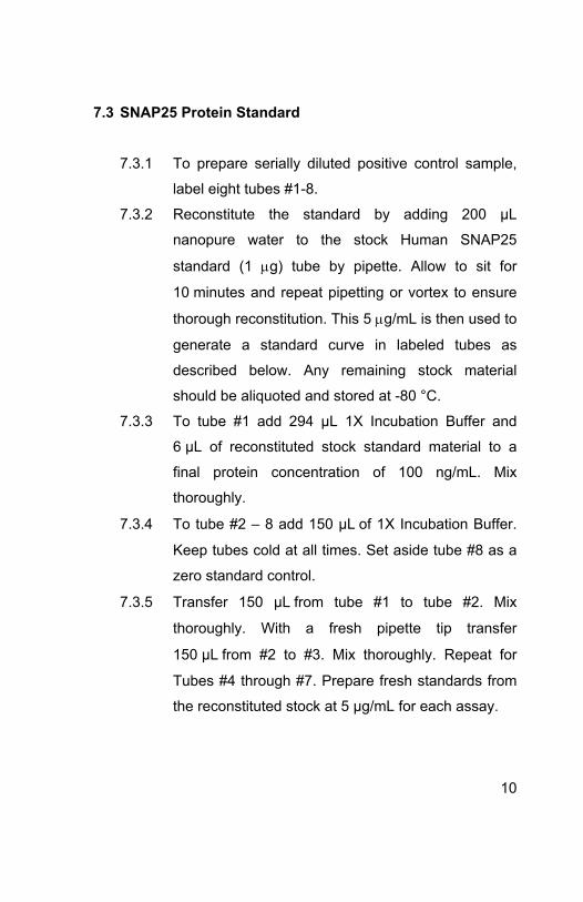
7.3 SNAP25 Protein Standard
7.3.1 To prepare serially diluted positive control sample,
label eight tubes #1-8.
7.3.2 Reconstitute the standard by adding 200 µL
nanopure water to the stock Human SNAP25
standard (1 g) tube by pipette. Allow to sit for
10 minutes and repeat pipetting or vortex to ensure
thorough reconstitution. This 5 g/mL is then used to
generate a standard curve in labeled tubes as
described below. Any remaining stock material
should be aliquoted and stored at -80 °C.
7.3.3 To tube #1 add 294 µL 1X Incubation Buffer and
6 µL of reconstituted stock standard material to a
final protein concentration of 100 ng/mL. Mix
thoroughly.
7.3.4 To tube #2 – 8 add 150 µLof 1X Incubation Buffer.
Keep tubes cold at all times. Set aside tube #8 as a
zero standard control.
7.3.5 Transfer 150 µLfrom tube #1 to tube #2. Mix
thoroughly. With a fresh pipette tip transfer
150 µLfrom #2 to #3. Mix thoroughly. Repeat for
Tubes #4 through #7. Prepare fresh standards from
the reconstituted stock at 5 µg/mL for each assay.
10

SNAP25 protein standard (100ng/mL)
2 3 4 5 6 7
150 l 150 l 150 l 150 l 150 l 150 l
1/2 1/4 1/8 1/16 1/32 1/64
1
8. Assay ProcedureEquilibrate all reagents and samples to room temperature before use. It is recommended all samples and standards be assayed in duplicate.
8.1 Prepare all reagents, working dilutions of controls and
samples as directed in the previous sections.
8.2 Remove unused microplate strips from the plate frame,
return them to the foil pouch containing the desiccant
pack, and seal.
8.3 Load 50 µL from tubes #1 – 8 into the wells for one strip.
Repeat as desired for replicates.
11

8.4 Add 50 µL of each sample per well. It is recommended to
include a dilution series of a control (normal) sample as a
reference.
8.5 Cover/seal the plate and incubate for 2 hours at room
temperature. If available use a plate shaker for all
incubation steps at 300 rpm.
8.6 Aspirate each well and wash, repeat this once more for a
total of three washes. Wash by aspirating or decanting
from wells then dispensing 300 µL1X Wash Buffer into
each well as described above. Complete removal of liquid
at each step is essential for good performance. After the
last wash, remove the remaining buffer by aspiration or
decanting. Invert the plate and blot it against clean paper
towels to remove excess liquid.
8.7 Immediately before use prepare sufficient (500 µL/strip
used) 1X SNAP25 Detector Antibody (step 6.4) in 1X
Incubation Buffer. Add 50 µL1X Detector Antibody to each
well used. Cover/seal the plate and incubate for 1 hour at
room temperature. If available use a plate shaker for all
incubation steps at 300 rpm.
8.8 Repeat the aspirate/wash procedure above.
8.9 Immediately before use prepare sufficient (500 µL/strip
used) 1X HRP Label in 1X Incubation Buffer (step 6.5).
Add 50 µL1X HRP Label to each well used. Cover/seal
the plate and incubate for 1 hour at room temperature. If
12

available use a plate shaker for all incubation steps at 300
rpm.
8.10 Repeat the aspirate/wash procedure above, however,
performing a total of five washes.
8.11 Add 100 µL TMB Development Solution to each empty
well and immediately record the blue color development
with elapsed time in the microplate reader prepared with
the following settings:
Mode: Kinetic
Wavelength: 600 nM
Time: up to 15 min.
Interval: 20 sec. - 1 min.
Shaking: Shake between readings
Alternative– In place of a kinetic reading, at a user defined, time
record the endpoint OD data at (i) 600 nm or (ii) stop the
reaction by adding 100 µL stop solution (1N HCl) to each
well and record the OD at 450 nm.
13

9. Data Analysis
Average the duplicate protein standard readings and plot against
their concentrations after subtracting the zero standard reading.
Draw the best smooth curve through these points to construct a
standard curve. Most plate reader software or graphing software can
plot these values and curve fit. A four parameter algorithm (4PL)
usually provides the best fit, though other equations can be
examined to see which provides the most accurate (e.g. linear, semi-
log, log/log, 4 parameter logistic). Read relative SNAP25
concentrations for unknown and control samples from the standard
curve plotted. Samples producing signals greater than that of the
highest standard should be further diluted and reanalyzed, then
multiplying the concentration found by the appropriate dilution factor.
14

TYPICAL STANDARD CURVE - For demonstration only.
SNAP25 Standard Curve
SNAP25 Protein Concentration (ng/mL)
mO
D/m
in (6
00 n
m)
0.1 1 10 1000.1
1
10
100
Figure 1. Example standard curve. A dilution series of recombinant SNAP25
protein (ab74529) in the working range of the assay (0.06 – 50 ng/mL)
15

TYPICAL EXPERIMENTAL RESULTS - For demonstration only.
Mouse Brain Homogenate
Mouse Brain Homogenate (g/mL)
mO
D/m
in (6
00 n
m)
1 10 100 10000.1
1
10
100
Figure 2. Example Mouse Brain Homogenate dilution series and working
range.
Typical working ranges for tissue extracts
Sample Type Range
Mouse Brain Homogenate Extract 2 – 250 µg/mL
Rat Brain Homogenate Extract 2 – 250 µg/mL
SENSITIVITY
Determined minimum detectable amount of SNAP25 (mean zero
standard n=24 + 2 standard deviations) = 58.9 pg/mL
16
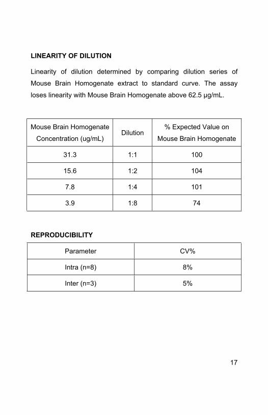
LINEARITY OF DILUTION
Linearity of dilution determined by comparing dilution series of
Mouse Brain Homogenate extract to standard curve. The assay
loses linearity with Mouse Brain Homogenate above 62.5 µg/mL.
Mouse Brain Homogenate
Concentration (ug/mL)Dilution
% Expected Value on
Mouse Brain Homogenate
31.3 1:1 100
15.6 1:2 104
7.8 1:4 101
3.9 1:8 74
REPRODUCIBILITY
Parameter CV%
Intra (n=8) 8%
Inter (n=3) 5%
17

RECOVERY
If assay is to be performed on serum samples or high concentration
extraction buffer samples, it is recommended to add equal
concentration serum or extraction buffer to the standard protein for
accurate results.
SNAP25
(ng/mL)10% FCS HGDMEM 10% NGS 50% RIPA
100 101% 90% 75%
33.3 96% 83% 80%
11.1 74% 59% 65%
3.7 79% 70% 61%
1.23 100% 90% 79%
18

Interference and Spike Recovery
SNAP25 Protein (ab74529) (ng/mL)
mO
D/m
in (6
00 n
m)
1 10 1000.1
1
10
100
Incubation Buffer10% FCS media10% NGS50% RIPA
Figure 3. Sample recovery under 3 different conditions compared with the
wash buffer from the kit. 10% NGS and 50% RIPA buffer decrease the
signal recovery by 20-25% on average.
19

10. Specificity
Species– Human, mouse and rat reactive. Others untested.
Figure 4. The detector antibody used in this kit specifically detects human
SNAP25 recombinant protein (lane 2) as determined by Western Blotting.
This antibody is species cross reactive for SNAP25 in Mouse Brain
Homogenate (lane 3), Rat Brain Homogenate (lane 5), and Human Brain
Homogenate (lane 7). SNAP25 is specifically expressed in brain tissue as
indicated by a lack of signal in Mouse Heart Homogenate (lane 4), Rat Heart
Homogenate (lane 6), Human Heart Homogenate (lane 8).
20

SNAP25 Assay Specificity
Tissue at 500 ug/mL
mO
D/m
in (6
00 n
m)
MHH HHH RHH MBH RBH0
10
20
30
40
Figure 5. Example Assay Tissue Specificity with Mouse Heart Homogenate
(MHH), Human Heart Homogenate (HHH), Rat Heart Homogenate (RHH),
Mouse Brain Homogenate (MBH), and Rat Brain Homogenate (RBH).
21

11. Troubleshooting
22
Problem Cause Solution
Inaccurate Pipetting Check pipettes
Poor standardcurve Improper standard
dilution
Prior to opening, briefly spin the stock standard tube and
dissolve the powder thoroughly by gentle mixing
Low SNAP25 concentration in sample
Use appropriate positive control
Incubation times too brief
Ensure sufficient incubation times; change to overnight standard/sample incubation
Sample degraded
Work with freshly extracted samples. Keep samples
cold before loading on the plate.
Low Signal
Inadequate reagent volumes or improper
dilution
Check pipettes and ensure correct preparation
Plate is insufficiently washed
Review manual for proper wash technique. If using a
plate washer, check all ports for obstructionsLarge CV
Contaminated wash buffer Make fresh wash buffer
Low sensitivity Improper storage of the ELISA kit
Store your reconstituted standards at -80°C, all other
assay components 4°C. Keep substrate solution
protected from light

UK, EU and ROWEmail: [email protected]: +44 (0)1223 696000www.abcam.com
US, Canada and Latin AmericaEmail: [email protected]: 888-77-ABCAM (22226)www.abcam.com
China and Asia Pacific Email: [email protected]: 108008523689 (中國聯通)www.abcam.cn
JapanEmail: [email protected]: +81-(0)3-6231-0940www.abcam.co.jp
23
Copyright © 2012 Abcam, All Rights Reserved. The Abcam logo is a registered trademark.
All information / detail is correct at time of going to print.