smith, frances (2017) antimicrobials and antimicrobial...
TRANSCRIPT
Smith, Frances (2017) Antimicrobials and antimicrobial resistance. MRes thesis, University of Nottingham.
Access from the University of Nottingham repository: http://eprints.nottingham.ac.uk/48053/2/Smith_Frances_MResThesis_FinalandRevised_ConfidentialRemoved.pdf
Copyright and reuse:
The Nottingham ePrints service makes this work by researchers of the University of Nottingham available open access under the following conditions.
This article is made available under the University of Nottingham End User licence and may be reused according to the conditions of the licence. For more details see: http://eprints.nottingham.ac.uk/end_user_agreement.pdf
For more information, please contact [email protected]
ANTIMICROBIALSANDANTIMICROBIALRESISTANCEMResThesis
FrancesSmith
CentreforBiomolecularSciences,UniversityofNottingham

2
TABLE OF CONTENTS Abbreviations...........................................................................................3
1. Introduction..........................................................................................4
2. Investigating the regulation of the Arginine-Specific Protease AaaA in Pseudomonas aeruginosa and Creating a Soluble Truncated Version for Cell-Free Inhibitor Screens..........................13
2.1. Abstract..................................................................................................142.2. Introduction............................................................................................152.3. Materials and Methods.........................................................................192.4. Results and Discussion.......................................................................24Acknowledgements......................................................................................31
3. Assessing FDA-approved Drugs for their Ability to Act as Antitubercular Agents..........................................................................32
3.1 Abstract...................................................................................................333.2. Introduction............................................................................................343.3. Materials and Methods.........................................................................363.4. Results....................................................................................................413.5. Discussion.............................................................................................56Acknowledgements......................................................................................61
4. Discussion: Perspective for the Next Three Years of Each Project.....................................................................................................62
Concluding Remarks............................................................................67
Reference List........................................................................................68

3
ABBREVIATIONS
ART–Antiretroviraltherapy
CF–Cysticfibrosis
CNS–Centralnervoussystem
CYP–CytochromeP450
DCS–D-cycloserine
FDA–FoodandDrugAdministration
HTS–Highthroughputscreening
ICU–Intensivecareunit
IC50–Inhibitoryconcentrationthatgives50%growthinhibition
IHF–Integrationhostfactor
MIC99–Minimuminhibitoryconcentrationthatgives99%growthinhibition
ORF–Openreadingframe
SDM–Site-directedmutagenesis
TB-Tuberculosis
4
1. INTRODUCTION
Antimicrobial resistance is rapidly emerging as a problem for
industrialised countries because current healthcare regimes are
unable to properly contain and treat resistance. Access to healthcare,
with good sanitation lowered the threat of many infectious diseases
in industrialised nations, however increase in drug resistance is now
becoming a global crisis and is threatening to revert health-care to
the state it was in the pre-antibiotics era1. The current global crisis of
increasing antibiotic resistance has been heralded as an “apocalyptic” threat2.
1. The Antibiotic Age
Antibiotics exploit the differences between prokaryotic and
eukaryotic cells and can be divided into different categories based on
their target. Cell wall biosynthesis is targeted by b-lactam antibiotics
and vancomycin. Moving inside the cell, DNA replication is prevented
by Quinolones, and DNA transcription is inhibited when rifampicin
binds to bacterial RNA polymerase. Protein synthesis, namely the
bacterial 70S-ribosome, is inhibited by macrolides, aminoglycosides
and tetracyclines. There is also a new class containing
oxazolidinones which act on the initiation step during protein
synthesis. Lastly, antibiotics target key metabolic processes such as
the biosynthesis of tetrahydrofolicacid which is inhibited by
sulphonamides and trimethoprim3. Some antibacterial agents have
multiple cellular targets, such as triclosan at bactericidal
concentrations4. At sub-lethal bacteriostatic concentrations triclosan specifically targets fatty acid biosynthesis4,5.
The first naturally occurring antibiotic to be used clinically was
discovered in 1928 by Alexander Fleming. At the time, the clinical
potential was unforeseen and initially lead to the dismissal of

5
Penicillin by Fleming and his peers shortly after its discovery. It
wasn’t until 1939 that it was re-discovered and mass produced as a
cure for gram-positive bacterial diseases. Typically, infections
caused by Staphylococcus, Streptococcus, Clostridium, and Listeria
are responsive to penicillin. P. aeruginosa infections can be treated
with penicillins from a more recent class, such as piperacillin 6. The
discovery of penicillin revolutionised the treatment of infectious
disease, this can be seen in the rapid decline in mortality rates due to infectious disease between the periods 1938 to 1952 in the USA7.
The discovery of penicillin engendered the biotechnology industry
we know today. Efforts to mass produce the chemical, produced by
Penicillum mould as a secondary metabolite, lead to development of
large-scale fermenters that are still used today8,9. The majority of
current antibiotics originate from natural biological sources, and are
usually secondary metabolites, produced by bacteria or mould that
grows in complex environments such as soil where competition for
available nutrients is high10. For example, Pseudomonas fluorescens
produces a phenazine antibiotic in the rhizosphere of wheat in order
to supress the fungal infection caused by Gaeumannomyces
graminis var. tritici11. Production of antibacterial agents provides a
fitness advantage by killing off competition, however contention
exists over this as a major role, as antibiotics are not naturally
produced at such high concentrations seen in therapeutic use10,12. A
few antibiotics, such as sulphonamides, trimethoprim and nitroimidazoles are of synthetic origin2.
Following on from penicillin, many more antibiotics have been
discovered, the last major class being lipopeptides in the early 2000s,
and before that, quinolones in 1960s13. The long break between the
introduction of quinolones and the present shows that all the “low-
hanging fruit” has been picked, and discovery of new antibiotics has
slowed down2. The absence of new effective antibiotics combined
with the rapid rise in bacterial resistance to current antibacterial

6
therapeutics is a huge problem, and if not addressed could lead to the end of the antibiotic era2,10,13,14.
2. The Rise in Antimicrobial Resistance
Antimicrobial resistance (AMR) is the abolishment of sensitivity to
current antibacterial and antifungal agents. For the purpose of this
study, AMR will refer only to bacterial resistance to antibiotics. AMR
is either acquired or intrinsic. Often antibiotic resistance strategies
are common and widely distributed in nature due to the natural
production of antibiotics as secondary metabolites10. This is not an
issue until bacteria are subjected to antibiotics in certain
environments, such as during therapeutic treatment. For example,
bacteria such as P. aeruginosa and M. tuberculosis naturally have
inherent resistance to a wide range of antibiotics3. Some bacterial
species are completely sensitive to antibiotics but will acquire
resistance by genetic modifications of their genome or by horizontal
gene transfer of mobile elements encoding resistance genes.
Resistance arises through selective pressure, simple Darwinian
evolution. Resistance may exist in a population of cells before
antibiotic is present in the environment, and with the exception of a
high fitness cost the resistant bacteria are no different than the
sensitive cells. Once an antibiotic selective pressure is introduced
however the resistance increases in frequency as sensitive cells die
and the resistant ones can thrive. Fleming himself warned against
the emergence of resistance to penicillin in 1945, having witnessed
how easily cells become insensitive to the antibiotic at non-lethal
concentrations15. Resistance has been reported to nearly all
antibiotics developed13.
Acquisition of resistance by horizontal gene transfer sees
transposons, plasmids, resistance islands, prophages, and integrons
containing resistance genes carried from resistant bacteria into
sensitive ones.3 These DNA elements are integrated into the
7
bacterial genome at specific sites using recombinases and
integrases. Resistance brings with it a fitness cost for the cell but
these are quickly negated through additional compensatory
mutations 3.
Intrinsic resistance is a natural insensitivity that is found in every
bacterium of a species, without any additional genetic mutation. The
prime example for intrinsic resistance is that exhibited by P.
aeruginosa, a multidrug resistant opportunistic pathogen. It has a
high level of intrinsic resistance due to restricted outer membrane
permeability, a b-lactamase encoded on the bacterial chromosome,
and the presence of, and overproduction of multi-drug-resistant
(MDR) efflux pumps3,16. Of these, the tripartite MexAB-OprM and
MexXY-OprM pumps have very broad specificity for their substrates.
Fluoroquinolones, tetracycline, macrolides, trimethoprim and b-
lactams (excluding imipenem) are all transported by the
constitutively-expressed MexAB-OprM, and in addition to the above,
aminoglycosides are transported by MexXY-OprM17. P. aeruginosa
efflux pumps export detergents, disinfectants, organic solvents and
dyes in addition to antibiotics3. With the exception of MexAB-OprM,
multi-drug efflux pumps are very tightly regulated17. Another example
of intrinsic resistance is that of Mycoplasma, as they are naturally
resistant to b-lactams as the cell wall inherently lacks the
peptidoglycan target3.
P. aeruginosa is also a model organism for physiological
resistance as it readily forms sessile biofilms3,18. The expression of
this type of intrinsic resistance is dependent on the environmental
growth conditions. Sessile biofilms can be seen in the lungs of
infected cystic fibrosis individuals, and the bacteria within often
cannot be cleared with antibiotics leading to persistent or recurring
infections19,20. Biofilms offer protection from antibiotics as they are a
complex assembly of stationary-phase bacteria surrounded by an
organised extracellular matrix that is difficult for antibiotics to
8
penetrate. Extracellular DNA, found in the biofilm matrix, plays a key
role in biofilm maturation and structure, but also conveys protection
from aminoglycoside antibiotics by binding to and sequestering
them21. During biofilm growth resistance genes are expressed that
are repressed during planktonic growth22. In addition to this, a high
number of bacteria within the biofilm exhibit a low metabolic state,
possibly due to the limited access to nutrients for growth. The
bacteria are persistors as they cannot be cleared by antibiotics that
target active growth, such as b-lactams.
Antimicrobial resistance is rapidly on the rise due to worldwide
overuse and misuse of antibiotics13. Use of antibiotics is not
regulated in many countries and they are available for purchase
online and prescription-free over the counter1,23. Easy access to
cheap antibiotics leads to inappropriate usage because the public
sees them as a quick and simple alternative to a wide range of
illnesses1,13,24. Another key contributor to antibiotic resistance is
incorrect prescriptions. This includes incorrect duration of treatment,
type of antibiotics, and unnecessary use. Inappropriate prescriptions
occur in up to 50% of cases13. Often the need for rapid treatment
means that there is limited time to determine the bacterial cause of
infection and administer the most suitable antibiotics accordingly. In
intensive care units (ICUs) in the US 30-60% of antibiotics
administered are inappropriate because of this need to treat patients
with bacterial infections quickly25. Patients in the ICU are prescribed
more antibiotics than necessary as there are often no defining clinical
symptoms of infection25. Aside from developing resistance,
inappropriate usage of antibiotics can have negative effects on the
patient, such as destroying the natural microflora that plays a key role in protection from disease.
Extensive use of antibiotics for agricultural practices is a massive
driver for resistance. They are used throughout the developing world
as growth promoters in livestock to give maximum yields of healthy,

9
quality product13,23,26. It is estimated that 80% of all antibiotics sold in
the US go to livestock, which are then eaten, resulting in transfer of
both antibiotics and resistant bacteria to humans26–28. These bacteria
then cause infections. Antibiotics excreted by the animals are spread
to the environment through ground water, surface run off and use of
excrement as fertilizer, exposing the environmental microbiome to
low levels of antibiotics and encouraging the development of
resistance13,26. Again, these can then be picked up by humans and
cause disease that is hard to treat. Antibacterial agents, such as
triclosan, are also used widely as preservatives, disinfectants and
antiseptics and again these contribute to the antibiotic resistance
crisis. For example, triclosan can be found in many household items
such as plastics, as well as in personal health-care items like toothpastes, but triclosan-resistance has been reported since 20004.
3. Tackling the AMR Crisis
AMR is both a clinical and a socioeconomic problem that is
estimated to result in a loss of $100 trillion USD by 2050 and cause
the deaths of 10 million people per year 3,29. At present 700,000
deaths each year globally are due to antibiotic-resistant infections29.
Avoiding this crisis can be achieved through a combination of policy and research.
One excellent example of policy preventing the spread of
disease-causing bacteria is of the instructions implemented to
prevent spread of multi-drug resistant C. difficile in hospitals. C.
difficile forms highly resistant spores, which can be transported easily
across hospitals to vulnerable immune-compromised patients. It was
recommended that new hand-washing procedures, isolation of
patients and thorough ward sterilisation with diluted bleach should be
implemented, and as a result nosocomial C.difficile infections were
reduced significantly30. Preventing the spread of antibiotic resistance
at the transmission step is fast and produces instant results.
10
However, care is needed when using disinfectants as there have
been reports of increased tolerance to antiseptics as a result of AMR.
Quinolone resistance, in the form of gyrase mutants in E. coli and
Salmonella decreases susceptibility to the disinfective agent triclosan by upregulation of the stress response31.
It is important to increase global surveillance and globally-
accessible databases for genetic fingerprinting profiles of drug-
resistant bacteria. Identifying bacteria can be done by a number of
ways, such as Multilocus sequence typing (MLST), PCR ribotyping,
multilocus variable tandem repeat analysis (MVLA)32. Keeping
identifiable information in world-wide databases will aid tracking the
spread of resistance across the globe, and data can be used to
predict the origin of resistance and also highlight potentially problematic pathogens3.
A vast amount of research is being undertaken to confront the
global issue of AMR. There is a strong focus on finding new drug
targets in order to develop new antibiotics2. Current antibiotics in use
target essential genes and disrupt bacterial growth, thus giving a
strong selective pressure for development of resistance. A new
approach is to screen for drugs that disrupt bacterial virulence rather
than bacterial growth as antivirulence agents should generate a far
weaker selective pressure for the development of resistance than
any current bactericidal or bacteriostatic antibiotics3. Antivirulence
drugs target secreted virulence factors and bacterial quorum-
sensing, the mechanism for bacterial communication that enables
attachment and colonisation of surfaces through the formation of
complex bacterial communities called biofilms. Examples of current
antivirulence drugs include Virstatin, which halts expression of the
cholera toxin and the co-regulated pilus in Vibrio cholerae, and B81-
2, an inhibitor of type IV secretion in Brucella abortus33,34. Targeting
virulence of pathogens instead of growth should allow bacteria to
remain within its host long enough for the adaptive immune system
to destroy it, thus conferring protection for the next time. It is worth

11
mentioning that mechanisms of resistance to antivirulence drugs
have already been reported, both in clinical isolates and
laboratories33–36. However, resistance seen today by traditional
antibiotics arises because cells are either killed directly or growth is
perturbed, thus resistant cells can reproduce rapidly in a cell-free
environment. This level and spread of resistance is unlikely to occur
upon use of antivirulence drugs, as cells will still grow in a
competitive, well-mixed population and the selection for resistance is low37.
Two important areas of AMR research are finding new drug
targets for exploitation, and drug discovery. The first project
undertaken during this research year involved the former, and the
second project, the latter. P. aeruginosa is a multi-drug resistant
pathogen that secretes a barrage of virulence factors into its
environment. One key virulence factor is the aminopeptidase
autotransporter AaaA, which generates a supply of arginine for
growth in oxygen-limited environments such as chronic wound
infections38. The expression of virulence factors must be tightly
controlled. During the first project, the regulation of AaaA was
investigated using a transcriptional reporter to find potential new drug
targets and to understand how and when expression occurs during
infection. In addition, AaaA was to be investigated as a target for
novel antivirulence drugs. In order to carry out structural studies of
AaaA for inhibitor binding site predictions, a soluble, active, truncated
version was needed. This was also to be used for cell-free inhibitor
screens against potential antivirulence agents. P. aeruginosa is a
common human pathogen that causes a broad spectrum of diseases.
It is vital to find effective antibiotics against it as it is commonly picked up in hospitals by immunocompromised patients.
The second project aimed to use high-throughput screening
methods to find current drugs for repurposing as antimycobacterial
agents. The FDA library contains over 1200 different small molecule
compounds, all licenced for use in a wide range of illnesses. These
12
illnesses are not necessarily infection-related, and the library
includes drugs used to treat depression, cancer, and arthritis to name
a few. One objective during this project was to find appropriate drugs
that would act as adjuncts to lower the dose needed for D-
cycloserine, a second-line drug used to treat TB resistant to front-line
antibiotics. The current treatment for drug-resistant tuberculosis is
long, costly and has serious side effects. By lowering the
concentration of D-cycloserine required, the negative side-effects
should be reduced too. It is important to find new antibiotics to treat
tuberculosis, as there has been a steady rise in the number of multi-drug resistant and extensively drug resistant cases.
13
2. INVESTIGATING THE REGULATION OF THE
ARGININE-SPECIFIC PROTEASE AAAA IN
PSEUDOMONAS AERUGINOSA AND CREATING A
SOLUBLE TRUNCATED VERSION FOR CELL-FREE
INHIBITOR SCREENS

14
2.1. ABSTRACT
Pseudomonas aeruginosa is a Gram-negative ubiquitous human
pathogen that infects wounds, burns, eyes and the lungs of cystic
fibrosis patients. Its intrinsic resistance to many antibiotics makes P.
aeruginosa notoriously difficult to treat. The gene pa0328 encodes an
autotransporter, AaaA, with arginine-specific aminopeptidase activity
and an important virulence factor. AaaA remains attached to the surface
of bacteria where it aids the uptake of nitrogen in environments with
limited free oxygen and nitrogen availability but plenty of peptides with
an N-terminal arginine. AaaA allows P. aeruginosa to evade the host
immune system, grow, and establish biofilms under anoxic conditions.
The regulation of such a virulence factor is complex and it is important
to understand which signals result in aaaA expression or repression.
These signals could be used as targets for, or mimicked by, novel
antivirulence drugs. We hypothesise that regulators IHF and RpoN bind
directly to the promoter of aaaA to alter its expression. By specifically
mutating these sites the control of said regulators can be determined
using a transcriptional reporter. AaaA is a key virulence factor for chronic
wound infections and its mutation perturbs virulence. Additionally, AaaA
sits on the surface of P. aeruginosa and is easily accessible to
antimicrobials making it an ideal target for antivirulence drugs. There is
a need for a high-throughput inhibitor screen to be developed, and for
this, a soluble truncated version of AaaA must be cloned, which was the
second aim of this project. Mutagenesis of the IHF and RpoN binding
sites was attempted using site-directed mutagenesis, but proved
repeatedly unsuccessful. Truncating aaaA was more successful but not fully achieved in the time frame.
15
2.2. INTRODUCTION
Pseudomonas aeruginosa is an important multidrug-resistant
pathogen that infects cystic fibrosis (CF) sufferers, wounds, eyes, and
thermal injuries. It is an opportunistic pathogen and a common cause of
hospital-acquired infection 38.
P. aeruginosa readily forms biofilms; bacterial communities
adhered to a surface and a common cause of corrosion and infection.
Biofilms commonly grow on industrial surfaces resulting in biofouling -
examples of this can be found from washing machines to the Mir space
station 39. From a clinical perspective, biofilms provide increased
protection from antibiotics and clearance by the host immune system.
Microcolonies show increased resistance to antibiotics due to the
physical barrier between interior cells and the environment 40. In
addition, antibiotics targeting active metabolism are less effective on
bacteria further within the biofilm as these cells have less access to nutrients and so exhibit a low metabolic state. 40.
P. aeruginosa is notorious as the primary cause of morbidity in
CF sufferers 38. Cystic fibrosis patients contract lung infections
intermittently during childhood, eventually these persist to become
chronic lung infections that can survive for decades 41. Studies show that
80 % of CF patients are infected with P. aeruginosa by adulthood 42.
These chronic lung infections are, in part, due to failure of antibiotic
treatment to clear the bacteria. As the infection continues through the
generations strains become mucoid, a phenotype associated with
increased antibiotic tolerance and increased morbidity and mortality in
CF patients 41. Formation of immobile biofilms from planktonic cells in
the CF lungs increases resistance to antibiotics 1000-fold 40,42. It is
therefore vital to investigate the biology of P. aeruginosa to better
understand how to treat these infections and prevent persistence of the disease.
16
AaaA is an important virulence factor of P. aeruginosa. It is an
autotransporter with arginine-specific aminopeptidase activity and is one
of the 20 most-produced enzymes in biofilms 38. It has previously been
shown to provide a strong virulence advantage in chronic wound
infection. The bacterial load of P. aeruginosa ∆aaaA in a mouse chronic
wound model was far lower than the wild type at both 2 and 8 days post-
infection, demonstrating its key role in immune evasion 38. The catalytic
domain of AaaA cleaves the amino terminus arginine from extracellular
peptides providing P. aeruginosa with an alternate nitrogen source when
environmental nitrogen is low. This enables the bacterium to grow and
provides a selective advantage. It can also aid survival in hypoxic
environments, such as within biofilms, as it provides arginine as an energy source through substrate level phosphorylation43.
Autotransporters are a large superfamily of virulence factors
found in Gram-negative bacteria 44. They are characterised by an N-
terminal signal peptide to allow passage across the inner membrane, a
central passenger domain that confers enzymatic function, and a 12-
stranded C-terminal β-barrel domain for translocation across the outer
membrane. Autotransporters, as the name suggests, were thought to
translocate the extracellular passenger domain independently of ATP or
other secretion systems using the covalently-linked C-terminal β-barrel.
However, recent evidence suggests that chaperone proteins, in
particular the Bam complex, are required for insertion of the β-barrel into
the outer membrane and subsequent translocation 44. Unlike many
autotransporters, AaaA remains attached to the cell surface via its
membrane-embedded β-barrel after crossing the outer membrane 38,45.
It is important to look for inhibitors of AaaA as it is such a crucial
virulence factor. AaaA is an ideal target for antimicrobials as it is surface-
associated and therefore easier for compounds to access. To screen for
potential AaaA inhibitors, a cell free inhibitor assay is required. This
project aimed to develop and express a version of AaaA which is soluble,
active, and can easily be purified with a polyhistidine tag. Wild-type AaaA
is a membrane-associated protein, and these are notoriously hard to

17
purify in large quantities. To overcome this, the membrane-bound b-
barrel domain is not included in the truncated version as it is not
necessary for catalytic function of AaaA. This should make the protein
easier to purify in large quantities. The truncated version (tAaaA3)
includes the N-terminal signal peptide to direct the peptide to the
periplasm where it should fold into its fully-functional form, and not form
toxic cytoplasmic inclusion bodies when overexpressed. Previous
versions created by Daniella Spencer (University of Nottingham) did not
contain the N-terminal signal peptide, were highly toxic, and readily
formed inclusion bodies in the cytoplasm (unpublished data). Overall,
tAaaA3 retains the catalytic passenger domain of AaaA in the periplasm
where it is predicted to be easier to purify and less toxic to the cell than
a cytosol-localised version. The Purified tAaaA3 can then be used in a
high-throughput antimicrobial screen, or for structural studies to investigate inhibitor binding sites.
Bacteria must tightly control the expression of genes in response
to environmental signals and their changing intracellular metabolism in
order to survive. This is commonly achieved by regulatory proteins that
bind to promoters and positively or negatively affect transcription. In one
recent study, the promoter region of aaaA was shown to be negatively
regulated by RpoN, RhlR, MvaT and MvaU, and positively regulated by
ArgR using a bioluminescent transcriptional reporter46. The promoter
region of aaaA also contains putative binding sites for a number of these
regulators, such as ArgR and RpoN, as well as a site for an additional
transcriptional regulator, IHF46. Although the regulatory effect has been
shown, it is not yet established if this is caused by direct binding of these
regulatory proteins to the aaaA promoter region46.
IHF is a global regulator conserved between Gram-negative
bacteria and belongs to the DNA-binding protein family DNABII 47. It
regulates many genes, including those involved in the biosynthesis of
known virulence factors 48,49. IHF has two subunits, IHFα and IHFβ,
which form two βsheet arms and are joined at one end 47. The

18
heterodimer binds DNA specifically, creating a sharp 180º bend and
facilitating interactions between DNA and other DNA binding proteins,
like RNA polymerase, by opening up the major groove 47,50. RpoN is an
alternative sigma factor which also regulates the expression of virulence
factors in P. aeruginosa 51. When rpoN mutants were challenged with a
mouse burn-wound model, they were 100-fold less virulent,
demonstrating its importance to the pathogen 51. Like IHF, RpoN binds
specifically to a consensus sequence in the promoter region of target
genes 52,53. It has been shown to regulate aliginate, rhamnolipid, and lipase biosynthesis as well as quorum sensing pathways 52,54.
In order to further understand the regulation of aaaA, the aim of
this project was to identify IHF and RpoN as two regulators that bind
directly to their respective predicted binding sites in the promoter region
of aaaA. The effect of IHF and RpoN on expression was to be assessed
using a bioluminescent transcriptional reporter fused with the aaaA
promoter region. Inactivation of IHF and RpoN binding motifs within the
promoter region of the reporter was to be carried out by engineering
mutations in their binding consensus sequences using site-directed
mutagenesis. The transcriptional reporters could then be used to compare transcription in the activated and inactivated promoter regions.
This project aimed to determine the role of IHF and RpoN in
regulating aaaA expression, and to create an expression system for a
truncated AaaA version for inhibitor screens.
19
2.3. MATERIALS AND METHODS
2.3.1. Bacterial Strains and Culture conditions
Strains were cultured in Lysogeny broth (LB) or grown on LB agar
plates at 37 οC. Liquid cultures were shaken at 250rpm and were used
directly for plasmid purification. Where appropriate, ampicillin was used
at a working concentration of 100 µg/ml and tetracycline was used at 30
µg/ml. All stock antibiotics were stored at -20 οC and were prepared at
50 mg/ml. Stocks of E. coli strains were made from LB broth and 25% glycerol, and stored at -80οC.
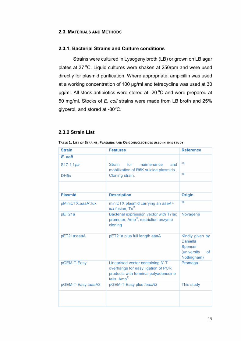
2.3.2 Strain List
TABLE1.LISTOFSTRAINS,PLASMIDSANDOLIGONUCLEOTIDESUSEDINTHISSTUDY
Strain Features Reference
E. coli
S17-1 lpir Strain for maintenance and mobilization of R6K suicide plasmids .
55
DH5a Cloning strain. 56
Plasmid Description Origin
pMiniCTX:aaaA’:lux miniCTX plasmid carrying an aaaA’-lux fusion, TcR
46
pET21a Bacterial expression vector with T7lac promoter, AmpR, restriction enzyme cloning
Novagene
pET21a:aaaA pET21a plus full length aaaA Kindly given by Daniella Spencer (university of Nottingham)
pGEM-T-Easy Linearised vector containing 3’-T overhangs for easy ligation of PCR products with terminal polyadenosine tails. AmpR.
Promega
pGEM-T-Easy:taaaA3 pGEM-T-Easy plus taaaA3 This study
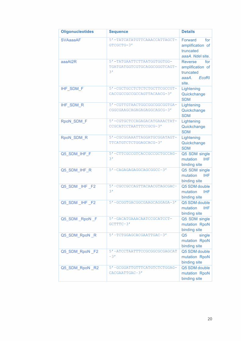
20
Oligonucleotides Sequence Details
SVAaaaAF 5’-TATCATATGTTCAAACCATTAGCT-GTCGCTG-3’
Forward for amplification of truncated aaaA. NdeI site.
aaaAt2R 5’-TATGAATTCTTAATGGTGGTGG-TGATGATGGTCGTGCAGGCGGGTCAGT-3’
Reverse for amplification of truncated aaaA. EcoRI site.
IHF_SDM_F 5’-CGCTGCCTCTCTCTGCTTCGCCGT-CACCGCCGCCGCCAGTTACAACG-3’
Lightening Quickchange SDM
IHF_SDM_R 5’-CGTTGTAACTGGCGGCGGCGGTGA-CGGCGAAGCAGAGAGAGGCAGCG-3’
Lightening Quickchange SDM
RpoN_SDM_F 5’-CGTGCTCCAGAGACATGAAACTAT-CCGCATCCTAATTTCCGCG-3’
Lightening Quickchange SDM
RpoN_SDM_R 5’-CGCGGAAATTAGGATGCGGATAGT-TTCATGTCTCTGGAGCACG-3’
Lightening Quickchange SDM
Q5_SDM_IHF_F 5’–CTTCGCCGTCACCGCCGCTGCCAG–3’
Q5 SDM single mutation IHF binding site
Q5_SDM_IHF_R 5’–CAGAGAGAGGCAGCGGCC–3’ Q5 SDM single mutation IHF binding site
Q5_SDM _IHF _F2 5’–CGCCGCCAGTTACAACGTAGCGAC–3’
Q5 SDM double mutation IHF binding site
Q5_SDM _IHF _F2 5’–GCGGTGACGGCGAAGCAGGAGA–3’ Q5 SDM double mutation IHF binding site
Q5_SDM _RpoN _F 5’–GACATGAAACAATCCGCATCCT-GCTTTC–3’
Q5 SDM single mutation RpoN binding site
Q5_SDM_RpoN _R 5’–TCTGGAGCACGAATTGAC–3’ Q5 single mutation RpoN binding site
Q5_SDM_RpoN _F2 5’–ATCCTAATTTCCGCGGCGCGAGCAT –3’
Q5 SDM double mutation RpoN binding site
Q5_SDM_RpoN _R2 5’–GCGGATTGTTTCATGTCTCTGGAG-CACGAATTGAC–3’
Q5 SDM double mutation RpoN binding site

21
2.3.2. DNA Manipulation
Plasmids were constructed by digestion of DNA with restriction
endonucleases and then ligation into the target vector backbone.
Agarose gel electrophoresis was carried out in 1X TAE buffer (80 mM
Tris-acetate pH 7.8, 19 mM EDTA) using a standard method 57. DNA
amplification by PCR was used to amplify fragments of DNA for cloning.
Q5 DNA Polymerase (NEB) was used for accurate amplification of the
DNA sequence. 2X Q5 Master Mix, nuclease-free water, 1.25 µl of each
10 mM oligonucleotide, and up to 100 ng of DNA template were added
to a total volume of 25 µl. PCR conditions were followed as
recommended by NEB. PCR clean-up was carried out using the Wizard
SV Gel and PCR Clean-Up System (Promega). Digestion was carried
out by mixing 1 µg DNA with 1 µl of each restriction enzyme, 2 µl of 10X
buffer and nuclease-free water to a total volume of 20 µl. For double-
digestion with NdeI, the enzyme was added alone and the reaction was
incubated at 37 ºC for 1 h before the second restriction endonuclease
was added and incubation continued for a further 1 h. Single-digest
controls were used when appropriate. Restriction digest fragments were
isolated from agarose gel using the Monarch DNA Gel Extraction kit
(NEB). A-tailing was necessary for cloning DNA fragments into the
vector P-GEM-T-Easy. To do this, 10 mM dATP, 0.2 µl Taq, 1 µl
Standard Taq Buffer, 7.8 µl cleaned-up DNA fragment, and nuclease-
free water to a total volume of 10 µl was incubated at 72 ºC for 20 min.
For ligation of DNA fragments to the target vector, DNA was mixed
together at a ratio of 5:1 insert:vector with 50 ng vector DNA. This was
incubated at 16 ºC with 1 µl T4 DNA ligase, T4 DNA ligase 10X buffer,
and nuclease-free water to a total volume of 10 µl for 16 h. The resulting
ligation mix was dialysed for 1 h and then transformed into
electrocompetent E. coli DH5α cells and grown overnight on selective
LB agar. Plasmid DNA was prepared using a plasmid purification kit (Qiagen). All DNA sequencing was performed by Source Bioscience.

22
2.3.3. Site-Directed Mutagenesis
Site-directed mutagenesis (SDM) was carried out using two different methods.
QuikChange Lightning Site-Directed Mutagenesis Kit (Aligent)
Primers were designed according to the QuikChange Lightning
protocol. A PCR reaction mixture was prepared containing Pfu Buffer
10X, 1µl 10 µM forward primer, 1µl 10 µM reverse primer, 50 ng template
DNA, 1µl 10 mM each dNTP, 1 µl Pfu DNA Polymerase, and nuclease-
free water to 50 µl. PCR was performed under the following thermocycling conditions:
Temperature (ºC) Time (s) 95 30 95 30 68 60 72 60 / kb 4 Stored indefinitely
Steps 2 to 4 were repeated for 12-16 cycles. The reaction mixture was
then treated with 2 µl DpnI and 10X Dpn1 Buffer (NEB). After gentle
mixing the reaction was incubated at 37 ºC for 20 min for digestion of
parental DNA. 15 µl of the reaction was then used to transform commercially competent E. coli TOP10 cells.
23
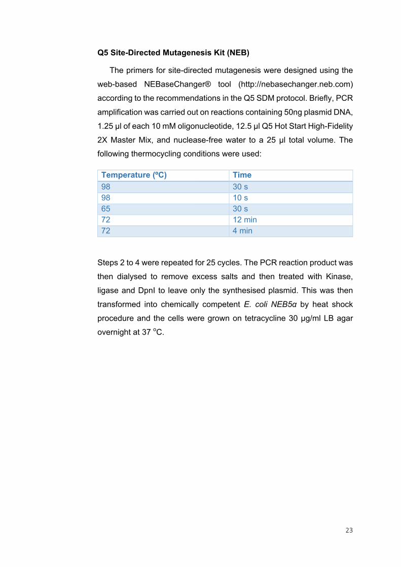
Q5 Site-Directed Mutagenesis Kit (NEB)
The primers for site-directed mutagenesis were designed using the
web-based NEBaseChanger® tool (http://nebasechanger.neb.com)
according to the recommendations in the Q5 SDM protocol. Briefly, PCR
amplification was carried out on reactions containing 50ng plasmid DNA,
1.25 µl of each 10 mM oligonucleotide, 12.5 µl Q5 Hot Start High-Fidelity
2X Master Mix, and nuclease-free water to a 25 µl total volume. The following thermocycling conditions were used:
Temperature (ºC) Time 98 30 s 98 10 s 65 30 s 72 12 min 72 4 min
Steps 2 to 4 were repeated for 25 cycles. The PCR reaction product was
then dialysed to remove excess salts and then treated with Kinase,
ligase and DpnI to leave only the synthesised plasmid. This was then
transformed into chemically competent E. coli NEB5α by heat shock
procedure and the cells were grown on tetracycline 30 µg/ml LB agar
overnight at 37 οC.

24
2.4. RESULTS AND DISCUSSION
2.4.1. In silico analysis of aaaA
In silico analysis of the aaaA promoter region was carried out
using BPROM (SoftBerry) a promoter prediction resource, the
PRODORIC database and PRODORIC Virtual Footprint binding site
prediction tool (http://www.prodoric.de). Promoter prediction was
attempted with BPROM (SoftBerry) but this returned no promoter sites.
The promoter was located instead by querying the PRODORIC
database. PRODORIC Virtual Footprint is a prediction tool which utilises
an extensive database of information on gene regulation in a number of
prokaryotic organisms. Such information is taken from scientific literature
and computational analysis and can be used to predict the regulation of
a target prokaryotic gene 58. A 500 bp region directly upstream of the
aaaA open reading frame (ORF) was input into the Virtual Footprint
algorithm to determine the binding sites for putative regulatory proteins.
Sequence analysis gave two predicted binding sites for Integrated host
factor (IHF); which can be found between the bases i) -253 to -238
relative to the start codon of aaaA on the (+) strand and ii) -148 to -133
on the (-) strand (Figure 1a). The latter lies on the correct strand and fit
with the IHF binding consensus sequence WCARNWNNTTR described
in previously published literature, where A/T is represented by a W, and
A/G is indicated by the letter R (Figure 1b). As a result this predicted binding sequence was chosen for site-directed mutagenesis 47,59.
2.4.2. Mutating binding sites in the promoter region of aaaA
To further understand the regulation of aaaA, substitution
mutations were to be made in the 500bp upstream region using site-
directed mutagenesis. The 500bp upstream region of aaaA has
previously been cloned onto a plasmid encoding the lux operon, called
miniCTX:paaaA:lux 46. Transcription of the lux genes is subsequently
under the control of the aaaA promoter and provides a simple,
25
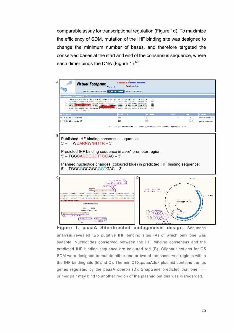
comparable assay for transcriptional regulation (Figure 1d). To maximize
the efficiency of SDM, mutation of the IHF binding site was designed to
change the minimum number of bases, and therefore targeted the
conserved bases at the start and end of the consensus sequence, where each dimer binds the DNA (Figure 1) 60.
Figure 1. paaaA Site-directed mutagenesis design. Sequence
analysis revealed two putative IHF binding sites (A) of which only one was
suitable. Nucleotides conserved between the IHF binding consensus and the
predicted IHF binding sequence are coloured red (B). Oligonucleotides for Q5
SDM were designed to mutate either one or two of the conserved regions within
the IHF binding site (B and C). The miniCTX:paaaA:lux plasmid contains the lux
genes regulated by the paaaA operon (D). SnapGene predicted that one IHF
primer pair may bind to another region of the plasmid but this was disregarded.
D:
A:
B:
C:
Published IHF binding consensus sequence: 5’ – WCARNWNNTTR – 3’ Predicted IHF binding sequence in aaaA promoter region: 5’ – TGGCAGCGGCTTGGAC – 3’ Planned nucleotide changes (coloured blue) in predicted IHF binding sequence: 5’ – TGGCGGCGGCGGTGAC – 3’
26
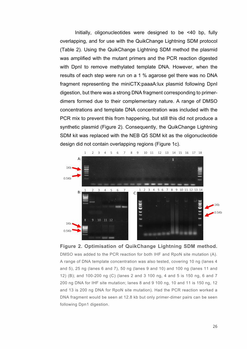
Initially, oligonucleotides were designed to be <40 bp, fully
overlapping, and for use with the QuikChange Lightning SDM protocol
(Table 2). Using the QuikChange Lightning SDM method the plasmid
was amplified with the mutant primers and the PCR reaction digested
with DpnI to remove methylated template DNA. However, when the
results of each step were run on a 1 % agarose gel there was no DNA
fragment representing the miniCTX:paaaA:lux plasmid following DpnI
digestion, but there was a strong DNA fragment corresponding to primer-
dimers formed due to their complementary nature. A range of DMSO
concentrations and template DNA concentration was included with the
PCR mix to prevent this from happening, but still this did not produce a
synthetic plasmid (Figure 2). Consequently, the QuikChange Lightning
SDM kit was replaced with the NEB Q5 SDM kit as the oligonucleotide design did not contain overlapping regions (Figure 1c).
Figure 2. Optimisation of QuikChange Lightning SDM method.
DMSO was added to the PCR reaction for both IHF and RpoN site mutation (A).
A range of DNA template concentration was also tested, covering 10 ng (lanes 4
and 5), 25 ng (lanes 6 and 7), 50 ng (lanes 9 and 10) and 100 ng (lanes 11 and
12) (B); and 100-200 ng (C) (lanes 2 and 3 100 ng, 4 and 5 is 150 ng, 6 and 7
200 ng DNA for IHF site mutation; lanes 8 and 9 100 ng, 10 and 11 is 150 ng, 12
and 13 is 200 ng DNA for RpoN site mutation). Had the PCR reaction worked a
DNA fragment would be seen at 12.8 kb but only primer-dimer pairs can be seen
following Dpn1 digestion.
A:
B: C:
1Kb
0.5Kb
1Kb
0.5Kb
1Kb
0.5Kb
123456789101112131415161718
1234567 1234567891011121314
89101112
27
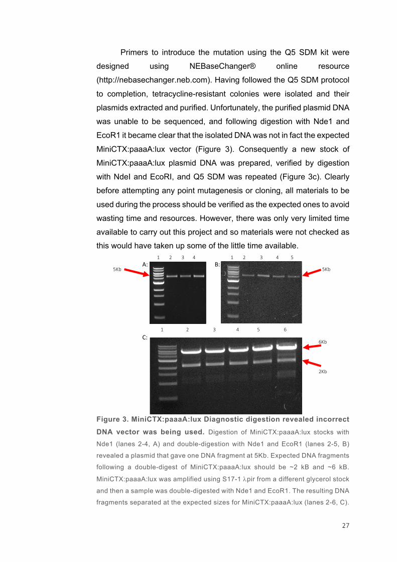
Primers to introduce the mutation using the Q5 SDM kit were
designed using NEBaseChanger® online resource
(http://nebasechanger.neb.com). Having followed the Q5 SDM protocol
to completion, tetracycline-resistant colonies were isolated and their
plasmids extracted and purified. Unfortunately, the purified plasmid DNA
was unable to be sequenced, and following digestion with Nde1 and
EcoR1 it became clear that the isolated DNA was not in fact the expected
MiniCTX:paaaA:lux vector (Figure 3). Consequently a new stock of
MiniCTX:paaaA:lux plasmid DNA was prepared, verified by digestion
with NdeI and EcoRI, and Q5 SDM was repeated (Figure 3c). Clearly
before attempting any point mutagenesis or cloning, all materials to be
used during the process should be verified as the expected ones to avoid
wasting time and resources. However, there was only very limited time
available to carry out this project and so materials were not checked as this would have taken up some of the little time available.
Figure 3. MiniCTX:paaaA:lux Diagnostic digestion revealed incorrect
DNA vector was being used. Digestion of MiniCTX:paaaA:lux stocks with
Nde1 (lanes 2-4, A) and double-digestion with Nde1 and EcoR1 (lanes 2-5, B)
revealed a plasmid that gave one DNA fragment at 5Kb. Expected DNA fragments
following a double-digest of MiniCTX:paaaA:lux should be ~2 kB and ~6 kB.
MiniCTX:paaaA:lux was amplified using S17-1 lpir from a different glycerol stock
and then a sample was double-digested with Nde1 and EcoR1. The resulting DNA
fragments separated at the expected sizes for MiniCTX:paaaA:lux (lanes 2-6, C).
A: B:
C:
1234 12345
123456
5Kb
2Kb
6Kb
5Kb
28
The Q5 SDM method was completed using the
MiniCTX:paaaA:lux vector and the plasmids from tetracycline-resistant
colonies were purified. However, sequence analysis revealed that
although the plasmids were correct, they did not carry the desired
mutation. This could be for a number of reasons. Firstly, the primer-
binding to the DNA may have been weak, or the annealing temperature
was wrong. This can be solved by performing the PCR on a number of
reactions with an annealing temperature gradient. Alternatively, the
primers can be redesigned to overlap each other, and the extra bases
will increase affinity for the DNA. If there was incomplete digestion of the
parental DNA, this could be fixed by increasing the KDL digestion time
from 5 min to 20 min although it is more likely that the DpnI enzyme was
not functional. Lastly, the template plasmid could simply be too large to
amplify with this method as it is 12,822 bp. The only way to overcome
this would be to mutate paaaA in a smaller vector and then clone it back
into MiniCTX:lux. An alternative method to tackle the overall project
question is through the use of gel mobility shift assays which have been used to show IHF binding to the phtD operon in P. syringae 47.
2.4.3. Cloning a soluble truncated version of the AaaA protein
It is notoriously difficult to obtain high quantities of purified,
functional membrane proteins. For this reason, a truncated version of
AaaA (tAaaA3) was designed to exclude the membrane-embedded β-
barrel domain, and include a polyhistidine tag for additional purification
using a nickel column.
Using the plasmid pET21a:aaaA a truncated version of aaaA was
PCR amplified. The oligonucleotides used have either an Nde1
restriction site or an EcoR1 site and were designed to include the N-
terminal signal peptide of AaaA and end the truncated protein on
hydrophilic amino acids to prevent the formation of inclusion bodies.
29
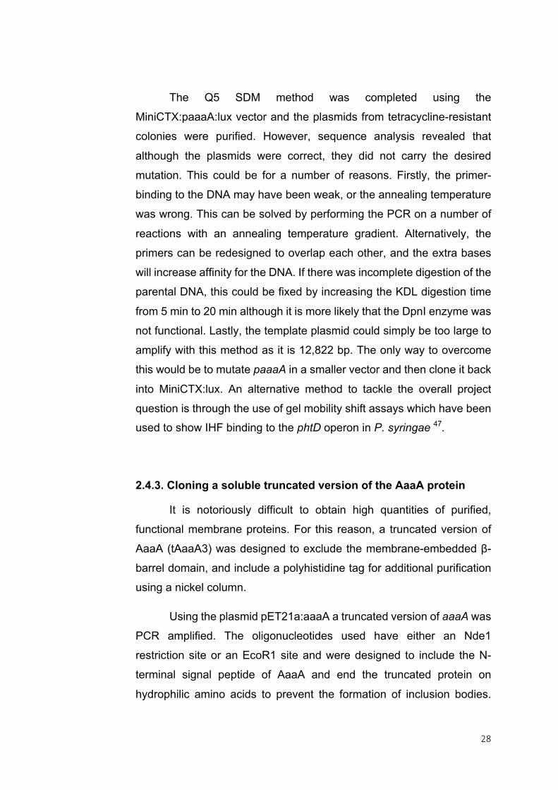
Inclusion of the signal peptide aimed to target tAaaA3 to the periplasm, where it will be less toxic to the cell (Figure 4).
Figure 4. Cartoon of Full length AaaA compared to truncated
version tAaaA3. AaaA sits tethered to the outer membrane of Pseudomonas
aeruginosa. The truncated version, tAaaA3, does not encode the β-barrel domain
but does include a polyhistidine tag to aid purification. It is thought that by
including the signal peptide tAaaA3 will be targeted to the periplasm and therefore
will not be toxic to the cells during over-expression.
The purified PCR product was dialysed, a poly-A tail was
introduced and the resultant fragment was ligated into pGEM-T-Easy.
Following a second dialysis step, pGEM-T-Easy:taaaA3 was
transformed into electrocompetent E. coli DH5α. Successful
transformants were ampicillin resistant. On the first attempt the taaaA3
fragment was significantly shorter than expected due to the reverse
primer annealing to a different position on the gene. After altering the
primer and repeating, plasmids from successful clones and were shown
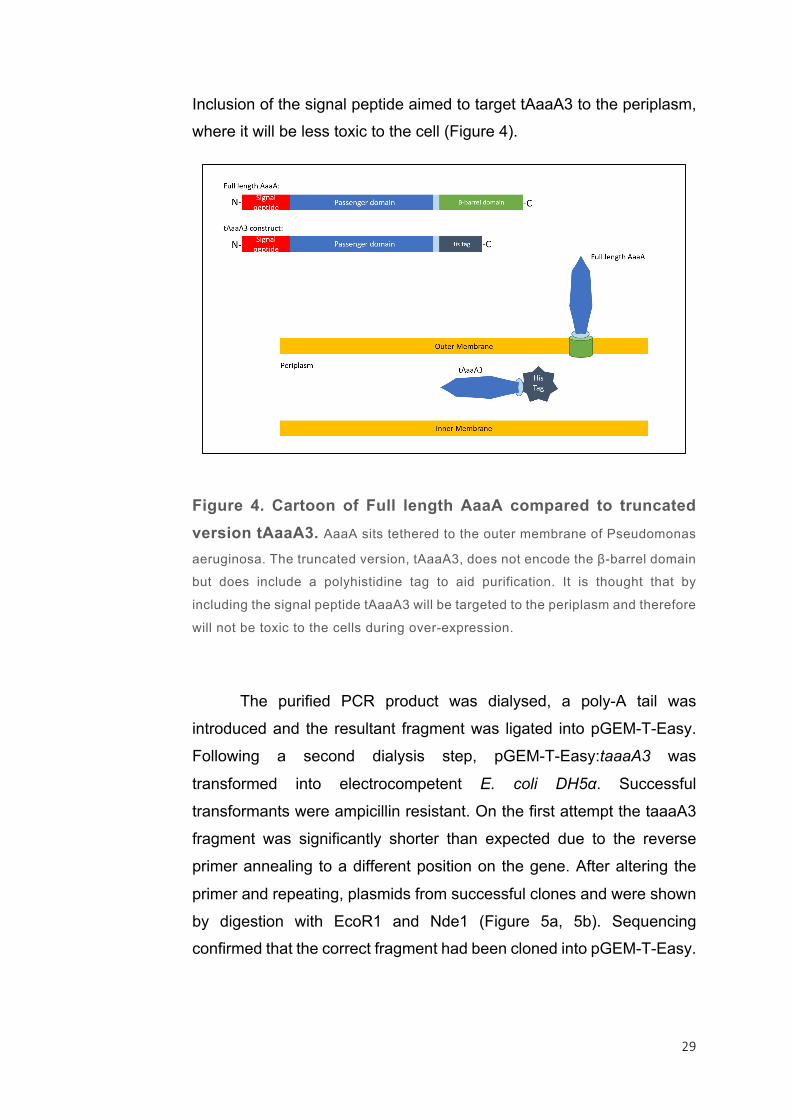
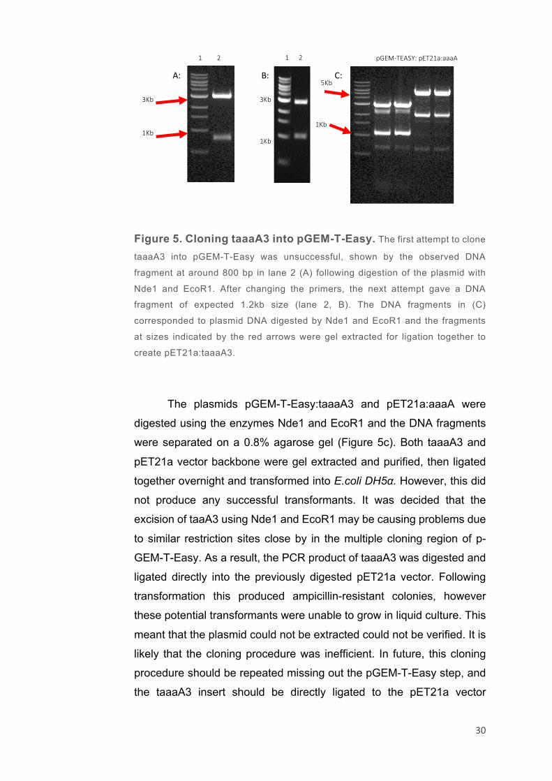
by digestion with EcoR1 and Nde1 (Figure 5a, 5b). Sequencing confirmed that the correct fragment had been cloned into pGEM-T-Easy.
30
Figure 5. Cloning taaaA3 into pGEM-T-Easy. The first attempt to clone
taaaA3 into pGEM-T-Easy was unsuccessful, shown by the observed DNA
fragment at around 800 bp in lane 2 (A) following digestion of the plasmid with
Nde1 and EcoR1. After changing the primers, the next attempt gave a DNA
fragment of expected 1.2kb size (lane 2, B). The DNA fragments in (C)
corresponded to plasmid DNA digested by Nde1 and EcoR1 and the fragments
at sizes indicated by the red arrows were gel extracted for ligation together to
create pET21a:taaaA3.
The plasmids pGEM-T-Easy:taaaA3 and pET21a:aaaA were
digested using the enzymes Nde1 and EcoR1 and the DNA fragments
were separated on a 0.8% agarose gel (Figure 5c). Both taaaA3 and
pET21a vector backbone were gel extracted and purified, then ligated
together overnight and transformed into E.coli DH5α. However, this did
not produce any successful transformants. It was decided that the
excision of taaA3 using Nde1 and EcoR1 may be causing problems due
to similar restriction sites close by in the multiple cloning region of p-
GEM-T-Easy. As a result, the PCR product of taaaA3 was digested and
ligated directly into the previously digested pET21a vector. Following
transformation this produced ampicillin-resistant colonies, however
these potential transformants were unable to grow in liquid culture. This
meant that the plasmid could not be extracted could not be verified. It is
likely that the cloning procedure was inefficient. In future, this cloning
procedure should be repeated missing out the pGEM-T-Easy step, and
the taaaA3 insert should be directly ligated to the pET21a vector
A: B: C: pGEM-TEASY:
taaaA3
pET21a:aaaA
1Kb
3Kb 3Kb
1Kb
1Kb
5Kb
12 12
31
backbone. Literature shows that each pET expression system enables
overexpression of a protein, but has varying degrees of effectiveness
depending on the protein in question. It is recommended that if protein
expression is inadequate in one pET system, then another, such as pET28a, may be preferential 61.
Continuation of this study should also focus on developing other
types of high-throughput inhibitor screens, for example, those used in 96
pin-lid biofilm assays 62. In addition to screening potential AaaA inhibitors
for their ability to penetrate and affect P. aeruginosa biofilms, 96 pin-lid
biofilm assays can be used to further understand the role AaaA has in
biofilm formation by comparing growth of mutant and wild-type in the
presence and absence of its substrate and under a variety of
environmental conditions.
The outcome from this project was the creation of the vector
pGEM-T-Easy:taaaA3, which is the penultimate step in the cloning
procedure for creating the expression vector pET21a:taaaA3. AaaA is
an important virulence factor in P. aeruginosa, and when absent alters
biofilm formation in vivo and in vitro 38. The truncated version, tAaaA3,
is designed to be a soluble, active version that will not form toxic
cytoplasmic inclusion bodies when overexpressed. Obtaining tAaaA3
will provide a functional version of AaaA for structural studies and
inhibitor binding site predictions. Understanding the regulation of aaaA
will help determine its role in the cell and ultimately aid the use of
inhibitors in P. aeruginosa infection treatment and prevention. However,
in the time frame given the IHF and RpoN predicted binding sites were
unable to be mutated by site-directed mutagenesis in the plasmid
MiniCTX:paaaA:lux. This process may be more successful in a smaller vector.
ACKNOWLEDGEMENTS I would like to thank Dr Kim Hardie, Daniella Spencer, James
Brown and lab C75 for all their help during this project.
32
3. ASSESSING FDA-APPROVED DRUGS FOR
THEIR ABILITY TO ACT AS ANTITUBERCULAR
AGENTS
33
3.1 ABSTRACT
Tuberculosis is the leading cause of morbidity and mortality
across the globe. It presents itself as a pulmonary infection but if left
untreated can spread to other organs. Current TB therapy is long and
expensive, and resistance to front-line antituberculars is increasing;
there are growing numbers of multi drug-resistant and extensively drug-
resistant cases. Second-line drugs used for drug-resistant TB, such as
D-cycloserine, have serious side-effects giving a poor quality of life and
often result in the treatment discontinuation. Part of this study aimed to
screen a sub-inhibitory concentration of D-cycloserine against the FDA
library to find compounds that act synergistically with D-cycloserine.
Ultimately these could reduce the therapeutic dosage of D-cycloserine
and thus the side-effects. From a library of over 1200 compounds, 50
hits were found. In a previous study the drug GBR12909 (Vanoxerine)
showed potential for repurposing as a new antitubercular. The genes
MSMEG_2240 and MSMEG_2038 were then found mutated in highly
vanoxerine-resistant strains of Mycobacterium smegmatis, highlighting
them as potential targets. The second arm of this study aimed to
establish whether these genes are targeted by vanoxerine and therefore
solve the mode-of-action of the drug. Instead, the study revealed that vanoxerine is not targeting, but is in fact activated by these enzymes.

34
3.2. INTRODUCTION
Tuberculosis (TB) is a common and serious disease that has
been recorded throughout history. Indeed, evidence exists as far back
as the ancient Egyptians whose mummified skeletons and soft tissue
contain ancient Mycobacterium tuberculosis complex DNA 63. TB is
caused by a group of bacteria, including Mycobacterium africanum,
Mycobacterium canettii, Mycobacterium caprae, Mycobacterium microti,
Mycobacterium pinnipedii, and the more well-known Mycobacterium
bovis BCG and Mycobacterium tuberculosis 64. The disease can either
present in an active form, or lie dormant in an asymptomatic, latent form
for many years 64. Current treatment involves six to nine months of
overseen therapy and a combination of four first-line drugs with adverse
side-effects 65.
TB is declining in industrialised countries due to access to
effective treatments and improvements in healthcare 66. Nevertheless,
TB remains the leading cause of both death and morbidity across the
world and in 2015 the World Health Organisation estimated that there
were 10.4 million new cases of TB globally 67,68. In 2008 one third of the
world’s population was estimated to be carrying latent TB, and in one
tenth of those people the disease will become active during their lifetime 66. This, coupled with the rise of multiply drug-resistant (MDR) and
extensively drug-resistant (XDR) strains has increased the pressure to
search for new antitubercular drugs. Second-line therapies for MDR and
XDR strains have serious side-effects; D-cycloserine (DCS) is notorious
for causing psychiatric problems such as anxiety, depression, delirium,
euphoria and hallucinations in up to 50% of patients as well as serious
seizures in 2% 69,70.
New drugs are estimated to cost $1.5 billion and take up to 17
years to reach patients; with only 10-30% successfully making it through
clinical trials71–73. Research into re-purposing current drugs for
alternative therapies, such as new antimicrobials, is growing - its appeal
35
coming from the decreased time frame and cost for trialling these drugs
to treat diseases73. Repurposing drugs for TB therapy is well-
established; previous work has shown that specific cephalosporins will
act synergistically with first-line drugs rifampicin and ethambutol, and
certain non-steroidal anti-inflammatory drugs and anti-arthritic drugs
have promising antitubercular effects74–76. High-throughput screening
(HTS) is a useful tool for drug-repurposing as it allows a massive number
of compounds to be tested for their potency against specific bacteria
under a short time frame. This study aimed to screen the FDA library
against M. bovis BCG and to compare it with a second screen containing
a sub-inhibitory concentration of D-cycloserine in order to find
compounds that act as adjuncts for DCS. Ultimately, these could be
used to lower the dose of DCS given to patients and therefore reduce the toxic side-effects.
A second arm of this project involves the mode-of-action studies
of Vanoxerine (GBR12909). Vanoxerine was discovered in a previous
screen of the FDA library by Panchali Kanvatirth (University of
Birmingham) as having potential as an antimycobacterial drug
(unpublished data). Vanoxerine (GBR-12909) was developed to be a
treatment for Parkinson’s disease, but was abandoned during clinical
trials due to a lack of efficacy. It has also been involved in clinical trials
for depression and cocaine addiction where it was established as safe
for humans in a tolerance study and phase 1 trial 77. During a project
carried out by Christopher Burke (University of Birmingham) two genes,
MSMEG_2038 and MSMEG_2240, were identified in the model
M. smegmatis as conferring high-levels of resistance to vanoxerine
(unpublished data). Both genes encode enzymes with a similar function
and were found to contain non-synonymous mutations in two mutants
growing in high concentrations of vanoxerine, suggesting that they are
the drug targets. This project aimed to discover the mode-of-action of
vanoxerine by over-expressing the genes in M. smegmatis and
determining the new minimum inhibitory concentration (MIC); a higher MIC than the wild-type would confirm that the hypothesis is correct.
36
3.3. MATERIALS AND METHODS
3.3.1. List of Strains
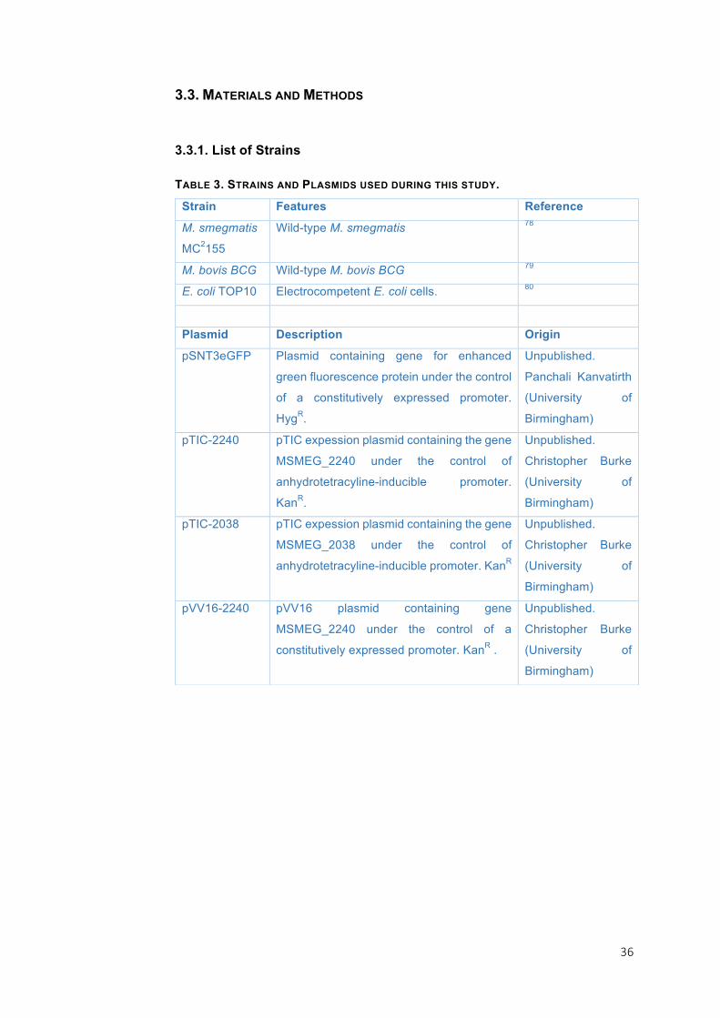
TABLE 3. STRAINS AND PLASMIDS USED DURING THIS STUDY.
Strain Features Reference
M. smegmatis
MC2155
Wild-type M. smegmatis 78
M. bovis BCG Wild-type M. bovis BCG 79
E. coli TOP10 Electrocompetent E. coli cells. 80
Plasmid Description Origin
pSNT3eGFP Plasmid containing gene for enhanced
green fluorescence protein under the control
of a constitutively expressed promoter.
HygR.
Unpublished.
Panchali Kanvatirth
(University of
Birmingham)
pTIC-2240 pTIC expession plasmid containing the gene
MSMEG_2240 under the control of
anhydrotetracyline-inducible promoter.
KanR.
Unpublished.
Christopher Burke
(University of
Birmingham)
pTIC-2038 pTIC expession plasmid containing the gene
MSMEG_2038 under the control of
anhydrotetracyline-inducible promoter. KanR
Unpublished.
Christopher Burke
(University of
Birmingham)
pVV16-2240 pVV16 plasmid containing gene
MSMEG_2240 under the control of a
constitutively expressed promoter. KanR .
Unpublished.
Christopher Burke
(University of
Birmingham)

37
3.3.3. Culturing M. smegmatis strains
M. smegmatis was grown in Middlebrook 7H9 broth + glycerol
0.5% with 0.05% tween 80 and selective antibiotic. Hygromycin was
used at 50 µg/ml and Kanamycin at 50 µg/ml to maintain plasmids.
Liquid cultures were incubated at 37 °C and shaken at 220 rpm. Cultures
were passaged every 5 days by adding 100 µl into 10 ml broth and were
passaged a maximum of 5 times. At this point, a new colony was
selected from an agar plate and used to begin a novel liquid culture.
3.3.4. Culturing M. bovis BCG strains
Liquid cultures were made from Middlebrook 7H11 broth + 0.5 %
glycerol with 0.05% Tween 80 and the correct selective antibiotic.
Kanamycin was added at a concentration of 25 µg/ml. Cultures were
incubated at 37 °C and 5% CO2, and passaged every 10 days by
inoculating 10 ml broth with 100 µl of the previous culture. After 5
passages, the next culture was started using a new colony from an agar plate.
3.3.5. M. smegmatis Liquid MIC99 Tests and M. bovis BCG Liquid MIC99 Tests
Liquid MIC99 Tests were carried out using 96-well flat-bottom
plates. Initially each well contained 100 µl of broth + 0.05% tween, apart
from column 2 which contained 200 µl plus the highest concentration of
drug. This was then serially diluted 9 times across the plate. The range
of concentrations for Vanoxerine began at 400 µM, DCS began at 400
µg/ml, and EMK compounds began at 800 uM. A stock of each
compound was kept at 10 mM. Cells were taken from an exponentially
growing culture and diluted to an initial 0D600 of 0.1. 100 µl of the culture
was added to each well to give a final OD600 of 0.05. When needed, 50
ng/ml anhydrotetracycline was added to induce the expression of genes
cloned on to the pTIC expression vector. This was then wrapped in tin
38
foil to prevent evaporation and incubated at 37 °C for 24 h. The
fluorescence of GFP-expressing strains was measured immediately by
excitation at 485 nm and emission at 520 nm with an automated
microtitre-plate reader, whereas wild-type strains were incubated with 30
µl 0.02% resazurin in each well for 4 h (M. smegmatis) and 24 h (M.
bovis BCG) using a previously published protocol, then fluorescence
was measured by excitation at 530 nm and emission at 590 nm 81. The
output was given as raw data for each microtire well and as a % survival
when compared with positive and negative controls, accompanied by a
Z-prime value. The Z-prime value assesses the quality and
reproducibility of the assay, it is a standard statistical measure used in
high throughput screening. It relies upon the means (µ) and standard
deviations (s) of the positive (p) and negative (n) controls:
Z-prime= 1-(3(sp+sn))/(µp-µn)
The Z-prime value determines the scale of separation between
the positive and negative controls in relation to the standard deviations.
Assays were only considered of good enough quality if the Z-prime value
was above 0.5, meaning that there are 12 standard deviations between
the mean of the positive and the mean of the negative controls.
Graphpad Prism was used for analysis and to determine the MIC99,
IC50 (the concentration that gives 50% survival) and highest non-
inhibitory concentration (NIC) for each data set using results given as % survival.
3.3.6. M. smegmatis Solid MIC Tests and M. bovis BCG Solid MIC Tests
Cell growth was tested against a range of concentrations of
Vanoxerine, from 0 µM to 100 µM. M. smegmatis was grown on 7H9
agar + 0.5% glycerol and 7H11 agar + 0.5% glycerol + 10% OADC was
used for M. bovis BCG. Plasmid-selective antibiotic was included in the
media when required. 2 µl of cells at OD 0.1, 0.01, 0.001 and 0.0001
39
were spotted onto the plate in a clockwise manner and the plates were
incubated at 37 °C for 4 days (M. smegmatis) or 37 °C + 5 CO2 for 10
days (M. bovis BCG). The MIC was determined as the plate where there was no growth for the inoculum of lowest cell concentration.
3.3.7. M. bovis BCG Vanoxerine-resistant Mutant Generation
Plates were made using the same media used for solid MIC
testing and containing 0X, 1X, 2.5X and 5X, and 10X the MIC of
Vanoxerine. 108 cells were spread on each plate, which was then
incubated at 37 °C + 5% CO2 for a minimum of 2 weeks or until the plates
dried out.
3.3.8. Transformation of plasmids into M. smegmatis and M. smegmatis pSNT3eGFP
Electrocompetent cells were made by growing a 50 ml culture to
OD 0.5, then cells were serially washed with 10% glycerol (v/v) and
harvested, each time resuspending in a smaller volume of 10% glycerol.
100 µl of electrocompetent cells were added to a 2mm electroporation
cuvette with 6 µl of the plasmid DNA. This was left on ice for 30 minutes
then electroporated at 1800V, and returned to the ice for 15 min before
250 µl tryptic-soy broth (TSB) was added and the cells. They were then
incubated for 4 h at 37 °C before spreading on TSB-agar plate with the
appropriate antibiotics. Cells were also spread onto a TSB-only plate to
act as a control. Plates were kept at 37 °C and successful transformants
were grown in a liquid culture and then stored at -80 °C in 25% glycerol.
3.3.9. Second-line drug screen against the FDA library
Two screens were carried out using M. bovis BCG against the
FDA library. 96-well microtiter plates were used; each well containing 10
µl 400 µM of each compound to which 100 µl M. bovis BCG cell culture
at OD600 0.05 was added. An inhibitory concentration of isoniazid was

40
added to the negative control wells. Both screens were carried out
simultaneously, with the second screen also containing 6.25 µg/ml DCS
in the cell culture. The 96-well plates were then incubated at 37 °C for
48 h in air-tight containers, then 30 µl 0.02% resazurin was added to
each well and the plates were replaced for a further 24 h 81. Once the
plates had been read, the two data sets were compared for significant changes in cell survival.

41
3.4. RESULTS
3.4.1. FDA Compound Screen for DCS Adjuncts
Determination of the D-cycloserine MIC99 and IC50 in M. bovis BCG and M. smegmatis
MIC can be defined as the minimum inhibitory concentration that
completely retards bacterial growth. Typically it is determined visually,
however for this study 96-well microtitre plates were used for
experiments to make the data more comparable and as a result the
process was automated and the MIC determined by an algorithm to
make it accurate and reproducible 82. As a result, any liquid MIC
determined during this study will be defined as the MIC99 that gives 99%
growth inhibition. Before performing the FDA-compound screen it was
necessary to determine the MIC99 of DCS for M. bovis BCG and a sub-
inhibitory concentration to use as a standard, the IC50. The MIC99 was
determined as 12.66 µg/ml and the IC50 as 11.66 µg/ml in M. bovis BCG
(Figure 6). As there was little difference between the MIC99 and the
IC50, the concentration 6.25 µg/ml was chosen for the FDA screen as this was the last concentration tested that gave 100% survival.
42
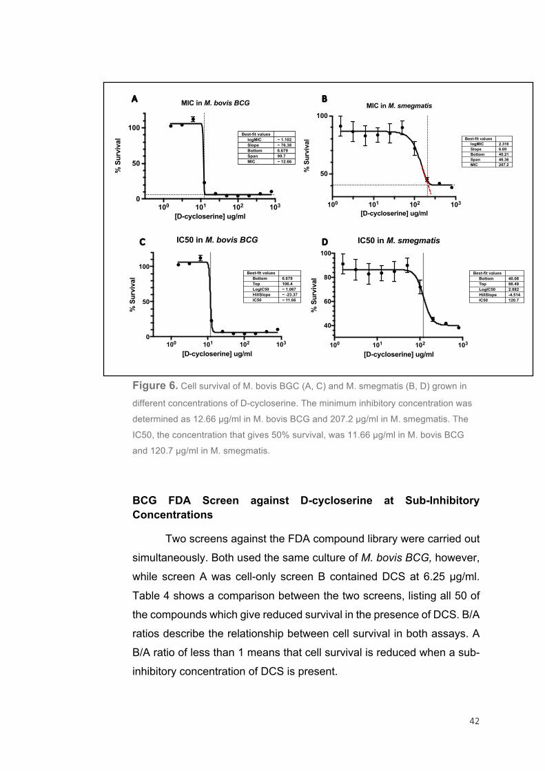
Figure 6. Cell survival of M. bovis BGC (A, C) and M. smegmatis (B, D) grown in
different concentrations of D-cycloserine. The minimum inhibitory concentration was
determined as 12.66 µg/ml in M. bovis BCG and 207.2 µg/ml in M. smegmatis. The
IC50, the concentration that gives 50% survival, was 11.66 µg/ml in M. bovis BCG
and 120.7 µg/ml in M. smegmatis.
BCG FDA Screen against D-cycloserine at Sub-Inhibitory Concentrations
Two screens against the FDA compound library were carried out
simultaneously. Both used the same culture of M. bovis BCG, however,
while screen A was cell-only screen B contained DCS at 6.25 µg/ml.
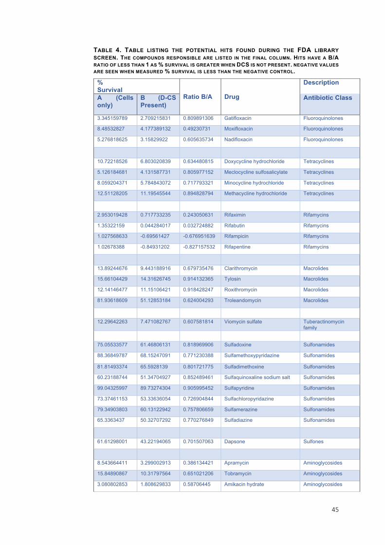
Table 4 shows a comparison between the two screens, listing all 50 of
the compounds which give reduced survival in the presence of DCS. B/A
ratios describe the relationship between cell survival in both assays. A
B/A ratio of less than 1 means that cell survival is reduced when a sub-
inhibitory concentration of DCS is present.
100 101 102 103
50
100
[D-cycloserine] ug/ml
% S
urvi
val Best-fit values
logMICSlopeBottomSpanMIC
2.3166.6940.2146.36207.2
MIC in M. smegmatis
100 101 102 103
40
60
80
100
[D-cycloserine] ug/ml
% S
urvi
val
IC50 in M. smegmatis
Best-fit valuesBottomTopLogIC50HillSlopeIC50
40.0886.492.082-4.514120.7
100 101 102 1030
50
100
[D-cycloserine] ug/ml
% S
urvi
val
Best-fit valueslogMICSlopeBottomSpanMIC
~ 1.102~ 76.386.67999.7~ 12.66
MIC in M. bovis BCG
100 101 102 1030
50
100
[D-cycloserine] ug/ml
% S
urvi
val
IC50 in M. bovis BCG
Best-fit valuesBottomTopLogIC50HillSlopeIC50
6.679106.4~ 1.067~ -23.37~ 11.66
A B
DC
43
A common theme from these HTS results is the presence of other
antibiotics. DCS lowers the minimum inhibitory concentration needed for
many Rifamycins, including the first-line TB drugs rifampicin and
rifabutin with B/A ratios of -0.68 and 0.03 respectively (Table 4)64. Of the
other first-line tuberculosis drugs, cell survival % for streptomycin
sulphate is lowered slightly when combined with DCS, shown by the B/A
ratio of 0.91. Isoniazid and pyrazinamide do not appear to be affected
as they gave B/A ratios of 1.02 and 1.00 respectively and cell survival
was close to 100 % for both screens. Other antimycobacterial drugs
appear to have synergy with DCS, such as dapsone which had a B/A
ratio of 0.70. Dapsone has been used to treat leprosy, caused by
Mycobacterium leprae, since 1945, and has also been approved for use against dermatitis herpetiformis by the FDA83,84.
A few antimicrobials also appear to have an increased killing
effect on M. bovis BCG when used in conjunction with DCS, for example,
there are two antifungal agents, Enilconazole and Clotrimazole listed as
hits (Table 4)85,86. They gave the B/A ratios of 0.95 and 0.94 respectively.
Pentamidine isethionate, with a B/A ratio of 0.90 is an antiparasitic drug
used to treat African trypanosomiasis and leishmaniasis87,88.
Interestingly, the b-lactamase inhibitor clavulanic acid had a B/A ratio of
0.80. Clavulanate is used as an adjuvant with b-lactam antibiotics to
resensitise penicillin-resistant bacteria89.
As well as antimicrobials, DCS appears to increase bactericidal
activity for a number of drugs licenced for other uses. The most dramatic
change can be found with Auranofin, a gold-containing compound
currently used as an antirheumatic, which had a B/A ratio of 0.17 (Table
4)73. Some drugs that appeared as hits in this screen affect the central
nervous system, such as Protriptyline hydrochloride which is used as a
tricyclic antidepressant, and zotepine, a second-generation
antipsychotic drug used in the treatment of schizophrenia90,91. It is
unlikely that either of these would be recommended for use with DCS as all three drugs affect the CNS69.

44
There are many other antibiotics in the FDA library that were not
affected by addition of DCS, and these were found across a range of
antibiotic classes. Such antibiotics include ceftazidime pentahydrate, a
third-generation cephalosporin which gave a B/A ratio of 1.02 and the
aminoglycoside sisomycin sulphate which had a B/A ratio of 0.98.
Additionally, imipenem, a carbapenem b-lactam antibiotic, had a B/A
ratio of 0.97, and dirithromycin, a macrolide antibiotic, gave a B/A ratio
of 1.1292–95. The B/A ratios vary slightly between these antibiotics but,
most importantly, all had a measured cell survival around 100 % (the
level of the positive control) for both screens. This means that they had no effect on cell survival in the presence or absence of DCS.
Some compounds appeared to interact antagonistically with
DCS. One such compound is the antibiotic vancomycin hydrochloride
which gave a B/A ratio of 1.4. Vancomycin is a glycopeptide antibiotic
often used as a last-resort antibiotic to treat serious drug-resistant gram-
positive infections96. A B/A ratio of 1.4 suggests that cell survival
increased when DCS was present in combination with vancomycin. The
data shows cell survival was 11.09 % with vancomycin and 15.63 % with
DCS and vancomycin together. Although these results show that cell
growth is inhibited significantly in both conditions, it also suggests that
the inhibitory properties of vancomycin are perturbed in the presence of
low levels of DCS, highlighting problems that may occur if combining the
drugs in a clinical setting. The most dramatic antagonistic effect is seen
with methiothepin maleate, an antipsychotic97. In the presence of sub-
inhibitory concentrations of DCS the cell survival went from 42.73% to
95.05%, giving a B/A ratio of 2.22 and completely negating any
antimycobacterial effects.
45
TABLE 4. TABLE LISTING THE POTENTIAL HITS FOUND DURING THE FDA LIBRARY SCREEN. THE COMPOUNDS RESPONSIBLE ARE LISTED IN THE FINAL COLUMN. HITS HAVE A B/A
RATIO OF LESS THAN 1 AS % SURVIVAL IS GREATER WHEN DCS IS NOT PRESENT. NEGATIVE VALUES ARE SEEN WHEN MEASURED % SURVIVAL IS LESS THAN THE NEGATIVE CONTROL.
% Survival
Ratio B/A Drug
Description
A (Cells only)
B (D-CS Present)
Antibiotic Class
3.345159789 2.709215831 0.809891306 Gatifloxacin Fluoroquinolones
8.48532827 4.177389132 0.49230731 Moxifloxacin Fluoroquinolones
5.276818625 3.15829922 0.605635734 Nadifloxacin Fluoroquinolones
10.72218526 6.803020839 0.634480815 Doxycycline hydrochloride Tetracyclines
5.126184681 4.131587731 0.805977152 Meclocycline sulfosalicylate Tetracyclines
8.059204371 5.784843072 0.717793321 Minocycline hydrochloride Tetracyclines
12.51128205 11.19545544 0.894828794 Methacycline hydrochloride Tetracyclines
2.953019428 0.717733235 0.243050631 Rifaximin Rifamycins
1.35322159 0.044284017 0.032724882 Rifabutin Rifamycins
1.027568633 -0.69561427 -0.676951639 Rifampicin Rifamycins
1.02678388 -0.84931202 -0.827157532 Rifapentine Rifamycins
13.89244676 9.443188916 0.679735476 Clarithromycin Macrolides
15.66104429 14.31626745 0.914132365 Tylosin Macrolides
12.14146477 11.15106421 0.918428247 Roxithromycin Macrolides
81.93618609 51.12853184 0.624004293 Troleandomycin Macrolides
12.29642263 7.471082767 0.607581814 Viomycin sulfate Tuberactinomycin family
75.05533577 61.46806131 0.818969906 Sulfadoxine Sulfonamides
88.36849787 68.15247091 0.771230388 Sulfamethoxypyridazine Sulfonamides
81.81493374 65.5928139 0.801721775 Sulfadimethoxine Sulfonamides
60.23188744 51.34704927 0.852489461 Sulfaquinoxaline sodium salt Sulfonamides
99.04325997 89.73274304 0.905995452 Sulfapyridine Sulfonamides
73.37461153 53.33636054 0.726904844 Sulfachloropyridazine Sulfonamides
79.34903803 60.13122942 0.757806659 Sulfamerazine Sulfonamides
65.3363437 50.32707292 0.770276849 Sulfadiazine Sulfonamides
61.61298001 43.22194065 0.701507063 Dapsone Sulfones
8.543664411 3.299002913 0.386134421 Apramycin Aminoglycosides
15.84890867 10.31797564 0.651021206 Tobramycin Aminoglycosides
3.080802853 1.808629833 0.58706445 Amikacin hydrate Aminoglycosides

46
9.105023948 8.306152429 0.91226036 Streptomycin sulfate Aminoglycosides
18.6606877 13.63130709 0.730482569 Gentamicine sulfate Aminoglycosides
8.278133373 6.317405769 0.763143753 Dihydrostreptomycin sulfate Aminoglycosides
44.14187837 37.30914166 0.845209652 Florfenicol Phenicols
32.35296157 28.95862445 0.895084191 Thiamphenicol Phenicols
4.857429464 3.100296499 0.638258676 Thiostrepton Thiopeptides
15.50424322 7.419426622 0.478541682 Linezolid Oxazolidinones
83.58896025 65.74426261 0.786518488 Sulfacetamide sodic hydrate
Not Currently Antibiotics
74.27600141 70.67780924 0.951556464 Enilconazole Antifungal agent85
66.72212454 64.22889044 0.962632573 Salmeterol
92.88983205 87.6520121 0.943612559 Protriptyline hydrochloride Tricyclic antidepressent (TCA)90
29.35858164 4.96622702 0.169157594 Auranofin antirheumatic agent73
58.6627251 46.90704673 0.799605655 Clavulanate potassium salt b-lactam drug that inhibits b-lactamase89
89.56264909 78.94338766 0.881432031 Topotecan
3.121447267 2.818781585 0.903036747 Pentamidine isethionate Antiparasitic agent88
69.34561864 59.14401216 0.852887512 Amiodarone hydrochloride
94.53179574 82.68924081 0.874724109 Cyclobenzaprine hydrochloride
0.079280865 0.063049364 0.795265848 Doxorubicin hydrochloride
93.18402104 82.76341653 0.888171766 Zotepine antipsychotic91
48.87354515 42.72754438 0.87424688 Perhexiline maleate
62.45088133 58.46523734 0.93617954 Clotrimazole Antifungal agent86

47
3.4.2. Mode-of-Action Studies of Vanoxerine
A similar HTS screen had been performed by Panchali Kanvatirth
(University of Birmingham) previously to find drugs within the FDA library
that have antimycobacterial activity against M. smegmatis and M. bovis
BCG. From this screen, the small-molecule compound vanoxerine
showed promising results. Christopher Burke (University of Birmingham)
raised two mutants in M. smegmatis against vanoxerine at 2.5X the MIC
by inoculating plates containing vanoxerine at 0, 1, 2.5, 5 and 10X the
MIC and incubating plates until growth appeared or the agar dried out,
the same method as used during this study. The mutants’ genomes were
isolated and sequenced by Panchali Kanvatirth (University of
Birmingham). Genes which contained non-synonymous mutations were
thought to be potential targets. Two genes, MSMEG_2038 and
MSMEG_2240 were found mutated in two separate vanoxerine-resistant
mutants. They encode a cytochrome p450 and a monoxygenase, both
have very similar functions and catalyse oxidation of their substrate98.
As a result, their wild-type counterparts were suggested to be the targets
of vanoxerine, and so by overexpressing them during cell survival
assays their role should be explained further. The second arm of this
project was to continue investigation into the mode of action of vanoxerine by overexpressing these genes in M. smegmatis.
GFP-Expressing Strains Were Chosen for the Cell Survival Assay
The plasmid pSNT3eGFP was transformed into M. smegmatis
and used for reading the cell survival in liquid cultures. This was
compared with growing wild-type M. smegmatis and then adding
resazurin, a compound that fluoresces when metabolised. The former was shown to give the most accurate results, as shown in Figure 7.
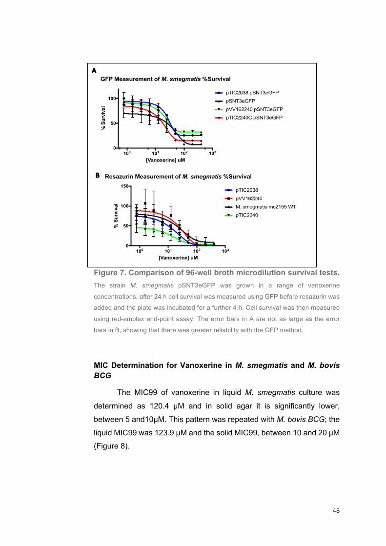
48
Figure 7. Comparison of 96-well broth microdilution survival tests.
The strain M. smegmatis pSNT3eGFP was grown in a range of vanoxerine
concentrations, after 24 h cell survival was measured using GFP before resazurin was
added and the plate was incubated for a further 4 h. Cell survival was then measured
using red-amplex end-point assay. The error bars in A are not as large as the error
bars in B, showing that there was greater reliability with the GFP method.
MIC Determination for Vanoxerine in M. smegmatis and M. bovis BCG
The MIC99 of vanoxerine in liquid M. smegmatis culture was
determined as 120.4 µM and in solid agar it is significantly lower,
between 5 and10µM. This pattern was repeated with M. bovis BCG; the
liquid MIC99 was 123.9 µM and the solid MIC99, between 10 and 20 µM (Figure 8).
100 101 102 1030
50
100
[Vanoxerine] uM
% S
urvi
val
GFP Measurement of M. smegmatis %Survival
pTIC2038 pSNT3eGFP
pSNT3eGFP
pVV162240 pSNT3eGFP
pTIC2240C pSNT3eGFP
100 101 102 1030
50
100
150
[Vanoxerine] uM
% S
urvi
val
Resazurin Measurement of M. smegmatis %Survival
pTIC2038
pVV162240
M. smegmatis mc2155 WT
pTIC2240
A
B
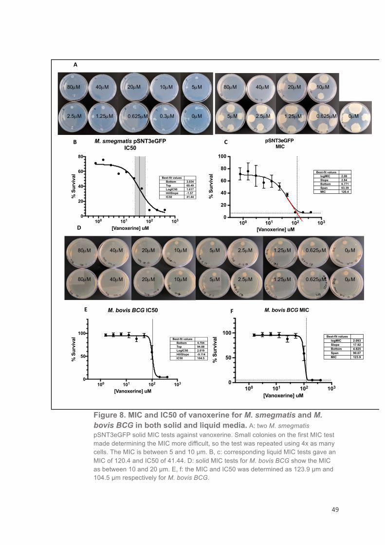
49
Figure 8. MIC and IC50 of vanoxerine for M. smegmatis and M. bovis BCG in both solid and liquid media. A: two M. smegmatis pSNT3eGFP solid MIC tests against vanoxerine. Small colonies on the first MIC test made determining the MIC more difficult, so the test was repeated using 4x as many cells. The MIC is between 5 and 10 µm. B, c: corresponding liquid MIC tests gave an MIC of 120.4 and IC50 of 41.44. D: solid MIC tests for M. bovis BCG show the MIC as between 10 and 20 µm. E, f: the MIC and IC50 was determined as 123.9 µm and 104.5 µm respectively for M. bovis BCG.
100 101 102 1030
20
40
60
80
[Vanoxerine] uM
% S
urvi
val
M. smegmatis pSNT3eGFPIC50
Best-fit valuesBottomTopLogIC50HillSlopeIC50
3.03469.491.617-1.5741.44
100 101 102 1030
20
40
60
80
100
[Vanoxerine] uM
% S
urvi
val Best-fit values
logMICSlopeBottomSpanMIC
2.082.846.77163.39120.4
pSNT3eGFP MIC
100 101 102 1030
50
100
[Vanoxerine] uM
% S
urvi
val Best-fit values
logMICSlopeBottomSpanMIC
2.09317.924.82590.07123.9
M. bovis BCG MIC
100 101 102 1030
50
100
[Vanoxerine] uM
% S
urvi
val
M. bovis BCG IC50
Best-fit valuesBottomTopLogIC50HillSlopeIC50
4.70494.882.019-9.114104.5
A
B C
E F
D
80µM 80µM 40µM 10µM 40µM 20µM 20µM 10µM 5µM
5µM 2.5µM 2.5µM 1.25µM 1.25µM 0.625µM 0.625µM 0.3µM 0µM 0µM
80µM
80µM
40µM
40µM
20µM
20µM
10µM
10µM
5µM
5µM
2.5µM
2.5µM
1.25µM
1.25µM
0.625µM
0.625µM
0µM
0µM
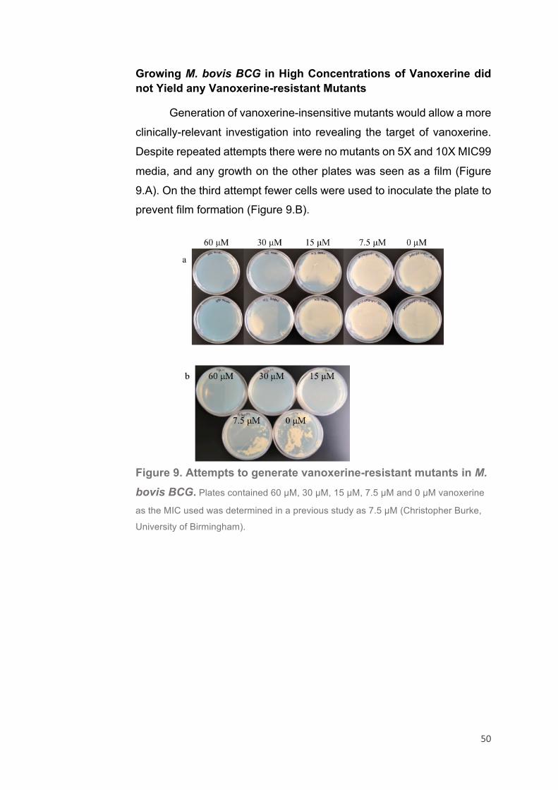
50
Growing M. bovis BCG in High Concentrations of Vanoxerine did not Yield any Vanoxerine-resistant Mutants
Generation of vanoxerine-insensitive mutants would allow a more
clinically-relevant investigation into revealing the target of vanoxerine.
Despite repeated attempts there were no mutants on 5X and 10X MIC99
media, and any growth on the other plates was seen as a film (Figure
9.A). On the third attempt fewer cells were used to inoculate the plate to prevent film formation (Figure 9.B).
Figure 9. Attempts to generate vanoxerine-resistant mutants in M. bovis BCG. Plates contained 60 µM, 30 µM, 15 µM, 7.5 µM and 0 µM vanoxerine
as the MIC used was determined in a previous study as 7.5 µM (Christopher Burke,
University of Birmingham).

51
Overexpression of CYP Genes Leads to an Increased Sensitivity to Vanoxerine in M. smegmatis
A number of plasmid samples prepared by Christopher Burke
(University of Birmingham) were tested at the commencement of this
study to ensure that the expected plasmids needed for this project were
present (Figure 10.A). Preps labelled pTIC-2240 C, pTIC-2038 C and D,
and pVV16-2240 C and D, were digested to remove the inserted gene,
either MSMEG_2240 or MSMEG_2038. The letters C and D are used to
indicate and identify plasmids from separate plasmid preps. DNA
fragments were separated on an agarose gel. The gene length of
MSMEG_2240 is 1209 bp and MSMEG_2038 is 1560 bp. Correct DNA
fragments confirmed the presence of the expected plasmids (Figure
10.A, lanes 2, 4, 5 and 6). As seen in Figure 10.A, there was no DNA in
the plasmid prep labelled pTIC2038 C (lane 3) so this was discarded.
Electrocompetent M. smegmatis and M. smegmatis pSNT3eGFP
were transformed with the verified expression plasmids pTIC2240 C,
pTIC2038 D and pVV16-2240 C and grown on selective agar plates.
Successful transformants were cultured and the MIC99 against
vanoxerine was determined for each. Liquid MIC99 tests revealed that
over-expression of MSMEG_240 and MSMEG_2038 had the opposite
effect to what was expected: the MIC99 decreased and the cells became more sensitive to vanoxerine (Figure 10. B, C, D, E).

52
Figure 10. Expression plasmids were verified by removing the gene fragment with
a double digest and separating the results on a gel (A). The plasmids were prepared
by Christopher Burke (University of Birmingham) using different methods, and the
letter at the end of each plasmid name corresponds to the method used. The plasmid
prep from lane 3 did not contain DNA. Correct plasmids were transformed into M.
smegmatis pSNT3eGFP and successful transformants were used to determine the
MIC for vanoxerine in a liquid MIC test. The MIC of M. smegmatis pSNT3eGFP was
120.4 µm (B) and the MIC of the expression strains containing pTIC6a-2240, pTIC6a-
2038 and pVV16-2240 was 48.33 µm, 52.44 µm and 52.3 µm respectively (C, D, E).
100 101 102 1030
20
40
60
80
100
[Vanoxerine] uM
% S
urvi
val Best-fit values
logMICSlopeBottomSpanMIC
2.082.846.77163.39120.4
M. smegmatis pSNT3eGFP MIC
100 101 102 1030
50
100
[Vanoxerine] uM
% S
urvi
val
Best-fit valueslogMICSlopeBottomSpanMIC
1.724.78425.4368.1252.44
M. smegmatis pSNT3eGFP pTIC2038MIC
100 101 102 1030
50
100
[Vanoxerine] uM%
Sur
viva
l Best-fit valueslogMICSlopeBottomSpanMIC
1.6843.18214.370.4548.33
M. smegmatis pSNT3eGFP pTIC2240MIC
100 101 102 10320
40
60
80
100
[Vanoxerine] uM
% S
urvi
val Best-fit values
logMICSlopeBottomSpanMIC
1.7183.5832.2359.0252.3
M. smegmatis pSNT3eGFP pVV162240 MIC
100 101 102 1030
20
40
60
80
100
[Vanoxerine] uM
% S
urvi
val Best-fit values
logMICSlopeBottomSpanMIC
2.082.846.77163.39120.4
M. smegmatis pSNT3eGFP MIC
100 101 102 1030
50
100
[Vanoxerine] uM
% S
urvi
val
Best-fit valueslogMICSlopeBottomSpanMIC
1.724.78425.4368.1252.44
M. smegmatis pSNT3eGFP pTIC2038MIC
100 101 102 1030
50
100
[Vanoxerine] uM
% S
urvi
val Best-fit values
logMICSlopeBottomSpanMIC
1.6843.18214.370.4548.33
M. smegmatis pSNT3eGFP pTIC2240MIC
100 101 102 10320
40
60
80
100
[Vanoxerine] uM
% S
urvi
val Best-fit values
logMICSlopeBottomSpanMIC
1.7183.5832.2359.0252.3
M. smegmatis pSNT3eGFP pVV162240 MIC
1234567
3Kb
1Kb
5Kb
A
B C
D E

53
Vanoxerine Analogues Exhibit Altered Activity on M. smegmatis
Due to the promising effects of vanoxerine on M. smegmatis and
M. bovis BCG, vanoxerine analogues were chemically synthesised
through a collaboration with another laboratory, and these were then
tested for their antimicrobial properties (Figure 11).
Figure 12.A shows that EMK007 and EMK006 have no
antimicrobial properties. Similarly, EMK017 and EMK019 did not inhibit
cell survival (Figure 12.B). The curves for vanoxerine (GBR12909) in
Figures A and B have different aspects because of background noise,
likely present because the turnover of GFP proteins produced by the
cells is too slow. The GFP protein is used in order to measure and quantify the population of cells using fluorescence.
EMK compounds that displayed activity against M. smegmatis
pSNT3eGFP can be found in Figure 12. C. In order to compare the
relative activities of these EMK compounds the results were normalised
separately for each compound using Graphpad Prism so that 0% was
defined as the smallest mean in each EMK compound data set, and
100% was the largest mean (Figure 12.C). Normalisation was carried
out because there was a wide range in values for % survival at non-
inhibitory and fully inhibitory concentrations between the EMK
compound assays, including those of the vanoxerine controls, due to
varying background levels of GFP. Therefore, the data sets have been
presented in a manner that makes them comparable. Figure 12.C shows
that EMK013, EMK014 and EMK016 only impacted growth under the
highest concentrations. Interestingly, EMK015 appears far more potent
at inhibiting cell growth than vanoxerine. Vanoxerine (GBR12909) was
chemically synthesised in the lab and labelled EMK014. However, this did not have the same MIC99 as the industrially-made chemical.
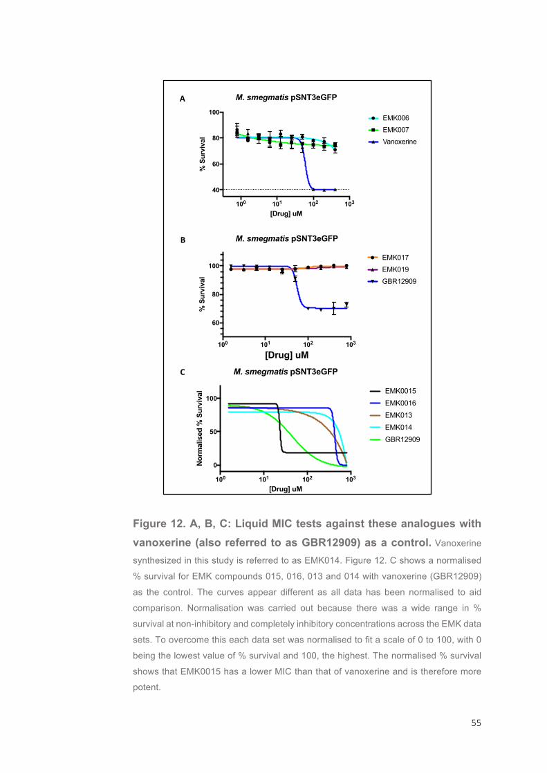
55
Figure 12. A, B, C: Liquid MIC tests against these analogues with
vanoxerine (also referred to as GBR12909) as a control. Vanoxerine
synthesized in this study is referred to as EMK014. Figure 12. C shows a normalised
% survival for EMK compounds 015, 016, 013 and 014 with vanoxerine (GBR12909)
as the control. The curves appear different as all data has been normalised to aid
comparison. Normalisation was carried out because there was a wide range in %
survival at non-inhibitory and completely inhibitory concentrations across the EMK data
sets. To overcome this each data set was normalised to fit a scale of 0 to 100, with 0
being the lowest value of % survival and 100, the highest. The normalised % survival
shows that EMK0015 has a lower MIC than that of vanoxerine and is therefore more
potent.
100 101 102 103
0
50
100
[Drug] uM
Nor
mal
ised
% S
urvi
val
M. smegmatis pSNT3eGFP
EMK0015
EMK0016
EMK013
EMK014
GBR12909
100 101 102 103
60
80
100
[Drug] uM
% S
urvi
val
M. smegmatis pSNT3eGFP
EMK017
EMK019
GBR12909
100 101 102 103
40
60
80
100
[Drug] uM
% S
urvi
val
M. smegmatis pSNT3eGFP
EMK006
EMK007
Vanoxerine
B
C
A

56
3.5. DISCUSSION
The FDA Compound Screen Revealed Drugs that Increase the Potency of DCS in Mycobacteria
Due to the efficacy of and lack of cross-resistance, DCS is an
effective second-line drug for MDR-TB treatment, yet 19-55% of patients
stop chemotherapy due to its highly toxic nature 69. Prescribing the
lowest dose of DCS that will cure tuberculosis is therefore a necessity,
and finding compounds that enable a lower dose to be administered
would both improve the patients’ quality of life and reduce treatment
discontinuation 99. The aim of this study was to find compounds that
reduce cell survival when a non-inhibitory concentration of DCS is also
present, thus showing synergy with DCS.
Current literature states the MIC of DCS as 15 µg/ml in M.
tuberculosis 100. In M. bovis BCG the MIC99 was determined as 12.66
µg/ml, but is nearly 18-fold higher in M. smegmatis emphasising the
greater clinical relevance of M. bovis BCG as a model. At 11.66 µg/ml
the IC50 was too similar to the MIC99 to use as a sub-inhibitory
concentration for testing with M. bovis BCG against the FDA library, instead 6.25 µg/ml DCS was used.
During this study 50 hits were identified; around 4% of the FDA
library. Most results were already known antibiotics, such as the
sulphonamides, moxifloxacin, gentamicine and thiamphenicol. The first-
line TB drugs rifampicin and rifabutin had promising synergistic effects
with DCS. Rifampicin is used in combination with ethambutol, isoniazid
and pyrazinamide for treatment against drug-susceptible tuberculosis64.
Rifabutin is used in the place of rifampicin for HIV/AIDS patients
undergoing first-line drug treatment, due to the complications that arise
when used in combination with anti-retroviral therapy (ART)64. Briefly,
rifampicins induce hepatic enzymes such as CYP3A which in turn lower
serum concentrations of protease inhibitors used in ART. Rifabutin has
a much lower effect on CYP3A induction than rifampicin and so is used
57
as a substitute for treating drug-susceptible tuberculosis in HIV/AIDS
patients64. However, clinical use of rifamycins in combination with DCS
would be unlikely, as DCS is used only in cases where TB is already
resistant to two first-line drugs. Other first-line drugs such as isoniazid
and pyrazinamide did not lower the cell survival from ~100 % when
combined with sub-inhibitory concentrations of DCS. Clavulanate
potassium salt also shows synergistic effects with DCS. Also known as
clavulanic acid, it is commonly used with penicillins as it is a beta-
lactamase inhibitor and can re-sensitise beta-lactam resistant bacteria 89. A synergistic effect with clavulanic acid has been shown in other drug-
repurposing studies with cephalosporins 75. There were a number of
antibiotics that had no effect on cell survival when in the presence or
absence of DCS. This is likely because M. bovis BCG is resistant to both the drugs and DCS at the concentrations used.
The most promising result is auranofin, an antirheumatic gold-
containing compound. With 40 µM auranofin present the survival of M.
bovis BCG was 21.35 % which was then reduced to 4.9 % in the
presence of 6.25 µg/ml of DCS. This study has demonstrated that
auranofin works synergistically with DCS as cell survival was reduced to
values close to known TB drugs such as rifampicin and rifabutin.
Auranofin is currently used as an antirheumatic drug, with a plasma half-
life of 15-25 days and a bioavailability of 40% 73,101. As mycobacteria
exist in the body in different metabolic states - actively growing cells
double only every 24 h and persistors remain dormant - the long half-life
is promising for TB therapy because the longer the drug remains active
in the body the more likely a successful outcome. 64.
There were a couple of cases where the cell survival was greater
in the screen containing DCS, suggesting that potential drug interactions
are antagonistic. This was most dramatically seen with methiothepin
maleate, where the inhibitory properties were perturbed completely in
the presence of DCS. Methiothepin maleate is an antipsychotic drug and
serotonin receptor antagonist97. It is hard to speculate why this occurs
without knowing the target of methiothepin maleate in the mycobacterial

58
cell. Vancomycin also produced a B/A ratio greater than 1, and this may
be easier to explain as both vancomycin and D-cycloserine target the
bacterial cell wall. D-cycloserine targets the D-Alanine-D-Alanine Ligase
Ddl in Mycobacterium tuberculosis, and Vancomycin targets terminal D-
Ala-D-Ala dipeptides in peptidoglycan precursors102,103. Therefore, with
sub-inhibitory concentrations of DCS present perhaps fewer D-Ala-D-
Ala dipeptides are synthesised and the antibacterial effects of
vancomycin are reduced.
The experiment was shown to be both reliable and repeatable as
compounds that have no effect on M. bovis BCG had values similar to
each other and to the positive control. The assay is of good quality as
the determined Z-prime values are extremely high. Continuation of this
project should focus on determining the mode-of-action of auranofin and
other hits that are not already known for being antimicrobials. Where
possible their chemical scaffold should be used to develop more potent analogues.
No Mutants were Raised against vanoxerine in M. bovis BCG
M. bovis BCG is a more clinically relevant model than M.
smegmatis. During this study, several attempts were made to raise
mutants against vanoxerine for mode-of-action studies. Despite
numerous attempts with different numbers of cells no mutants were
raised, and there was no growth at all on either the 5X and 10X MIC99
plate. Bacterial films formed on plates, which allowed growth on higher
concentrations of vanoxerine by reducing the concentration at the
interface. It is likely that the due to the low mutation frequency of
Mycobacteria, particularly in the slow-growing strains, mutants could not
be raised 104,105. In the future, more attempts should be made to raise
mutants against high concentrations of vanoxerine, then their genomes
should be sequenced and any genes containing non-synonymous mutations would highlight potential targets of vanoxerine.
59
Solid and Liquid MIC Tests Give Different Results
The M. smegmatis pSNT3eGFP MIC of vanoxerine, determined
by liquid and solid MIC tests, is 120.4 µM and 10µM respectively. Such
a large difference is unexpected but may be due to a preferred medium for cell growth (liquid over solid) giving faster growth and a higher MIC.
Vanoxerine is a Prodrug Metabolised and Activated by Cytochrome P450s
Vanoxerine is a high affinity inhibitor of dopamine uptake, it binds
strongly to dopamine transporters and dissociates slowly making it a
strong contender as a cocaine antagonist. In humans, it is metabolised
by cytochrome p450s (CYPs), a broad family of enzymes which catalyse
the oxidation of their substrate and are often involved in drug
biotransformation 106,107. The genes involved in this study,
MSMEG_2240 and MSMEG_2038 encode a CYP and a flavin-binding
mono-oxygenase respectively. Both enzyme families have very similar
activity profiles; they catalyse oxidative metabolism of their substrates.
As shown, overexpression of the genes MSMEG_2240 and
MSMEG_2038 result in a significant decrease in the MIC99 towards
vanoxerine. This suggests that instead of targeting these proteins,
vanoxerine is actually a prodrug which is metabolised to an active form
in the cell by CYPs, much like isoniazid 108. This study provides strong
evidence that, with respect to vanoxerine, either these two enzymes are
oxidising the molecule separately or are distinct steps in the same metabolic process.
These genes were highlighted as potential targets because they
were mutated in two vanoxerine-resistant strains. These results show
that mutations in these genes confer resistance not because they are
the target, but because they are the activators. The overexpression of

60
these genes in M. smegmatis is less clinically relevant than M. bovis
BCG but is useful for mode-of-action studies.
As vanoxerine is metabolised by human cytochrome p450s
including CYP3A4, clinical use with rifampicin may have unforeseen
effects as the front-line drug induces CYP3A4 activity 64. Therefore,
metabolism-based pharmacokinetic studies and drug-drug interactions
are important before the drugs are used concomitantly 106. For example,
inducing CYP3A4 with rifampicin may increase the presence of active
vanoxerine, but this could have negative effects on the patient and would complicate calculating the correct dosage.
Comparing Vanoxerine Analogues
As it is likely that vanoxerine is being oxidized, the chemical
scaffold was used to design and create a number of similarly-related
chemicals, as seen in Figure 11. A, in order to compare MICs and
determine where the oxidation reaction is occurring on the compound.
These mode-of actions studies were unfortunately impeded by the fact
that the EMK compounds were not soluble in 40% DMSO, and so the
negative results could be due to a lack of solubility and not because of
the altered structure. It was necessary to dissolve the compounds in
40% DMSO, not 100% DMSO, as a starting volume of 16 µl or 32 µl
10mM stock solution would mean that an inhibitory concentration of
DMSO is also used. In future, these vanoxerine analogues should be
remade with a salt present to aid solubility. Vanoxerine purchased from
Sigma Aldrich was a salt containing dihydrochloride, and dissolved
easily.
Despite problems with solubility, initial experiments revealed that
EMK015 is far more potent than vanoxerine at inhibiting the growth of M.
smegmatis. This result is promising, and once a soluble salt version is
available further testing is needed against M. bovis BCG to see if it has
potential as an antimycobacterial agent. The chemical scaffold of
61
vanoxerine should continue to be used to generate analogues, not only
to find the site of oxidation, but also to create new compounds that may have a higher inhibitory effect than vanoxerine, much like EMK015.
ACKNOWLEDGEMENTS
I would like to thank Dr. Luke Alderwick for his guidance during this
project, as well as Stephen Bethnell for all his help with M. smegmatis,
Chris Burke for creating the plasmids used in this study and everyone
who helped with this project. I would also like to thank Eleni for producing the vanoxerine analogues during our collaboration.
62
4. DISCUSSION: PERSPECTIVE FOR THE NEXT
THREE YEARS OF EACH PROJECT
Antimicrobials and Antimicrobial resistance is a rapidly emerging
global crisis. Resistance to almost every antibiotic developed has now
been reported and the spread of resistance leading to global outbreaks
of disease is well documented30,109–111. The efficacy of current antibiotics
is now in decline, yet the production of new compounds is also waning
due to a lack of discoveries, economic pressures and increased
regulatory measures making it harder to get novel antibiotics to the
market. A world-wide effort to implement new policies to control
resistance development, and restart research into discovery and development of new compounds is greatly needed13.
The studies undertaken in this thesis were designed to address
specific antimicrobial and antimicrobial resistance themes. The AMR
umbrella covers a broad range of research areas. The first project
focused on finding new drug targets by targeting the gram-negative cell
envelope, surface and secreted antigens, and proteins involved in host
interactions. Targeting AaaA was an unconventional approach to
eradicating bacterial pathogens, as inhibition of this would not be
bactericidal or bacteriostatic but would perturb virulence and make P.
aeruginosa more susceptible to antibiotic clearance38. The second
project tackled research into discovery of novel antitubercular agents,
also using an unconventional approach as work involved repurposing
drugs from an FDA library of small-molecule compounds licenced for alternate uses in the USA.
There were a number of limitations for the project “Investigating
the regulation of arginine-specific protease AaaA in P. aeruginosa and
creating a soluble truncated version for cell free inhibitor screens”. The
main limitation being the strict time constraint of 16 weeks for the entire
project. Such time constraints often limit what can be achieved during a
project, and cloning in particular takes a long time due to the need for
63
optimisation of each step in the process. The overall aim for the project
was to clone and express the histidine-tagged truncated version of
AaaA, which contained the N-terminal signal domain and the central
passenger/catalytic domain but did not include the C-terminal b-barrell
domain. Previous versions of the truncated AaaA protein created by
Daniella Spencer (University of Nottingham) were unsuccessful due to
large inclusion bodies forming in the cytoplasm, hence the need for an
N-terminal signal to direct the protein to the periplasm where it should
be folded and active but less toxic to the cell. The truncated gene was
successfully cloned into pGEM-T-Easy vector, the final step before using
restriction digest and ligation to move the DNA fragment into the
expression vector, pET21a. Should this project be continued for an
additional 3 years as a PhD project, there will be far more time available to complete the cloning and expression of the truncated AaaA protein.
If expression of taaaA3 is toxic to the cell, there are a number of
alternate methods that can be used to obtain a soluble truncated AaaA
protein. The polyhistidine tag could be moved to the N-terminus after the
signal peptide, as currently it is cloned into the C-terminus. Additionally,
the current truncated version could be expressed in a pseudomonas
vector so that if tAaaA3 is toxic to E. coli cells, then cloning could be
done in E. coli but ultimately the protein will be expressed and purified
in P. aeruginosa. Alternatively, purification of both the truncated version
and for the soluble and active form of the full AaaA version could be optimised.
Progression of P. aeruginosa infection to the chronic state in the
CF lung is met with a number of physiologic and metabolic changes in
order for the bacteria to adapt to its environment and survive112. Through
progression of the disease P. aeruginosa faces aerobic, microaerobic
and anaerobic conditions and must adapt its metabolism accordingly19.
Oxygen is preferentially used as a terminal electron acceptor for
substrate catabolism, but under anaerobic conditions P. aeruginosa can
use available nitrogen from denitrification or fermentation of arginine

64
instead, as an alternate electron acceptor113–115. Denitrification occurs
when nitrogen oxides are available in the environment, but in some
conditions the only available source of nitrogen is from the fermentation
of arginine to ornithine114. Even under these conditions P. aeruginosa
grows and forms biofilms. AaaA is an arginine-specific aminopeptidase
and cleaves amino terminal arginine from peptides providing an
alternate nitrogen source38. It is needed for establishment of chronic but
not acute infections, so expressing the truncated version of AaaA in
conditions mimicking that of a chronic infection should increase the
tolerance to tAaaA by the P. aeruginosa cell, and higher levels could be
purified. Following purification, structural analysis will be performed to
investigate inhibitor binding sites.
Understanding when AaaA is most useful for P. aeruginosa
should be one of the aims of the PhD project. A high-throughput screen,
such as a 96 well pin-lid assay, can be used to screen biofilm growth
under a number of conditions mimicking those found in the CF lung116.
These approaches could be done in parallel with protein purification. The
pin-lid assay can also be used to screen for novel inhibitors of the AaaA
protein. A wild-type, aaaA mutant and complemented strain can be
assessed for biofilm formation in the presence of novel inhibitory
molecules designed to target AaaA.
The other part of this project was investigating the transcriptional
regulation of aaaA. Two separate approaches and a very wide range of
conditions were tested during optimisation of site-directed mutagenesis
but unfortunately neither yielded the correctly mutated IHF site in the
MiniCTX:aaaA’:lux plasmid. Due to the large size of the plasmid in
question (13Kb) SDM should be far more successful if the promoter
region was mutated in a smaller vector and then transferred back into
the MiniCTX::lux plasmid. This process has more steps but SDM should
be successful in a smaller plasmid. To back up SDM, gel mobility shift
assays could be carried out in parallel. These have been used to show
IHF binding to the phtD operon in P. syringae 47. If successful, SDM
should be carried out in other regulatory binding sites in the promoter of

65
aaaA, including NarL, ArgR and RpoN. NarL is part of the two-
component system NarX-NarL, which is responsible for sensing and
responding to concentrations of nitrate and nitrite117,118. It is unsurprising,
therefore that its binding site can be found in the aaaA promoter. RpoN
negatively regulates the expression from the aaaA promoter and again
is involved in transcriptional control of a number of genes for nitrogen
utilization46. Interestingly, most isolates found in the CF lung infections
have rpoN mutations not found in strains collected from other habitats119.
ArgR regulates arginine, and has been shown to positively regulate transcription from PaaaA46.
Once all four binding sites have been mutated, the
bioluminescent reporters can be used to determine aaaA transcription
compared with the wild-type. Bioluminescence can be quantified so that
levels of expression can be analysed. In addition, transcription of the
mutated reporters can be compared in different strain backgrounds,
including and ArgR and RpoN mutant. Mutated transcriptional reporters
can also be tested for expression and biofilm defects using the 96 pin-
lid assay in different growth conditions. These experiments should help
identify regulators that directly interact with the promoter of aaaA and the conditions in which they are needed.
The second project undertaken involved assessing FDA-
approved drugs for their ability to act as antitubercular agents.
Additionally, the mode of action of the drug vanoxerine was investigated
in M. smegmatis. Vanoxerine has been trialled for depression,
parkinsonism, and cocaine dependency and showed potential as an
antimycobacterial agent in a previous screen of the FDA library77.
Collaboration with another laboratory allowed the testing of newly
synthesised compounds based on the chemical-scaffold of vanoxerine
(EMK compounds) in order to find a version with a higher activity. Again,
time constraints meant that these compounds could not be fully
investigated.

66
If this were to be carried on as a PhD project, then the EMK
compounds should all be retested for their efficacy once soluble salt
versions are available. This should be done using a similar method as
before, with 96-well microtiter plates for MIC99 broth dilution tests.
Comparison of IC50 and MIC99 with those determined for vanoxerine
should point to more potent vanoxerine-based compounds. Repurposing
chemical scaffolds has been used before to develop new antitubercular
drugs. Analogues of Lobenzarit, an anti-arthritic drug, were generated in
a targeted approach to drug discovery and tested for their ability to inhibit
the M. tuberculosis enzyme anthranilate phosphoribosyl-transferase and
this was met with success76. The changes in MIC99 with each change
of the chemical scaffold can also be used to determine which areas of
the molecule are essential for oxidisation by CYP proteins in mycobacteria.
The target of vanoxerine and mode of action also needs to be
identified in M. smegmatis and M. bovis BCG. I suggest the way forward
is through computational methods. There are two in silico approaches
for identification of drug-target interaction: ligand-based and target-
based virtual screening. As we know the chemical structure of
vanoxerine but not the target, a receptor-based approach should be
applied. Target –based screening is also known as reverse ligand-
protein docking and involves one ligand being screened for its ability to
bind to an array of clinically relevant macromolecular targets120,121. The
binding ability is then scored and ranked with the highest the most likely
to bind. However, this method would be limited to the 3D-structures
stored in databases such as protein drug target database (PDTD) and
Target Fishing Dock (TarFisDock) and would not be consigned solely to
M. tuberculosis proteins120,121. Another approach is through genomic
screens of resistant mutants. Mutants to vanoxerine should be raised in
high concentrations of up to 10 X the MIC99 in both M. smegmatis and
M. bovis BCG. Their genomes should be sequenced and genes with
non-synonymous mutations should be identified in each mutant. Genes
which are mutated across all strains indicate a potential vanoxerine

67
target and should be investigated further. This could be done through
overexpression in wild-type strains, the method employed during this
study. Alternatively, isothermal titration calorimetry (ITC) could be used
to measure biochemical interactions between vanoxerine and a number
of proteins in a crude mixture122. ITC can be used with gel-filtration
chromatography to “fish” for target proteins within a complex sample of
macromolecules122. By separating out proteins with gel-filtration
chromatography, each fraction can then be tested for ligand-binding
using ITC. Those that bind can be identified using N-terminal amino acid
sequencing. Once the target of vanoxerine and its derivatives has been
identified, structural analysis can be performed along with in silico
ligand-binding screens for additional inhibitor scaffolds.
CONCLUDING REMARKS
Drug discovery and development is becoming increasingly harder
due to scientific bottlenecks, increased regulation from approval bodies
and economic circumstance. Using unusual approaches to drug
discovery can fast-track the production of new antimicrobials, as is the
case with drug repurposing. There is a strong need for antimicrobial
agents that bacteria can’t develop resistance to and the best way to
address this is by developing drugs that do not create a strong selection
for resistance. Antivirulence drugs are ideal in this respect as they do
not target essential biological processes. It is naïve, however, to assume
that bacteria will never develop resistance to any of the antibiotics they
are presented with. Careful control over the use of antibiotics should be
employed to prevent the development and spread of resistance if we are to avoid the upcoming antibiotic apocalypse.
68
REFERENCE LIST
1. Michael, C. A., Dominey-Howes, D. & Labbate, M. The Antimicrobial
Resistance Crisis: Causes, Consequences, and Management. Front.
Public Heal. 2, 1–8 (2014).
2. Wright, G. D. Something old, something new: revisiting natural
products in antibiotic drug discovery. Can. J. Microbiol. 60, 147–154
(2014).
3. Normark, B. H. & Normark, S. Evolution and spread of antibiotic
resistance. J. Int. Med. 252, 91–106 (2002).
4. Yazdankhah, S. P. et al. Triclosan and Antimicrobial Resistance in
Bacteria: An Overview. Microb. Drug Resist. 12, 83–90 (2006).
5. Russell, A. D. Whither triclosan? J. Antimicrob. Chemother. 53, 693–
695 (2004).
6. Meyers, B. R., Hirschman, S. Z., Strougo, L. & Srulevitch, E.
Comparative study of piperacillin, ticarcillin, and carbenicillin
pharmacokinetics. Antimicrob. Agents Chemother. 17, 608–611 (1980).
7. Armstrong, G. L., Conn, L. A. & Pinner, R. W. Trends in Infectious
Disease Mortality in the United States During the 20th Century. JAMA
281, 61 (1999).
8. Sykes, R. Penicillin: From discovery to product. Bulletin of the World
Health Organization (2001). doi:10.1590/S0042-96862001000800016
9. Drews, J. Drug Discovery: A Historical Perspective. Science (80-. ).
287, 1960–1964 (2000).
10. Sengupta, S., Chattopadhyay, M. K. & Grossart, H. P. The multifaceted
roles of antibiotics and antibiotic resistance in nature. Front. Microbiol.
4, 1–13 (2013).
11. Thomashow, L. S. & Weller, D. M. Role of a phenazine antibiotic from
Pseudomonas fluorescens in biological control of Gaeumannomyces
graminis var. tritici. J. Bacteriol. 170, 3499–3508 (1988).
69
12. Davies, J. What are antibiotics? Archaic functions for modern activities.
Mol. Microbiol. 4, 1227–1232 (1990).
13. Ventola, C. L. The antibiotic resistance crisis. Pharm. Ther. 40, 277–83
(2015).
14. Golkar, Z., Bagasra, O. & Gene Pace, D. Bacteriophage therapy: A
potential solution for the antibiotic resistance crisis. J. Infect. Dev.
Ctries. 8, 129–136 (2014).
15. Fleming, A. Nobel Lecture: Penicillin. Nobel Lect. Physiol. or Med.
1942-1962 83–93 (1945).
16. Li, X. Z., Nikaido, H. & Poole, K. Role of MexA-MexB-OprM in antibiotic
efflux in Pseudomonas aeruginosa. Antimicrob. Agents Chemother. 39,
1948–1953 (1995).
17. Depardieu, F., Podglajen, I., Leclercq, R., Collatz, E. & Courvalin, P.
Modes and modulations of antibiotic resistance gene expression. Clin.
Microbiol. Rev. 20, 79–114 (2007).
18. Moradali, M. F., Ghods, S. & Rehm, B. H. A. Pseudomonas aeruginosa
Lifestyle: A Paradigm for Adaptation, Survival, and Persistence. Front.
Cell. Infect. Microbiol. 7, (2017).
19. Tramper-Stranders, G. A. et al. Initial Pseudomonas aeruginosa
infection in patients with cystic fibrosis: Characteristics of eradicated
and persistent isolates. Clin. Microbiol. Infect. 18, 567–574 (2012).
20. Tolker-Nielsen, T. Biofilm Development. Microbiol. Spectr. 3, (2015).
21. Wilton, M., Charron-Mazenod, L., Moore, R. & Lewenza, S.
Extracellular DNA acidifies biofilms and induces aminoglycoside
resistance in Pseudomonas aeruginosa. Antimicrob. Agents
Chemother. 60, 544–553 (2016).
22. McElroy, K. E. et al. Strain-specific parallel evolution drives short-term
diversification during Pseudomonas aeruginosa biofilm formation. Proc.
Natl. Acad. Sci. U. S. A. 111, E1419-27 (2014).
23. Editorials. The antibiotic alarm. Nature 495, 141 (2013).
70
24. Van Duong, D., Binns, C. W. & Van Le, T. Availability of antibiotics as
over-the-counter drugs in pharmacies: a threat to public health in
Vietnam. Trop. Med. Int. Heal. 2, 1133–1139 (1997).
25. Luyt, C.-E., Bréchot, N., Trouillet, J.-L. & Chastre, J. Antibiotic
stewardship in the intensive care unit. Crit. Care 18, 480 (2014).
26. Bartlett, J. G., Gilbert, D. N. & Spellberg, B. Seven ways to preserve
the Miracle of antibiotics. Clin. Infect. Dis. 56, 1445–1450 (2013).
27. Spellberg, B. & Gilbert, D. N. The future of antibiotics and resistance: A
tribute to a career of leadership by John Bartlett. Clin. Infect. Dis. 59,
S71–S75 (2014).
28. Gross, M. Antibiotics in crisis. Curr. Biol. 23, R1063–R1065 (2013).
29. O’Neill, J. Antimicrobial Resistance : Tackling a crisis for the health and
wealth of nations. (2014).
30. McDonald, L. C. et al. An Epidemic, Toxin Gene-Variant Strain of
Clostridium difficile. N. Engl. J. Med. 353, 2433–2441 (2005).
31. Mark A. Webber, Michelle M. C. Buckner, Liam S. Redgrave, Gyles Ifill,
Lesley A. Mitchenall, Carly Webb, Robyn Iddles, Anthony Maxwell, L.
J. V. P. Quinolone-resistant gyrase mutants demonstrate decreased
susceptibility to triclosan. J. Antimicrob. Chemother. 72, 2755–2763
(2017).
32. Rupnik, M., Wilcox, M. H. & Gerding, D. N. Clostridium difficile
infection: new developments in epidemiology and pathogenesis. Nat.
Rev. Microbiol. 7, 526–36 (2009).
33. Hung, D. T. Small-Molecule Inhibitor of Vibrio cholerae Virulence and
Intestinal Colonization. Science (80-. ). 310, 670–674 (2005).
34. Smith, M. A. et al. Identification of the binding site of Brucella VirB8
interaction inhibitors. Chem. Biol. 19, 1041–1048 (2012).
35. García-Contreras, R. et al. Resistance to the quorum-quenching
compounds brominated furanone C-30 and 5-fluorouracil in
Pseudomonas aeruginosa clinical isolates. Pathog. Dis. 68, 8–11
(2013).
71
36. Maeda, T. et al. Quorum quenching quandary: resistance to
antivirulence compounds. ISME J. 6, 493–501 (2012).
37. Allen, R. C., Popat, R., Diggle, S. P. & Brown, S. P. Targeting
virulence: can we make evolution-proof drugs? Nat. Rev. Microbiol. 12,
300–308 (2014).
38. Luckett, J. C. A. et al. A Novel Virulence Strategy for Pseudomonas
aeruginosa Mediated by an Autotransporter with Arginine-Specific
Aminopeptidase Activity. PLoS Pathog. 8, (2012).
39. Kim, W. et al. Spaceflight Promotes Biofilm Formation by
Pseudomonas aeruginosa. PLoS One 8, 1–8 (2013).
40. Ciofu, O., Mandsberg, L. F., Wang, H. & Høiby, N. Phenotypes
selected during chronic lung infection in cystic fibrosis patients:
Implications for the treatment of Pseudomonas aeruginosa biofilm
infections. FEMS Immunol. Med. Microbiol. 65, 215–225 (2012).
41. Pritt, B., O’Brien, L. & Winn, W. Mucoid Pseudomonas in cystic
fibrosis. Am. J. Clin. Pathol. 128, 32–34 (2007).
42. George, A. M., Jones, P. M. & Middleton, P. G. Cystic fibrosis
infections: Treatment strategies and prospects. FEMS Microbiol. Lett.
300, 153–164 (2009).
43. Gamper, M., Zimmermann, A. & Haas, D. Anaerobic regulation of
transcription initiation in the arcDABC operon of Pseudomonas
aeruginosa. J. Bacteriol. 173, 4742–4750 (1991).
44. Bernstein, H. D. Looks can be deceiving: Recent insights into the
mechanism of protein secretion by the autotransporter pathway. Mol.
Microbiol. 97, 205–215 (2015).
45. Dautin, N. & Bernstein, H. D. Protein secretion in gram-negative
bacteria via the autotransporter pathway. Annu. Rev. Microbiol. 61, 89–
112 (2007).
46. Paredes-Osses, E. A Novel Virulence Strategy for Pseudomonas
aeruginosa Mediated by an Autotransporter with Arginine-Specific
Aminopeptidase Activity. University of Nottingham (2014).
72
doi:10.1371/journal.ppat.1002854
47. Arvizu-Gómez, J. L., Hernández-Morales, A., Pastor-Palacios, G.,
Brieba, L. G. & Álvarez-Morales, A. Integration Host Factor (IHF) binds
to the promoter region of the phtD operon involved in phaseolotoxin
synthesis in P. syringae pv. phaseolicola NPS3121. BMC Microbiol. 11,
90 (2011).
48. Sieira, R., Comerci, D. J., Pietrasanta, L. I. & Ugalde, R. A. Integration
host factor is involved in transcriptional regulation of the Brucella
abortus virB operon. Mol. Microbiol. 54, 808–822 (2004).
49. Stonehouse, E., Kovacikova, G., Taylor, R. K. & Skorupski, K.
Integration host factor positively regulates virulence gene expression in
Vibrio cholerae. J. Bacteriol. 190, 4736–4748 (2008).
50. Freundlich, M., Ramani, N., Mathew, E., Sirko, A. & Tsui, P. The role of
integration host factor In gene expression in Escherichia coli. Mol.
Microbiol. 6, 2557–2563 (1992).
51. Hendrickson, E. L., Plotnikova, J., Mahajan-miklos, S., Rahme, L. G. &
Ausubel, F. M. Differential Roles of thee Pseudomonas aeruginosa
PA14 rpoN Gene in Pathogenicity in Plants, Nematodes, Insects, and
Mice. J. Bacteriol. 183, 7126–7134 (2001).
52. Thompson, L. S., Webb, J. S., Rice, S. A. & Kjelleberg, S. The
alternative sigma factor RpoN regulates the quorum sensing gene rhlI
in Pseudomonas aeruginosa. FEMS Microbiol. Lett. 220, 187–195
(2003).
53. Barrios, H., Valderrama, B. & Morett, E. Compilation and analysis of
sigma(54)-dependent promoter sequences. Nucleic Acids Res. 27,
4305–4313 (1999).
54. Studholme, D. J. & Buck, M. The biology of enhancer-dependent
transcriptional regulation in bacteria: Insights from genome sequences.
FEMS Microbiol. Lett. 186, 1–9 (2000).
55. Simon, R., Priefer, U. & Pühler, A. A Broad Host Range Mobilization
System for In Vivo Genetic Engineering: Transposon Mutagenesis in
Gram Negative Bacteria. Nat. Biotechnol. 1, 784–791 (1983).

73
56. Grant, S. G., Jessee, J., Bloom, F. R. & Hanahan, D. Differential
plasmid rescue from transgenic mouse DNAs into Escherichia coli
methylation-restriction mutants. Proc. Natl. Acad. Sci. 87, 4645–4649
(1990).
57. Sambrook, J., Fritsch, E. & Mantiatis, T. Molecular Cloning: A
Laboratory Manual. (Cold Spring Harbour, 1989).
doi:10.3724/SP.J.1141.2012.01075
58. Münch, R. et al. PRODORIC: Prokaryotic database of gene regulation.
Nucleic Acids Res. 31, 266–269 (2003).
59. Abdou, L., Chou, H. T., Haas, D. & Lu, C. D. Promoter recognition and
activation by the global response regulator CbrB in Pseudomonas
aeruginosa. J. Bacteriol. 193, 2784–2792 (2011).
60. Wozniak, D. J. & Carolina, N. Integration host factor and sequences
algD transcription start site are required for Integration Host Factor and
Sequences Downstream of the Pseudomonas aeruginosa algD
Transcription Start Site Are Required for Expression. J. Bacteriol. 176,
5068–5076 (1994).
61. Kaplan, R. S. High-level bacterial expression of mitochondrial transport
proteins. J. Bioenerg. Biomembr. 28, 41–7 (1996).
62. Ceri, H. et al. The Calgary Biofilm Device : New Technology for Rapid
Determination of Antibiotic Susceptibilities of Bacterial Biofilms The
Calgary Biofilm Device : New Technology for Rapid Determination of
Antibiotic Susceptibilities of Bacterial Biofilms. J. Clin. Microbiol. 37,
1771 (1999).
63. Zink, A. R. et al. Characterization of Mycobacterium tuberculosis
Complex DNAs from Egyptian Mummies by Spoligotyping. J. Clin.
Microbiol. 41, 359–367 (2003).
64. Zumla, A., Nahid, P. & Cole, S. T. Advances in the development of new
tuberculosis drugs and treatment regimens. Nat. Rev. Drug Discov. 12,
388–404 (2013).
65. Podany, A. T. & Swindells, S. Current strategies to treat tuberculosis.
F1000Research 5, 1–8 (2016).
74
66. Ouellet, H., Johnston, J. B. & Montellano, P. R. O. De. The
Mycobacterium tuberculosis Cytochrome P450 System. Arch.
Biochem. Biophys. 493, 82–95 (2011).
67. WHO. Global Tuberculosis Report 2016. (2016).
68. Hong, W., Chen, L. & Xie, J. Molecular basis underlying
Mycobacterium tuberculosis D-cycloserine resistance . Is there a role
for ubiquinone and meraquinone metabolic pathways? Expert Opin.
Ther. Targets 18, 691–701 (2014).
69. Saraf, G. et al. Cycloserine induced delirium during treatment of multi-
drug resistant tuberculosis ( MDR-TB ). Egypt. J. Chest Dis. Tuberc.
64, 449–451 (2015).
70. Carroll, M. W. et al. Frequency of adverse reactions to first- and
second-line anti-tuberculosis chemotherapy in a Korean cohort. Int. J.
Tubercular Lung Dis. 167, 961–966 (2012).
71. Woodcock, J. & Woosley, R. The FDA Critical Path Initiative and Its
Influence on New Drug. Annu. Rev. Med. 59, 1–12 (2008).
72. Pessetto, Z., Weir, S., Sethi, G., Broward, M. & Godwin, A. Drug
Repurposing for Gastrointestinal Stromal Tumor. Mol. Cancer Ther. 12,
1299–1309 (2013).
73. Roder, C. & Thomson, M. J. Auranofin : Repurposing an Old Drug for a
Golden New Age. Drugs R. D. 15, 13–20 (2015).
74. Maitra, A. et al. Repurposing drugs for treatment of tuberculosis: a role
for non-steroidal anti-inflammatory drugs. Br. Med. Bull. 118, 145–155
(2016).
75. Ramón-García, S. et al. Repurposing clinically approved
cephalosporins for tuberculosis therapy. Sci. Rep. 6, 34293 (2016).
76. Evans, G. L. et al. Repurposing the chemical scaffold of the anti-
arthritic drug Lobenzarit to target tryptophan biosynthesis in
Mycobacterium tuberculosis. ChemBioChem 15, 852–864 (2014).
77. Obejero-paz, C. A. et al. Quantitative Profiling of the Effects of
Vanoxerine on Human Cardiac Ion Channels and its Application to
75
Cardiac Risk. Sci. Rep. 5, 1–12 (2015).
78. Etienne, G. et al. The cell envelope structure and properties of
Mycobacterium smegmatis mc2155: Is there a clue for the unique
transformability of the strain? Microbiology 151, 2075–2086 (2005).
79. Philipp, W. J. et al. Physical mapping of Mycobacterium bovis BCG
Pasteur reveals differences from the genome map of Mycobacterium
tuberculosis H37Rv and from M. bovis. Microbiology 3135–3145
(1996).
80. Stenmark, P. et al. A new member of the family of di-iron carboxylate
proteins. Coq7 (clk-1), a membrane-bound hydroxylase involved in
ubiquinone biosynthesis. J. Biol. Chem. 276, 33297–33300 (2001).
81. Taneja, N. K. & Tyagi, J. S. Resazurin reduction assays for screening
of anti-tubercular compounds against dormant and actively growing
Mycobacterium tuberculosis , Mycobacterium bovis BCG and
Mycobacterium smegmatis. Antimicrob. Chemother. 288–293 (2007).
doi:10.1093/jac/dkm207
82. Lambert, R. J. & Pearson, J. Susceptibility testing: accurate and
reproducible minimum inhibitory concentration (MIC) and non-inhibitory
concentration (NIC) values. J. Appl. Microbiol. 88, 784–790 (2000).
83. Pickert, A. & Raimer, S. An evaluation of dapsone gel 5% in the
treatment of acne vulgaris. Expert Opin. Pharmacother. 10, 1515–1521
(2009).
84. Bhat, R. M. & Prakash, C. Leprosy: An overview of pathophysiology.
Interdiscip. Perspect. Infect. Dis. 2012, (2012).
85. Karlstam, E. & Pringle, J. Enilconazole Treatment of Horses with
Superficial Aspergillus Spp. Rhinitis. J. Vet. Intern. Med. 22, 1239–
1242 (2008).
86. Sawyer, P., Brogden, R., Pinder, R., Speight, T. & Avery. Clotrimazole:
a review of its antifungal activity and therapeutic efficacy. Drugs 9,
424–447 (1975).
87. Sundar, S. (Banaras H. U. & Chakravarty, J. (Banaras H. U. An Update
76
on Pharmacotherapy for Leishmaniasis Shyam. Expert Opin.
Pharmacother. 16, 237–252 (2015).
88. Babokhov, P., Sanyaolu, A. O., Oyibo, W. A., Fagbenro-Beyioku, A. F.
& Iriemenam, N. C. A current analysis of chemotherapy strategies for
the treatment of human African trypanosomiasis. Pathog. Glob. Health
107, 242–252 (2013).
89. Hugonnet, J.-E. & Blanchard, J. Irreversible Inhibition of the
Mycobacterium tuberculosis beta-lactamase by Clavulanate.
Biochemistry 46, 11998–12004 (2007).
90. Biggs, J., Holland, W. & Sherman, W. Steady-state protriptyline levels
in an outpatient population. Am. J. Psychiatry 132, 960–962 (1975).
91. Subramanian, S. et al. Zotepine versus other atypical antipsychotics for
schizophrenia. Cochrane database Syst. Rev. (2010).
doi:10.1002/14651858.CD006628.pub3
92. Farina, A., Porrà, R., Cotichini, V. & Doldo, A. Stability of reconstituted
solutions of ceftazidime for injections: An HPLC and CE approach. J.
Pharm. Biomed. Anal. 20, 521–530 (1999).
93. Lode, H., Kemmerich, B. & Koeppe, P. Comparative clinical
pharmacology of gentamicin, sisomicin, and tobramycin. Antimicrob.
Agents Chemother. 8, 396–401 (1975).
94. Lyon, J. Imipenem/cilastin: the first carbapenem antibiotic. Drug Intell.
Clin. Pharmacol. 19, 895–899 (1985).
95. Wintermeyer, S., Abdel-Rahman, S. & Nahata, M. Dirithromycin: A
New Macrolide. Ann. Pharmacother. 30, 1141–1149 (1996).
96. Kahne, D., Leimkuhler, C., Lu, W. & Walsh, C. Glycopeptide and
lipoglycopeptide antibiotics. Chem. Rev. 105, 425–448 (2005).
97. National Center for Biotechnology Information. Methiothepin Maleate.
PubChem Compound Database doi:CID=5358812
98. Cashman, J. R. Some distinctions between flavin-containing and
cytochrome P450 monooxygenases. Biochem. Biophys. Res.
Commun. 338, 599–604 (2005).
77
99. Lange, C. et al. Management of patients with multidrug-
resistant/extensively drug-resistant tuberculosis in Europe: a TBNET
consensus statement. Eur. Respir. J. 44, 23–63 (2014).
100. Desjardins, C. A. et al. Genomic and functional analyses of
Mycobacterium tuberculosis strains implicate ald in D-cycloserine
resistance. Nat. Genet. 48, 544–551 (2016).
101. Kean, W. F., Hart, L., Buchanan, W. W., Et, K. & Auranofin, A. L.
Auranofin. Br. J. Rheumatol. 36, 560–572 (1997).
102. Prosser, G. A. & Carvalho, L. P. S. De. Reinterpreting the Mechanism
of Inhibition of Mycobacterium tuberculosis D-Alanine:D-Alanine Ligase
by D-Cycloserine. Biochemistry 52, 7145–7149 (2013).
103. Nikolaidis, I., Favini-Stabile, S. & Dessen, A. Resistance to antibiotics
targeted to the bacterial cell wall. Protein Sci. 23, 243–259 (2014).
104. Outhred, A. C. et al. Identifying Likely Transmission Pathways within a
10-Year Community Outbreak of Tuberculosis by High-Depth Whole
Genome Sequencing. PLoS One 11, 1–15 (2016).
105. Sandegren, L. et al. Genomic Stability over 9 Years of an Isoniazid
Resistant Mycobacterium tuberculosis Outbreak Strain in Sweden.
PLoS One 6, 1–12 (2011).
106. Cherstniakova, S. A. et al. METABOLISM OF VANOXERINE, 1-[2-
[BIS(4-FLUOROPHENYL)METHOXY]ETHYL]-4-(3- PHENYLPROPYL)
PIPERAZINE, BY HUMAN CYTOCHROME P450 ENZYMES. Drug
Metab. Dispos. 29, 1216–1220 (2001).
107. Ogu, C. C. & Maxa, J. L. Drug interactions due to cytochrome P450.
BUMC Proc. 13, 421–423 (2000).
108. Montellano, P. R. O. De. Cytochrome P450-activated prodrugs. Futur.
Med Chem. 5, 213–228 (2013).
109. Zhang, R., Eggleston, K., Rotimi, V. & Zeckhauser, R. J. Antibiotic
resistance as a global threat: evidence from China, Kuwait and the
United States. Glob. Heal. 2, 6 (2006).
110. Poulou, A. et al. Outbreak caused by an ertapenem-resistant, CTX-M-

78
15-producing Klebsiella pneumoniae sequence type 101 clone carrying
an OmpK36 porin variant. J. Clin. Microbiol. 51, 3176–3182 (2013).
111. Pépin, J. et al. Emergence of fluoroquinolones as the predominant risk
factor for Clostridium difficile-associated diarrhea: a cohort study during
an epidemic in Quebec. Clin. Infect. Dis. 41, 1254–1260 (2005).
112. Sousa, A. & Pereira, M. Pseudomonas aeruginosa Diversification
during Infection Development in Cystic Fibrosis Lungs—A Review.
Pathogens 3, 680–703 (2014).
113. Frimmersdorf, E., Horatzek, S., Pelnikevich, A., Wiehlmann, L. &
Schomburg, D. How Pseudomonas aeruginosa adapts to various
environments: A metabolomic approach. Environ. Microbiol. 12, 1734–
1747 (2010).
114. Hoboth, C. et al. Dynamics of Adaptive Microevolution of Hypermutable
Pseudomonas aeruginosa during Chronic Pulmonary Infection in
Patients with Cystic Fibrosis. J. Infect. Dis. 200, 118–130 (2009).
115. Hassett, D. J. et al. Pseudomonas aeruginosa hypoxic or anaerobic
biofilm infections within cystic fibrosis airways. Trends Microbiol. 17,
130–138 (2009).
116. Junker, L. M. & Clardy, J. High-throughput screens for small-molecule
inhibitors of Pseudomonas aeruginosa biofilm development.
Antimicrob. Agents Chemother. 51, 3582–3590 (2007).
117. Benkert, B. et al. Nitrate-responsive NarX-NarL represses arginine-
mediated induction of the Pseudomonas aeruginosa arginine
fermentation arcDABC operon. Microbiology 154, 3053–3060 (2008).
118. Schreiber, K. et al. The anaerobic regulatory network required for
Pseudomonas aeruginosa nitrate respiration. J. Bacteriol. 189, 4310–
4314 (2007).
119. Kresse, A. U., Dinesh, S. D., Larbig, K. & Römling, U. Impact of large
chromosomal inversions on the adaptation and evolution of
Pseudomonas aeruginosa chronically colonizing cystic fibrosis lungs.
Mol. Microbiol. 47, 145–158 (2003).
79
120. Kharkar, P. S., Warrier, S. & Gaud, R. S. Reverse docking: a powerful
tool for drug repositioning and drug rescue. Future Med. Chem. 6,
333–42 (2014).
121. Li, H. et al. TarFisDock: A web server for identifying drug targets with
docking approach. Nucleic Acids Res. 34, 219–224 (2006).
122. Zhou, X., Sun, Q. & Kini, R. M. A universal method for fishing target
proteins from mixtures of biomolecules using isothermal titration
calorimetry. Protein Sci. 17, 1798–1804 (2008).