small conductance calcium-activated potassium (sk
TRANSCRIPT

Wright State University Wright State University
CORE Scholar CORE Scholar
Browse all Theses and Dissertations Theses and Dissertations
2009
Small Conductance Calcium-activated Potassium (SK) Channels Small Conductance Calcium-activated Potassium (SK) Channels
in Mammalian Spinal Motoneurons in Mammalian Spinal Motoneurons
Zhihui Deng Wright State University
Follow this and additional works at: https://corescholar.libraries.wright.edu/etd_all
Part of the Biomedical Engineering and Bioengineering Commons
Repository Citation Repository Citation Deng, Zhihui, "Small Conductance Calcium-activated Potassium (SK) Channels in Mammalian Spinal Motoneurons" (2009). Browse all Theses and Dissertations. 917. https://corescholar.libraries.wright.edu/etd_all/917
This Dissertation is brought to you for free and open access by the Theses and Dissertations at CORE Scholar. It has been accepted for inclusion in Browse all Theses and Dissertations by an authorized administrator of CORE Scholar. For more information, please contact [email protected].

SMALL CONDUCTANCE CALCIUM-ACTIVATED
POTASSIUM (SK) CHANNELS IN MAMMALIAN SPINAL
MOTONEURONS
A dissertation submitted in partial fulfillment of the
requirements for the degree of
Doctor of Philosophy
By
ZHIHUI DENG
M.S., Tianjin Medical University, 1998
B.S., Shandong Medical University, 1993
2009
Wright State University

COPYRIGHT BY
ZHIHUI DENG
2009
All Rights Reserved

ii
Wright State University
SCHOOL OF GRADUATE STUDIES
March 9, 2009
I HEREBY RECOMMEND THAT THE DISSERTATION PREPARED UNDER MY
SUPERVISION BY Zhihui Deng ENTITLED Small conductance calcium-activated
potassium (SK) channels in mammalian spinal motoneurons BE ACCEPTED IN
PARTIAL FULFILLMENT OF THE REQUIREMENTS FOR THE DEGREE OF
Doctor of Philosophy.
Robert E.W. Fyffe, Ph.D.
Dissertation Director
Gerald M. Alter, Ph.D.
Director, BMS Ph.D. Program
Joseph F. Thomas, Jr., Ph.D.
Dean, School of Graduate Studies
Committee on Final Examination:
Robert E.W. Fyffe, Ph.D.
Kathrin Engisch, Ph.D.
Paula Bubulya, Ph.D.
Timothy C. Cope, Ph.D.
David R. Cool, Ph.D.
iii
ABSTRACT
Deng, Zhihui, Ph.D., Biomedical Sciences Ph.D. Program, Wright State University, 2009.
Small conductance calcium-activated potassium (SK) channels in mammalian spinal
motoneurons.
Three homologous small conductance calcium-activated potassium (SK)
channel subunits (SK1, SK2 & SK3) are expressed in distinct and overlapping patterns in
mammalian central nervous system. SK channels likely mediate the medium
afterhyperpolarization (mAHP), which plays an essential role in regulating neuron
repetitive firing frequency. In spinal motoneurons (MNs) the mAHP duration is shorter
on average in fast (F-type) MNs than that in slow (S-type) MNs. To better understand the
molecular basis for mAHP, we determined the expression and sub-cellular distribution of
SK channels in normal, axonally-injured, and developing spinal MNs in vivo using
immunohistochemistry and quantitative confocal imaging techniques.
SK2 and SK3 channels are clustered on the surface membrane of MN soma
and proximal dendrites. SK clusters are post-synaptically localized at synapses associated
with cholinergic C-terminals. In complementary pattern, SK2-immunoreactive (-IR) and
SK3-IR clusters are expressed in different subpopulations of rat and mouse spinal α-MNs;
on average, SK3-IR MNs are smaller than SK2-IR MNs. Comparison of SK3 expression
in rat soleus versus gastrocnemius MNs, together with intracellular electrophysiological
data suggests that SK3-IR MNs are S-type whereas SK2-IR MNs are F-type. Moreover a
subpopulation of motor axon terminals innervating slow muscle fibers expresses SK3
channels.

iv
In postnatal developing mouse MNs, differential expression of SK2 and SK3
channels becomes apparent around the same time that muscle fiber differentiation occurs
(around P9). The SK channel clustering develops in concert with the establishment and
maturation of pre-synaptic cholinergic C-terminals, corresponding to the maturation of
motor function.
Injury to the motor axon results in a decreased AHP duration in S-type MNs
but an increased AHP duration or no change in F-type MNs. Here, we characterized the
effects on SK3 channel clustering in rat spinal MNs following nerve crush. SK3 clusters
appear unaltered until the 3rd day after axotomy. By the 8th day post-injury, the average
sizes of SK3 clusters are much smaller than in the normal control MNs. In contrast, co-
localized Kv2.1 clusters start to fragment and become reduced in size within hours
following injury, suggesting differential regulation and dynamics of discrete channel
populations at these synapses.

v
TABLE OF CONTENTS
ABSTRACT ....................................................................................................................... iii
TABLE OF CONTENTS .................................................................................................... v
LIST OF FIGURES ............................................................................................................ x
LIST OF TABLES ........................................................................................................... xiii
AKNOWLEDGMENTS .................................................................................................. xiv
DEDICATION .................................................................................................................... xv
CHAPTER I Background and Significance ................................................................. 1
I SK channels .................................................................................................................. 1
i) What are SK channels? ............................................................................................ 1
ii) Cloned and native SK channels .............................................................................. 1
iii) Ca++
sensitivity of SK channels............................................................................. 3
iv) Calmodulin constitutively bound with SK ............................................................ 4
v) Modulation of SK channel Ca++
sensitivity ............................................................ 5
vi) Heteromeric co-assembly of different SK subunits in expression system ............ 6
vii) SK channels in mammalian neurons .................................................................... 7
II Spinal motoneurons and motor units ......................................................................... 10
i) An introduction to spinal motoneurons ................................................................. 10
ii) Control of motoneuron excitability ...................................................................... 10
iii) Motor unit types .................................................................................................. 17
III Afterhyperpolarization (AHP) in motoneuron ......................................................... 20
i) Medium AHP in motoneurons............................................................................... 21
ii) Modulation of motoneuron mAHP....................................................................... 23

vi
IV Postnatal development of motoneurons ................................................................... 27
i) Postnatal anatomical development of motoneurons .............................................. 27
ii) Postnatal maturation of synaptic inputs on motoneurons ..................................... 28
iii) Postnatal differentiation of motor units ............................................................... 29
iv) Postnatal development of intrinsic motoneuron membrane properties ............... 30
V Responses of motoneurons to axonal injury ............................................................. 32
i) Motoneuron survival after axotomy ...................................................................... 32
ii) Synapse stripping ................................................................................................. 33
iii) Morphological changes of motoneurons after axotomy ...................................... 34
iv) Electrophysiological properties of axotomized adult motoneurons .................... 35
CHAPTER II Hypothesis and Specific Aims ................................................................... 38
CHAPTER III Materials & methods................................................................................. 42
Tissue preparation ......................................................................................................... 42
Immunohistochemistry / immunofluorescence ............................................................. 43
Electron microscopy pre-embedding immunoassay ..................................................... 44
Western blotting ............................................................................................................ 45
Electrophysiological intracellular recording of MNs ................................................... 46
Fluorogold retrograde tracing ....................................................................................... 47
Peripheral nerve crush................................................................................................... 47
Analysis of en face ion channel clusters using ImagePro Plus 5.1 ............................... 48
Statistical analysis ......................................................................................................... 48
CHAPTER IV Aim I. The expression, distribution and membrane organization of SK
channels in spinal motoneurons ........................................................................................ 51

vii
Introduction ................................................................................................................... 51
Methods & materials ..................................................................................................... 54
Results ........................................................................................................................... 55
1. The specificity of SK antibodies ........................................................................... 55
2. Distribution of SK immunoreactivity in spinal cord............................................. 56
3. Differential expression of SK channels in rat and mouse motoneurons ............... 57
4. Expression of SK3 in presynaptic motor axon terminals ...................................... 59
Discussion ..................................................................................................................... 59
CHAPTER V Aim II. The expression of SK channels in distinct types of rat spinal
motoneurons ...................................................................................................................... 83
Introduction ................................................................................................................... 83
Methods & materials ..................................................................................................... 85
Results ........................................................................................................................... 87
1. SK3 expression in rat soleus versus gastrocnemius motoneurons ........................ 87
2. SK3 expression in intracellularly recorded motoneurons ..................................... 87
3. SK3 expression in rat fast versus slow motor axon terminals .............................. 88
Discussion ..................................................................................................................... 89
1. Soleus motoneurons versus gastrocnemius motoneurons ..................................... 89
2. Intracellular recording from fast motoneurons versus slow motoneurons............ 90
3. Motor axon terminals of slow motoneurons versus fast motoneurons ................. 92
4. Mechanism underlying the differential mAHP in motoneurons ........................... 92
CHAPTER VI Aim III The synaptic localization of SK channel clusters in spinal
motoneurons .................................................................................................................... 103

viii
Introduction ................................................................................................................. 103
Methods & materials ................................................................................................... 105
Results ......................................................................................................................... 106
1. Postsynaptic location of SK channel clusters ..................................................... 106
2. Specific localization of SK clusters at cholinergic synapses .............................. 106
3. Co-localization of SK channels and Kv2.1 channels .......................................... 108
Discussion ................................................................................................................... 109
CHAPTER VII Aim IV Postnatal maturation of SK channels in spinal motoneurons 130
Introduction ................................................................................................................. 130
Methods & materials ................................................................................................... 131
Results ......................................................................................................................... 132
1. Postnatal development of SK channels ............................................................... 132
2. Postnatal development of VAChT-IR C-type synapses in motoneurons............ 133
3. Postnatal development of Kv2.1 channels in motoneurons ................................ 134
Discussion ................................................................................................................... 134
CHAPTER VIII Aim V Modulation of SK channel expression in spinal motoneurons
following axonal injury ................................................................................................... 150
Introduction ................................................................................................................. 150
Methods & materials ................................................................................................... 152
Results ......................................................................................................................... 153
1. The modulation of SK3 channel expression in motoneurons after nerve crush . 153
2. The alteration of Kv2.1 channel expression in motoneurons after nerve crush .. 154
Discussion ................................................................................................................... 155

ix
1. Regulation of Kv2.1 clusters following axotomy ............................................... 155
2. Regulation of SK clusters following axotomy .................................................... 156
CHAPTER IX SUMMARY ........................................................................................... 164
BIBLIOGRAPHY ........................................................................................................... 167

x
LIST OF FIGURES
Figure 1-1 Structure of SK channel subunit .................................................................... 2
Figure 1-2 Multi-protein organization of SK channel in cell surface membrane ............... 6
Figure 1-3 Action potential elicited from neonatal rat hypoglossal MN [Lape & Nistri
2000] ................................................................................................................................. 20
Figure 3-1 Quantitative analysis of Kv2.1-IR clusters from confocal image. .................. 49
Figure 4-1 Western blotting of rat spinal cord membrane proteins. ................................. 63
Figure 4-2 Low magnification images of rat lumbar spinal cord and the SK3
immunofluorescence. ........................................................................................................ 65
Figure 4-3 SK3-IR motoneurons in rat lumbar spinal cord. ............................................. 67
Figure 4-4 SK3-IR motoneurons expressing CGRP and CHAT immunoreactivity. ........ 69
Figure 4-5 Dual immunofluorescence for SK3 and Map2 in rat lumbar spinal
motoneurons. ..................................................................................................................... 71
Figure 4-6 SK3 immunofluorescence in mouse and cat lumbar spinal motoneurons. ..... 73
Figure 4-7 Differential expression of SK2 and SK3 channels in different subpopulations
of mouse spinal motoneurons. .......................................................................................... 75
Figure 4-8 SK2-IR and SK3-IR spinal alpha-motoneurons in Hb9-eGFP mouse. ........... 77
Figure 4-9 Expression of SK3 in a subpopulation of intra-spinal motor axon terminals. 79
Figure 4-10 Expression of SK3 in a subpopulation of motor axon terminals at
neuromuscular junctions. .................................................................................................. 81
Figure 5-1 SK3 expression in motoneurons innervating slow versus fast muscles. ......... 97
Figure 5-2 Intracellular recording of rat spinal motoneurons. .......................................... 99
Figure 5-3 SK3 immunofluorescence at NMJs innervating slow muscle fibers. ........... 101

xi
Figure 6-1 Synaptic localization of SK3-IR clusters in rat spinal motoneurons. ........... 114
Figure 6-2 Postsynaptic SK3-IR clusters in rat spinal motoneurons are specifically
located at cholinergic synapses apposed to presynaptic C-terminals. ............................ 116
Figure 6-3 At ultra-structural level, SK3-IR is restricted to the postsynaptic membrane at
cholinergic synapses in spinal motoneurons. .................................................................. 118
Figure 6-4 Postsynaptic SK2-IR clusters are located at cholinergic synapses in mouse
spinal motoneurons. ........................................................................................................ 120
Figure 6-5 SK3-IR clusters are not associated with any glutamatergic, serotonergic or
inhibitory (glycinergic/ GABAergic) synapses in rat spinal motoneurons. .................... 122
Figure 6-6 Postsynaptic SK3-IR clusters are co-localized with Kv2.1 clusters in spinal
motoneurons. ................................................................................................................... 124
Figure 6-7 Postsynaptic SK2-IR clusters are co-localized with Kv2.1 channel clusters in
spinal motoneurons. ........................................................................................................ 126
Figure 6-8 Quantitative analysis of SK3-IR cluster size in rat spinal motoneurons. ...... 128
Figure 7-1 Expression of SK2 channels in postnatal developing mouse motoneurons. . 138
Figure 7-2 Expression of SK3 channels in postnatal developing mouse motoneurons. . 140
Figure 7-3 Postnatal development of presynaptic cholinergic C-terminals and
postsynaptic SK2 channels. ............................................................................................ 142
Figure 7-4 Postnatal development of presynaptic cholinergic C-terminals and
postsynaptic SK3 channels. ............................................................................................ 144
Figure 7-5 Postnatal development of Kv2.1 channels and SK2 channels in motoneurons.
......................................................................................................................................... 146

xii
Figure 7-6 Postnatal development of Kv2.1 channels and SK3 channels in motoneurons.
......................................................................................................................................... 148
Figure 8-1 SK3 channels in axonally injured motoneurons............................................ 160
Figure 8-2 Kv2.1 channels in axonally injured motoneurons. ........................................ 162

xiii
LIST OF TABLES
Table 1 Tissue, fixation, sectioning and antibodies for immunofluoresence in Aim I .... 62
Table 2 Tissue, fixation, sectioning and antibodies for immunofluoresence in Aim II... 95
Table 3 The membrane properties of 10 recorded MNs ................................................... 96
Table 4 Tissue, fixation, sectioning and antibodies for immunofluoresence in Aim III 113
Table 5 Tissue, fixation, sectioning and antibodies for immunofluoresence in Aim IV 137
Table 6 Regulation of SK3 cluster sizes in rat spinal motoneurons after axonal injury 158
Table 7 Regulation of Kv2.1 cluster sizes in rat spinal motoneurons after axonal injury
......................................................................................................................................... 159

xiv
AKNOWLEDGMENTS
I wish to thank my advisor Rober E.W. Fyffe, for his patience and guidance throughout
all these years. His stimulating suggestions and encouragement have helped me during
this research. I am deeply grateful for his academic, emotional and spiritual support.
I would like to express my warm and sincere thanks to the other members of my
dissertation committee: Dr. Tim Cope, Dr. Kathy Engisch, Dr. Paula Bubulya, and Dr.
David Cool for their valuable suggestions and help. I am grateful for their encouragement
and support on my research and studies.
I would like to give my sincere thanks to Katie Bullinger for conducting intracellular
recording, to Shannon Romer for immunostaining of recorded motoneurons, and to
Ricardo Zerda for his great help with EM experiments. I also would like to thank Dr.
Ping Gao and Dr. Halm for their assistance with Western blotting.
I truly appreciate the valuable suggestions, comments and help offered by Dr. Francisco
Alvarez and other members of Dr. Fyffe‟s laboratory with whom I have worked.
Finally, I would like to thank all my family and friends who have given me
encouragement and support which make this dissertation possible.

xv
DEDICATION
To my husband Shihong who encourages and supports me,
brings me love and happiness, and shares every moment of joy and
sorrow with me…
To our dear baby boy Joshua who brings us lots of Joy and
happiness…
To my parents who give me incredible inspiration and
unconditional love and support throughout all these years…
To my brother Yuwen and my parents-in-law, who have been
tremendous supportive during my difficult times and helped me go
through them…

- 1 -
CHAPTER I Background and Significance
I SK channels
i) What are SK channels?
Potassium (K+) channels are the most diverse ion channel family which is
composed of functional distinct subfamilies including voltage-gated K+ (Kv) channels,
calcium (Ca++
)-activated K+ (Kca) channels, and others. In response to different stimuli,
K+ channels mediate outward K
+ currents with different kinetics. Unlike Kv channels
which are driven solely by the changes of membrane potential, Kca channels are
activated by a rise of cytosolic Ca++
concentration.
According to their electrophysiological, pharmacological, and molecular
structural properties, Kca channels may be divided into three subfamilies: BK (large
conductance Kca), IK (intermediate conductance Kca) and SK (small conductance Kca)
channels. In contrast to the large BK single-channel conductance ~250 pS, cloned SK
channels exhibit unitary conductance ~10 pS [Vergara et al, 1998].
ii) Cloned and native SK channels
Three homologous SK subunits (SK1, SK2 & SK3) have been cloned. Their
sequences code proteins of 561 (hSK1), 580 (rSK2) and 553 (rSK3) amino acids
respectively. The trans-membrane segments of three isoforms are highly conserved, but
the sequences and length of N- and C- termini are divergent. Similar as the members of

- 2 -
Kv channels, SK channels express tetrameric structure of four pore forming subunits;
each subunit consist of six trans-membrane segments (S1~S6) with N- and C-termini
inside the cell. The pore forming domain is located between S5 and S6 [Kohler et al.,
1996]. The following cartoon demonstrates the structure of SK subunit.
Figure 1-1 Structure of SK channel subunit
At least three isoforms of SK2 subunit (SK2-std, SK2-short and SK2-long) were
identified in mouse brain [Strassmaier et al., 2005; Murthy et al., 2008]. When
transfected into COS cells, the homomeric SK2-std channels were distributed uniformly
on cell surface whereas the homomeric SK2-long channels clustered into punctae
[Strassmaier et al., 2005]. The elevated expression of SK2-short was suggested to be
related to aging and Alzheimer‟s disease [Murthy et al., 2008].
Direct phosphorylation of K+ channels is known as a post-translational
modification mechanism underlying the modulation of channel kinetics, K+ current
amplitude, or channel density in plasma membrane [Jonas & Kaczmarek 1996]. For
example, Kv2.1 channels, which are believed to mediate delayed rectifier in mammalian
central neurons, have up to 60 potential phosphorylation sites [Park et al., 2006]. Kv2.1
channels are constitutively highly phosphorylated and a graded dephosphorylation can be
triggered by neuron activity or injury. The Kv2.1 dephosphorylation results in an

- 3 -
alteration of channel expression and biophysical properties [Misonou et al., 2004, 2005;
Mohapatra & Trimmer 2006; Park et al., 2006]. (see section II for more details)
Similarly, multiple potential phosphorylation sites that may be targeted by protein
kinases were shown in primary sequences of cloned SK channels [Kohler et al., 1996].
The direct phosphorylation of several serine residues within C-terminus of SK2 channel
by AMP-dependent protein kinase (PKA) led to a reduction of the cell surface expression
of these channels in transfected COS cells [Ren et al., 2006]. Recent studies showed the
plasticity of native SK channels in hippocampal CA1 cells and amygdale pyramidal
neurons that long term potentiation (LTP) induced a protein kinase A (PKA) dependent
internalization of postsynaptic SK2 channels [Lin et al., 2008; Faber et al., 2008]. In
contrast to the internalization/ recycling of SK channels following phosphorylation,
Kv2.1 distribution in cell surface membrane is reorganized that Kv2.1 clusters disperse
when dephophorylated [Misonou et al., 2004, 2005; Mohapatra & Trimmer 2006].
So far, cloned and native SK channels are the only known target of bee venom
apamin, which makes it an important tool for the study of these channels. Three cloned
SK channels express different sensitivity to apamin: SK2 and SK3 channels show higher
affinity to apamin than SK1 channels [Kohler et al., 1996; Hirschberg et al., 1998].
iii) Ca++ sensitivity of SK channels
Calcium ions function as ubiquitous intracellular second messengers.
Transmembrane transporters, intracellular Ca++
store system, and many Ca++
binding
proteins in the cytoplasm maintain intracellular free Ca++
ion concentration at very low
level (10-100nM) in eukaryotic cells, which makes Ca++
suitable as an intracellular
messenger for rapid cellular responses. SK channels are highly Ca++
sensitive in a

- 4 -
voltage-independent manner. Their gating with the application of Ca++
is rapid, occurring
within a few milliseconds. All three cloned SK channels exhibit similar dose-response
relationship: Ca++
concentration for half-maximal activation (K0.5) is about 0.3-0.7 μM
[Kohler et al., 1996; Hirschberg et al., 1998; Xia et al., 1998].
The Ca++
sources activating SK channels in neurons include distinct subtypes
(including L-type, N-type, P/Q-type) of voltage-dependent Ca++
channels (VDCC),
intracellular Ca++
stores and Ca++
permeable ionotropic neurotransmitter receptors [Li &
Bennett 2007; Marrion & Tavalin 1998; Stocker 2004; Bowden et al., 2001]. SK channels
are activated by increased intracellular Ca++
during an action potential and their activity
contributes to medium duration afterhyperpolarization (mAHP), thus they play an
essential role in neuron firing properties [Zhang& Krnjevic 1987; Sah & Faber 2002;
Faber & Sah 2003].
iv) Calmodulin constitutively bound with SK
SK channel subunits are constitutively associated with calmodulin (CaM). SK
channel protein does not directly bind to Ca++
when cytosolic Ca++
concentration
increases; instead, calmodulin works as Ca++
sensor by binding to Ca++
[Xia et al., 1998;
Maylie et al., 2004; Stocker 2004]. SK channel activation by Ca++
binding to CaM results
in channel opening whereas channel deactivation is the reverse process by dissociation of
Ca++
from CaM [Bildl et al., 2004; Pedarzani et al., 2001; Xia et al., 1998].
CaM binds to SK subunit through CaM binding domain (CaMBD) and this
constitutive interaction between SK and CaM is very stable. Crystallographic studies
showed a dimeric complex: two CaMBDs are arranged in an antiparallel configuration,
and two CaMs are woven around CaMBDs symmetrically at each end. Thus, each CaM

- 5 -
molecule contacts both subunits of the CaMBD dimmer. Therefore the tetrameric SK
channels function as dimers-of-dimers. Upon Ca++
binding to CaM, a large
conformational rearrangement of CaM occurs, which induces the conformational changes
of associated transmembrane domains of the channel protein. As a result, the ionic
conductance pore selective for K+ is triggered to open leading to hyperpolarization of
membrane potential [Maylie et al., 2004; Schumacher et al., 2001].
The calcium independent interaction between SK subunit and CaM is also
suggested to be necessary for the cell surface expression of the channels. When this
constitutive interaction was interrupted by site mutations (SK2:64/67), SK2 channels fail
to target cell surface membrane in transfected COS cells [Lee et al., 2003; Maylie et al.,
2004].
v) Modulation of SK channel Ca++ sensitivity
In addition to CaM, the cytoplasmic domains of SK channel protein also interact
with protein kinase CK2 and protein phosphatase 2A (PP2A) forming a multi-protein
complex [Bildl et al., 2004; Allen 2007]. CaM is a substrate for CK2 [Marin 1999], and
CK2 was demonstrated to be able to efficiently phosphorylate CaM associated with SK
CaMBD, but SK CaMBD was not a substrate for CK2. The phosphorylation of CaM by
CK2 may reduce the Ca++
sensitivity of SK channels [Bildl et al., 2004; Allen 2007]. The
shift of Ca++
sensitivity was suggested to result mainly from the accelerated deactivation
kinetics of SK channels, which reflects the stability of interaction between Ca++
and CaM
[Bildl et al., 2004; Schumacher et al., 2001; Wissmann et al., 2002]. The following
cartoon demonstrates the microdomain multi-protein organization of SK channel in
cytoplasmic membrane (not in proportion).

- 6 -
Figure 1-2 Multi-protein organization of SK channel in cell surface membrane
Recent evidence showed this mechanism in native neurons. Neurotransmitters
including noradrenaline were found to increase neuronal excitability by reducing SK2
channel calcium sensitivity gating in dorsal root ganglion cells, and this effect is through
CK2-dependent phosphorylation of SK2-bound CAM [Maingret et al., 2008].
In addition, some drugs (eg. 1-EBIO, CyPPA & NS4591) may increase Ca++
sensitivity of SK channels, which brings a clue for the mechanism underlying the positive
regulation of native SK channels [Hougaard et al., 2007].
vi) Heteromeric co-assembly of different SK subunits in expression system
The interaction between co-expressed different SK subunits was studied and
shown to cause alteration in protein trafficking, cell surface membrane targeting, and
channel pharmacological properties. The expression of rSK1 gene alone in mammalian
cell lines does not form functional channels. When rSK1 and rSK2 were co-expressed,
the SK channel current magnitude was larger, and apamin sensitivity was reduced
compared with the cells expressing rSK2 only [Benton et al., 2003]. The rSK3 can form
functional heteromeric channels with rSK2 but not with hSK1 subunit, since hSK1 may
interrupt functional SK3 assemblies on cell surface [Monaghan et al., 2004].
- 7 -
These in vitro studies suggested the possibility of heteromeric assembly of SK
channels in vivo. However, in vivo studies showed controversial results that some
suggested SK channels exclusively form homomeric channels [Sailer et al., 2002]
whereas others suggested the co-assembly of different SK subunits in mouse brain
[Strassmaier et al., 2005].
vii) SK channels in mammalian neurons
As mentioned before, SK channels mediate medium AHP following action
potential and thus determine neuron firing properties (detailed discussion in next section).
In addition, SK channels were also shown involved in dendritic excitability, synaptic
integration and neurotransmission in mammalian neurons.
Persistent inward currents (PICs) amplify synaptic inputs and govern the dendritic
excitability in motoneurons. PICs are voltage-activated and facilitated by
neuromodulatory inputs of monoamines including serotonin and other neurotansmitters
such as glutamate and acetylcholine [Svirskis & Hounsgaard 1998]. Plateau potential, a
latent firing property of motoneurons, is PICs dependent. A persistent calcium current
(Ca++
PIC) and a persistent sodium current (Na+ PIC) in dendritic tree plays a major role
in generating PICs. Low voltage-activated L-type Ca++
channels contribute to Ca++
PIC,
but the channel mediating Na+ PIC has not been identified [Heckman et al., 2003; Li &
Bennett 2003; Schwindt & Crill 1980]. Recent evidences showed that in some central
neurons including spinal motoneurons, dendritic SK channels may be activated by
dendritic L-type Ca++
channels. The activation of these SK channels terminates Ca++
PIC
and thus participates in dendritic excitability. Therefore, these dendritic SK channels are
spatially and functionally different from the SK channels mediating mAHP which are

- 8 -
believed to locate around soma and be activated by N, P-type Ca++
channels [Li &
Bennett 2007; Bond et al, 2005; Cai et al., 2004].
Long-term potentiation (LTP) in different brain areas such as hippocampus and
lateral amygdala is believed to underlie learning and memory. LTP requires the activation
of ionotropic NMDA receptors. Recent studies showed that SK channels were co-
localized and tightly coupled to NMDA receptors in hippocampus CA1 neurons and
lateral amygdala pyramidal neurons. The activation of these SK channels by Ca++
entry
via NMDA receptors reduced the amplitude of Ca++
transient and thus forms a negative
feedback loop to depress synaptic potential [Ngo-Anh et al., 2005; Faber et al., 2005].
The plasticity of these SK channels by internalization from the postsynaptic membrane
was mediated by protein kinase A (PKA) and suggested to contribute to LTP [Lin et al.,
2008; Faber et al., 2008].
In addition, SK3 channels were found to be expressed in cholinergic motor axon
terminals at rat neuromuscular junctions (NMJs) [Roncarati et al., 2001] and
glutamatergic presynaptic terminals of cultured mouse hippocampal neurons [Obermair
et al., 2002]. These results suggest that SK3 may involve in the regulation of excitatory
synaptic neurotransmission.
SK channel knock-out transgenic mouse lines were constructed, and the animals
lacking any one of the SK genes (SK1, SK2, or SK3) were viable. Whole cell patch clamp
recording showed that only SK2 channels are necessary for the mAHP in hippocampal
CA1 neurons [Bond et al., 2004]. Unfortunately no available information shows the
properties of motoneurons in SK knock-out mice.

- 9 -
Three SK channels (SK1, SK2 & SK3) have been shown to be expressed in
distinct and overlapping patterns in mammalian central nervous system [Stocker et al.,
2000, Sailer et al., 2004]. However, the expression of SK channels in many neurons
including motoneurons is still unknown, though electrophysiological evidences suggested
the existence and the functions of these channels in motoneurons [Zhang et al., 1987;
Hounsgaard et al., 1988; Viana et al., 1993; Powers et al., 1999; Sawczuk et al., 1997;
Miles et al., 2005]. . This dissertation will address this issue aiming to illustrate the
molecular basis for the recorded AHP and dendritic SK currents and to shed light on the
mechanisms underlying motoneurons excitability and firing properties (see aim I).

- 10 -
II Spinal motoneurons and motor units
i) An introduction to spinal motoneurons
Spinal motoneurons, described as “final common pathway” by C. S. Sherrington,
transmit signals from central nervous system to their target skeletal muscles. There are
two major types of motoneuron in mammalian spinal cord: larger -MN and smaller γ-
MN. -MNs innervate extrafusal muscle fibers throughout the skeletal muscles whereas
γ-MNs innervate intrafusal muscle fibers within muscle spindles. A third type of
motoneuron, β-MN, innvervates both extrafusal and intrafusal muscle fibers. All
motoneurons discussed in this study are -MNs, which contribute to voluntary skeletal
muscle contraction and muscle tone.
Motoneuron pool is defined as the population of motoneurons innervating a
particular muscle. The topographic distribution of sciatic motoneuron pools have been
identified in cat and rat lumbar spinal cord. The sciatic motoneurons form a continuous
cell column in the dorsolateral quadrant of ventral horn, within which the motoneurons of
each sciatic branches occupy spatially distinct and overlapping sub-compartments [Burke
1981; Swett et al 1986].
ii) Control of motoneuron excitability
Motoneuron excitability and firing properties are essential in motor control.
Motoneuron input-output function describes the relationship between synaptic inputs (or
injected currents) onto motoneurons and resulting firing of motoneurons. The
transformation of synaptic inputs to the initiation of action potential depends on: i)
motoneuron type (S-type or F-type) and its membrane properties; ii) location of synaptic

- 11 -
contacts; iii) effects of neuromodulators on synaptic transmission and repetitive firing
properties [Rekling et al., 2000].
On the one hand, different types of synaptic inputs (excitatory versus inhibitory)
onto motoneurons regulate their excitability and firing behavior; on the other hand, the
intrinsic properties of motoneuron including ionic channels shape their response to
synaptic inputs.
A) Synaptic input onto motoneurons
In the past 30 years Robert Fyffe and colleagues have been using both light and
electron microscopy to study synapses in spinal motoneurons and inhibitory interneuron
Renshaw cells. They demonstrated the quantity and distribution of Ia afferent boutons
[Brown & Fyffe 1981; Fyffe & Light 1984] and inhibitory glycine/GABAergic terminals
from Renshaw cells and other inhibitory interneurons [Fyffe 1991; Alvarez et al., 1996,
1997] contacting motoneurons. They also quantitatively analyzed the descending
serotonergic synapses upon motoneurones [Alvarez et al., 1998].
Each mammalian -MN receives tens of thousand synaptic contacts of various
origins, and the majority of synaptic inputs are located on dendrites which constitutes ~97%
of total membrane surface area of the motoneuron [Brannstrom 1993; Cullheim et al.,
1987].
Synaptic inputs on motoneurons can be divided into two categories which
influence their excitability and firing properties in different ways. 1) The synapses acting
via ionotropic receptors, such as glutamate and glycine/GABA, cause depolarizing
(excitatory) or hyperpolarizing (inhibitory) postsynaptic potential. 2) The synapses

- 12 -
working through metabotropic receptors, such as amines and peptides, change
motoneuron intrinsic input/ output properties [Kernell 1999; Rekling et al 2000].
According to ultrastructural studies, there are distinct types of presynaptic
boutons (S, F, C and others) contacting motoneurons [Conradi et al., 1979; Kellerth et al.,
1979; Lagerback et al., 1986; Johnson, 1986]. S-type boutons, which contain spherical
synaptic vesicles, distribute more frequently on distal dendrites than on proximal
dendrites. Descending serotonergic boutons and most glutamatergic boutons including
large Ia primary afferent terminals are S-type [Conradi et al., 1983; Fyffe & Light 1984;
Ornung et al., 1998]. F-type boutons, which contain pleomorphic or flattened synaptic
vesicles, are the most numerous type of synapse on motoneuron somatic membrane and
are assumed to be inhibitory terminals such as glycinergic and GABAergic terminals
arising from inhibitory interneurons [Fyffe 1991; Ornung et al., 1996]. C-terminals, large
cholinergic boutons exhibiting postsynaptic sub-surface cisternae, arise from a group of
interneurons located close to the central canal of spinal cord [Maxwell et al., 2003].
Small S-type and F-type boutons contact both -MNs and γ-MNs whereas large C-type
and Ia primary afferent boutons exclusively target -MNs [Lagerback, 1985; Lagerback
et al., 1986; Johnson, 1986; Ichiyama et al., 2006].
B) Ionic channels in motoneuron
Like all other central neurons, motoneuron has its unique combination of ionic
channels which matches its specific physiological role. Motoneurons express diverse
ionic channels in their surface membrane including Na+, K
+ and Ca
++ channels.
(1) Na+ channels

- 13 -
Voltage-gated Na+ channels play an essential role in neuron excitability. Several
Na+ currents were found in motoneurons but the Na
+ channels underlying these currents
are not well defined yet. Fast inactivating TTX sensitive Na+ current is responsible for
action potentials initiation and propagation in all motoneurons [Rekling et al., 2000]. A
TTX-sensitive persistent Na+ current contributes to persistent inward current (PIC),
which produces a prolonged depolarization, amplifies excitatory synaptic inputs, and is
turned off by inhibitory inputs to motoneurons [Heckman et al., 2003; Li & Bennett
2003]. Recent studies suggested that the Na+ PIC plays an essential role in action
potential initiation, determining firing frequency and sustaining repetitive firing in
motoneurons [Lee & Heckman 2001; Kuo et al., 2006; Harvey et al., 2006; Li & Bennett
2007]. Several subtypes of voltage-gated Na+ channels including Nav1.5, Nav1.8 and
Nav1.9 are activated at more hyperpolarized potential (lower than -60mV) and show
slower inactivation kinetics than fast inactivating Na+ channels [Diss et al., 2004].
However, the Na+ channel subtypes mediating PIC remain unclear [Kiss 2008]. In
addition, there exists a TTX insensitive Na+ current in motoneurons [Rioult-Pedotti 1997].
Since motoneurons are not ideal for voltage-clamp techniques, the biophysical properties
of Na+ channels in motoneurons are difficult to study. One type of TTX-sensitive Na
+
channel, with a conductance of 14.0 pS, was found in the soma of neonatal rat spinal
motoneurones [Safronov & Vogel 1995].
(2) K+ channels
There are many K+ currents contributing to the integrated function of
motoneurons. K+ currents are responsible for shaping action potential and determining
the sub-threshold membrane behavior and firing properties of motoneurons. Moreover,

- 14 -
neuromodulators target these K+ currents and regulate motoneuron excitability. Several
K+ currents in motoneurons such as delayed rectifier and Ca
++ dependent K
+ currents
have been studied extensively.
Delayed rectifier (Ik), which is activated by depolarization, contributes to the
falling phase of action potential and fast AHP. Kv2.1 channels (Shab) are abundant in
spinal motoneurons and might be the major contributor to the delayed rectifier in
motoneurons. Kv2.1 was shown to form small irregularly shaped and large disc-like
clusters in the surface membrane of soma and proximal dendrites in -MNs. Prominent
disc-like Kv2.1 clusters were invariably apposed to presynaptic cholinergic C-terminals
and co-localized with postsynaptic muscarinic (m2) receptors [Muennich & Fyffe 2004].
Recent studies illustrated the dynamic regulation of native and cloned Kv2.1
channels in hippocampal neurons and HEK cells induced by neuron acitivity, glutamate
or muscarine stimulation, ischemia and CO2 application. Kv2.1 channels are
constitutively maintained highly phosphorylated. All the above events cause a graded
dephosphorylation of Kv2.1 and lead to a dispersing of Kv2.1 clusters in cell surface
membrane and a hyperpolarizing shift of channel activation. This effect was calcineurin
(PP2B) dependent and resulted from a calcium influx or release from intracellular
calcium store [Misonou et al., 2004, 2005; Mohapatra & Trimmer 2006; Park et al.,
2006]. Similar Kv2.1 de-clustering effect was observed in spinal motoneuorns induced by
sciatic stimulation, glutamate or muscarine application [unpublished results from Fyffe
lab]. If the motoneuron injury may induce similar modulation of Kv2.1 clusters will be
examined in aim V.

- 15 -
Ca++
-activated K+ channels are gated by increased intracellular Ca
++, which play
an essential role in determining firing properties of motoneurons and serve to terminate
long bursts of action potentials. Two types Ca++
-activated K+ channels, large conductance
BK channels and small conductance SK channels are expressed in motoneurons. SK
channels were shown to mediate medium AHP in motoneurons [Zhang et al., 1987;
Hounsgaard et al., 1988; Viana et al., 1993; Powers et al., 1999; Sawczuk et al., 1997;
Miles et al., 2005]. As discussed before, electrophysiological evidences suggested that
there exist dendritic SK channels in motoneurons which were spatially and functionally
different from the SK channels mediating mAHP [Li & Bennett 2007; Bond et al, 2005;
Cai et al., 2004]. The expression and sub-cellular distribution of SK channels will be
studied in this dissertation. Moreover, how motoneuron injury may cause the regulation
of SK expression in cell surface membrane will also be examined in aim V.
(3) Ca++
channels
Voltage-dependent Ca++
conductances in motoneurons contribute to
afterdepolarization (ADP) and afterhyperpolarization (AHP) and thus play a role in
determining the duration of action potential. In addition, voltage-dependent persistent
inward calcium currents (Ca++
PIC), which are facilitated by monoamines, play a major
role in enhancing excitatory synaptic inputs and sustaining repetitive firing [Heckman et
al., 2003]. Ca++
PIC may also be promoted by other neurotransmitters such as glutamate
and acetylcholine [Svirskis & Hounsgaard 1998].
Voltage-dependent Ca++
channels (VDCCs) may be classified into low voltage-
activated (LVA) and high voltage-activated (HVA) channels; HVA channels may be

- 16 -
subdivided into L-type, P/Q-type, N-type and others by pharmacological study [Lacinova
2005].
Cav1.3 (or α1D), a subtype of L-type Ca++
channels, is activated by relatively
hyperpolarized membrane potential compared with other HVA VDCCs and has been
shown to contribute to Ca++
PIC in spinal motoneurons [Carlin et al., 2000; Alaburda et
al., 2002; Simon et al., 2003; Elbasiouny et al., 2006]. Electrophysiological studies
demonstrated a dendritic origin of Ca++
PIC [Bennett et al., 1998; Carlin et al., 2000;
Hultborn et al., 2003]. Computational models of cat spinal motoneuron suggested that
Cav1.3 channels mediating Ca++
PIC are localized in the dendritic tree around hundreds of
micrometers from soma [Elbasiouny et al., 2005; Bui et al., 2006]; in addition, these
channels form discrete hot spots instead of uniform density distribution [Bui et al., 2006;].
Immunohistochemical studies also showed the dendritic location of Cav1.3 channels in
spinal motoneurons of cats, rats, mice and turtles but their results are of variation. Cav1.3
channels were shown to distribute mainly in the soma and proximal dendrites of rat
motoneurons whereas in the dendritic tree of mouse motoneurons [Westenbroek et al.,
1998; Carlin et al., 2000]. In turtle motoneurons, Cav1.3 channels form clusters in the cell
membrane of soma, proximal and distal dendrites. Cav1.3 clusters were always located at
synaptic sites [Simon et al., 2003]. At the ultrastructural level, Cav1.3 were found in the
soma and dendrites of cat motoneuron, associated with both intracellular organelles and
plasma membrane [Zhang et al., 2008]. Dendritic SK channels activated by Ca++
PIC
were suggested to be close (within 100nm) to Cav1.3 channels in motoneuron dendritic
tree [Li & Bennett 2007].

- 17 -
N-type and P/Q- type Ca++
channels were suggested to activate SK currents
mediating mAHP during motoneuron firing [Bayliss et al., 1995; Li & Bennett 2007].
Since action potentials are initiated in axon initial segment and hillock [Oomura &
Maeno 1963], and AHP is initiated within millisecond after initial spike, N-, P/Q-type
channels and SK channels account for mAHP are assumed to localize close to
motoneuron cell body. The cellular distribution of SK channels in motoneuron will be
investigated in this dissertation.
iii) Motor unit types
Motor unit, a single motoneuron and all its innervated muscle fibers, is the
functional unit of motor activity. Motor units may be classified into fast (F-type) and
slow (S-type) types according to their twitch properties including twitch half-relaxation
time (RT1/2) and twitch time-to-peak tension (TPT). Slow motor units have longer
RT1/2 than fast units; however, there is some overlapping between F and S units in TPT.
Slow motor units are fatigue resistant and their contraction tends to produce smaller
tention than that of fast units. Fast motor units may further be subdivided into fast twich
fast fatiguing (FF), fast twitch with intermediate fatigue resistance (FI), and fast twitch
fatigue resistance (FR). Histochemical properties of muscle fibers, such as myofibrillar
ATPase, mitochondrial oxidative enzymes, and myosin heavy chain isoforms, can also be
used to distinguish muscle unit types [Burke 1981; Gardiner 1993; Staron et al 1999].
Most mammalian muscles are heterogeneous because they contain diverse
histochemical types of muscle fibers, so the motoneuorn pools innervating these muscles
are also heterogeneous. As an exceptional example, cat soleus is histochemically
homogeneous slow muscle. Rat gastrocnemius (fast) and soleus (slow) muscles will be

- 18 -
used in this study, both of which are shown heterogeneous. Staron and colleagues studied
muscle fiber composition of hindlimb muscles of adult rats by identifying myosin heavy
chain (MHC) isoforms of muscle fibers. Over 80% soleus muscle fibers (81.9+7.4%)
were type I (slow type). They separated gastrocnemius muscle into pure deep-red (GDR)
and superficial-white (GSW) portions. Over 99% GSW fibers were type II (fast type)
whereas over 65% GDR fibers were fast type. In gastrocnemius muscle, slow fibers were
the smallest; in contrast, slow fibers were the largest within soleus muscle [Staron et al
1999].
Though there is no anatomical characteristic that may clearly distinguish
motoneurons innervating different types of motor units, on average, fast motoneurons are
larger than slow motoneurons. During movements distinct types of motor units in
different muscles are recruited in an orderly fashion. Within heterogeneous muscles,
motor units are recruited sequentially related to motor unit types: S →FR →FF, which
means from low to high force output, high to low resistance to fatigue, and from small to
large motoneurons referred to as “size principle” [Burke 1981]. “Size principle” was
proposed by Elwood Henneman about 50 years ago that the orderly recruitment of
motoneurons within a pool is based on neuron sizes, with the smaller neurons activated first
and de-recruited last. There is still disagreement on the mechanisms underlying this orderly
recruitment including active presynaptic inputs onto motoneurons and motoneuron
intrinsic properties [Mendell 2005].
Motoneurons of different type motor units may also be distinguished by their
electrophysiological properties. Generally, slow motoneurons have slower axonal
conduction velocity, larger and longer AHP, lower rheobase, higher input resistance and
greater overshoot of action potential than fast motoneurons. However, these differences

- 19 -
are quantitative rather than qualitative, and there is always overlapping of all these
properties between motoneuron types [Burke 1981; Zengel et al., 1985; Gardiner 1993].
The differential AHP expressed by distinct types of motoneurons is of interest in
this dissertation. Better understanding how distinct types of motoneurons express SK
channels will provide insight into the molecular basis for differential AHP in
motoneurons (aim II).

- 20 -
III Afterhyperpolarization (AHP) in motoneuron
An action potential is comprised of several distinct components including a fast
initial spike, afterdepolarization (ADP), and afterhyperpolarization (AHP). The currents
that contribute to AHP display three distinct kinetic components: fast (IfAHP), medium
(ImAHP) and slow (IsAHP).
Figure 1-3 Action potential elicited from neonatal rat hypoglossal MN [Lape & Nistri 2000]
Fast AHP results primarily from the activation of voltage-gated K+ channels and
large conductance Ca++
-activated K+ (BK) channels. It contributes to the falling phase of
action potential in spinal motoneurons but not in hypoglossal motoneurons [McLarnon
1995; Viana et al., 1993]. Medium AHP is mediated by small conductance Ca2+
-activated
potassium (SK) channels. Slow AHP is commonly seen following a train of spikes. It is
activated by a rise of Ca++
but none of the known SK channels or BK channels underlies
IsAHP. So far the ionic channels underlying IsAHP is not identified yet [Sah and Faber,
2002; Bond et al., 2004].
Initial spike
fAHP
ADPmAHP
Initial spike
fAHP
ADPmAHP

- 21 -
i) Medium AHP in motoneurons
Medium AHP in motoneurons is independent of membrane potential, and may be
blocked by bee venom apamin indicating it is mediated by SK channels [Zhang et al.,
1987; Hounsgaard et al., 1988; Viana et al., 1993; Powers et al., 1999; Sawczuk et al.,
1997; Miles et al., 2005]. Medium AHP does not contribute to the repolarization of action
potential, but it plays an important role in the repetitive firing of neurons. Medium AHP
is activated rapidly, and its decay reflects the reduction of intracellular Ca++
levels back
to normal, so its time course is Ca++
dependent. The greater the calcium entry, the larger
the peak amplitude is, and the slower the current decays [Sah 1992 & 1996; Faber & Sah,
2003]. Close co-localized voltage-gated Ca++
channels, Ca++
permeable neurotransmitter
receptors, and Ca++
release from intracellular stores are proposed to be responsible for the
activation of SK channels in neurons [Marrion & Tavalin 1998; Bowden et al., 2001;
Stocker 2004; Bond et al., 2005; Li & Bennett 2007].
The AHP conductance recruited by single action potential in cat lumbar
motoneurons was wide spread (0.3-1.4 μS; 0.7±0.3 μS) [Manuel et al., 2005]. During the
repetitive firing of motoneurons, the mAHP conductance increases progressively
following successive spikes until saturation. An earlier study of cat spinal motoneurons
showed that the maximal AHP conductance (1.78 - 3.46 μS) due to the summation was
1.8-2.7 times larger than the conductance recruited by a single action potential
[Baldissera et al., 1978]. The cytoplasmic calcium is accumulated during repetitive firing
which leads to AHP conductance summation. The deactivation kinetics of SK channels
also contributes to the AHP summation because the complete AHP decay is not allowed
between spikes during high frequency firing resulting in accumulated SK activation until
saturation [Teagarden et al., 2008].

- 22 -
The maximal AHP conductance and the kinetics of SK channels determines the
gain (the slope of F-I curve) of motoneurons. Unlike many other central neurons,
Motoneurons express a limited range of firing rate that their steady state firing rate is
confined at 10-50 Hz [Meunier 2005; Manuel et al., 2005]. Motoneurons innervating
slow twitch motor units have lower firing frequency than those innervating fast twitch
motor units. The matching between motoneurons firing rate and the contractile properties
of target muscle fibers is mostly attributable to the medium AHP [Kernell 1999].
Spike frequency adaptation (SFA) is a prominent property of many neurons,
including motoneurons. After the onset of firing, there are several phases (initial, early,
late) of SFA during which firing rate drops. The mechanism underlying SFA is not very
clear, but medium AHP was assumed to play a role in SFA [Baldissera et al., 1974;
Kernell 1999]. Some studies suggested that mAHP is responsible for the initial SFA in
rat hypoglossal motoneurons [Powers et al., 1999] but is not necessary for the SFA in
mouse lumbar spinal motoneurons [Miles et al., 2005].
The duration of the mAHP varies with spinal motoneuron types, and it is closely
correlated with the contraction time of its innervated muscle fibers. On average, slow (S-
type) motoneurons express larger and longer mAHP than fast (F-type) motoneurons
[Gardinar 1993; Zengel et al., 1985]. The medium AHP conductance in slow
motoneurons was shown similar as that in fast motoneurons. Since slow motoneurons
have lower input conductance, it is explained that why they express higher AHP
amplitude than fast motoneurons [Manuel et al., 2005]. However, why slow motoneurons
have longer AHP decay time than fast motoneurons remains unknown. In this

- 23 -
dissertation, how different types of motoneurons express SK channels will be studied
aiming to shed light on mechanism underlying the differential AHP in motoneurons.
ii) Modulation of motoneuron mAHP
As mentioned before, motoneurons receive thousands of synaptic inputs, which
may be divided into two categories: the synapses causing depolarizing (excitatory) or
hyperpolarizing (inhibitory) postsynaptic potential via ionotropic receptors, and the
synapses modulating motoneuron intrinsic input/output properties through metabotropic
receptors. One of these neuromodulating effects is the regulation of the amplitude and
duration of AHP which leads to the alteration of motoneuron firing properties. Serotonin
and acetylcholine have been shown to act as neuromodulators regulating motoneuron
AHP [Kernell 1999; Rekling et al 2000].
A) Serotonergic synapses
Serotonin (5-HT) is a potent neuromodulator in mammalian central nervous
system. Spinal motoneurons receive direct descending serotonergic innervation from
Raphe premotor neurons in brain stem. Fyffe and colleagues showed that each cat lumbar
spinal motoneuron receives about 1,500 (1,570 ± 487) 5-HT-immunoreactive bouton
contacts with the vast majority apposed to dendrites rather than soma [Alvarez et al.,
1998].
Serotonin may increase motoneuron excitability by inhibiting mAHP [Van
Dongen et al 1986; Bayliss et al., 1995; Grunnet et al., 2004], hyperpolarizing the
threshold for action potential [Fedirchuk & Dai 2004], facilitating Ca++
PIC [Perrier &
Hounsgaard 2003] and Na+
PIC [Harvey et al., 2006]. Pharmacological studies showed
that 5-HT2 receptors mediated the facilitation of PIC and plateau potentials in

- 24 -
motoneurons [Perrier & Hounsgaard 2003; Harvey et al., 2006; Perrier & Cotel 2008].
The mechanism underlying the mAHP inhibition of serotonin in motoneurons is
uncertain though 5-HT1A receptors were suggested to mediate this modulatory effect
[Bayliss et al., 1995; Grunnet et al., 2004].
As a G protein coupled receptor, the activation of 5-HT1A receptor was shown to
inhibit Ca++
influx via N- and P-type channels in rat hypoglossal motoneurons. This
effect of serotonin might be responsible for the reduction of mAHP mediated by SK
channels in hypoglossal motoneurons [Bayliss et al., 1995]. Similarly, the activation of 5-
HT1A receptors was suggested to reduce mAHP in turtle and lamprey spinal
motoneurons [Perrier & Cotel 2008; Grunnet et al., 2004; Hill et al., 2003]. In rat spinal
motoneurons N- and P-type Ca++
channels were also suggested to mediate calcium
currents activating SK channels underlying mAHP [Li & Bennett 2007]; however,
serotonin was shown to have no appreciable effect on high voltage activated Ca++
currents including N- and P-type channels [Berger & Takahashi 1990]. Future study is
needed on the mechanism underlying serotonin-induced AHP modulation in mammalian
spinal motoneurons.
B) Cholinergic C-terminals
Spinal motoneurons receive synaptic input from large cholinergic C-terminals
over their soma and proximal dendritic membrane. C-terminals arise from a
subpopulation of cholinergic interneurons located close to the central canal [Maxwell et
al, 2003]. These cholinergic synapses are characterized of sub-surface cisternae, which
are continuous with rough endoplasmic reticulum. Postsynaptic muscarinic m2 receptors
were shown to appose to large cholinergic C-terminals in motoneurons. [Hellstrom et al,

- 25 -
2003]. In addition to C-terminals, motoneurons also receive cholinergic synaptic inputs
from nearby homonymous motoneurons [Cullheim et al., 1977].
Voltage gated Kv2.1 channels were observed to localize postsynaptic to C-
terminals and co-localize with M2 receptors in spinal motoneuorns [Muennich & Fyffe
2004]. Cholinergic stimulation in cultured hippocampal neurons induced a dispersing of
Kv2.1 clusters in cell surface membrane and a hyperpolarizing shift of channel activation
via the dephosphorylation of Kv2.1 channels. This effect was calcineurin dependent and
resulted from a calcium release from sub-surface cisternae via muscarinic receptors
[Mohapatra & Trimmer 2006]. Similar de-clustering effects were observed in vitro
neonatal spinal motoneuorns by muscarine application [Katie‟s SfN poster 2007;
unpublished results from Fyffe lab].
The activation of M2 receptor may lead to a reduction of AHP and thus increase
the input-output gain (slope of f-I curve) of motoneurons [Brownstone 2006; Lape et al.,
2000]. The mechanism underlying the decreased AHP by M2 receptors activation in
motoneurons is not clear. If the calcium sequestion/release is involved in this modulation
needs to be clarified in future study.
As discussed before, direct phosphorylation of K+ channels is a post-translational
modification mechanism underlying the modulation of channel kinetics, K+ current
amplitude, or channel density in plasma membrane [Jonas & Kaczmarek 1996]. SK
channels have multiple potential phosphorylation sites and the direct phosphorylation of
cloned and native SK2 channel by PKA was shown to cause an internalization of these
channels from cell surface membrane [Kohler et al., 1996; Ren et al., 2006; Lin et al.,
2008; Faber et al., 2008]. At the same time, constitutively bound calmodulin is a substrate

- 26 -
for CK2 and its phosphorylation may reduce SK channel Ca++
sensitivity [Bildl et al.,
2004; Allen 2007]. Neurotransmitters such as noradrenaline was found to inhibit SK2
channels by a CK2 dependent calmodulin phosphorylation resulting in the reduction of
calcium sensitivity of channel gating in dorsal root ganglion cells [Maingret et al., 2008].
Whether these mechanisms are involved in the SK channel modulation by acetylcholine
and/or serotonin needs to be clarified in future study.
In order to explore the mechanism underlying the effects of these
neuromodulators on spinal motoneurons, the synaptic localization of SK channels in
motoneuron plasma membrane will be examined to detect if they are spatially associated
with any specific types of presynaptic terminals. The expected results may shed light on
the signaling pathway of neuromodulation in motoneurons (see aim III).

- 27 -
IV Postnatal development of motoneurons
During the early postnatal period, the motor function matures with both growth
and reorganization in mammalian central nervous system. The mature motor skill
depends on the complicated networks of neurons and the complex intrinsic properties of
individual motoneurons. Functioning as the final common pathway, proper postnatal
maturation of both intrinsic properties and synaptic connectivity of spinal motoneurons is
essential for the development of motor output control.
At birth motoneurons have higher excitability, express abundant spontaneous
activity, and receive both appropriate and inappropriate synaptic contacts. With
increasing age during postnatal development, motoneuron excitability decreases with
increased cell sizes, membrane properties change, and the synaptological organization
onto motoneuron matures [Carrascal et al., 2005; Wilson et al, 2004; Vinay et al., 2000;
Ulfhake & Cullheim 1988a; Conradi & Ronnevi 1977].
i) Postnatal anatomical development of motoneurons
Ulfhake and colleagues studied the anatomical development of cat hind limb
motoneurons after birth. The postnatal increase of cell size varied among motoneurons,
resulting in a wider cell size range in adult cats than that in kittens. On average,
motoneuron soma area doubled and total dendritic membrane area increased 400%
postnatally [Ulfhake & Cullheim 1988a].
More than half of the terminal dendritic branches of newborn kitten motoneurons
possessed growth cones, filopodia, and/or fusiform processes [Ulfhake & Cullheim
1988b]. During postnatal development, the dendritic branching structure of motoneuron

- 28 -
remodeled and the spatial distribution of dendritic branches changed; however, there was
no net change in the number of dendrites or in the degree of dendritic branching [Ulfhake
et al 1989].
ii) Postnatal maturation of synaptic inputs on motoneurons
Vinay and colleagues showed that neonatal rat motoneurons received both
appropriate and inappropriate connections from the periphery, and supraspinal control
was weak and acted mainly through polysynaptic connections. During the 1st postnatal
week, inappropriate sensomotor contacts disappeared, and supraspinal control increases
gradually because of myelin formation of descending axons. They suggested that the
spontaneous activity of motoneurons and primary afferent terminals may play a role in
the refinement of sensomotor connections and neuron maturation [Vinay et al., 2000].
As discussed before, spinal motoneurons receive synaptic input of large
cholinergic C-terminals, which arise from a subpopulation of cholinergic interneurons
located close to the central canal. Wilson and colleagues demonstrated that these
presynaptic C-terminals develop on the membrane of soma and proximal dendrites of
mouse spinal motoneurons after birth, which contributes to postnatal motor function
maturation. Postsynaptic potassium channel subunit Kv2.1 occurred during early
postnatal period in concert and colocalized with the maturation of presynaptic C-
terminals [Wilson et al, 2004].
Conradi and Ronnevi studied the synaptic inputs onto initial axon segment (IS) of
cat spinal -MNs. During the first postnatal week, the IS received the boutons of
different ultrastructural types, and these boutons disappeared during the second postnatal
week [Conradi & Ronnevi 1977].

- 29 -
iii) Postnatal differentiation of motor units
The maturation of motoneuron properties is associated with the differentiation of
muscle fibers. The duration of motoneuron AHP, which limits the maximal firing
frequency of motoneurons, is positively correlated to the contraction time of their
innervated muscle fibers.
All skeletal muscles show slow contraction in new born kitten, and the
contraction speed of gastrocnemius muscle becomes progressively faster during postnatal
development whereas soleus muscle shows little changes. In contrast, the AHP duration
in soleus motoneurons becomes progressively longer with age whereas that in
gastrocnemius motoneurons remains unchanged [Huizar 1975]. Postnatal changes in the
contractile properties of skeletal muscles were thus shown to be independent of the
duration changes of AHP in their innervating motoneurons. Since AHP is an important
determinant for firing frequency, it is not clear whether postnatal muscle differentiation is
independent of the firing pattern maturation of its innervating motoneurons.
The average size of adult soleus motoneurons is smaller than that of
gastrocnemius motoneurons. During early postnatal period (around P10), the difference
of the soma sizes between kitten soleus and gastrocnemius motoneurons was found to be
significant. The differentiation of motoneurons into γ-MNs and -MNs was suggested to
occur earlier than the motoneuron differentiation into slow (tonic) and fast (phasic) types,
so it was supposed to occur before P10 in kitten [Sato 1977]. Moreover, motoneurons in
different motoneuron pools were shown to mature differentially [Vinay et al., 2000].
Similar as the motoneuron differentiation in kitten, rat muscle fibers differentiate into
different types around P7-P12 [Ishihara & Taguchi 1991; Picquet et al., 1997].

- 30 -
iv) Postnatal development of intrinsic motoneuron membrane properties
Compared with adult rat motoneurons, neonatal rat motoneurons have higher
input resistance, longer duration of action potential, lower maximal firing frequency,
higher gain (slope of f-I curve), and no clear threshold for repetitive firing [Fulton1986;
Carrascal et al., 2005]. As for kitten spinal motoneurons, with increasing age, the resting
membrane potential changes toward more hyperpolarized; the EPSPs evoked by dorsal
root stimulation in motoneurons decreases; action potential amplitude increases but
duration decreases [Kerllerth 1971].
The development of AHP in neonatal motoneurons differs between different
species. The AHP of neonatal rat spinal motoneurons is longer than that in adult
motoneurons; in contrast, the AHP of kitten spinal motoneurons is shorter than that in
adult cat motoneurons [Carrascal et al., 2005; Fulton 1986; Huizar 1975].
Gao and colleagues studied the ionic currents underlying the action potential in
embryonic and postnatal rat spinal motoneurons and suggested a large increase of the
density of existing voltage-gated ion channels rather than expressing new channel types.
Among these channels, Ca++
dependent K+ current (Ikca) was shown a significant
postnatal increase resulting the shorter action potential duration and the AHP generation
[Gao & Ziskind-Conhaim 1998]. Delayed rectifier K+ currents were developmentally
regulated and shown to play an essential role in the maturation of action potential from
long duration Ca++
dependent to a brief Na+ dependent in Xenopus spinal neurons
including motoneurons [Ribera 1999]. Kv2 channels were suggested to function as
delayed rectifier channels in vertebrate neurons [Blaine & Ribera 2001].

- 31 -
The high voltage-activated (HVA) Ca++
currents including L-type, which mediate
PIC and are assumed to be responsible for the activation of Ca++
dependent K+ current
(Ikca), increase during perinatal period in rat motoneurons [Vinay et al., 2000]. Similarly,
in mouse motoneurons, L-type Ca++
channels were found to develop both physiologically
and anatomically during early postnatal period (before P18), which parallels the motor
function development [Jiang 1999].
The development of the ionic currents underlying motoneuron membrane
properties is very important for the maturation of motor function. Since mAHP is an
important determinant for motoneuron firing properties and SK channels mediate mAHP,
studying the development of SK channels in motoneurons at different postnatal stage will
shed light on the postnatal maturation of motoneuron properties. This question will be
explored in aim IV.

- 32 -
V Responses of motoneurons to axonal injury
Peripheral nerves are subject to different types of injury which may lead to the
dysfunction of motor and sensory system. In order to find effective solution for functional
recovery following nerve injury, the responses of motoneurons to axotomy and the
factors that determine motoneuron survival and axon regeneration have been studied
broadly. Following sections contain some important results showing our current
understanding of the overall effects of axotomy on motoneurons.
i) Motoneuron survival after axotomy
Two key factors determine motoneuron survival following axotomy: the age and
the location of injury. Motoneuron survival becomes less target dependent during
postnatal development [Pollin et al., 1991]. The vulnerability of rat motoneurons to nerve
injury was shown to decline rapidly during the first week (P7) after birth. The critical
period when axonal regenerative capability started was around two weeks (P14) [Chan et
al., 2002]. The location of injury is another determinant factor for motoneuron survival;
proximal axon injury causes higher motoneuron death rate than distal axon injury [Dai et
al., 2000].
A recent study showed the electrophysiological properties of axotomized rat
neonatal motoneurones that the electrical properties were profoundly altered shortly after
axotomy. There was a marked increase in motoneurone excitability associated with a
shift in the motoneurone firing pattern from a predominantly phasic pattern to a tonic
pattern [Mentis et al., 2007].
The mechanism underlying the different fate of adult and neonatal motoneurons
following axotomy has been explored. The neonatal motoneuron death is likely due to the

- 33 -
deprivation of the critical target-derived trophic factors for motoneuron survival;
moreover, the responses of neurotrophin factor receptors such as glial cell line-derived
neurotrophic factor (GDNF) receptor GFRalpha1 in motoneurons were also suggested to
be crutial for motoneuron fate after axonal injury [Honma et al., 2002]. Axotomy causes
an increased intracellular Ca++
concentration; up-regulated Ca++
buffering proteins such
as Calbindin and Parvalbumin in axotomized motoneurons were shown to facilitate the
survival of injured motoneurons [Krebs et al., 1997; Dassesse et al., 1998; Dekkers et al.,
2004].
ii) Synapse stripping
Another predominant sign after axotomy is the loss of synaptic inputs onto
motoneurons [Gonzalez-Forero et al., 2004; Tiraihi et al., 2004]. Ultrastructural studies
showed that excitatory (S type) terminals were eliminated more than inhibitory (F type)
terminals after motoneuron axonal injury, and this preferential glutamatergic inputs
elimination was suggested to help diminishing excitotoxicity of axotomy [Linda et al.,
2000].
A transient increase of F type boutons on motoneurons at 3-week and 6-week
after axotomy was observed in a cat study; however, at 12-week the number of boutons
decreased to half of normal level [Brannstrom et al., 1998]. Two years after re-
innervation, synaptic covering recovered, but the distribution of S type and F type
terminals was different compared with normal control [Brannstrom et al., 1999].
A number of studies also showed the modulation of postsynaptic receptors in
motoneurons after axotomy. AMPA receptor subunits are differentially regulated in
axotomized motoneurons that GluR1 subunits were increased whereas GluR2/3 and

- 34 -
GluR4 subunits were decreased [Garcia Del Cano et al., 2000; Alveraz et al., 2000].
Metabotropic glutamate receptor 1a was downregulated after axotomy [Alveraz et al.,
1997]. Abundance of glycine receptor GlyR subunits and gephyrin decrease sharply in
axotomized facial motoneurons [Eleore et al., 2005].
iii) Morphological changes of motoneurons after axotomy
The survival rate of newborn motoneurons is much lower than that of adult
motoneurons following axonal injury. There are noticeable morphological changes in
neonatal rat spinal motoneurons within 24 hours post axotomy. The changes include
soma shrinking, the thickness of dendrites decreasing, and the length of dendritic
branches decreasing [Peyghambari et al., 2005; Mentis et al., 2007]. At birth, the entire
somato-dendritic surface of rat spinal motoneurons is covered with fine filopodial
growth-associated processes, which are eliminated during the first postnatal week.
Following neonatal axotomy, the postnatal elimination of growth associated processes
was halted and axonal-like processes sprouted from soma and proximal dendrites in some
axotomized motoneurons. These results indicate that synaptic interaction with target
muscle during early postnatal period is essential for the maturation of motoneurons
[Dekkers et al., 1998].
Several studies focused on the responses of adult cat neck motoneurons to
axotomy. In contrast to other motoneurons, 11-17 weeks after injury, the dendrite area
increased, the total number of dentritic branches increased, and growth cone-like
lamellipodia and filopodia formed [Rose & Odlozinski, 1998]. 8-12 weeks post injury
axon-like structures formed from distal motoneuron dendrites [Rose et al., 2001], and 20-
35 weeks post injury some of these axon-like structures acquired synaptic connections

- 35 -
[MacDermid et al., 2004]. Permanent transection of motor nerve may cause a gradual
elimination of whole axon collateral system in injured spinal motoneurons [Havton et al.,
1990].
Morphological changes were also studied at ultrastructural level which aims to
understand what really occur in degenerating and regenerating motoneurons following
axotomy. The reported results include cytoskeleton enlargement, chromatolysis, and the
changes of organelles including Nissl bodies, Golgi bodies, and mitochondia [McIlwain
et al., 2005; Guntinas-Lichius et al., 1996; Johnson et al., 1989].
iv) Electrophysiological properties of axotomized adult motoneurons
Besides the alteration of synaptic inputs onto axotomized motoneuron, the
intrinsic membrane properties of motoneurons were also changed after axonal injury,
resulting in altered neuron excitability and firing properties.
Neuronal excitability may be indirectly measured by rheobase, the threshold
current of a long depolarizing current pulse for eliciting an action potential. Following
axotomy the adult motoneurons showed a decreased rheobase in cats and rats [Kuno et al.,
1974; Gustafsson 1979; Foehring et al., 1986; Nakanishi et al., 2005; Bichler et al., 2007].
This change was suggested to result from increased input resistance due to the elevated
specific membrane resistivity, decreased cell size and altered dendritic geometry of
injured motoneurons [Gustafsson & Pinter 1984; Foehring et al., 1986]. The modulation
of ionic (K+, Na
+, Ca
++) conductance in cell surface membrane at sub-threshold or
threshold membrane potential level might also contribute to the reduction in rheobase
[Titmus & Faber 1990].

- 36 -
Axotomy resulted in a differential modulation of AHP duration in cat
motoneurons that slow (S-type) motoneurons decrease AHP duration whereas fast (F-
type) motoneurons slightly increase AHP duration. The differences in this
electrophysiological property between fast and slow motoneurons became less prominent
in axotomized animals compared with normal control, resulting in an axotomy-induced
de-differentiation of spinal -MNs [Kuno et al., 1974; Foehring et al., 1986]. The
duration of AHP in rat motoneurons innervating fast (gastrocnemius) muscles was also
observed to increase following axotomy [Gardiner & Seburn 1997]. The slopes of the
frequency-current (f-I) curves for first interspike intervals and steady-state firing rates
were steeper in cat axotomized motoneurons than those in normal control [Gustafsson
1979]. A significant reduction of AHP conductance was demonstrated in cat axotomized
motoneurons and suggested to be major determinant for their altered AHP and firing
pattern, though several other factors may also play a role in motoneuron property changes
[Gustafsson 1979; Titmus & Faber 1990].
In addition, axotomized motoneurons also showed decreased axonal conduction
velocity and increased amplitude of overshoot of action potential compared to normal
control [Kuno et al., 1974; Foehring et al., 1986; Gardiner et al., 1997].
The mechanism underlying axotomy-induced electrical properties changes in
motoneurons has been studied but many issues are still unresolved. Pinter and colleagues
used botulinum toxin to block the release of acetylcholine at the neuromuscular junctions
(NMJs). Their results showed that the existence of neurotransmission at a small number
of motor terminals was sufficient to prevent axotomy-like changes in motoneuron
electrical properties, but the complete elimination of neurotransmission at all motor

- 37 -
terminals resulted in axotomy-like changes [Pinter et al., 1991]. Later, Cope and
colleagues showed that inhibition of acetylcholine release from motor terminals using
botulinum toxin only resulted in minor changes in motoneuron properties; in contrast, the
blockade of acetylcholine receptors at endplates using α–bungarotoxin induces
motoneuron properties changes equivalent to axotomy. Their results suggested a
retrograde signaling mechanism that maintains normal motoneuron excitability which
depended on the activation of acetylcholine receptors at NMJs [Nakanishi et al., 2005].
Decreased AHP conductance in axotomized motoneurons was observed in
electrophysiological study and was suggested to play a major role in their altered AHP
and firing pattern [Gustafsson 1979; Titmus & Faber 1990]. This electrical membrane
properties change might result from the altered expression and/or the gating properties of
ionic channels mediating AHP in motoneurons. Since SK channels mediate medium AHP,
studying SK expression and organization in axotomized motoneurons will shed light on
the mechanisms underlying changed AHP and firing properties in motoneurons induced
by axonal injury (see aim V).

- 38 -
CHAPTER II Hypothesis and Specific Aims
Spinal alpha-motoneurons ( -MNs), also known as “final common pathway”,
transmit signals from central nervous system to their target skeletal muscles and thus
control voluntary muscle contraction and muscle tone. Motoneuron excitability and firing
properties are essential in motor control, and their underlying mechanisms have been
studied extensively. Medium afterhyperpolarization (mAHP) and dendritic persistent
inward currents (PICs) are two important properties controlling motoneuron excitability
and firing behavior.
Medium AHP, which occurs following an action potential, determines the gain
(the slope of F-I curve) of motoneurons and thus plays an essential role in their repetitive
firing [Meunier 2005; Manuel et al., 2005]. The duration of the mAHP varies with
motoneuron types; on average, slow (S-type) motoneurons express larger and longer
mAHP than fast (F-type) motoneurons [Burke 1981; Zengel et al., 1985; Gardiner 1993].
The amplitude and duration of AHP in motoneurons is subjected to the modulating effect
of neuromodulators such as serotonin and acetylcholine [Kernell 1999; Rekling et al
2000]. SK channels are suggested to mediate ionic currents underlying mAHP in
motoneurons [Zhang et al., 1987; Hounsgaard et al., 1988; Viana et al., 1993; Powers et
al., 1999; Sawczuk et al., 1997; Miles et al., 2005].
Persistent inward currents (PICs) amplify synaptic inputs and play an essential
role in controlling motoneuron excitability. PICs are enhanced by monoamines including
serotonin and other neurotansmitters such as glutamate and acetylcholine [Svirskis &
Hounsgaard 1998]. A calcium current (Ca++
PIC) mediated by L-type Ca++
channels and

- 39 -
a sodium current (Na+ PIC) are the major components of PICs [Heckman et al., 2003; Li
& Bennett 2003; Schwindt & Crill 1980]. Recent physiological observation suggested
that there exist SK channels in dendrites which may be activated by dendritic Ca++
PIC in
motoneurons. The activation of these SK channels decreases PICs and thus plays a role in
the control of motoneuron excitability [Li & Bennett 2007]. Therefore, there exist two
spatially and functionally distinct subpopulations of SK channels in same motoneurons.
Though there are numerous electrophysiological evidences for the existence of
SK channels in motoneurons, so far the understanding of SK channel expression and
distribution in motoneurons is very poor. The molecular basis for the recorded AHP and
dendritic SK currents need to be defined. This dissertation will address these issues.
Development and injury are two important fields in neuroscience; many
properties of developing and injured motoneurons have been characterized. Motor
function matures during early postnatal period and the development of AHP after birth
contributes to the maturation of motoneuron firing properties [Carrascal et al., 2005;
Fulton 1986; Huizar 1975]. However, the mechanism underlying the maturation of AHP
in developing mototneurons is to be clarified. Peripheral nerves are subject to different
types of injury and axotomy results in a differential modulation of AHP duration in
motoneurons. Slow motoneurons were shown to reduce AHP whereas fast motoneurons
increase AHP or do not change after axonal injury [Kuno et al., 1974; Gustafsson et al.,
1984]. The mechanism underlying this differential modulation of AHP in motoneurons is
unknown. This dissertation will also explore these questions.
I hypothesize that SK channels are expressed differentially in distinct types of
motoneurons which is responsible for their differential AHP. I also hypothesize that SK

- 40 -
channel maturation after birth underlies the postnatal development of AHP in
motoneurons. Finally, I hypothesize that the regulation of SK channel expression induced
by axotomy is responsible for the AHP modulation following injury.
The following aims are proposed in order to test my hypotheses:
I. Characterize the expression, distribution and membrane organization of SK channels
in spinal motoneurons;
II. Determine the differential expression of SK channels in distinct types of spinal
motoneurons;
III. Address the synaptic localization of SK channel clusters spinal motoneurons;
IV. Reveal the postnatal maturation of SK channel clusters in spinal motoneurons;
V. Investigate the modulation of SK channel expression by axonal injury in spinal
motoneurons;
Aim I will characterize the expression of different SK channel isoforms in rat,
mouse and cat lumbar spinal cord, in vivo. One transgenic mouse model (Hb9-eGFP) will
be employed for the identification of -motoneurons. Immunohistochemistry and
confocal microscopy will be performed and the antibodies against SK2 and SK3 channels
will be used. This aim will be continued in aim III which is focused on the synaptic
localization of SK channels. The results in this aim will lay the foundation for the next
four aims in this dissertation.
In aim II, the expression of SK channels in distinct types (slow & fast) of spinal
motoneurons will be determined. This study will use rats (Sprague-Dawley or Wistar).
Fluorogold retrograde tracing will be employed to indentify specific motoneuron pools in
spinal cord. In addition, in vivo intracellular recording of single motoneurons will be

- 41 -
performed for the physiological identification of motoneuron type followed by
immunohistochemical staining for detecting SK expression. The expected results will test
our hypothesis if SK channels are expressed differentially in distinct types of
motoneurons.
Aim III will address the synaptic localization of SK channels in the surface
membrane of spinal motoneurons. To identify different subsets of synapses in
motoneurons, antibodies against several pre-synaptic and post-synaptic markers will be
used. The results in this aim will provide us insight into the mechanism of AHP and its
modulation in motoneurons.
In aim IV, mouse pups of different age will be used to follow the postnatal
development of SK channels in motoneuron membrane. The expected results of the
postnatal maturation of SK2 and SK3 channel clusters in the surface membrane of
motoneurons will shed light on the mechanism of postnatal AHP development in
mototneurons.
One peripheral nerve crush model using Wistar rats will be employed in aim V in
order to study the modulation of SK channels induced by axonal injury in spinal
motoneurons. At different time points after the nerve crush animals will be sacrificed for
the immunohistochemical study. The results are expected to explain the
electrophysiological observation of the changes of AHP in injured motoneurons.

- 42 -
CHAPTER III Materials & methods
Tissue preparation
Sprague-Dawley rats, Wistar rats, CBA mice, or Hb9-eGFP mice of both
sexes were euthanized with an overdose of pentobarbital, Nembutal, or euthenal, and
perfused transcardially with vascular rinse phosphate buffer (0.01M phosphate buffer
with 0.8% NaCl, 0.025% KCl and 0.05% NaHCO3, pH 7.4) followed by 4%
paraformaldehyde in 0.1M phosphate buffer (pH 7.4). Spinal cords and
gastrocnemius/soleus musles were quickly removed and post-fixed in the same fixative
for 2 ~ 4 hours at room temperature. Tissue was stored in 15% sucrose at 4oC C until
sectioning on a freezing/sliding microtome or cryostat. Transverse sections of lumbar
spinal cord (usually L4 or L5 segments) and neuromuscular junction rich regions of the
gastrocnemius/soleus muscles were cut at 20 m to 50 m thickness and collected in PBS
(0.01M phosphate buffered saline; pH 7.3).
For electron microscopy, Sprague-Dawley rats were perfused transcardially
with 3% paraformaldehyde and 1% gluteraldehyde in 0.1M phosphate buffer (pH 7.4).
Lumbar spinal cord tissue was post-fixed in the same fixative overnight at room
temperature. Then tissue was stored in 0.01M PBS (pH 7.3).
All animal procedures were performed according to the Laboratory Animal
Use Committee at Wright State University.

- 43 -
Immunohistochemistry / immunofluorescence
Transverse spinal cord or muscle sections were obtained using
freezing/sliding microtome or Cryostate microtome. Sections were cut at 30~60μm
thickness for floating procedure whereas at 10~20μm thickness for on-slide staining.
Two sets of antibodies are used: a primary antibody directed against the
protein of interest, and a secondary dye-coupled antibody recognizing the primary
antibody. The primary antibodies may be the same ones as those used in Western blotting.
In dual or triple immunolabeling, sections were rinsed with PBS (0.01M
phosphate buffered saline; pH 7.3), blocked with normal horse/goat serum (10% in PBS)
and then incubated in primary antibody mixtures diluted in PBS-T (0.01M PBS with 0.1%
Triton-X-100, pH 7.3). Sections were incubated in primary antibodies overnight at 4oC,
and immunoreactivity was detected with Fluorescein (FITC), Cy3-, or Cy5- conjugated
secondary antibodies (Jackson ImmunoResearch Laboratories) directed against the
appropriate primary antibody species. Sections were mounted and cover-slipped under
Vectashield mounting medium (Vector Laboratories).
When the respective antibodies were raised in same host species (rabbit), dual
immunolabeling of SK3 and SK2 (or SK and CGRP) employed a sequential process.
After the incubation of the first (anti-SK3) primary antibody, the immunoreactivity was
detected with FITC-conjugated goat anti-rabbit secondary antibody (Fab fragment). Then
all available binding sites were blocked by excess Fab fragment followed by the
incubation in the second (anti-SK2) primary antibody. Finally Cy3-conjugated goat anti-
rabbit secondary antibody was used to detect these sites.

- 44 -
Electron microscopy pre-embedding immunoassay
Ricardo Zerda helped with this study. Lumbar spinal cord tissue was sectioned
at 50~100μm thickness using Vibratome. Sections were treated with 1% NaBH4 in PBS
for 30mins to reduce glutaraldehyde and increase the permeability and affinity of primary
antibody; then sections were rinsed with PBS thoroughly.
Sections were incubated with the polyclonal rabbit anti-SK3 (1:100;
Chemicon) in PBS for 3 days at 4ºC on shaker. Immunoreactive site were then revealed
using avidin-biotin complex (ABC kits from Vector Laboratories) followed by
peroxidase protocals using DAB as substrate (0.02% diaminobenzidine and 0.01% H2O2
in 0.05M Tris Buffer) with reaction time of 10 minutes.
Next, German Silver Intensification was performed to enhance the reaction
products. Sections were treated with 2.5% glutaraldehyde in H2O for 30mins followed by
tris maleic acid rinse for 3 times and nanopure H2O rinse for 10mins. Then sections were
incubated in Silver Nitrate solution for 10mins at 60 ºC in dark room followed by
nanopure H2O rinse for 10mins and 0.05% Gold Chloride solution for 5mins to stabilize
silver staining. Another nanopure H2O rinse for 10mins and 2.5% Sodium Thiosulfate
wash for 3mins to dissolve unbound silver were followed by 10mins nanopure H2O rinse
and 0.01M PBS.
Then, sections were post-fixed using 0.25% Osmium Tetroxide with 1%
potassium ferricyanide for 20mins at 4ºC with light protection followed by a graded
series of dehydration with ethanol and then 2 x 5mins propylene oxide.
Finally, section were incubated in 1:1 propylene oxide / Epon-Araldite
overnight followed by flat-embedding in Epon-Araldite using coated coverslips and

- 45 -
drying for 2 days in 60 ºC oven. Motoneuron areas in lamina IX with immuno-labeling
were selected for ultrathin sectioning (60-70nm) using Sorvall MT 6000 ultramicrotome.
Serial sections were collected onto grids, contrasted with 2% Uranyl Acetate and Lead
Citrate for 5mins each, and viewed using Philips EM201 transmission electron
microscopy.
Western blotting
Western blot can detect the protein of interest in a protein mixture and give
information about the molecular weight of the protein. This method is dependent on a
highly specific antibody directed against the target protein. In this dissertation western
blotting was used to test the specificity of polyclonal antibodies rabbit anti-SK2 and anti-
SK3.
Isolated lumbar spinal cords of Sprague-Dawley rats were disrupted by
sonication in a buffered solution containing protease inhibitors (on ice). The isolation
solution contained 178mM Na+, 1.5mM Mg2+, 153mM Cl-, 50mM HEPES, 10mM
EDTA, 10% glycerol, 1% Triton X-100, and 1.0mM 4-(2-aminoethyl) benzenesulfonyl
fluoride, as well as 1.54M aprotinin, 23.5M leupeptin, and 14.6M pepstatin A. In order to
obtain membrane protein samples, spinal cord tissue samples were centrifuged at 6,000 g
for 10 min at 4°C and then the resulting supernatant was centrifuged at 100,000 g for 60
min at 4°C. Protein content was determined using the Bradford method.
Protein samples were electrophoresed using 9% acrylamide SDS-PAGE gel
and transferred to polyvinylidene difluoride (PVDF) membranes. The membranes were
blocked with 10% non-fat dry milk in TBS-T (TBS with 0.1% Tween 20), followed by
- 46 -
incubation with primary antibody (anti-SK2 or anti-SK3; rabbit 1:1,000; Alomone) for 2
hours and then with horseradish peroxidase-conjugated secondary antibody. Membranes
were developed (90 s) with LumiGLO (Cell Signaling Technology, Beverly, MA) and
then detected using Fuji film.
Electrophysiological intracellular recording of MNs
This in vivo electrophysiological study was performed by Katie Bullinger in
Dr. Tim Cope‟s lab. Adult female Wistar rats were anesthetised with isoflurane (4-5% in
100% O2, inhalation in induction chamber) and maintained by isoflurane (1-3% in 100%
O2, through tracheal cannula). Respiratory rate, end tital pCO2, Heart rate, O2 saturation
and core body temperature were monitored. The lumbar spinal cord was exposed dorsally
by laminectomy. Spinal MG motoneurons were identified by antidromic stimulation of
MG muscle nerves placed on a stimulating hook electrode. Motoneurons were impaled
with microelectrodes and the intrinsic membrane properties were measured, including
Rheobase, Input resistance and AHP ½ decay time. To determine rheobase, depolarizing
current pulses (50 ms) were delivered intracellularly in 1 nA steps until the motoneuron
fired. To determine AHP, depolarizing current pulses (0.5 ms) greater than rheobase were
given intracellularly to elicit single action potentials. The AHP ½ decay time is defined as
the time for the membrane potential to decay from the peak of the hyperpolarization to
the one-half of that level. To measure input resistance, intracellular injection of
hyperpolarized current pulses (50 ms) was used and the resulting membrane potential

- 47 -
change was recorded. After the intracellular recording the motoneuronss were filled with
neurobiotin and the animals were transcardially perfused for immunoassays.
Fluorogold retrograde tracing
Lori Goss helped with the surgeries and fluorogold injection. Fluorogold
retrograde tracing was used to identify specific spinal motoneuron pools. Survival
surgeries were performed on rats anesthetized with isoflurane. An incision was made on
the left hindlimb to expose the gastrocnemius or soleus muscles, which were then
injected in multiple sites with 4% fluorogold. 3 days later animals were transcardially
perfused for immunohistochemical assays described as above.
Peripheral nerve crush
Lori Goss helped with the surgeries and fluorogold injection. Two survival
surgeries were performed using asceptic procedures on rats anesthetized by isoflurane. A
skin incision was made on the posterior aspect of the left hindlimb to expose muscles or
nerves, and then closed by staples. In the first surgery gastrocnemius and soleus muscles
were injected in multiple sites with fluorogold, to retrogradely label motoneurons. Three
days later in the second surgery the tibial nerve was crushed with fine jewlers forceps. 0,
1, 3 or 8 days after nerve damage, the animals were transcardially perfused and the
lumbar spinal cords were harvested for immunoassays.

- 48 -
Analysis of en face ion channel clusters using ImagePro Plus 5.1
For the measurement of SK and Kv2.1 cluster size, only the „en face‟ ion
channel clusters were analyzed, which was ensured by the selection of single optical
section and the creation of an area of interest (AOI) containing „en face‟ clusters. A
threshold of 25%-33% of maximal fluorescence intensity was used to distinguish
immuno-labeled clusters from the background. The manually measured smallest clusters
in confocal images are 0.05μm2 or 0.222μm in diameter which corresponds to about 4
pixels (pixel size 0.111μm in diameter), so they are clearly different from background
noise. See figure 3-1 for the details.
Statistical analysis
The statistical analysis of motoneuron average sizes was conducted using t-
test to compare SK3-IR motoneurons with SK2-IR motoneurons or Non SK3-IR
motoneurons. The statistical significance level was set at p<0.05.
Two-factor ANOVA was conducted with factors TIME and SIDE for Kv2.1
and SK3 clusters analysis after nerve crush. Wright State University Statistical
Consulting Center helped with this analysis.

- 49 -
Figure 3-1 Quantitative analysis of Kv2.1-IR clusters from confocal image.
ImagePro Plus 5.1 was used for the analysis. A: One motoneuron labeled with NeuN
(green) expresses Kv2.1 clusters (red) in the surface membrane of soma and proximal
dendrites. This is a superimposed image of multiple optical sections taken on confocal
microscopy. B: One single optical section from the motoneuron in A shows Kv2.1-IR
clusters in the surface membrane of motoneuron. C: Create an area of interest (AOI) to
restrict measurement of en face Kv2.1 clusters only. D: Select the range of fluorescence
intensity from 0 to 4095 for display and set a threshold 25% of maximal intensity (1024 ~
4095) for cluster measurement. E: Then the program selects all clusters from the
background that contain fluorescence intensity greater than the threshold. The out-of-
focus halo is excluded. F: All the selected clusters are numbered and their areas and
diameters are measured.

- 50 -
A B
C D
E F
A B
C D
E F

- 51 -
CHAPTER IV Aim I. The expression, distribution and membrane organization of SK channels in
spinal motoneurons
Introduction
Compared with many other central neurons, the gain (the slope of frequency-
current curve) in motoneurons is low which matches the contractile properties of their
target muscle fibers. Spinal motoneurons innervating slow twitch motor units express
lower firing rate than those innervating fast motor units. The medium
afterhyperpolarization (mAHP) following action potential plays an important role in
limiting steady state firing frequency of motoneurons [Kernell, 1999; Manuel et al., 2005;
Meunier 2005]. The duration of the mAHP varies with motoneuron types; on average,
slow motoneurons express larger and longer mAHP than fast motoneurons. Differential
mAHP in spinal α-MNs innervating slow and fast twitch motor units have been well
characterized in cat and rat [Burke 1981; Zengel et al., 1985; Gardiner, 1993]. However,
the molecular basis underlying the recorded currents remains unknown.
Based on previous results of other researchers, SK channels are believed to
mediate the ionic currents underlying mAHP in motoneurons [Zhang et al., 1987;
Hounsgaard et al., 1988; Viana et al., 1993; Powers et al., 1999; Sawczuk et al., 1997;
Miles et al., 2005]. Thus, understanding the expression, distribution, and membrane

- 52 -
organization of SK channels will bring insight into the mechanisms underlying the
excitability and firing properties of motoneurons. The work in this dissertation addresses
this issue.
Three SK channels (SK1, SK2 & SK3) have been cloned [Kohler et al., 1996] and
shown to be expressed in distinct and overlapping patterns in mammalian central nervous
system [Stocker et al., 2000, Sailer et al., 2004]. However, the expression of SK channels
in many neurons is still unknown. So far the structural and functional properties of SK
channels in vivo motoneurons are not clear yet. What types of SK channels are expressed
in motoneurons needs to be clarified; furthermore, the cellular distribution and membrane
organization of these channels is also desirable for understanding the mAHP properties in
motoneurons. On one hand, the spatial distribution of SK channels over somatic and
dendritic membrane of motoneurons governs their mAHP characteristics. On the other
hand, how SK channels are organized in the surface membrane, i.e., forming clusters or
diffusively distributed, is important in determining SK channels Ca++
gating properties
and thus influencing the mAHP properties during motoneuron discharge.
Recent electrophysiological observation suggested that there also exist SK
channels in dendrites which were spatially and functionally different from those
mediating mAHP in motoneurons. These dendritic SK channels could be activated by
persistent inward calcium current (Ca++
PIC) mediated by L-type Ca++
channels in
motoneurons [Li & Bennett 2007]. This will also be examined in this
immunohistochemical study.
My hypothesis is that motoneurons express SK channels and the subcellular
distribution and the membrane organization of SK channels may explain the

- 53 -
electrophysiological recording of mAHP currents in motoneurons. In this aim, the
immunoreactivity against SK2 and SK3 channels will be studied in rat, mouse and cat
spinal cord using immunohistochemistry and confocal microscopy. Motoneurons
innervating lower limbs that located in the ventral horn (Rexed‟s lamina IX) of lumbar
spinal cord (L4-5 in rats & mice; L6-7 in cats) are the focus of my study. Neuronal
marker NeuN will be used to reveal all the neurons in lamina IX. Microtubule associated
protein 2 (MAP2) will be employed to label motoneuron dendrites in the same area.
Immunoreactivity against choline acetyltransferase (CHAT) and calcitonin gene-related
peptide (CGRP) will be used to identify motoneurons in lamina IX [Barber et al., 1984;
Réthelyi et al., 1989].
The homeodomain protein HB9 is expressed selectively by motoneurons in
developing vertebrates and consolidates the identity of postmitotic motoneurons. In Hb9-
eGFP transgenic mice motoneurons are supposed to be identifiable by the expression of
enhanced green fluorescent protein (eGFP) [Arber et al., 1999; Wichterle et al., 2002].
During the postnatal development of these transgenic mice, the GFP intensity in spinal
motoneurons is decreasing, and the GFP is only observed in spinal α-MNs rather than γ-
MNs in adult animals [Alvarez unpublished results]. In this study adult Hb9-eGFP
transgenic mice will be used to identify spinal α-MNs.
Since other investigators observed the expression of SK3 in some excitatory
presynatic terminals including motor axon terminals innervating muscles [Roncarati et al.,
2001; Obermair et al., 2002], the presence of SK channels in motor axon terminals in
spinal cord and neuromuscular junctions (NMJs) will be tested in this study. Renshaw
cells receive excitatory motor axon collaterals and mediate recurrent inhibition of

- 54 -
homonymous and synergistic α-MNs [Baldissera et al., 1981]. Most Renshaw cells are
located in the ventral portion of Rexed‟s lamina VII in spinal cord [Thomas and Wilson
1965], and they may be identified by calbindin D28K immunoreactivity [Carr et al.,
1998]. The Renshaw cell area (ventral lamina VII) contains a high density of small to
medium sized (2.26 +/- 0.94 microm in diameter) cholinergic motor axon terminals
contacting Renshaw cell dendrites [Fyffe 1991; Alvarez et al., 1999]. The SK expression
in these intra-spinal motor axon terminals will be examined as well as that in motor axon
terminals at NMJs in muscles.
The results in this aim will lay the foundation for the next four aims in this
dissertation.
Methods & materials
The specificity of polyclonal antibodies rabbit anti-SK2 and anti-SK3 were tested
by pre-adsorption test using immunohistochemistry (rats, mice, & cats) and Western
blotting (rats). The peptide antigen used are SK2 (amino acid residues 542-559, from a
putative intracellular C-terminal part of SK2; Alomone); and SK3 (amino acid residues 2-
21, from a putative intracellular N-terminal part of SK3; Alomone & Chemicon). The
protocols for immunohistochemistry and western blotting have been described (see
Chapter III).
Adult Spray-Dawley rats, CBA mice, Hb9-eGFP transgenic mice and cats were
used for this in situ study. Lumbar spinal cords and soleus/gastrocnemius muscles were
processed for analysis. Tissue preparation has been described in Chapter III. The
immunohistochemical procedures performed in this aim are listed in Table 1.

- 55 -
The primary antibodies used to label SK channels include anti-SK2 (Alomone;
rabbit 1:100) and anti-SK3 (Alomone, rabbit 1:100; or Chemicon, rabbit 1:1,000). The
neuronal markers used are anti-NeuN (neuronal nuclei; Chemicon; mouse 1:400), anti-
calbindinD28K (Swant; mouse 1:1,000), and Map2 (microtubule associated protein 2;
Upstate; mouse 1:200). Anti-CHAT (Chemicon; Goat 1:500) or anti-CGRP (Peninsula;
rabbit 1:100) were used to identify motoneurons and anti-VAChT (vesicular
acetylcholine transportor; BD Pharmigen; Goat 1:5,000) to label cholinergic motor axon
terminals.
Immunoreactivity of primary antibodies was detected with Cy3- , Fluorescein
(FITC), or Cy5- conjugated secondary antibodies (Jackson ImmunoResearch
Laboratories). After immunostaining, the sections are examined with confocal
microscopy (Olympus Fluoview) and subsequent analysis is performed using ImagePro
Plus and CorelDraw software.
Results
1. The specificity of SK antibodies
The specificity of SK2 and SK3 antibodies were confirmed using western blotting.
Western blots of the membranes prepared from rat spinal cord showed that the anti-SK3
recognizes a major protein band of ~70 kDa molecular weight. The pre-incubation using
the peptide antigen (amino acid residues 2-21 of intracellular sequence of human SK3;
Alomone) abolished the staining of membranes. Similarly as SK3, anti-SK2 recognizes a
a major protein band of ~72 kDa molecular weight. The pre-incubation of the peptide
antigen (amino acid residues 542-559, from a putative intracellular C-terminal part of

- 56 -
SK2; Alomone) abolished the staining of membranes (figure 4-1). No cross-reactivity
between SK2 and SK3 was observed.
The pre-adsorption control test also confirmed the specificity of the SK antibodies
(see below).
2. Distribution of SK immunoreactivity in spinal cord
Transverse sections of rat and mouse spinal cord (L4 ~ L5) with Nissl staining are
shown in figure 4-2, in which motoneuron area (lamina IX) and Renshaw cell area
(ventral lamina VII) are illustrated. The SK3-like immunoreactivity (SK3-IR) was
observed in whole gray matter of rat lumbar spinal cord, with the highest intensity in
superficial dorsal horn, motoneuron area (lamina IX) and Renshaw cell area (ventral
lamina VII) (figure 4-2). The distribution of SK3-IR in mouse spinal cord is similar to
that in rat tissue (figure 4-6). This result is consistent with previous studies from other
researchers (Sailer et al., 2004; Mongan et al., 2005). All SK3-like immunoreactivity was
abolished with peptide antigen pre-absorption (hSK3 amino acid 2-21; Alomone).
Higher magnification confocal images show that, in lamina IX motoneuron area,
SK3-IR forms large disk-shaped clusters in the surface membrane of soma and proximal
dendrites of a subpopulation of neurons (figure 4-3). The en face SK3 clusters in single
optical sections showed aggregates of smaller punctae with discrete bright spots within
each cluster (figure 6-8). There was also relatively small amount SK3 immunoreactivity
within cytoplasm. The SK3-IR neurons were confirmed to be motoneurons using dual
labeling of choline acetyltransferase (CHAT) or calcitonin gene-related peptide (CGRP)
(figure 4-4). Approximately, each SK3-IR motoneurons express about 50 SK3 clusters
totally and half of which are located on cell body. Double labeling with dendritic marker

- 57 -
Map2 showed that there was no detectable SK3 immunoreactivity in the intermediate and
distal dendrites of motoneurons (figure 4-5).
In the parallel study using cat spinal cords, SK3-IR also forms large clusters in the
surface membrane of soma and proximal dendrites of motoneurons in lamina IX.
However, unlike the differential expression of SK3 in motoneurons of rats and mice,
SK3-IR clusters were seen in almost all presumed α-motoneurons in lamina IX of cat
lumbar spinal cord (figure 4-6).
In ventral lamina VII Renshaw cell area and lamina IX, SK3 immunoreactivity
also appears in punctate fashion and seems to be expressed by presynaptic terminals (see
below).
Unfortunately anti-SK2 did not work with rat or cat tissue, so SK2-IR was
analyzed only in mouse spinal cord. The SK2-IR was observed in whole gray matter of
mouse spinal cord. Higher magnification images show that SK2-IR forms clusters over
the surface membrane of soma and proximal dendrites of neurons in lamina IX
motoneuron area, similar as SK3-IR in the same area. However, the SK2-IR clusters
seem to be expressed by different subpopulation of motoneurons (see below).
3. Differential expression of SK channels in rat and mouse motoneurons
Using dual labeling of SK3 with neuronal marker NeuN, we found that in lamina
IX only about 23% (73/316) of presumed α-motoneurons expressed SK-IR clusters. The
intensity of NeuN-IR in motoneurons is not uniform that some neurons exhibit higher
level of cytoplasmic staining than others. Noticeably, SK3-IR motoneurons expressed
less NeuN intensity than those lacking SK3-IR (figure 4-3).

- 58 -
More interestingly, on average, the soma size of motoneurons expressing SK3-IR
clusters was smaller than that of neurons lacking SK3-IR clusters. The difference
between the mean soma diameters of rat SK3-IR motoneurons (36.7 + 3.4 µm; n=61) and
that of motoneurons without SK3-IR clusters (40.2 + 3.5 µm; n=183) was of statistical
significance (P<0.001) (figure 4-3). The cross-sectional areas of motoneuron cell bodies
also showed similar results (SK3-IR MNs: 1066.2 ± 179.2 µm2, n=61; non-SK3 MNs:
1263.6 ± 205.1 µm2, n=183; p<0.01).
The SK3-IR motoneurons are presumed to be α-MNs rather than γ-MNs
according to the size range of rat hindlimb α-MNs reported by other investigators [Chen
& Wolpaw, 1994; Ishihara et al., 2001; Bose et al., 2005; Ichiyama et al., 2006].
Moreover, all SK3-IR motoneurons receive synaptic inputs of large cholinergic C-
terminals (see aim III for details), which do not target γ-MNs [Lagerback, 1985;
Lagerback et al., 1986; Johnson, 1986; Ichiyama et al., 2006]. In addition, Hb9-eGFP
transgenic mice were used to confirm SK3-IR motoneurons are α-MNs (figure 4-8).
Since larger α-MNs do not express SK3, if they express SK2 is an important
question to be answered. SK2 immunoreactivity in mouse spinal cord also appeared
clusters over the surface membrane of α-MN soma and proximal dendrites in lamina IX.
Dual labeling of SK2 and SK3 showed that SK2-IR and SK3-IR clusters are expressed, in
complementary pattern, in different subpopulations of spinal α-MNs (figure 4-7). No co-
expression of two SK isoforms in any motoneurons was observed. Similar as the results
in rat, SK3-IR motoneurons exhibit comparatively weaker NeuN staining than SK2-IR
motoneurons. As expected, the mean soma diameter of SK2-IR motoneurons (29.7 + 3.2

- 59 -
um; n=87) was larger than that of SK3-IR motoneurons (27.3 + 3.6 um; n=72), and the
difference was of statistical significance (p<0.001) (figure 4-7).
4. Expression of SK3 in presynaptic motor axon terminals
SK3 was reported to target some excitatory presynaptic terminals by other
investigators [Roncarati et al., 2001; Obermair et al., 2002], so if SK channels are
trafficked to motor axon terminals is examined in this study. In ventral lamina VII
cholinergic motor axon recurrent collaterals contacting Renshaw cells were revealed by
VAChT immunoreactivity [Fyffe, 1991; Alvarez et al., 1999]. Numerous SK3-IR punctae
were observed in this Renshaw cell area and almost all of them were colocalized with
VAChT (98.7%; n=163). However, only a small portion of VAChT-IR terminals
exhibited SK3-IR in this area (figure 4-9), which showed that a minority of motor
terminals express SK3. Dual labeling of SK3 with calbinfinD28K revealed that some
SK3-IR punctae were in apposition to the dendrites of Renshaw cells (figure 4-9), which
indicated that those SK3-IR punctae are motor axon terminals. Similarly, a small portion
of motor axon terminals at neuromuscular junctions also exhibited SK3-IR (figure 4-10).
In contrast, SK2 was not observed in any presynaptic motor axon terminals.
Discussion
Using immunohistochemistry, it was shown that spinal motoneurons express SK
channels, which is consistent with the electrophysiological observation that the mAHP in
motoneurons may be blocked by apamin [ Zhang and Krnjevic, 1987; Hounsgaard et al.,
1988; Viana et al., 1993; Powers et al., 1999; Sawczuk et al., 1997; Miles et al., 2005].
SK channels form large clusters in the surface membrane of motoneurons soma and
proximal dendrites. The high magnification images showed that the SK clusters were

- 60 -
composed of aggregates of smaller punctae with discrete bright spots within each clusters.
This characteristic immunoreactivity of SK clusters was similar as that of Kv2.1 macro-
clusters reported by our lab [Muennich & Fyffe 2004]. The small amount SK
immunoreactivity within cytoplasm is presumed to represent the intracellular
compartments involved in channel production and trafficking as well as channel
internalization and recycling. Interestingly, in rat and mouse SK2 and SK3 channels are
expressed differentially in distinct subpopulations of spinal motoneurons. Since there is a
significant difference of sizes between SK2-IR motoneurons and SK3-IR motoneurons,
whether the differential expression of SK channels is related to motoneuron types will be
examined in aim II.
It was expected that the findings in cats would resemble the results from rats and
mice. However, we didn‟t see the differential expression of SK3 in cat motoneurons. This
different result in cats raises the question: if it represents the real difference in the SK
channel expression or if it is due to the methodology and experiment condition.
The epitope of the antibody is amino acid 2-21 of human SK3. The sequence of the N-
terminus of the channel gene containing this epitope is shown to be well preserved across
different species including rat, mouse, pig and human (from the company data sheets),
though the cat SK3 channel is not cloned. The specificity of SK3 is confirmed in rat
using western blot. The antigen pre-adsorption control was also performed in cats and all
the SK3 staining was blocked by SK3 peptide. So our results suggested a different SK
expression in spinal motoneurons among different species.
Recent electrophysiological evidences showed that there exist dendritic SK
channels located close to L-type Ca++
channels mediating Ca++
PIC [Li & Bennett 2007,

- 61 -
Bond et al, 2005]. However, in this study, no SK3-like or SK2-like immunoreactivity was
observed in the intermediate and distal dendrites of motoneurons revealed by MAP2
immunoreactivity besides the large SK clusters in proximal dendrites. Since the
computational models of motoneuron suggested that Cav1.3 channels mediating Ca++
PIC
are localized in the dendritic tree around hundreds of micrometers from soma
[Elbasiouny et al., 2005; Bui et al., 2006], the SK clusters in proximal dendrites can not
account for the dendritic SK currents found in Li‟s study.
The failure of identifying dendritic SK channels may result from: (i) The
limitation of the current technical and experiment conditions. The ion channel density
might be too low for the current technique to detect; (ii) SK1 channels mediate the
recorded currents in Li‟s eletrophysiological finding, which was not characterized in this
immunohistochemical study due to the lack of available good antibody against SK1
channels.
A minority of presynaptic motor axon terminals was found to display SK3
immunoreactivity, which is partially consistent with previous report from Roncarati and
colleagues who observed that mature motor axon terminals at NMJs in hind limb muscles
expressed SK3 [Roncarati et al., 2001]. The mechanism underlying the differential
expression of SK3 in motor terminals is not clear; whether it is related to motor unit types
will be studied in aim II. Since the muscle fiber types were not addressed in Roncarati‟s
study, one possible explanation is that they investigated one specific type (eg. S-type)
muscle fibers by chance whereas the motor axon terminals innervating other type (F-type)
muscle fibers were lacking SK3 and not involved in their study.

- 62 -
Table 1 Tissue, fixation, sectioning and antibodies for immunofluoresence in Aim I
Tissue Fixation Sections Primary Abs Secondary Abs 5
Single
immunofluorescence
for SK3
Rat spinal
cord
4%
paraformaldehyde
with 0 post-fix
40 µm
floating
sections
SK3 (rabbit
1:100)2
Cy3 anti-rabbit IgG
1:300;
Dual
immunofluorescence
for NeuN and SK3
Rat spinal
cord
4%
paraformaldehyde
with 0 post-fix
40 µm
floating
sections
NeuN (mouse
1:400)1
SK3 (rabbit
1:100)2
FITC anti-mouse
IgG 1:200
Cy3 anti-rabbit IgG
1:300
Dual
immunofluorescence
for NeuN and SK3
Mouse
spinal
cord
4%
paraformaldehyde
with 2 hours post-
fix
40 µm
floating
sections
NeuN (mouse
1:400)1
SK3 (rabbit
1:100)2
FITC anti-mouse
IgG 1:200
Cy3 anti-rabbit IgG
1:300
Dual
immunofluorescence
for NeuN and SK3
Cat spinal
cord
4%
paraformaldehyde
with overnight post-
fix
40 µm
floating
sections
NeuN (mouse
1:400)1
SK3 (rabbit
1:100)2
FITC anti-mouse
IgG 1:200
Cy3 anti-rabbit IgG
1:300
Dual
immunofluorescence
for CGRP and SK3
Rat spinal
cord
4%
paraformaldehyde
with 0 post-fix
40 µm
floating
sections
SK3 (rabbit
1:100)2
CGRP (rabbit
1:100)6
Cy3 Fab fragment
anti-rabbit 1:50
FITC anti-rabbit
IgG 1:200
Dual
immunofluorescence
for CHAT and SK3
Rat spinal
cord
4%
paraformaldehyde
with 0 post-fix
40 µm
floating
sections
SK3 (rabbit
1:100)2
CHAT (Goat
1:500)1
Cy3 anti-rabbit IgG
1:300;
FITC anti-goat
1:200
Dual
immunofluorescence
for SK2 and SK3
Mouse
spinal
cord
4%
paraformaldehyde
with 2 hour post-fix
40 µm
floating
sections
SK2 (rabbit
1:100)2
SK3 (rabbit
1:100)2
Cy3 Fab fragment
anti-rabbit 1:50
FITC anti-rabbit
IgG 1:200
Single
immunofluorescence
for SK3
Hb9-eGFP
mouse
4%
paraformaldehyde
with 2 hour post-fix
40 µm
floating
sections
SK3 (rabbit
1:100)2
Cy3 anti-rabbit IgG
1:300
Single
immunofluorescence
for SK2
Hb9-eGFP
mouse
4%
paraformaldehyde
with 2 hour post-fix
40 µm
floating
sections
SK2 (rabbit
1:100)2
Cy3 anti-rabbit IgG
1:300
Dual
immunofluorescence
for SK3 and VAChT
Rat spinal
cord
4%
paraformaldehyde
with 0 post-fix
40 µm
floating
sections
SK3 (rabbit
1:100)2
VAChT (goat
1:5,000)3
Cy3 anti-rabbit IgG
1:300
FITC anti-goat IgG
1:200
Dual
immunofluorescence
for SK3 and
CalbindinD28K
Rat spinal
cord
4%
paraformaldehyde
with 0 post-fix
40 µm
floating
sections
SK3 (rabbit
1:100)2
Calbindin
(mouse 1:1,000)4
Cy3 anti-rabbit IgG
1:300
FITC anti-mouse
IgG 1:200
Dual
immunofluorescence
for SK3 and VAChT
Rat
soleus/MG
muscle
4%
paraformaldehyde
with 2 hour post-fix
60 µm
floating
sections
SK3 (rabbit
1:100)2
VAChT (goat
1:5,000)3
Cy3 anti-rabbit IgG
1:300
FITC anti-goat IgG
1:200 1 Chemicon; USA
2 Alomone; Jerusalem
3 BD Pharmingen; San Diego, CA
4 Swant; Bellinzona, Switzerland
5 Jackson ImmunoResearch Laboratories; West Grove, PA
6 Peninsula; Belmont, California

- 63 -
Figure 4-1 Western blotting of rat spinal cord membrane proteins.
The specificity of SK2 and SK3 antibodies was tested using western blotting. Membrane
proteins extracted from rat lumbar spinal cord were separated by electrophoresis using 9%
acrylamide SDS-PAGE gel. Ponceau stain was used to demonstrate separated proteins,
which were then transferred to the membranes. Western blots of the membranes showed
that the anti-SK3 (Alomone; rabbit 1:1000) recognizes a major protein band of ~70 kDa
molecular weight (A), and anti-SK2 (Alomone; rabbit 1:1000) recognizes a major protein
band of ~72 kDa molecular weight (B). There is a faint band of ~60 kDa revealed in both
blots, which may result from non-specific binding of primary or secondary antibody.
Another possible explanation for this extra band is that SK channel proteins were
fragmented during the experiment and the faint protein band represents a portion of SK
proteins.

- 64 -
SK3 Ponceau
stain
9% acrylamide
SDS gel
A
SK3 Ponceau
stain
9% acrylamide
SDS gel
A
SK2 Ponceau
stain
B
SK2 Ponceau
stain
B

- 65 -
Figure 4-2 Low magnification images of rat lumbar spinal cord and the SK3 immunofluorescence.
A: Semi-transverse section of rat lumbar spinal cord with Nissl staining. Approximate
lamina boundaries in the ventral horn are marked with dashed lines including motoneuron
area in Lamina IX and Renshaw cell area in ventral Lamina VII.
B: Confocal image of SK3 immunofluorescence in rat lumbar spinal cord. SK3-like
immunoreactivity (SK3-IR) was present in all laminae of rat lumbar spinal cord. The
highest levels of SK3 labeling intensity are in the superficial dorsal horn and in lamina IX.
Within lamina IX motoneuron area and lamina VII, SK3-IR appears to be expressed in
both pre- and post-synaptic neuronal structures. SK3-IR is prominent in a subpopulation
of presumed motoneuron (boxed area). The higher magnification image of the boxed area
shows the SK3-IR form clusters in the surface membrane of motoneuron soma and
proximal dendrites.

- 66 -
A
LIX
LVIII Renshaw
cell area
A
LIX
LVIII Renshaw
cell area
Sk3
B200μm
Sk3
B200μm

- 67 -
Figure 4-3 SK3-IR motoneurons in rat lumbar spinal cord.
A: Dual immunofluorescence for NeuN and SK3 in lamina IX motoneuron area of rat
lumbar spinal cord. Motoneuron cell bodies and proximal dendrites are revealed by NeuN
immunoreactivity (green). A small portion of motoneurons (arrows) express SK3-IR
clusters (red) on their surface membrane while other motoneurons do not exhibit SK3
immunoreactivity. SK3-IR motoneurons are comparably smaller and have less NeuN
intensity than others.
B: Quantitative analysis of SK3-IR motoneuron sizes versus those lacking SK3-IR in rat
lumbar spinal cord. Motoneurons with average diameter greater than 30μm were
analyzed. The distribution of the soma average diameter of SK3-IR motoneurons is
represented by red bars whereas blue bars show motoneurons lacking SK3
immunoreactivity. The mean soma diameter of SK3-IR motoneurons (36.7 + 3.4 μm;
n=61) was significantly smaller than that of motoneurons without SK3-IR clusters (40.2
+ 3.5 μm; n=183), P<0.001.

- 68 -
Sk3NeuN
A Sk3NeuN
A
0%
5%
10%
15%
20%
25%
30%
35%
40%
<31 31-34 34-37 37-40 40-43 43-46 >46
Motoneuron soma average diameter (μ m)
Pe
rce
nta
ge
of
MN
so
bse
rve
d
SK3-IR
Non-SK3-IR
B
0%
5%
10%
15%
20%
25%
30%
35%
40%
<31 31-34 34-37 37-40 40-43 43-46 >46
Motoneuron soma average diameter (μ m)
Pe
rce
nta
ge
of
MN
so
bse
rve
d
SK3-IR
Non-SK3-IR
0%
5%
10%
15%
20%
25%
30%
35%
40%
<31 31-34 34-37 37-40 40-43 43-46 >46
Motoneuron soma average diameter (μ m)
Pe
rce
nta
ge
of
MN
so
bse
rve
d
SK3-IR
Non-SK3-IR
B

- 69 -
Figure 4-4 SK3-IR motoneurons expressing CGRP and CHAT immunoreactivity.
In lamina IX motoneuron area of rat lumbar spinal cord, SK3-IR neurons are confirmed
to be motoneurons by their immunoreactivity against CGRP (calcitonin gene-related
peptide) and CHAT (choline acetyltransferase).
A: Dual immunofluorescence for CGRP (green) and SK3 (red) in motoneurons. Four
motoneurons are revealed by CGRP immunoreactivity, one of which expresses SK3-IR
clusters in the surface membrane of its soma and proximal dendrites (arrow).
B: Dual immunofluorescence for CHAT (green) and SK3 (red) in motoneurons. Three
motoneurons exhibit CHAT immunoreactivity, one of which expresses SK3-IR clusters
in the surface membrane of its soma and proximal dendrites (arrow).

- 70 -
A
Sk3CGRP
A
Sk3CGRP
B
Sk3CHAT
B
Sk3CHAT

- 71 -
Figure 4-5 Dual immunofluorescence for SK3 and Map2 in rat lumbar spinal motoneurons.
Motoneuron dendrites are revealed by immuoreactivity against Map2 (microtubule
associated protein 2) (green). There are 6 motoneuron cell bodies surrounded by neuropil
in this confocal image and three of them express SK3-IR clusters in their soma and
proximal dendrites. There is no detectable SK3 immunoreactivity in the intermediate and
distal dendrites of motoneurons labeled with Map2.

- 72 -
Map2
Sk3
Map2
Sk3

- 73 -
Figure 4-6 SK3 immunofluorescence in mouse and cat lumbar spinal motoneurons.
A. Confocal image of SK3 immunofluorescence in mouse lumbar spinal motoneurons. In
lamina IX a group of neurons are revealed by NeuN immunoreactivity (green). Among
these presumed motoneurons one comparably smaller motoneuron (arrow) express SK3-
IR clusters in its soma and proximal dendrites while other larger ones lack SK3
immunoreactivity.
B. Confocal image of SK3 immunofluorescence in cat lumbar spinal motoneurons. In
lamina IX motoneuron area a group of neurons are revealed by NeuN immunoreactivity
(green). Different from rat and mouse, almost all the presumed cat motoneurons express
SK3-IR clusters in their soma and proximal dendrites.

- 74 -
Mouse
A Sk3NeuN
Mouse
A Sk3NeuN
Cat
B Sk3NeuN
Cat
B Sk3NeuN

- 75 -
Figure 4-7 Differential expression of SK2 and SK3 channels in different subpopulations of mouse spinal motoneurons.
A: Dual immunofluorescence for SK2 and SK3 in mouse spinal motoneurons. Two
motoneurons (asterisks) are revealed in this confocal image that the larger one
exclusively expresses SK2-IR clusters (green) whereas the smaller one SK3-IR clusters
(red) in the surface membrane of their soma and proximal dendrites.
B: Quantitative analysis of SK3-IR motoneuron sizes versus SK2-IR motoneurons in
mouse spinal cord. The diagram shows the distribution of soma average diameter of SK3-
IR motoneurons (red bars) versus SK2-IR motoneurons (blue bars). On average, SK2-IR
motoneurons are larger than SK3-IR motoneurons. The mean soma diameter of SK2-IR
motoneurons (29.7 + 3.2 μm; n=87) was significantly greater than that of motoneurons
with SK3-IR clusters (27.3 + 3.6 μm; n=72), P<0.001.

- 76 -
*
*
Sk2 Sk3
A
*
*
Sk2 Sk3
A
B
0%
5%
10%
15%
20%
25%
30%
19-21 21-23 23-25 25-27 27-29 29-31 31-33 33-35 35-37
Motoneuron soma average diameter (μm)
Pe
rce
nta
ge o
f M
Ns
observ
ed
SK2-IR
SK3-IR
B
0%
5%
10%
15%
20%
25%
30%
19-21 21-23 23-25 25-27 27-29 29-31 31-33 33-35 35-37
Motoneuron soma average diameter (μm)
Pe
rce
nta
ge o
f M
Ns
observ
ed
SK2-IR
SK3-IR
0%
5%
10%
15%
20%
25%
30%
19-21 21-23 23-25 25-27 27-29 29-31 31-33 33-35 35-37
Motoneuron soma average diameter (μm)
Pe
rce
nta
ge o
f M
Ns
observ
ed
SK2-IR
SK3-IR
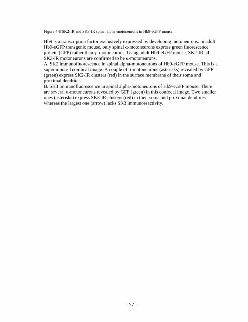
- 77 -
Figure 4-8 SK2-IR and SK3-IR spinal alpha-motoneurons in Hb9-eGFP mouse.
Hb9 is a transcription factor exclusively expressed by developing motoneurons. In adult
Hb9-eGFP transgenic mouse, only spinal α-motoneurons express green fluorescence
protein (GFP) rather than γ–motoneurons. Using adult Hb9-eGFP mouse, SK2-IR ad
SK3-IR motoneurons are confirmed to be α-motoneurons.
A. SK2 immunofluorescence in spinal alpha-motoneurons of Hb9-eGFP mouse. This is a
superimposed confocal image. A couple of α-motoneurons (asterisks) revealed by GFP
(green) express SK2-IR clusters (red) in the surface membrane of their soma and
proximal dendrites.
B. SK3 immunofluorescence in spinal alpha-motoneurons of Hb9-eGFP mouse. There
are several α-motoneurons revealed by GFP (green) in this confocal image. Two smaller
ones (asterisks) express SK3-IR clusters (red) in their soma and proximal dendrites
whereas the largest one (arrow) lacks SK3 immunoreactivity.

- 78 -
SK2Hb9A
**
SK2Hb9A
**
Hb9SK3
B
*
*
Hb9SK3
B
*
*

- 79 -
Figure 4-9 Expression of SK3 in a subpopulation of intra-spinal motor axon terminals.
A. Dual immunofluorescence for VAChT and SK3 in Renshaw cell area of rat lumbar
spinal cord. Cholinergic motor axon terminals in ventral laminal VII Renshaw cell area
are revealed by VAChT immunoreactivity (green). A minority of VAChT-IR motor
terminals express SK3 (red) resulting in yellowish color that indicates the co-localization
of the two staining whereas other terminals lack SK3 immunoreactivity. Conversely,
almost all SK3-IR punctae exhibit VAChT immunoreactivity. The intensity of the
VAChT and SK3 staining on a motor axon terminal was measured indicated by the white
line in the image. The boxed diagram shows the measurement of the fluorescence
intensity that the peaks of the two labeling overlap indicating co-localization.
B. Dual immunofluorescence for CalbindinD28K and SK3 in Renshaw cell area of rat
lumbar spinal cord. Several Renshaw cells (asterisks) are revealed by CalbindinD28K
immunoreactivity (green).Renshaw cell dendrites are contacted by some SK3-IR punctae
(red), indicating these SK3-IR punctae are motor axon terminals.

- 80 -
SK3VAChT
ASK3
VAChTA
SK3CalbindinB
*
*
*
*
SK3CalbindinB
*
*
*
*

- 81 -
Figure 4-10 Expression of SK3 in a subpopulation of motor axon terminals at neuromuscular junctions.
Similar as intra-spinal motor axon terminals, SK3 channels are expressed in a
subpopulation of motor axon terminals at neuromuscular junctions (NMJs). Two NMJs in
rat gastrocnemius muscle are revealed by presynaptic VAChT immunoreactivity (green).
One presynaptic motor axon terminal (A1) expresses SK3 (red, A2) while the other one
(B1) lacks SK3 immunoreactivity (B2).

- 82 -
VAChT
VAChT SK3
SK3
A1 A2
B1 B2
VAChT
VAChT SK3
SK3
A1 A2
B1 B2

- 83 -
CHAPTER V Aim II. The expression of SK
channels in distinct types of rat spinal
motoneurons
Introduction
The amplitude and duration of the mAHP varies with motoneurons innervating different
types of motor units. On average, slow (S-type) motoneurons express larger and longer
mAHP than fast (F-type) motoneurons. Since SK channels are proposed to mediate
mAHP, investigating the expression and membrane organization of SK channels in
distinct types of motoneurons will be of great importance for our understanding of
differential AHP expressed by spinal motoneurons.
Though there is no anatomical characteristic that may clearly distinguish different types
of motoneurons, generally, fast motoneurons are larger than slow motoneurons [Burke
1981]. Based on the results in aim I, SK2 and SK3 channels are expressed, in
complementary pattern, by different subpopulations of rat and mouse spinal motoneurons.
On average, SK3-IR motoneurons are smaller than SK2-IR motoneurons. In order to
explain the differential AHP expressed by distinct types of motoneurons, it is important
to determine if the differential expression of SK channels in motoneurons is related to the
motor unit types. My hypothesis is that slow motoneurons express SK3 channels whereas

- 84 -
fast motoneurons express SK2 channels. To determine the expression of SK channels in
distinct types of motoneurons is the focus of this study.
As discussed before, most mammalian muscles are heterogeneous containing diverse
histochemical types of muscle fibers. For example, in the rat, soleus muscle is slow and
gastrocnemius muscle is fast; however, the muscle fibers of both muscles are not
histochemically pure. Muscle fiber type composition studies of myosin heavy chain
(MHC) isoforms showed that the majority (>80%) of rat soleus muscle fibers are slow
type expressing MHCI whereas rat gastrocnemius muscle fibers are primarily (> 80%)
fast type expressing MHCII [Staron et al 1999]. In this study, SK3 expression in rat
soleus motoneurons and gastrocnemius motoneurons will be examined respectively.
Fluorogold retrograde tracing will be used to identify specific motoneuron pools in
lumbar spinal cord. The expected results will shed light on the SK expression in
motoneurons innervating distinct types of motor units.
Another important technique, electro-physiological recording, will be used here
to identify motoneuron types. Intracellular recording is a powerful technique that enables
the electrophysiological membrane properties of single motoneurons to be determined.
Generally, slow motoneurons have slower axonal conduction velocity, larger and longer
AHP, higher input resistance and greater overshoot of action potential than fast
motoneurons; however, there is always overlapping of all these properties between
motoneuron types [Burke 1981; Zengel et al., 1985; Gardiner 1993]. In this study
membrane properties including input resistance, rheobase, the amplitude and duration of
AHP will be recorded for the identification of motoneuron types. Intracellular recording

- 85 -
followed by intracellular filling of neurobiotin and immunocytochemistry will be
performed to explore the SK expression in recorded motoneurons.
Aim I also showed that a small portion of presynaptic motor axon terminals in
rat spinal cord and neuromuscular junctions displayed SK3-IR immunoreactivity. This
result is partially consistent with the results from other investigators who did not examine
muscle fiber types in their study [Roncarati et al., 2001]. According to our results it is of
interest to understand what type of muscle fibers SK3-IR terminals innervate and whether
it is related to motor unit types. Neuromuscular junctions in rat soleus (slow) muscle and
gastrocnemius (fast) muscles will be examined separately. The expected results will
provide us insight on the differential expression of SK3 in presynaptic motor axon
terminals.
Methods & materials
Adult Spray-Dawley and wistar rats were used for the in situ study of the SK3
expression in soleus motoneurons and gastrocnemius motoneurons. Retrograde labeling
of spinal motoneurons by fluorogold injection into muscles (soleus or gastrocnemius)
was performed for the identification of specific motoneuron pools in lumbar spinal cord.
The surgery and fluorogold injection was performed by Lori Goss. Three days after
fluorogold injection lumbar spinal cords were harvested for immunohistochemical assays.
Protocols for fluorogold injection are described in Chapter III.
Intracellular recording of in vivo motoneurons was performed in Dr. Tim Cope‟s
lab in collaboration with Katie Bullinger. Adult Wistar rats were anesthetised with

- 86 -
isoflurane. Spinal motoneurons identified by antidromic stimulation of medial
gastrocnemius (MG) muscle nerve were intracellularly recorded and filled with
neurobiotin after the measurement of membrane properties including input resistance,
rheobase, and AHP amplitude and duration. Animals were then transcardially perfused
and spinal cords containing recorded and neurobiotin-filled motoneurons were processed
for immunohistochemical analysis. Neurobiotin was revealed by Alexa Fluor 405
conjugated Streptavidin (Vector Laboratories; 1:50), then the sections were incubated in
primary antibodies followed by fluorescence conjugated secondary antibodies.
For the study of SK3 expression in motor axon terminals in muscle, neuromuscular
junctions (NMJs) in soleus and gastrocnemius muscles of adult Spray-Dawley rats were
analyzed. NMJs were revealed by immunoreactivity against α-bungarotoxin which
targets postsynaptic acetylcholine receptors in muscle fibers. Anti-Myosin type I (Sigma;
mouse 1:500) was used to identify slow type muscle fibers. At the same time, SK3
immunoreactivity was examined.
Immunoreactivity of primary antibodies was detected with Cy3- , Fluorescein
(FITC), or Cy5- conjugated secondary antibodies (Jackson ImmunoResearch
Laboratories). All immunohistochemical procedures performed in this aim are listed in
Table 2. The immunostaining results were examined with confocal microscopy (Olympus
Fluoview).

- 87 -
Results
1. SK3 expression in rat soleus versus gastrocnemius motoneurons
Soleus motoneurons and gastrocnemius (MG/LG) motoneurons in rat lumbar
spinal cords were identified by retrogradely labeled fluorogold which was revealed using
epi-fluorescence microscope with UV filter (figure 5-1). Since both α-MNs and γ-MNs
were labeled with fluorogold, and only α-MNs are of interest in this study, all presumed
γ-MNs were discarded whose soma average diameters were less than 25 um [Chen &
Wolpaw, 1994; Ishihara et al., 2001; Bose et al., 2005; Ichiyama et al., 2006].
A total of 133 soleus α-MNs were observed and (63%) 84 of them expressed SK3-IR
clusters; in contrast, 37 out of 149 (25%) MG/LG α-MNs exhibited SK3-IR clusters. The
SK3 immunoreactivity was then arbitrarily divided into 2 subgroups: strong and weak.
Among the 84 SK3-IR soleus motoneurons, 50 (60%) expressed strong SK3 intensity
whereas 34 (40%) weak. As for the 37 SK3-IR MG/LG motoneurons, 25 (68%) exhibited
strong SK3 immunoreactivity and 12 (32%) weak (figure 5-1).
2. SK3 expression in intracellularly recorded motoneurons
In vivo electrophysiological recordings were performed in Dr. Tim Cope‟s lab in
collaboration with Katie Bullinger. 10 medium gastrocnemius (MG) motoneurons were
recorded followed by immunohistochemical analysis of SK3 expression. Shannon Romer
was of great help with the immunostaining and imaging on confocal microscopy.
Motoneuron types were classified by their AHP ½ decay time (presumptive
slow MN > 20ms; presumptive fast MN < 20ms) according to the previous results of rat
gastrocnemius motoneuorns [Gardiner 1993].

- 88 -
The electric membrane properties (rheobase & AHP ½ decay time) and SK3
immunoreactivity of the 10 recorded motoneurons are listed in Table 3, which are ranked
by AHP ½ decay time. Among the 10 recorded motoneurons, one presumptive slow
motoneuron (rheobase 1.3nA, AHP 1/2 decay time 25.0ms) expressed SK3 clusters over
its surface membrane of soma and proximal dendrites (figure 5-2). Eight presumable fast
motoneurons did not exhibit SK3 immunoreactivity, one of which was shown in figure 5-
2. These results support the hypothesis that slow motoneurons rather than fast
motoneurons express SK3 channels.
However, one presumable fast motoneuron with rheobase 12nA and AHP ½
decay time 15.7ms exhibited SK3 clusters. This result challenges the above hypothesis.
3. SK3 expression in rat fast versus slow motor axon terminals
Since a subpopulation of motor axon terminals was found to express SK3
channels in aim I, the SK3 expression in motor axon terminals at neuromuscular
junctions (NMJs) was studied here. NMJs in rat soleus and MG/LG muscles were
examined using immunohistochemistry. The NMJs were labeled by α-bungarotoxin,
which targets postsynaptic nicotinergic acetylcholine receptors. Slow muscle fibers were
identified with myosin type I and fast muscle fibers were lack of myosin I. All observed
NMJs were randomly selected from muscle sections. The majority of NMJs innervating
slow type muscle fibers exhibited presynaptic expression of SK3 (figure 5-3). In contrast,
SK3 expression was seldom observed in NMJs innervating fast type muscle fibers. This
finding of SK3 in slow rather than fast motor axon terminals suggests a differential
expression of SK channels in distinct types of spinal motoneurons.

- 89 -
Discussion
1. Soleus motoneurons versus gastrocnemius motoneurons
In this study two different sets of experiments were performed in order to test if
the differential SK3-IR cluster expression in rat motoneurons is related to motor unit
types. The first one detected the SK3 expression in rat soleus versus MG/LG
motoneurons. As a result, the majority (63%) of soleus motoneurons expressed SK3-IR
clusters; in contrast, only a minority (25%) of gastrocnemius motoneurons expressed SK3.
The exact proportion of each motor unit type in both motoneuron pools are not
clear, but the proportion of each muscle fiber type in both muscles were studied that more
than 80% of rat soleus muscle fibers are slow whereas more than 80% rat gastrocnemius
muscle fibers are fast [Staron et al 1999]. Electrophysiological study also showed the
consistent results of the proportion of each motor unit type (slow type 16.7% whereas fast
type 83.3%) in rat MG [Kanda & Hashizume 1992]. Based on these previous reports, our
results indirectly suggest that slow motoneurons rather than fast motoneurons tend to
express SK3 channels, though our numbers do not perfectly match the proportion of each
muscle fiber types.
One important thing needs to be mentioned here is the technical consideration.
The fluorogold retrograde tracing technique is not 100% reliable that the possibility of
injected fluorogold leaking into adjacent muscles may affect the final results due to the
juxtaposition of soleus and gastrocnemius muscles.

- 90 -
2. Intracellular recording from fast motoneurons versus slow motoneurons
The physiological properties of gastrocnemius motoneuorns in cat and rat
indicate that slow motoneurons expresse higher input resistance, lower rheobase and
longer AHP ½ decay time than fast motoneurons [Zengel et al., 1985; Gardiner 1993].
However, there is always overlapping of all these properties between distinct motoneuron
types, so none of these properties is perfect predictor for classifying motor units. Another
thing needs to be considered is the variation of these electrophysiological parameters, for
example, rheobase is of physiological variation that it varies during recording. Compared
to rheobase, the measurement of AHP is relatively more stable.
Zengel and colleagues studied cat medial gastrocnemius motoneuron properties
and suggested that rheobase was the best individual predictor for classifying motor units
as fast twitch fast fatiguing (FF), fast twitch with intermediate fatigue resistance (FI), fast
twitch fatigue resistance (FR) or slow twitch fatigue resistant (S). The AHP ½ decay time
was the most accurate predictor in discriminating between slow and fast motoneurons
[Zengel et al., 1985]. Unfortunately there is not as much information of motoneuron
properties available for rat as for cat. Gardiner studied rat gastronemius motoneurons and
found that slow motoneurons always had AHP ½ decay time longer than 20ms whereas
fast motoneurons never had the value greater than 19ms [Gardiner 1993]. The cutoff
point of AHP ½ decay time (20ms) between slow and fast units set by Gardiner was used
in this study for classifying motoneurons. The absolute definition of motoneurons can
only be obtained by determining the mechanical twitching properties or myosin types of
their innervated muscle fibers, which was not available in this study.
- 91 -
So far, only one recorded motoneuron was typed as slow by its long AHP ½
decay time (25.0ms) and low rheobase (1.3nA). This presumptive “slow” motoneuron
expressed SK3 clusters in its soma and proximal dendrites as expected. In contrast, eight
out of the nine (89%) presumable “fast” motoneurons did not express SK3 channels. All
these expected results supported our proposed hypothesis.
Unexpectedly, one presumable fast motoneuron (AHP ½ decay time 15.7ms)
expressed SK3 clusters. As a fact, the classification of motoneuron types based on their
membrane properties is not 100% reliable, and there always exists uncertainty when
using any motoneuron physiological properties as predictors. The SK3-IR “fast”
motoneuron had AHP ½ decay time 15.7ms, which is close to the cutoff point (20ms), so
there is a possibility that it is a slow rather than fast motoneuron.
With current data, it is difficult to draw a conclusion due to the following
reasons:
1) The size of our sample (n=10) is too small; only one presumptive slow motoneuron
was recorded. More recorded motoneurons in future study, especially slow type, will
bring more information for a conclusion;
2) There is always overlapping of motoneuron membrane properties innervating
different types of motor units, so using the cutoff point of 20ms AHP ½ decay time to
discriminate slow and fast motor units is not 100% reliable and the mistyping is
inevitable;
3) The limitation of current techniques: there may be mistakes when using
immunohistochemistry to identify recorded neurons because of the possible leaking
of neurobiotin into adjacent neurons.

- 92 -
Though it is too early to conclude that SK3 expression is expressed by slow
rather than fast motoneurons, the present results are promising. There was a clear bias of
SK3 expression in: i) retrogradely labeled soleus versus gastronemius motoneurons; ii)
physiologically identified fast versus slow recorded motoneurons. But in each case, the
identity of “fast” and “slow” is not absolute. Future study is needed for a conclusion.
3. Motor axon terminals of slow motoneurons versus fast motoneurons
Our results show that a subpopulation of motor axon terminals in ventral horn
spinal cord and neuromuscular junctions express SK3 channels. This is consistent with
the previous report [Roncarati et al., 2001]. We suggested that motor axon terminals of
slow motoneurons rather than those of fast motoneurons express SK3 channels. This
result suggests a differential expression of SK channels in nerve terminals by
motoneurons innervating distinct types of motor units. However, it is unknown if what is
expressed in axon terminals such as SK3 channel is also expressed in motoneuron cell
body.
4. Mechanism underlying the differential mAHP in motoneurons
Though more data are required for a conclusion, our results in this aim
suggested a differential expression of SK channels in rat spinal motoneurons that slow
type is more likely to express SK3 whereas fast type SK2. Whether this may explain the
differential AHP expressed by distinct types of motoneurons is a question that needs to be
answered. The mAHP conductance in slow motoneurons was shown similar as that in fast
motoneurons [Manuel et al., 2005]. The lower input conductance of slow motoneurons
may explain their higher AHP amplitude; however, why slow motoneurons have longer

- 93 -
AHP duration is unknown. Why SK3 in slow motoneurons mediates longer AHP than
SK2 does in fast motoneurons needs to be explored in future study.
In order to confirm that SK3 is responsible for the mAHP in slow motoneuron
and SK2 for fast ones, the deletion of SK3 or SK2 genes would be the ideal model. As
discussed before, SK channel knock-out transgenic mouse lines have been constructed
[Bond et al., 2004]. However, few studies, if any, have been performed in mouse for the
electrophysiological recording of motoneurons. Unfortunately, no knock-out rat is
available at this time.
As discussed in Background, the biophysical properties of three cloned SK
isoforms (SK1, SK2, & SK3) in expression system are not different from each other,
including their unitary conductance and kinetics of calcium activation. However, the
environment of in vivo motoneurons is different from the isolated cells in expression
system, which lack many regulatory proteins and scaffording proteins in native
motoneurons. If any other proteins interact with SK channel clusters and contribute to the
different kinetics of SK3 and SK2 channels in rat and mouse motoneurons needs to be
examined in future study.
Another possibility is that the clustering of SK channels results in different Ca++
sensitivity and kinetics of SK3 channels from that of SK2 channels. As discussed in
Background, the phosphorylation state of calmodulin constitutively bound to SK2 or SK3
channels may be regulated by CK2 / PP2A leading to altered channel Ca++
sensitivity
[Bildl et al., 2004; Allen 2007]. When SK channels aggregate into clusters in motoneuron
surface membrane, the phosphorylation state of calmodulin might be changed and this
change is not at the same level for SK2 and SK3 channels leading to different Ca++

- 94 -
sensitivity and channel kinetics. The exact mechanism of differential AHP in
motoneurons needs to be clarified in future study.
Since the differential expression of SK channels was not observed in cat, the
mechanism underlying the differential AHP in cat motoneurons might be different from
that in rodents.

- 95 -
Table 2 Tissue, fixation, sectioning and antibodies for immunofluoresence in Aim II
Tissue Fixation Sections Primary Abs Secondary Abs6
Dual
Immunofluorescence
for SK3 and NeuN
Rat spinal cord
after Fluorogold
injection
4%
paraformaldeh
yde with 2
hour post-fix
40 µm
floating
sections
NeuN (mouse
1:400)1
SK3 (Alomone;
rabbit 1:100)2
FITC anti-mouse
IgG 1:200
Cy3 anti-rabbit
IgG 1:300
Triple
immunofluorescence
for SK3, VAChT,
and Neurobiotin
Rat spinal cord
after intracellular
recording and
neurobiotin
filling of MNs
4%
paraformaldeh
yde with 2
hour post-fix
40 µm
floating
sections
SK3 (rabbit
1:100)2
VAChT (goat
1:5,000)3
Alexa Fluor 405
Streptavidin
1:508
Cy3 anti-rabbit
IgG 1:300
Cy5 anti-goat
IgG 1:50
Triple
immunofluorescence
for SK3, kv2.1, and
Neurobiotin
Rat spinal cord
after intracellular
recording and
neurobiotin
filling of MNs
4%
paraformaldeh
yde with 2
hour post-fix
40 µm
floating
sections
SK3 (rabbit
1:100)2
Kv2.1 (mouse
1:1,000)4
Alexa Fluor 405
Streptavidin
1:508
Cy3 anti-rabbit
IgG 1:300
FITC anti-mouse
IgG 1:200
Triple
immunofluorescence
for α-bungarotoxin,
SK3, and myosin
type I
Rat soleus/
gastrocnemius
muscles
4%
paraformaldeh
yde with 4
hour post-fix
20 µm on-
slide
sections
SK3 (rabbit
1:100)2
Myosin I (mouse
1:500)5
α-bungarotoxin-
Alexa 488
(1:200)7
Cy3 anti-mouse
IgG 1:300
Cy5 anti-rabbit
IgG 1:50
__________________________________________________
1 Chemicon; USA
2 Alomone; Jerusalem
3 BD Pharmingen; San Diego, CA
4 Neuromab; Davis, Califormia
5 Sigma-Aldrich; St. Louis, MO
6 Jackson ImmunoResearch Laboratories; West Grove, PA
7 Molecular Probes; Carlsbad, California
8 Vector Laboratories; Burlingame, CA

- 96 -
Table 3 The membrane properties of 10 recorded MNs
Recorded
MN
Rheobase
(nA)
AHP 1/2
decay (ms)
AHP amplitude
(mv) SK3-IR
Putative
identity
1 26 8.5 1.17 no Fast
2 16 9.1 0.64 no Fast
3 9 9.6 1.85 no Fast
4 11 9.7 1.13 no Fast
5 9 10.2 1.98 no Fast
6 28 11.6 1.02 no Fast
7 10 15 1.65 no Fast
8 12 15.7 1.76 Yes Fast
9 3.7 18.9 3.75 no Fast
10 1.3 25.0 3.99 Yes Slow

- 97 -
Figure 5-1 SK3 expression in motoneurons innervating slow versus fast muscles.
A. Gastrocnemius motoneurons retrogradely labeled with fluorogold. 3 motoneurons
(asteristks) are revealed by fluorogold staining under UV light after the fluorogold
injection into gastocnemius muscles. Only the brightest presumed alpha-
motoneurons were selected for this study. Motoneurons with average diameter
less than 25 um were discarded.
B. SK3 expression in soleus motoneurons versus gastrocnumius motoneurons. A
total of 133 soleus motoneurons and 149 gastrocnemius motoneurons were
analyzed for the SK3 expression analysis. A majority (63%) of soleus
motoneurons express SK3 whereas a minority (25%) of gastrocnemius
motoneurons do so. The SK3 immunoreactivity is arbitrarily divided into strong
and weak, and 60% SK3-IR soleus motoneurons and 68% SK3-IR MG/LG
motoneurons express strong SK3 intensity.

- 98 -
0%
20%
40%
60%
80%
100%
Soleus MN LG/MG MN
Non SK3
weak SK3
strong SK3
B
0%
20%
40%
60%
80%
100%
Soleus MN LG/MG MN
Non SK3
weak SK3
strong SK3
0%
20%
40%
60%
80%
100%
Soleus MN LG/MG MN
Non SK3
weak SK3
strong SK3
B
A
200μm
LIX
A
200μm
LIX
200μm
LIX

- 99 -
Figure 5-2 Intracellular recording of rat spinal motoneurons.
A. Triple immunofluorescence for Neurobiotin, VAChT and SK3 in a recorded „slow‟
motoneuorn. This recorded motoneuron was classified as a slow motoneuron due
to its low rheobase (1.3nA) and long AHP ½ decay time (25.0ms).
B. The measurement of rheobase and AHP for the recorded motoneuron shown in A.
(i) Action potential elicited by intracellular current injection to determine AHP ½
decay time. The AHP ½ decay time was defined as the time for the membrane
potential to decay from the peak of the hyperpolarization to the one-half of that
level. (ii) To determine rheobase, depolarizing current pulses (50 ms) were
delivered intracellularly in 1 nA steps until the motoneuron fired.
C. Triple immunofluorescence for Neurobiotin, Kv2.1 and SK3 in a recorded „fast‟
motoneuorn. This recorded motoneuron was classified as a fast motoneuron due
to its higher rheobase (10nA) and shorter AHP ½ decay time (15.5ms).
D. The measurement of rheobase and AHP for the recorded motoneuron shown in C.
(i) Action potential elicited by intracellular current injection to determine AHP ½
decay time. (ii) To determine rheobase, depolarizing current pulses (50 ms) were
delivered intracellularly in 1 nA steps until the motoneuron fired.

- 100 -
SK3
NeurobiotinVAChT
A
SK3
NeurobiotinVAChT
A
Bi ii
2 m
V
25 ms
10 ms10
mv
10 ms1
nA
Bi ii
2 m
V
25 ms
10 ms10
mv
10 ms1
nA
CSK3
Kv2.1Neurobiotin
CSK3
Kv2.1Neurobiotin
Di ii
2 m
V
15.5 ms
10 ms10 m
v
10 ms
2 n
A
Di ii
2 m
V
15.5 ms
10 ms10 m
v
10 ms
2 n
A
- 101 -
Figure 5-3 SK3 immunofluorescence at NMJs innervating slow muscle fibers.
A. Transverse section of rat soleus muscle fibers with triple immunofluorescence for
a-bungarotoxin, Myosin type I and SK3. Slow muscle fibers are identified with
immunoreactivity against myosin I (red). Two NMJs are revealed by
immunostaining of a-bungarotoxin (green). The immunofluorescence of one NMJ
(arrow) is demonstrated in higher magnification images shown in B.
B. Triple immunofluorescence for a-bungarotoxin, Myosin type I and SK3 in the
NMJ shown in A. The NMJ labeled with a-bungarotoxin (green, B1) innervates
slow muscle fiber stained with Myosin I (red, B2) and expresses SK3 (white, B3).
B4 is the merging image of the triple immunolabeling.
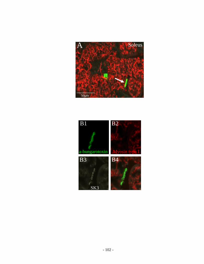
- 102 -
SK3
Myosin type Ia-bungarotoxin
B1 B2
B3 B4
SK3
Myosin type Ia-bungarotoxin
B1 B2
B3 B4
A Soleus
50μm
A Soleus
50μm

- 103 -
CHAPTER VI Aim III The synaptic localization of SK channel clusters in spinal motoneurons
Introduction
Each mammalian -motoneuron receives thousands of synaptic contacts of
various origins, some of which modulate motoneuron intrinsic input/output properties by
working through metabotropic receptors. One of these modulating effects is the
regulation of the amplitude and duration of AHP that leads to the alteration of
motoneuron firing properties [Kernell 1999; Rekling et al 2000].
Medium AHP in motoneuron was found to be reduced by serotonin and
muscarine/acetylcholine, and the activation of 5-HT1A receptors and M2 receptor were
suggested to mediate their modulatory effect respectively [Van Dongen et al 1986;
Bayliss et al., 1995; Grunnet et al., 2004; Brownstone 2006; Lape et al., 2000]. However,
the exact signaling pathway of these neuromodulation is unknown.
Medium AHP in motoneurons may be blocked by apamin indicating it is
mediated by SK channels [Zhang et al., 1987; Hounsgaard et al., 1988; Viana et al., 1993;
Powers et al., 1999; Sawczuk et al., 1997; Miles et al., 2005]. Based on to the results of
aim I, SK2 and SK3 channels form clusters over the surface membrane of soma and
proximal dendrites of motoneuorns. If this characteristic organization contributes to the
neuromodulatory effects of neurotransmitters is an important question to be answered.

- 104 -
My hypothesis is that SK clusters in motoneuron surface membrane are
located at synaptic sites in association with one specific type of presynaptic terminals, for
example, cholinergic C-terminals, because of the large size of SK clusters. This aim will
focus on examining the synaptic localization of SK clusters in motoneurons. The
expected results will provide insight into the mechanism underlying the neuromodulation
in motoneurons.
In order to identify different subsets of synapses, several pre-synaptic or post-
synaptic markers will be used to reveal cholinergic, serotonergic, glutamatergic, and
glycine/GABAergic synapses in motoneurons. Antibody against vesicular acetylcholine
transporter (VAChT) reveals presynaptic cholinergic bountons including large C-
terminals contacting α-motoneurons in lamina IX and motor axon collaterals contacting
Renshaw cells in lamina VII [Alvarez et al., 1999; Deng & Fyffe 2004]. Antibody against
5-HT labels descending serotonergic inputs from Raphe neurons in brainstem [Alvarez et
al., 1998]. Antibodies against vesicular glutamate transporters (VGLUT1 & 2) reveal
glutamatergic boutons that VGLUT1 labels primary afferent terminals and VGLUT2
descending and segmental glutamate inputs onto motoneurons [Alvarez et al., 2004].
Gephyrin is the clustering protein for glycine receptors and antibody against gephyrin
post-synaptically reveals glycine/GABAergic synapses in motoneurons and interneurons
[Alvarez et al., 1997].
Voltage gated potassium Kv2.1 channel, the contributor to the delayed rectifier,
was abundant and distributed in clusters in the surface membrane of soma and proximal
dendrites of -motoneurons. Large Kv2.1 clusters were apposed to presynaptic

- 105 -
cholinergic C-terminals and colocalized with postsynaptic muscarinic (m2) receptors
[Muennich & Fyffe 2004]. Kv2.1 will also be tested in this study.
Methods & materials
Dual labeling of SK (SK2 or SK3) channels combined with one of synaptic
markers was performed for this immunohistochemical study.
The antibodies against pre-synaptic markers include: 1) anti-Synaptophysin
(Calbiochem/Oncogene; mouse 1:200) un-specifically labels presynaptic terminals; 2)
anti-VAChT (vesicular acetylcholine transportor; BD Pharmigen; Goat 1:5,000) labels
cholinergic presynaptic terminals; 3) anti- 5-HT (Dr. Pearson; Guinea Pig 1:5,000) labels
serotonergic presynaptic terminals; 4) anti-VGLUT1 (Chemicon; Guinea pig 1:5,000),
and anti-VGLUT2 (Chemicon; Guinea pig 1:5,000) label glutamatergic presynaptic
terminals.
The antibody against gephyrin (Alexis; mouse 1:100) post-synaptically labels
inhibitory glycine/GABAergic synapses. The antibody against Kv2.1 (Upstate
Biotechnology; mouse 1:250) labels voltage-gated Kv2.1 channels.
All immunohistochemical procedures performed in this aim are listed in Table 4.
The resulting immunostaining was visualized and imaged using confocal microscopy.
The SK3 cluster sizes were analyzed using ImagePro sofeware, and only the en face
clusters in individual optical sections were selected for this analysis (detailed method in
Chapter III).

- 106 -
The synaptic localization of SK was confirmed at ultra-structural level using
electron microscopy. Pre-embedding immunostaining of SK3 channel (Chemicon; rabbit
1:100) was performed on vibratom sections of rat lumbar spinal cord (see Chapter III for
details). The resulting labeling was visualized on Philips EM201 transmission electron
microscopy. Ricardo Zerda helped with the EM study.
Results
1. Postsynaptic location of SK channel clusters
Double labeling of presynaptic protein synaptophysin with SK3 revealed a close
correspondence between presynaptic boutons and SK3-IR clusters in the soma and
proximal dendrites of rat spinal motoneurons. Each SK3-IR cluster was apposed to (not
colocalized with) a presynaptic terminal revealed by synaptophysin, though some
presynaptic boutons were not apposed to any SK3-IR clusters on the surface membrane
of motoneurons. Using mouse spinal cord similar results were observed for SK2 channels.
This finding suggests a postsynaptic location of SK channel clusters in the surface
membrane of motoneurons. Moreover, the pre-synaptic terminals apposing SK-IR
clusters are the largest ones compared with those not associating any SK channels (figure
6-1). The co-localization of SK clusters with Kv2.1 channels also confirm the
postsynaptic location of SK clusters in motoneurons (see below).
2. Specific localization of SK clusters at cholinergic synapses
In order to identify what type of synapses SK clusters are localized at, several pre-
and post-synaptic markers were used to label different subsets of synapses on

- 107 -
motoneurons. Cholinergic C-terminals contacting the soma and proximal dendrites of -
motoneurons were revealed by the immunoreactivity against VAChT, which are the
largest boutons among various synaptic inputs onto motoneurons [Alvarez et al., 1999;
Deng & Fyffe 2004]. Double labeling of SK3 with VAChT showed that the majority
(94.9%; n=1051) of SK3-IR clusters in rat motoneurons are associated with VAChT-IR
presynaptic C-terminals. The immunoreactivity against VAChT and SK3 was closely
apposed to rather than overlapping with each other, which is consistent with the results of
synaptophysin indicating a postsynaptic rather than presynaptic localization of SK3-IR
clusters (figure 6-2). Similar results were observed for SK2-IR clusters in mouse spinal
motoneurons (figure 6-4).
The electron microscopy results confirmed the postsynaptic localization of SK3
channels at cholinergic synapses in rat spinal motoneurons. The immunoreactivity against
SK3 targeted the postsynaptic membrane apposed to presynaptic C-terminals. There was
some SK3 immunoreactivity within cytoplasm, which may demonstrate SK3 protein
trafficking to and recycling from motoneuron surface membrane (figure 6-3).
Then serotonergic, glutamatergic and glycine/GABAergic synapses were also
examined in order to test if postsynaptic SK3 clusters are particularly associated with
cholinergic C-terminals. Dual labeling of SK3 with 5-HT showed no close apposition of
SK3-IR clusters with any serotonergic bountons (figure 6-5C). Two subsets of
glutamatergic boutons were revealed by two isoforms of vesicular glutamate transporter
VGLUT1 and VGLUT2; as a result, none of SK3-IR clusters in motoneurons was found
to associate with any presynaptic glutamatergic terminals (figure 6-5AB). Inhibitory
glycine/GABAergic synapses were labeled by immunoreactivity against gephyrin, and no

- 108 -
overlapping of post-synaptic gephyrin immunoreactivity and SK3-IR clusters was
observed (figure 6-5D).
3. Co-localization of SK channels and Kv2.1 channels
Voltage gated Kv2.1 channel, the contributor to the delayed rectifier, forms
clusters in the surface membrane of soma and proximal dendrites of -motoneurons.
Large Kv2.1 clusters are postsynaptic and apposed to pre-synaptic cholinergic C-
terminals and co-localized with muscarinic (m2) receptors. The post-synaptic location of
large Kv2.1 clusters was confirmed at ultra-structural level using electron microscopy
[Muennich & Fyffe 2004]. Since SK-IR clusters were found to be post-synaptically
localized at cholinergic synapses, they were expected to co-localize with kv2.1 channels.
Then double labeling of SK3 combined with Kv2.1 was performed using rat spinal cords
and the results showed the perfect overlapping of the immunoreactivity against the two
ion channels in motoneurons (figure 6-6). This result confirms the post-synaptic location
of SK channel clusters in motoneurons.
It needs to be mentioned that all motoneuorns of both large and small sizes
expressed kv2.1-IR clusters whereas only a subpopulation of motoneurons exhibited
SK3-IR clusters. In SK3-IR motoneurons, only large kv2.1-IR clusters co-localize with
SK3-IR clusters, and small kv2.1-IR clusters do not contain SK3 immunoreactivity
(figure 6-6). Totally 341 SK3-IR clusters in 30 motoneurons were examined and all of
them were co-localized with Kv2.1. Similar results were observed for SK2-IR clusters in
mouse spinal motoneurons (figure 6-7).
The sizes of en face SK3-IR clusters in rat motoneurons were then quantitatively
analyzed. The range of the maximal diameters of SK3-IR clusters was from 1.29 μm to

- 109 -
7.14 μm (mean SD 3.51 0.88; n=245); and the cluster areas were from 1.03 μm2 to
15.62 μm2 (mean SD 6.75 2.68; n=245). This result of SK3-IR cluster sizes is
comparable to that of large Kv2.1 clusters in rat motoneurons [Muennich & Fyffe 2004]
(figure 6-8).
Discussion
In this immunohistochemical study SK2-IR and SK3-IR clusters in motoneurons
were shown to be specifically localized post-synaptically at cholinergic synapses
associated with C-terminals, which was confirmed at ultra-structural level using electron
microscopy. The co-localization of SK clusters with Kv2.1 channels at these synaptic
sites are also good evidences for the post-synaptic location of SK clusters. None of the
observed SK3-IR or SK2-IR clusters in motoneurons was localized at serotonergic,
glutamatergic, or glycine/GABAergic synapses. No evidence of presynaptic localization
of SK immunoreactivity was found at any synapses in motoneurons.
M2 muscarinic acetylcholine recepors (mAChR) were shown to enrich beneath C-
terminals in the postsynaptic membrane and suggested to be the major muscarinic
acetylcholine receptors in spinal motoneurons [Rekling et al., 2000; Hellström et al., 2003].
Voltage gated Kv2.1 channels were found to be co-localized with M2 receptors at these
sites [Muennich & Fyffe 2004]. Recent studies showed the dynamic regulation of Kv2.1
localization in cell surface membrane and channel biophysical properties plays an
important role in the control of neuron excitability [Misonou et al., 2004, 2005;
Mohapatra & Trimmer 2006]. In this study SK channels were observed to be localized in

- 110 -
the same postsynaptic domains, which may bring a new clue of the modulatory effect of
acetylcholine/ muscarine on motoneurons.
The cholinergic synapses in motoneuorns formed by large C-terminals are
characterized of sub-surface cisternae, which are endoplasmic reticulum-derived
organelles rich in ryanodine receptors, the intracellular calcium release channels
[Antonucci 2001]. The activation of postsynaptic M2 receptors leads to a reduction of
mAHP and increases the input-output gain (slope of f-I curve) in motoneurons
[Brownstone 2006; Lape et al., 2000]. One possible explanation is the activation of m2
receptors results in the alteration of calcium sequestration into and/or release from
subsynaptic calcium store, which reduces the free Ca++
ion concentration at those
microdomains and thus decreases SK channel activation resulting in AHP reduction.
However, several evidences suggested the activation of M2 receptors leads to an
increased calcium release from SCC [Zhou et al., 2003]. The cholinergic modulation of
Kv2.1 clusters in cultured hippocampal neurons was shown to result from calcium release
from subsurface cistern via muscarinic receptors causing a dispersing of Kv2.1 clusters in
cell surface membrane [Mohapatra & Trimmer 2006]. Similar Kv2.1 de-clustering effects
were also observed in neonatal spinal motoneuorns by muscarine application [Katie‟s
SfN poster 2007; unpublished results from Fyffe lab]. Since the co-localization of Kv2.1
and SK clusters at cholinergic synapses, if the calcium sequestion/release is involved in
the acetylcholine/ muscarine modulation of SK channel / AHP in motoneurons needs to
be clarified in future study.
In rat motor axon terminals the activation of M2 receptors was suggested to
inhibit neurotransmission by decreasing protein kinase A (PKA) activity, and P/Q-type

- 111 -
calcium channels were involved in this muscarine regulatory effect [Santafe et al., 2006].
However, other researchers suggested a membrane delimited pathway of (N- type and
P/Q-type) Ca++
channel modulation by G protein-coupled neurotransmitter receptors
including M2 receptors. G-protein βγ dimmers can directly bind to and inhibit these Ca++
channels by shifting their gating toward depolarizing membrane potential [Herlitze et al.,
1996; Ikeda 1996; Shapiro et al., 1999]. N-type and P/Q- type Ca++
channels were
suggested to activate SK channels mediating mAHP during motoneuron firing [Bayliss et
al., 1995; Li & Bennett 2007]. The activation of postsynaptic M2 receptors at C-terminals
sites in motoneuron could lead to a reduction of Ca++
influx through N-type and P/Q-
type channels resulting in a decreased mAHP via SK channels. However, the sub-cellular
localization of these Ca++
channels in motoneurons is not defined yet. At the other hand,
this fast modulation by G-protein βγ dimmers binding to ion channels might also occur to
SK channels at the same sites.
SK channels have multiple potential phosphorylation sites and the direct
phosphorylation of cloned and native SK2 channel by protein kinase A (PKA) causes an
internalization of these channels from cell surface membrane [Kohler et al., 1996; Ren et
al., 2006; Lin et al., 2008; Faber et al., 2008]. Though PKA activity was found to be
inhibited by M2 activation [Santafe et al., 2006], other phosphorylation pathways were
also observed to be initiated by the activation of M2 receptors [Zhou et al., 2003], so
there exist a possibility that direct phosphorylation of SK channels is involved in
muscarinic/ acetylcholine regulation in motoneurons.
Constitutively bound calmodulin is a substrate for CK2 and its phosphorylation
may reduce SK channel Ca++
sensitivity [Bildl et al., 2004; Allen 2007].

- 112 -
Neurotransmitters such as noradrenaline was found to inhibit SK2 channels by a CK2
dependent reduction of calcium sensitivity of channel gating in dorsal root ganglion cells
[Maingret et al., 2008]. Muscarine/acetylcholine might decrease the calcium gating of SK
channels in the same way through calmodulin phosphorylation by CK2.
As a conclusion, our results of SK localization at C-terminal synapses indicate an
important role of postsynaptic M2 muscarinic receptors in the AHP modulation of
motoneurons. However, the detailed molecular signaling pathway of acetylcholine/
muscarine regulation of motoneuron excitability and firing properties is yet to be fully
defined in future study.
So far the expression of SK channels in normal adult spinal motoneurons has been
studied. Postnatal maturation of motoneuron functions and their changes induced by
axonal injury are two important areas in neuroscience. How motoneuron firing properties
develop after birth and what changes occur after the axon is injured are fundamental
questions need to be clarified. Since SK channels mediate mAHP, investigating SK
channels in neonatal motoneurons and injured motoneurons will help answer these
questions. Aim IV and aim V will focus on SK channels in developing and injured
motoneurons respectively.

- 113 -
Table 4 Tissue, fixation, sectioning and antibodies for immunofluoresence in Aim III
Tissue Fixation Sections Primary Abs Secondary Abs8
Dual
immunofluorescence
for SK3 and
Synaptophysin
Rat spinal
cord
4%
paraformaldehyde
with 0 hour post-fix
20 µm on-
slice sections
SK3 (rabbit
1:100)1
Synaptophysin
(mouse 1:200) 2
FITC anti-
mouse IgM
1:200
Cy3 anti-rabbit
IgG 1:300
Dual
immunofluorescence
for SK3 and VAChT
Rat spinal
cord
4%
paraformaldehyde
with 0 hour post-fix
40 µm
floating
sections
SK3 (rabbit
1:100)1
VAChT (goat
1:5,000)3
FITC anti-goat
IgG 1:200
Cy3 anti-rabbit
IgG 1:300
Dual
immunofluorescence
for SK3 and
VGLUT1
Rat spinal
cord
4%
paraformaldehyde
with 0 hour post-fix
40 µm
floating
sections
SK3 (rabbit
1:100)1
VGLUT1
(Guinea pig
1:5,000)4
FITC anti-
Guinea pig IgG
1:200
Cy3 anti-rabbit
IgG 1:300
Dual
immunofluorescence
for SK3 and
VGLUT2
Rat spinal
cord
4%
paraformaldehyde
with 0 hour post-fix
40 µm
floating
sections
SK3 (rabbit
1:100)1
VGLUT2
(Guinea Pig
1:5,000)4
FITC anti-
Guinea pig IgG
1:200
Cy3 anti-rabbit
IgG 1:300
Dual
immunofluorescence
for SK3 and 5-HT
Rat spinal
cord
4%
paraformaldehyde
with 0 hour post-fix
40 µm
floating
sections
SK3 (rabbit
1:100)1
5-HT (Guinea
Pig 1:5,000)5
FITC anti-
Guinea pig IgG
1:200
Cy3 anti-rabbit
IgG 1:300
Dual
immunofluorescence
for SK3 and
Gephyrin
Rat spinal
cord
4%
paraformaldehyde
with 0 hour post-fix
40 µm
floating
sections
SK3 (rabbit
1:100)1
Gephyrin
(mouse 1:100)6
FITC anti-
mouse IgM
1:200
Cy3 anti-rabbit
IgG 1:300
Dual
immunofluorescence
for SK3 and kv2.1
Rat spinal
cord
4%
paraformaldehyde
with 0 hour post-fix
40 µm
floating
sections
SK3 (rabbit
1:100)1
Kv2.1 (mouse
1:200)7
FITC anti-
mouse IgM
1:200
Cy3 anti-rabbit
IgG 1:300
Dual
immunofluorescence
for SK2 and VAChT
Mouse
spinal
cord
4%
paraformaldehyde
with 2 hour post-fix
40 µm
floating
sections
SK2 (rabbit
1:100)1
VAChT (goat
1:5,000)3
FITC anti-goat
IgG 1:200
Cy3 anti-rabbit
IgG 1:300
Dual
immunofluorescence
for SK2 and kv2.1
Mouse
spinal
cord
4%
paraformaldehyde
with 2 hour post-fix
40 µm
floating
sections
SK2 (rabbit
1:100)1
Kv2.1 (mouse
1:200)7
FITC anti-
mouse IgM
1:200
Cy3 anti-rabbit
IgG 1:300 1 Alomone; Jerusalem
2 Calbiochem/Oncogene; Cambridge, MA
3 BD Pharmingen; San Diego, CA
4 Chemicon; USA
5 Dr. J. Pearson; Wright State University, Dayton, OH
6 Alexis; San Diego, CA
7 Upstate; Lake Placid, NY
8 Jackson ImmunoResearch Laboratories; West Grove, PA

- 114 -
Figure 6-1 Synaptic localization of SK3-IR clusters in rat spinal motoneurons.
A. One spinal motoneuron (asterisk) in rat spinal cord is surrounded by presynaptic
boutons labeled with synaptophysin (green).
B. The same motoneuron expresses SK3-IR clusters (red) in the surface membrane
of its soma and proximal dendrites.
C. Merging image of the immunofluorescence for synaptophysin and SK3 shows a
close correspondence between presynaptic boutons and postsynaptic SK3-IR
clusters. Each postsynaptic SK3-IR cluster is closely apposed by a synaptophysin-
IR presynaptic terminal although some presynaptic boutons on the motoneuron
are not associated with any SK3-IR clusters.

- 115 -
Synaptophysin
SK3
*
*
*
mergingA
B
CSynaptophysin
SK3
*
*
*
mergingA
B
C

- 116 -
Figure 6-2 Postsynaptic SK3-IR clusters in rat spinal motoneurons are specifically located at cholinergic synapses apposed to presynaptic C-terminals.
A. One motoneuron cell body is surrounded by presynaptic cholinergic C-terminals
labeled with immunofluorescence for VAChT (green).
B. The same motoneuron expresses SK3-IR clusters (red) in its surface membrane.
C. The merging image of the immunofluorescence for VAChT and SK3 shows the
close apposition of the two immunolabeling indicating that SK-IR clusters are
postsynaptically associated with presynaptic C-terminals.
D. The analysis of VAChT and SK3 staining intensity across one SK3-IR cluster/C-
terminal indicated by the white line in C. The measurement of the fluorescence
intensity shows the two labeling closely appose to each other.

- 117 -
*
*
*
A
B
C VAChTSk3
*
*
*
A
B
C VAChTSk3
DD

- 118 -
Figure 6-3 At ultra-structural level, SK3-IR is restricted to the postsynaptic membrane at cholinergic synapses in spinal motoneurons.
A. Electron microscopy image of one cholinergic synapses on motoneuron proximal
dendrite (M) formed by presynaptic large C-terminal (asterisk), which contains
spherical synaptic vesicles. This type of synapse is characterized of sub-surface
cistern. Distinct membrane labeling (arrows) of SK3 is postsynaptic and
associated with subsurface cisternae. Some cytoplasmic labeling is visible in the
motoneuron close to the synaptic membrane (within 0.5um).
B. Another C-terminal (asterisk) forming synapse on motoneuron soma (M) contains
spherical synaptic vesicles and expresses characteristic subsurface cistern.
Distinct membrane labeling of SK3 is clearly postsynaptic (arrow).
C. One more C-terminal (asterisk) synapse on motoneuron is postsynaptically
labeled with SK3 (arrows). There is some cytoplasmic labeling around the
synaptic site.
Notably, it appears that the immunolabeled synaptic sites illustrated in these
electronmicrographs are closely associated with one or more mitochondria. The
significance of the presence of mitochondria so close to the region of SK3-IR is unclear,
but it is known that mitochondria may serve as an intracellular calcium store, same as the
characteristic sub-surface cistern at these synapses.

- 119 -
A
*
**
M
M
M
B
C
0.5 mµ
0.5 mµ
0.5 mµ
A
*
**
M
M
M
B
C
0.5 mµ
0.5 mµ
0.5 mµ

- 120 -
Figure 6-4 Postsynaptic SK2-IR clusters are located at cholinergic synapses in mouse spinal motoneurons.
Same as SK3 channels, SK2-IR clusters are also associated with presynaptic cholinergic
C-terminals in the surface membrane of spinal motoneuron soma and proximal dendrites.
A. One motoneuron cell body is surrounded by presynaptic cholinergic C-terminals
labeled with immunofluorescence for VAChT (green).
B. The same motoneuron expresses SK2-IR clusters (red) in its surface membrane.
C. The merging image of the immunofluorescence for VAChT and SK2 shows the
close apposition of the two immunolabeling indicating that SK2-IR clusters are
postsynaptically associated with presynaptic C-terminals.
D. The analysis of VAChT and SK2 staining intensity across one SK2-IR cluster/C-
terminal indicated by the white line in C. The measurement of the fluorescence
intensity shows the two labeling closely appose to each other.

- 121 -
*
VAChTSk2
A
B
C
*
*
**
VAChTSk2
A
B
C
*
*
*
DD

- 122 -
Figure 6-5 SK3-IR clusters are not associated with any glutamatergic, serotonergic or inhibitory (glycinergic/ GABAergic) synapses in rat spinal motoneurons.
A. Dual immunofluorescence for VGLUT1 and SK3 in rat spinal motoneurons. Two
motoneuorns (asterisks) expressing SK3-IR clusters (red) are surrounded by
glutamatergic presynaptic boutons labeled with VGLUT1 (green). No SK3-IR
clusters are associated with any VGLUT1-IR terminals.
B. Dual immunofluorescence for VGLUT2 and SK3 in rat spinal motoneurons. One
motoneuron (asterisk) exhibits SK3-IR clusters (red) in the surface membrane of
its soma and receives some glutamatergic presynaptic inputs stained with
VGLUT2 (green). There is no close apposition of any SK3-IR clusters with
VGLUT2-IR terminals. This is a superimposed confocal image and the yellowish
spots are not the co-localization of the two immunolabeling in single optical
sections.
C. Dual immunofluorescence for 5-HT and SK3 in rat spinal motoneurons. One
motoneuron (asterisk) expresses SK3-IR clusters (red) in the surface membrane of
its soma and proximal dendrites. Descending serotonergic boutons are labeled
with 5-HT (green). No close apposition of SK3-IR clusters with 5-HT-IR
terminals is observed. This is a superimposed confocal image and the yellowish
spot is not the co-localization of the two immunostaining in single optical sections.
D. Dual immunofluorescence for Gephyrin and SK3 in rat spinal motoneurons. One
motoneuron (asterisk) expresses SK3-IR clusters (red) in the surface membrane of
its soma and proximal dendrites. Inhibitory glycinergic synapses are labeled with
postsynaptic marker Gephyrin (green). There is no overlapping of SK3 with
Gephyrin in the surface membrane of the motoneuron. Since this is a
superimposed confocal image, a couple of SK3-IR clusters are out of register.

- 123 -
Gephyrin
*
VGLUT1
**
5-HT
*
VGLUT2
*SK3
SK3
SK3
SK3
A
B
C
D
Gephyrin
*
VGLUT1
**
5-HT
*
VGLUT2
*SK3
SK3
SK3
SK3
A
B
C
D

- 124 -
Figure 6-6 Postsynaptic SK3-IR clusters are co-localized with Kv2.1 clusters in spinal motoneurons.
A. Superimposed confocal image of Kv2.1 immunostaining shows three rat spinal
motoneurons express Kv2.1 clusters (green) in the surface membrane of their
soma and proximal dendrites.
B. The same confocal image with SK3 staining shows that only the smaller
motoneuron (astrisk) express SK3-IR clusters (red) whereas two larger ones lack
SK3 immunoreactivity.
C. The merging image for Kv2.1 and SK3 immunofluorescence indicates that all
SK3-IR clusters are co-localized with Kv2.1-IR clusters showing yellowish color.
D. Single optical section of the SK3-IR motoneuron shown in A-C. The fluorescence
intensity of VAChT and SK3 staining across one cluster indicated by the white
line was quantitatively analyzed (E).
E. The analysis of the fluorescence intensity across the cluster shown by the white
line in D indicates the two immunolabeling overlap (co-localize) with each other.

- 125 -
**
*
A
B
C
Kv2.1
SK3 Merging
**
*
A
B
C
Kv2.1
SK3 Merging
*
D E
*
D E

- 126 -
Figure 6-7 Postsynaptic SK2-IR clusters are co-localized with Kv2.1 channel clusters in spinal motoneurons.
Same as SK3-IR clusters, SK2-IR clusters in mouse spinal motoneurons are co-localized
with Kv2.1-IR clusters.
A. Confocal image of Kv2.1 immunofluorescence shows that two mouse spinal
motoneurons (asterisks) express Kv2.1 clusters (green) in the surface membrane
of their soma and proximal dendrites.
B. The same confocal image with SK2 immunostaining shows that the two
motoneurons also express SK2-IR clusters (red).
C. The merging image for Kv2.1 and SK2 immunofluorescence indicates that all
SK2-IR clusters are co-localized with Kv2.1-IR clusters showing yellowish color.
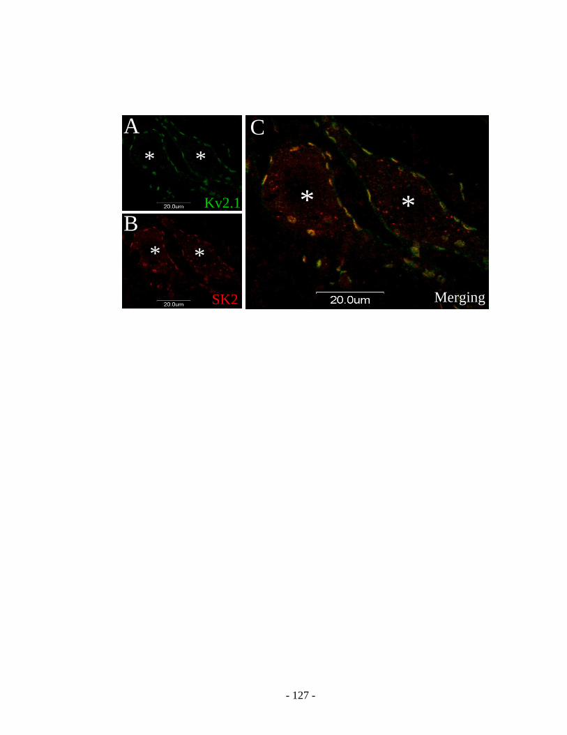
- 127 -
SK2
Kv2.1
Merging
* *
A
B
C
* *
* *
SK2
Kv2.1
Merging
* *
A
B
C
* *
* *
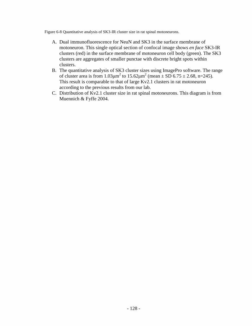
- 128 -
Figure 6-8 Quantitative analysis of SK3-IR cluster size in rat spinal motoneurons.
A. Dual immunofluorescence for NeuN and SK3 in the surface membrane of
motoneuron. This single optical section of confocal image shows en face SK3-IR
clusters (red) in the surface membrane of motoneuron cell body (green). The SK3
clusters are aggregates of smaller punctae with discrete bright spots within
clusters.
B. The quantitative analysis of SK3 cluster sizes using ImagePro software. The range
of cluster area is from 1.03μm2 to 15.62μm
2 (mean ± SD 6.75 ± 2.68, n=245).
This result is comparable to that of large Kv2.1 clusters in rat motoneuron
according to the previous results from our lab.
C. Distribution of Kv2.1 cluster size in rat spinal motoneurons. This diagram is from
Muennich & Fyffe 2004.

- 129 -
Sk3NeuN
A
Sk3NeuN
A
BDistribution of SK3 cluster sizes in rat spinal MNs
0%
10%
20%
30%
40%
0-1.9 2.0-3.9
4.0-5.9
6.0-7.9
8.0-9.9
10-11.9
12-13.9
>14
Cluster area (μm2)
Perc
en
tage
of
To
tal
clu
ste
rs
BDistribution of SK3 cluster sizes in rat spinal MNs
0%
10%
20%
30%
40%
0-1.9 2.0-3.9
4.0-5.9
6.0-7.9
8.0-9.9
10-11.9
12-13.9
>14
Cluster area (μm2)
Perc
en
tage
of
To
tal
clu
ste
rs
Distribution of SK3 cluster sizes in rat spinal MNs
0%
10%
20%
30%
40%
0-1.9 2.0-3.9
4.0-5.9
6.0-7.9
8.0-9.9
10-11.9
12-13.9
>140%
10%
20%
30%
40%
0-1.9 2.0-3.9
4.0-5.9
6.0-7.9
8.0-9.9
10-11.9
12-13.9
>14
Cluster area (μm2)
Perc
en
tage
of
To
tal
clu
ste
rs
Distribution of Kv2.1 cluster size in rat spinal MNs
~ from Muennich & Fyffe 2004 ~
C
Distribution of Kv2.1 cluster size in rat spinal MNs
~ from Muennich & Fyffe 2004 ~
C

- 130 -
CHAPTER VII Aim IV Postnatal maturation of SK channels in spinal motoneurons
Introduction
Rat spinal motoeneurons are produced on embryonic E11 to E14 day; about
half of generated motoneurons die before birth around E22. The motor behavior is
immature at birth and develops rapidly during the first 2 weeks [Vinay et al., 2000]. From
birth to P4 or P5, neonates start to be able to lift heads; from then until P12 or P13, pups
are able to lift their trunks and walk; from then until P16, they developed adult-like
locomotion, and complex motor acts [Geisler et al., 1993]. Similarly, at around P10 mice
start to be able to bear weight and walk with their abdomens suspended, which is
considered to be motor functionally mature [Jiang 1999].
The ionic currents underlying the motoneuron intrinsic membrane properties
are very important for motor function. Understanding how the ion channels mediating
these currents develop after birth will shed light on the mechanism underlying the
postnatal maturation of motoneuron properties. Medium AHP is an important
determinant for motoneuron firing properties and SK channels are believed to mediate
mAHP, so studying the development of SK channels in motoneurons will provide us
insight into the maturation of spinal motor control.
Based on the results in aim I and III, SK2 and SK3 channels form clusters at
cholinergic synapses over the surface membrane of soma and proximal dendrites in

- 131 -
motoneurons, where SK clusters are co-localized with voltage gated Kv2.1 channels and
M2 receptors. The mechanism of this specific synaptic organization is unknown, but it is
supposed to play an important role in determining the intrinsic membrane properties of
motoneurons. This aim will focus on the postnatal development of SK channel clusters at
cholinergic synapses, which is expected to shed light on the mechanism underlying the
maturation of motoneuron intrinsic properties.
The synaptic inputs onto spinal motoneurons also mature during early
postnatal period. There are inappropriate monosynaptic contacts from primary afferent
fibers on rat spinal motoneurons at birth and around seven days (P7) the majority
synaptic connections are appropriate [Vinay 2000]. Similarly, cholinergic synapses
formed by C-terminals on spinal motoneurons show a postnatal development which
corresponds to the maturation of pup‟s motor function [Wilson 2004; Ronnevi 1979]. If
the formation of SK clusters parallels the development of the cholinergic C-type synapses
in motoneurons will be investigated in this aim. At the same time, Kv2.1 channels will
also be examined at distinct postnatal ages. The expected results will provide valuable
information for understanding the mechanism underlying postnatal maturation of
motoneuron firing properties.
Methods & materials
Neonatal and adult CBA mice of both sexes will be used for this in vivo study.
Lumbar spinal cords of animals at distinct postnatal ages will be harvested for the
immunohostochemical analysis. The day of birth is considered P0, and the animals will

- 132 -
be grouped in one of the following age categories: P1-2, P5-6, P8-10, P13-14 and adult (>
6 weeks). The protocols for tissue preparation and immunohistochemistry are described
in Chapter III.
The primary antibodies against ionic channels will be used including anti-SK2
(Alomone; rabbit 1:100), anti-SK3 (Alomone; rabbit 1:100), and anti-Kv2.1 (Upstate
Biotechnology; mouse 1:250). In addition, neuronal marker anti-NeuN (neuronal nuclei;
Chemicon; mouse 1:400) and presynaptic marker anti-VAChT (vesicular acetylcholine
transportor; BD Pharmigen; Goat 1:5,000) will be employed in this study. All these
antibodies have been used in previous aims. All immunohistochemical procedures
performed in this aim are listed in Table 5. The resulting immunostaining will be
examined and imaged on confocal microscopy.
Results
The development of SK channels was investigated by studying the
immunoreactivity against SK2 and SK3 at distinct postnatal ages. At the same time, the
immunoreactivity against VAChT and Kv2.1 were also examined in order to illustrate the
maturation of pre-synaptic cholinergic C-terminals and post-synaptic Kv2.1 channels in
motoneurons respectively.
1. Postnatal development of SK channels
The immunoreactivity against SK2 in motoneurons was evident shortly after
birth (P1-2) showing discrete punctea over the surface membrane of soma and proximal
dendrites of presumed α-motoneurons in lamina IX which were revealed with NeuN

- 133 -
immunoreactivity. Some NeuN-IR presumed α-motoneurons were lack of SK2-IR
clusters. At that time the intensity of NeuN immunoreactivity in motoneuorns did not
show much difference. Then the number and the size of SK2 clusters in motoneurons
increased whereas the size of motoenurons also increased. Gradually immunoreactivity
against NeuN in presumed α-motoneurons started to show differences. At P8-10 SK2-IR
started to demonstrate differential expression among motoneurons that some
motoneurons with weaker NeuN intensity did not exhibit SK2-IR clusters. At P13-14 the
SK2 immunoreactivity in motoneurons resembled adult pattern (figure 7-1).
In contrast, no obvious SK3 immunoreactivity is visible after birth though a
few SK3-IR clusters were occasionally observed in some motoneurons from P2 to P6,
and these clusters were of very low intensity of immunoreactivity. At around P8 the
immunoreactivity against SK3 increased and started to focus on a subpopulation of
motoneurons with comparatively weaker NeuN immunoreactivity. It was around that
time motoneurons began to show differential NeuN intensity. A small portion of
presumed α-motoneuron exhibited SK3-IR clusters in the surface membrane of their
soma and proximal dendrites whereas others were lacking of SK3. At P10 it was evident
that SK3-IR motoneurons were smaller and expressed less NeuN intensity than those
lacking SK3. At P13-14 SK3 immunoreactivity looked mature and resembled adult
motoneurons (figure 7-2).
2. Postnatal development of VAChT-IR C-type synapses in motoneurons
VAChT-labeled cholinergic presynaptic terminals showed small punctea on
motoneurons and in the neuropil surrounding motoneurons at birth. At P1-2, SK2-IR
clusters in motoneurons were associated with some VAChT-IR bontons; however, the

- 134 -
sizes of these cholinergic terminals were so tiny compared with SK2 clusters that no
close apposition was observed. Then the size and the number of VAChT-IR boutons
contacting motoneurons gradually increased. The close apposition of SK2-IR and SK3-IR
clusters to VAChT-IR cholinergic presynaptic terminals was demonstrated by around P6
and the mismatching was seldom observed at this time. At P13-14 the VAChT-IR C-
terminals showed adult-like distribution and organization and their association with SK
clusters looked mature (figure 7-3; figure 7-4). Our results were consistent with previous
report in mice and rats [Wilson et al., 2004; Wetts & Vaughn 2001].
3. Postnatal development of Kv2.1 channels in motoneurons
At birth the immunoreactivity against Kv2.1 channels was shown a
continuous labeling of the surface membrane of motoneuron soma and proximal
dendrites. Then Kv2.1 channels started to form clusters, and at P5-6 Kv2.1-IR clusters
were evident in the surface membrane, and some of the Kv2.1 clusters were co-localized
with SK2-IR clusters. At P8-10 the continuous labeling of membrane disappeared and
there appeared both large and small Kv2.1-IR clusters instead. At that time all observed
SK2-IR and SK3-IR clusters were co-localized with Kv2.1. By P13-14 the
immunoreactivity against Kv2.1 and their association with VAChT-IR C-terminals and
SK channels appeared mature [Muennich & Fyffe 2004] (figure 7-5; figure 7-6).
Discussion
This study showed a different time course of SK2 versus SK3 expression after
birth. SK2 channels were expressed and showed clusters in cytoplasm membrane at birth

- 135 -
whereas SK3 channel expression was not evident. At P8 SK3 expression became evident
and showed clusters in cytoplasm membrane. In contrast, voltage gated Kv2.1 channels
were expressed at birth but the sub-cellular organization in cell surface membrane was
not mature. At P8-10 distribution and organization of Kv2.1 matured showing macro- and
micro- clusters in cytoplasm membrane instead of continuous expression. Though the two
different types of K+ channels are co-localized at same sites in motoneurons, they show
differential development after birth before clustering at cholinergic synapses.
Differential expression of SK channels appeared to become mature-like at
around P8-10 according to the immunohistochemical results in this study. This is about
the same time that muscle fiber differentiation occurs in rat. The studies of rat hindlimb
muscle fibers showed differentiating into slow contracting and fast contracting types at
around P7-P12 [Ishihara & Taguchi 1991; Picquet et al., 1997]. Our result suggests a
concurrent development of motoneurons and their target muscles that SK channel
maturation in motoneurons is in concert with muscle fiber differentiation. Motoneurons
that will differentiate into distinct types may start to show differential mAHP at this age.
However, whether mtoneuron properties guide the differentiation of its‟ target muscle
fibers or vice versa needs to be clarified in future study. Our result also supports the
hypothesis that the differential expression of SK channels in motoneurons is related to
motor unit types.
This study examined the postnatal development of both pre- and post-synaptic
components at cholinergic synapses in motoneurons. The development of post-synaptic
ionic (Kv2.1 & SK) channels occurred in concert with pre-synaptic cholinergic C-
terminals contacting motoneurons. At around P10 all three synaptic components showed

- 136 -
close apposition and co-localization. At the same time the pups start to be able to bear
weight and walk with their abdomens suspended, which is considered to be motor
functionally mature [Jiang 1999]. The time course of the postnatal development of the
components at cholinergic synapses corresponds to the maturation of motor function.

- 137 -
Table 5 Tissue, fixation, sectioning and antibodies for immunofluoresence in Aim IV
Tissue Fixation Sections Primary Abs Secondary Abs 5
Dual
immunofluorescence
for NeuN and SK3
mouse pup
spinal cord at
different ages
4%
paraformaldehyde
with 1 hour post-
fix
20 µm
on-slice
sections
NeuN (mouse
1:400)1
SK3 (rabbit 1:100)2
FITC anti-mouse
IgG 1:200
Cy3 anti-rabbit
IgG 1:300
Dual
immunofluorescence
for SK3 and kv2.1
mouse pup
spinal cord at
different ages
4%
paraformaldehyde
with 1 hour post-
fix
20 µm
on-slice
sections
SK3 (rabbit 1:100)2
Kv2.1 (mouse
1:200)3
FITC anti-mouse
IgG 1:200
Cy3 anti-rabbit
IgG 1:300
Dual
immunofluorescence
for SK3 and VAChT
mouse pup
spinal cord at
different ages
4%
paraformaldehyde
with 1 hour post-
fix
20 µm
on-slice
sections
SK3 (rabbit 1:100)2
VAChT (goat
1:5,000)4
Cy3 anti-rabbit
IgG 1:300
FITC anti-goat
IgG 1:200
Dual
immunofluorescence
for NeuN and SK2
mouse pup
spinal cord at
different ages
4%
paraformaldehyde
with 1 hour post-
fix
20 µm
on-slice
sections
NeuN (mouse
1:400)1
SK2 (rabbit 1:100)2
FITC anti-mouse
IgG 1:200
Cy3 anti-rabbit
IgG 1:300
Dual
immunofluorescence
for SK2 and kv2.1
mouse pup
spinal cord at
different ages
4%
paraformaldehyde
with 1 hour post-
fix
20 µm
on-slice
sections
SK2 (rabbit 1:100)2
Kv2.1 (mouse
1:200)3
FITC anti-mouse
IgG 1:200
Cy3 anti-rabbit
IgG 1:300
Dual
immunofluorescence
for SK2 and VAChT
mouse pup
spinal cord at
different ages
4%
paraformaldehyde
with 1 hour post-
fix
20 µm
on-slice
sections
SK2 (rabbit 1:100)2
VAChT (goat
1:5,000)4
Cy3 anti-rabbit
IgG 1:300
FITC anti-goat
IgG 1:200
___________________________________ 1 Chemicon; USA
2 Alomone; Jerusalem or Millipore/Chemicon/Upstate; Temecula, CA
3 NeuroMab; Davis, CA
4 BD Pharmingen; San Diego, CA
5 Jackson ImmunoResearch Laboratories; West Grove, PA

- 138 -
Figure 7-1 Expression of SK2 channels in postnatal developing mouse motoneurons.
A. Dual immunofluorescence for SK2 and NeuN in spinal motoneuorns of P2 mouse.
In lamina IX several motoneuorns are revealed by NeuN immunoreactivity. Each
NeuN-IR motoneuron expresses a few SK2-IR clusters in the surface membrane
of soma and promixal dendrites.
B. Dual immunofluorescence for SK2 and NeuN in spinal motoneuorns of P5 mouse.
The sizes of motoneurons and the number of SK2-IR clusters on each motoneuorn
increases.
C. Dual immunofluorescence for SK2 and NeuN in spinal motoneuorns of P8 mouse.
The range of motoneuron sizes increases at this age, so does the NeuN intensity.
Most motoneurons express SK2-IR clusters whereas one motoneuron (asterisk)
with weaker NeuN staining lacks SK2 immunoreactivity.
D. Dual immunofluorescence for SK2 and NeuN in spinal motoneuorns of P10
mouse. The SK2 and NeuN staining is more mature-like that larger motoneurons
with brighter NeuN staining express SK2-IR clusters whereas smaller
motoneurons (asterisks) with weaker NeuN labeling lack SK2 immunoreactivity.

- 139 -
A
B
C D*
*
**
A
B
C D*
*
**

- 140 -
Figure 7-2 Expression of SK3 channels in postnatal developing mouse motoneurons.
A. Dual immunofluorescence for SK3 and NeuN in spinal motoneuorns of P2 mouse.
In lamina IX several motoneuorns are revealed by NeuN immunoreactivity. No
obvious SK3 immunoreactivity is visible at this age though a few SK3-IR clusters
were occasionally observed in motoneurons.
B. Dual immunofluorescence for SK3 and NeuN in spinal motoneuorns of P5 mouse.
The sizes of motoneurons increases but there is still little SK3 immunoreactivity.
C. Dual immunofluorescence for SK3 and NeuN in spinal motoneuorns of P9 mouse.
NeuN intensity in motoneurons varies at this age and one motoneuron (asterisk)
with weaker NeuN staining expresses SK3-IR clusters while other brighter
motoneurons lack SK3 immunoreactivity.
D. Dual immunofluorescence for SK3 and NeuN in spinal motoneuorns of P13
mouse. The SK3 and NeuN staining is more mature-like that smaller motoneuron
(asterisks) with weaker NeuN intensity express SK3-IR clusters whereas larger
motoneurons with brighter NeuN staining lack SK3 immunoreactivity.

- 141 -
AB
CD
* *
AB
CD
* *

- 142 -
Figure 7-3 Postnatal development of presynaptic cholinergic C-terminals and postsynaptic SK2 channels.
A1-3: Dual immunofluorescence for SK2 and VAChT in P2 mouse spinal cord. One
motoneuron expresses a few SK2-IR clusters (red) in its surface membrane (A1). Some
small VAChT-IR punctae (green) contact this motoneuron (A2). The merging image (A3)
shows the mismatching of the two labeling though each SK2-IR cluster is associated with
some small VAChT-IR punctae.
B1-3: Dual immunofluorescence for SK2 and VAChT in P5 mouse spinal cord. One
motoneuron expresses SK2-IR clusters (red) and receives some VAChT-IR bountons
(green). Since the presynaptic bountons are larger compared to P2, the two labeling start
to match each other.
C1-3: Dual immunofluorescence for SK2 and VAChT in P8 mouse spinal cord. The size
of both postsynaptic SK2-IR clusters (C1) and presynaptic VAChT-IR terminals (C2) are
larger, and the two labeling start to show close apposition.
D1-3: Dual immunofluorescence for SK2 and VAChT in P13 mouse spinal cord. The
number and size of the presynaptic VAChT-IR bountons (D2) and postsynaptic SK2-IR
clusters (D1) are mature-like and the two labeling are closely apposed to each other (D3).

- 143 -
A3A2A1
B1 B2 B3
C1 C2 C3
D1 D2 D3
A3A2A1
B1 B2 B3
C1 C2 C3
D1 D2 D3

- 144 -
Figure 7-4 Postnatal development of presynaptic cholinergic C-terminals and postsynaptic SK3 channels.
A1-3: Dual immunofluorescence for SK3 and VAChT in P1 mouse spinal cord. One
motoneuron expresses few SK3-IR clusters with weak immunoreactivity at this age (A1).
Some small VAChT-IR punctae (green) contact this motoneuron (A2).
B1-3: Dual immunofluorescence for SK3 and VAChT in P8 mouse spinal cord. One
motoneuron expresses some visible SK3-IR clusters (red) and receives larger presynaptic
VAChT-IR bountons (green). The merging image shows the good matching of the two
immunolabeling (B3).
C1-3: Dual immunofluorescence for SK3 and VAChT in P10 mouse spinal cord. The size
and the number of both postsynaptic SK3-IR clusters (red) in motoneuron and
presynaptic VAChT-IR boutons (green) increase, and the two labeling start to show close
apposition (C3).
D1-3: Dual immunofluorescence for SK3 and VAChT in P13 mouse spinal cord. The
presynaptic VAChT-IR bountons (D2) and postsynaptic SK2-IR clusters (D1) in
motoneuron are mature-like and the two labeling are closely apposed to each other (D3).

- 145 -
D1 D2 D3
A3A2A1
B1 B2 B3
C1 C2 C3
D1 D2 D3
A3A2A1
B1 B2 B3
C1 C2 C3

- 146 -
Figure 7-5 Postnatal development of Kv2.1 channels and SK2 channels in motoneurons.
A1-3: Dual immunofluorescence for SK2 and Kv2.1 in one spinal motoneuron of P2
mouse. The motoneuron expresses a few SK2-IR clusters (A1). Different from SK2, the
immunoreactivity against Kv2.1 channels shows a continuous labeling of the surface
membrane of motoneuron soma and proximal dendrites (A2). A3 is the merging image of
the two immunostaining.
B1-3: Dual immunofluorescence for SK2 and Kv2.1 in one spinal motoneuron of P5
mouse. At this age Kv2.1 channels start to form clusters (B2), and the merging image
shows some co-localization of the two channel clusters (B3).
C1-3: Dual immunofluorescence for SK2 and Kv2.1 in one spinal motoneuron of P8
mouse. The continuous labeling of Kv2.1in cell surface membrane disappears; instead,
there appear both large and small Kv2.1-IR clusters (C2). The merging image indicates
the co-localization of SK2-IR and Kv2.1-IR clusters (C3).
D1-3: Dual immunofluorescence for SK2 and Kv2.1 in one spinal motoneuron of P13
mouse. At this age both types of ion channels look mature-like and all SK2-IR clusters
are co-localized with Kv2.1 clusters (D3).

- 147 -
A3A2A1
B1 B2 B3
C1 C2 C3
D1 D2 D3
A3A2A1
B1 B2 B3
C1 C2 C3
D1 D2 D3

- 148 -
Figure 7-6 Postnatal development of Kv2.1 channels and SK3 channels in motoneurons.
A1-3: Dual immunofluorescence for SK3 and Kv2.1 in two spinal motoneurons of P1
mouse. The motoneurons express continuous Kv2.1 immunoreactivity in their surface
membrane (A2). There is little SK3 immunoreactivity at this age (A1).
B1-3: Dual immunofluorescence for SK3 and Kv2.1 in one spinal motoneuron of P6
mouse. SK3 immunoreactivity (B1) is still weak and Kv2.1 channels start to form
clusters (B2).
C1-3: Dual immunofluorescence for SK3 and Kv2.1 in one spinal motoneuron of P8
mouse. The motoneuron expresses visible SK3-IR cluters in its surface membrane (C1).
Kv2.1 immunoreactivity shows both large and small clusters in the surface membrane of
motoneuron soma and proximal dendrites.
D1-3: Dual immunofluorescence for SK3 and Kv2.1 in one spinal motoneuron of P13
mouse. Both types of ion channel clusters are mature-like and all SK3-IR clusters are co-
localized with Kv2.1 clusters (D3).

- 149 -
A3A2A1
B1 B2 B3
C1 C2 C3
D1 D2 D3
A3A2A1
B1 B2 B3
C1 C2 C3
D1 D2 D3

- 150 -
CHAPTER VIII Aim V Modulation of SK channel expression in spinal motoneurons following
axonal injury
Introduction
Motoneurons serve to transmit neuronal commands from central nervous system
to skeleton muscles and thus control voluntary movement. Motoneuron axonal injury
resulting from peripheral nerve traumas interrupts this pathway and leads to the
dysfunction of motor control and muscle atrophy. Understanding the responses of
motoneurons to injury and the key factors determining neuron survival and axon
regeneration is of great importance for designing pharmaceutical and surgical
intervention for recovery. The long-term goal of this study is to define the structural and
functional properties underlying the excitability of motoneurons and the changes induced
by axonal injury, which will provide insight to help establish effective treatment of
traumatic nerve injuries.
The expression, distribution or physiological properties of intrinsic ionic channels
in motoneurons are subject to different types of modulation under certain circumstances
such as increased neuronal activity and ischemia [Misonou et al., 2006; Misonou et al.,
2005]. The modulation of specific ion channels underlying intrinsic currents may lead to
the increased excitability and changed firing pattern of motoneurons after axotomy.
Axotomy results in a differential modulation of AHP duration in cat motoneurons
that slow (S-type) motoneurons decrease AHP duration whereas fast (F-type)

- 151 -
motoneurons slightly increase AHP duration, leading to a de-differentiation of spinal -
MNs [Kuno et al., 1974; Gustafsson 1979; Gustafsson & Pinter 1984; Foehring et al.,
1986]. The duration of AHP in rat motoneurons innervating fast (gastrocnemius)
muscles was also observed to increase following axotomy [Gardiner & Seburn 1997].
The gain (slope of the frequency-current curve) was higher in cat axotomized
motoneurons than those in normal control [Gustafsson 1979]. A significant reduction of
AHP conductance was suggested to play a major role in the altered AHP and discharge
pattern in axotomized motoneurons [Gustafsson 1979; Titmus & Faber 1990]. However,
the mechanism underlying this modulation is unknown.
The time course of the AHP changes following axon injury has also been studied.
The decreased AHP duration in cat axotomized soleus motoneurons was observed within
10-20 days after injury; in contrast, the AHP duration in cat MG motoneurons started to
show increase about 4 weeks after axotomy [Kuno et al., 1974]. Similar results in rats
were reported. The AHP ½ decay time in rat tibial motoneurons and MG motoneurons
did not show changes 3 day and 5 day after nerve crush respectively [Bichler et al., 2007;
Nakanishi et al., 2005]. 4 weeks after axotomy the AHP ½ decay time in rat
gastrocnemius motoneurons was found to increase [Gardiner & Seburn 1997].
Since SK channels mediate ionic currents underlying medium AHP, axotomy-
induced reduction of AHP conductance in motoneurons may result from the down-
regulated gene expression, reorganized cell surface localization, and/or altered calcium
gating of SK channels. Based on the results in aim I, SK3 channels might be expressed
exclusively by slow motoneurons in rats and mice. The reduced AHP in slow
motoneurons following axon injury may result from the decreased SK3 channel density

- 152 -
in cell surface membrane, and/or decreased Ca++
sensitivity of SK3 channels. This study
will test the hypothesis that the axonal injury induced mAHP reduction in slow
motoneurons results from the decreased SK3 expression in cell surface membrane.
The results in previous aims show that SK3 clusters co-localize with Kv2.1
channels in somatic and proximal dendritic membrane of spinal motoneurons. Kv2.1
clusters were found to disperse following neuron activity and ischemic injury [Misonou
et al., 2006; Misonou et al., 2005]. How Kv2.1 clusters in spinal motoneurons are
modulated after axonal injury will also be tested in this aim. The expected results will
shed light on the mechanism underlying the modulation of ionic channels in motoneurons
by axonal injury.
Methods & materials
8 Adult wistar rats were used for this in vivo study. Lori Goss helped with the
surgeries and fluorogold injection. Two survival surgeries were performed in order to
make the axotomy model. In the first surgery, fluorogold was injected into left side soleus
muscles to retrogradely label soleus motoneurons in lumbar spinal cord. Three days later
a second surgery will be performed that left side tibial nerve was isolated and crushed for
5 seconds using forceps. The detailed procedure is described in Chapter III.
At each time points (0, 1, 3, 8 days) following nerve crush 2 animals were
transcardially perfused with fixative and the spinal cords were harvested for
immunoassay. Anti-SK3 (Alomone; rabbit 1:100) and anti-Kv2.1 (Upstate Biotechnology;
mouse 1:250) were used for this in situ immunohistochemical study. The resulting
- 153 -
immuno-staining was visualized and imaged on confocal microscopy. The en face SK3
and Kv2.1 clusters were analyzed using ImagePro software. The motoneurons in the right
side of the same spinal cord sections were selected for internal normal control. The
method of en face clusters analysis is described in Chapter III.
Results
We observed a differential regulation of postsynaptic SK clusters and Kv2.1
clusters over the surface membrane of motoneuron soma and proximal dendrites. The
presynaptic cholinergic C-terminals did not show apparent stripping/ retracting from
injured motoneurons.
1. The modulation of SK3 channel expression in motoneurons after nerve crush
The SK3 immunoreactivity in rat lumbar spinal motoneurons at different time
points (0, 1, 3, and 8 days) following nerve crush was visualized and imaged using
confocal microscopy. From the day of injury till 3 days following axotomy the SK3
clusters in the surface membrane of motoneuron soma and proximal dendrites did not
show much difference compared with normal control. Till the 8th day after injury the
SK3 clusters were still evident but the sizes were strikingly decreased compared with the
normal control. The smaller SK3 clusters did not seem resulting from cluster dissociation
because the immunoreactivity did not show any dispersion/diffusion and the number of
clusters in each motoneuron was not increased compared with the normal control (figure
8-1).

- 154 -
Motoneurons from both injury (left) side and control (right) side were sampled for
this study. The en face clusters were analyzed using ImagePro. The mean sizes (µm2) of
SK3 clusters at day 0 (mean + SD 7.2 + 3.8, n=67), day 1 (mean + SD 6.5 + 2.8, n=29),
and day 3 (mean + SD 7.2 + 3.6, n=76) following injury were not significant different
from the control side. The mean cluster sizes (µm2) at day 8 (mean + SD 3.3 + 2.1, n=71)
after injury were statistically significantly smaller than control side. The sizes of control
side SK3 clusters in all 8 animals are not statistically different from each other. See table
6 and figure 8-1 for details.
2. The alteration of Kv2.1 channel expression in motoneurons after nerve crush
In contrast to the comparatively slow modulation of SK3 after injury, Kv2.1
clusters started to show changes hours after nerve crush in the same day of injury. The
Kv2.1 clusters showed dispersing and started to distribute diffusely instead of
aggregating in the surface membrane of motoneurons. The en face cluster areas were
analyzed using ImagePro. However the mean cluster area (µm2) at day 0 (mean + SD
0.27 + 0.70, n=407) was not statistically different from the control side. From day 1 to
day 8 following injury the dispersing of Kv2.1 clusters continued and the average area of
Kv2.1 clusters decreased gradually. At day 1, 3 and 8 the Kv2.1 cluster areas (µm2) were
(mean + SD 0.24 + 0.57, n=482), (mean + SD 0.25 + 0.57, n=355), and (mean + SD 0.17
+ 0.33, n=691) respectively, and they were statistically different from the control side
(p<0.05). The sizes of control side Kv2.1 clusters in all 8 animals are not statistically
different from each other. See table 7 and figure 8-2 for details.

- 155 -
Discussion
This study showed the modulation of Kv2.1 and SK3 channel expression after
nerve crush with different time courses and in different ways in spite of the co-
localization of the two types of ion channels in motoneuorns. Kv2.1 clusters in cell
surface membrane started to break up within hours following injury, and the dispersing
continues in the first week. At the 8th
day the immuoreactivity of Kv2.1 was diffuse and
all large clusters disappeared. In contrast, SK3 clusters did not show any change till the
3rd
day after axotomy. At the 8th
day the average sizes of SK3 clusters were much smaller
than the normal control, but the immunoreactivity against SK3 did not show any
dispersion/diffusion, suggesting SK3 clusters did not dissociate as Kv2.1 did.
1. Regulation of Kv2.1 clusters following axotomy
Recent studies of in vivo or cultured hippocampal neurons showed the dynamic
regulation of Kv2.1 localization in cell surface membrane and channel biophysical
properties, which plays an important role in the control of neuron excitability. Kv2.1
channels are constitutively maintained highly phosphorylated. Various factors such as
neuronal activity, glutamate or muscarine stimulation, and ischemia may induce a graded
Kv2.1 dephosphorylation resulting in a dispersion of Kv2.1 clusters in cell surface
membrane and a hyperpolarizing shift of channel activation within minutes. These fast
modulatory effects involve calcium influx or release from intracellular calcium store and
the activation of calcineurin (PP2B) [Misonou et al., 2004, 2005; Mohapatra & Trimmer
2006]. Similar effects were observed in vitro or in vivo spinal motoneuorns that Kv2.1
clusters in surface membrane break up after sciatic nerve stimulation and glutamate or
muscarine application [unpublished results from Fyffe lab].

- 156 -
Using FRAP and quantum dot tracking techniques O‟Connell and colleagues
found that Kv2.1 channel is mobile in cell surface membrane but restrained within a
dynamic microdomain, perimeter fence, rather than tethering to a static scaffolding
protein. Cytoskeletal protein actin may play a role in cluster maintenance and localization
[O‟Connell et al., 2006]. Kv2.1 channel proteins within clusters may interact with each
other by C terminus in a phosphorylation dependent way. The de-phosphorylation could
interrupt this interaction leading to cluster dissociation and altered channel voltage-
dependent gating properties [Mohapatra & Trimmer 2006].
In this study we observed that axotomy caused Kv2.1 cluster dispersion in
motoneurons similar as the effect led by neuron activity, glutamate or acetylcholine/
muscarine stimulation, and ischemia. However, the dynamic modulation of Kv2.1
clusters induced by axotomy expressed a slower and longer time course starting from
hours and lasting for more than one week following injury. The Kv2.1 channel de-
clustering may result from the channel protein de-phosphorylation, and the channel
gating properties may be altered at the same time as discussed before. The
hyperplolarizing shift of Kv2.1 channels may result in a decrease of neuron excitability
and thus provide a protective mechanism from hyper-excitability after injury.
2. Regulation of SK clusters following axotomy
Compared to the faster regulation of Kv2.1 clusters, SK3 clusters did not show
changes within 3 days after injury. At the 8th
day the sizes of SK3 clusters were much
smaller than the normal control and it did not seem to result from the cluster dissociation
as Kv2.1 clusters did. The more stable SK clusters than Kv2.1 clusters suggest different
mechanisms for channel clustering and regulation at the same microdomains in

- 157 -
motoneuron surface membrane. In contrast to the mobile Kv2.1 proteins within clusters,
SK channels may be tethered to scaffolding proteins such as PDZ domain proteins which
cluster and organize postsynaptic neurotransmitter receptors and ion channels [Kim &
Sheng 2004; O‟Connell et al., 2006]. As a result, SK channels do not show lateral
mobility as Kv2.1 when they are regulated by injury or any other factors. The smaller SK
clusters at 8th
day may result from the internalization of channel proteins, but the exact
molecular mechanism needs to be clarified in future study.
Our results are in consistent with the electrophysiological observation of no AHP
changes within 5 days after axotomy in rat motoneurons [Bichler et al., 2007; Nakanishi
et al., 2005]. 4 weeks after axotomy the AHP ½ decay time was shown to change in rat
gastrocnemius motoneurons [Gardiner & Seburn 1997].The immunohistochemical results
in this study suggest that AHP conductance in axotomized motoneurons may be
decreased as early as 8 days. However, so far there is no available electrophysiological
data to confirm this anatomic observation.
Tetrodotoxin (TTX) is a potent neurotoxin that blocks action potentials in nerves
by binding to the pores of voltage gated Na+ channels in surface membrane. The duration
of AHP in cat soleus motoneurons was found to decrease within 8 days after TTX block
of soleus nerve, which mimics the transection of soleus nerve [Czeh et al., 1978].
However, the study in rats showed different results that TTX block did not induce
axotomy like changes in motoneuron properties [Gardiner & Seburn 1997].

- 158 -
Table 6 Regulation of SK3 cluster sizes in rat spinal motoneurons after axonal injury
Control side Injury side
Mean ± SD (μm2) Number of clusters Mean ± SD(μm
2) Number of clusters
Day 0 7.1 ± 3.1 n=71 7.2 ± 3.8 n=67
Day 1 6.7 ± 2.3 n=65 6.5 ± 2.8 n=29
Day 3 7.1 ± 2.7 n=75 7.2 ± 3.6 n=76
Day 8 7.0 ± 2.7 n=53 3.3 ± 2.1* n=71
*Significantly different from control side, P<0.001.

- 159 -
Table 7 Regulation of Kv2.1 cluster sizes in rat spinal motoneurons after axonal injury
Control side Injury side
Mean ± SD (μm2) Number of clusters Mean ± SD(μm
2) Number of clusters
Day 0 0.32 ± 0.85 n=335 0.27 ± 0.68 n=572
Day 1 0.32 ± 0.66 n=444 0.24 ± 0.57* n=482
Day 3 0.39 ± 0.83 n=440 0.25 ± 0.57** n=355
Day 8 0.33 ± 0.94 n=906 0.17 ± 0.33*** n=691
*Significantly different from control side, P<0.05;
** Significantly different from control side, P<0.01;
*** Significantly different from control side, P<0.001.

- 160 -
Figure 8-1 SK3 channels in axonally injured motoneurons.
A. Confocal images of SK3 immunofluorescence in injured motoneurons. Four
motoneurons from 4 different animals identified as axonally injured with their fluorogold
staining. The left side tibial nerves were crushed in 4 animals, and the spinal cords of the
4 animals were harvested at different time points (0, 1, 3 or 8 days after injury). At Day 0,
1, and 3, the SK3-IR clusters in the surface membrane of motoneuron soma and proximal
dendrites do not show much difference compared with control side. At day 8, SK3
clusters are still evident but the sizes are strikingly decreased compared with control side.
B. Quantitative analysis of SK3-IR cluster sizes in injured motoneurons. Motoneurons
from both injury (left) side and control (right) side were sampled for this study. The en
face clusters were analyzed using ImagePro. The mean sizes (μm2) of SK3 clusters at day
0 (mean + SD 7.2 + 3.8, n=67), day 1 (mean + SD 6.5 + 2.8, n=29), and day 3 (mean +
SD 7.2 + 3.6, n=76) following injury were not significant different from the control side.
The mean cluster sizes at day 8 (mean + SD 3.3 + 2.1, n=71) after injury were
statistically significantly smaller than control side.

- 161 -
A
B
Day 0 Day 1 Day 3 Day 80
2
4
6
8
SK
3 c
luste
r are
a (μ
m2)
Control side
Injury side
*
B
Day 0 Day 1 Day 3 Day 80
2
4
6
8
SK
3 c
luste
r are
a (μ
m2)
Control side
Injury side
*
Day 0 Day 1 Day 3 Day 80
2
4
6
8
Day 0 Day 1 Day 3 Day 80
2
4
6
8
SK
3 c
luste
r are
a (μ
m2)
Control side
Injury side
Control side
Injury side
*

- 162 -
Figure 8-2 Kv2.1 channels in axonally injured motoneurons.
A. Confocal images of Kv2.1immunofluorescence in injured motoneurons. Four
motoneurons from 4 different animals identified as axonally injured with their fluorogold
staining. The left side tibial nerves were crushed in the 4 animals, and the spinal cords of
the 4 animals were harvested at different time points (0, 1, 3 or 8 days after injury). The
immunofluorescence of Kv2.1in the 4 motoneurons is shown here. Kv2.1 clusters started
to break up hours after nerve crush in the same day of injury. From day 1 to day 8
following injury the dispersing of Kv2.1 clusters continued and the average area of Kv2.1
clusters decreased gradually
B. Quantitative analysis of Kv2.1-IR cluster sizes in injured motoneurons. The en face
cluster areas were analyzed using ImagePro. The mean cluster area (μm2) at day 0 (mean
+ SD 0.27 + 0.70, n=407) was not statistically different from the control side. At day 1, 3
and 8 the Kv2.1 cluster areas were statistically different from the control side (p<0.05).

- 163 -
A
0.0
0.1
0.2
0.3
0.4
0.5
Day 0 Day 1 Day 3 Day 8
Kv2.1
clu
ste
r are
a (μ
m2) Control side
Injury side
B
* *
*
0.0
0.1
0.2
0.3
0.4
0.5
Day 0 Day 1 Day 3 Day 8
0.0
0.1
0.2
0.3
0.4
0.5
Day 0 Day 1 Day 3 Day 8
Kv2.1
clu
ste
r are
a (μ
m2) Control side
Injury side
Control side
Injury side
B
* *
*

- 164 -
CHAPTER IX SUMMARY
To better understand the molecular basis for mAHP currents and their
regulation, we used immunohistochemistry and quantitative confocal imaging techniques
to determine the expression and sub-cellular distribution of SK channels in normal,
developing, and axonally-injured spinal motoneurons.
Several major findings emerged from my studies.
Rat and mouse spinal motoneurons differentially express specific SK channel
isoforms. Analysis of motoneuron size, intrinsic electrophysiological properties,
and target muscle profiles, as well as SK-labeling of neuromuscular junctions on
type-identified skeletal muscle fibers, supported my hypothesis that slow
motoneurons express SK3 whereas fast motoneurons express SK2. This
differential expression pattern may be partly responsible for the different AHP
properties in these neurons, although much future study is needed for an
explanation of how SK2 and SK3 mediate differential mAHP in distinct types of
motoneurons in rodents. Interestingly, I did not observe differential SK expression
in cat motoneurons, suggesting that different mechanisms underly differential
AHP properties in cat motoneurons.
I determined that SK2 and SK3 channels form clusters over the surface membrane
of the motoneuron soma and proximal dendrites. The SK clusters are
postsynaptically localized at cholinergic synapses associated with presynaptic C-
terminals and co-localized with Kv2.1 channels and M2 receptors. This
observation provides clues for a potential mechanism underlying acetylcholine
regulation of motoneuron firing properties; however, the detailed molecular

- 165 -
signaling pathway for acetylcholine/ muscarine modulation needs to be clarified
in future study.
Differential expression of SK2 & SK3 channels in motoneurons becomes
apparent around the same time that muscle fiber differentiation occurs. Postnatal
development of the key components components (Kv2.1, SK, and C-terminal) at
cholinergic synapses in motoneurons corresponds to the maturation of motor
function.
Axonal injury induces the modulation of SK3 and Kv2.1 clusters in rat
motoneurons with different time courses and in different ways. SK3 expression is
dramatically decreased 8 days after injury, which might explain the AHP
reduction in slow motoneurons.

- 166 -
The following cartoons demonstrate the SK2 and SK3 channels in normal,
developing and axonally injured rat and mouse spinal motoneurons.
P1 AdultP9
SK2
SK3
Normal After Axotomy
?

- 167 -
BIBLIOGRAPHY
Alaburda A, Perrier JF, Hounsgaard J. Mechanisms causing plateau potentials in spinal
motoneurones. Adv Exp Med Biol. 2002; 508:219-26.
Alvarez FJ, Taylor-Blake B, Fyffe RE, De Blas AL, Light AR. Distribution of immunoreactivity
for the beta 2 and beta 3 subunits of the GABAA receptor in the mammalian spinal
cord. J Comp Neurol. 1996 Feb 12; 365(3):392-412.
Alvarez FJ, Dewey DE, Carr PA, Cope TC, Fyffe RE. Downregulation of metabotropic glutamate
receptor 1a in motoneurons after axotomy. Neuroreport. 1997; 8(7): 1711-6.
Alvarez FJ, Dewey DE, Harrington DA and Fyffe REW. Cell-type specific organization of
glycine receptor clusters in the mammalian spinal cord. J Comp Neurol. 379(1997)
150-170.
Alvarez FJ, Pearson JC, Harrington D, Dewey D, Torbeck L and Fyffe REW. Distribution of 5-
hydroxytryptamine-immunoreactive boutons on alpha-motoneurons in the lumbar
spinal cord of adult cats. J Comp Neurol. 393(1998) 69-83.
Alvarez FJ, Dewey DE, McMillin P, Fyffe RE. Distribution of cholinergic contacts on Renshaw
cells in the rat spinal cord: a light microscopic study. J Physiol. 1999 Mar 15; 515 ( Pt
3):787-97.
Alvarez FJ, Fyffe RE, Dewey DE, Haftel VK, Cope TC. Factors regulating AMPA-type
glutamate receptor subunit changes induced by sciatic nerve injury in rats. J Comp
Neurol. 2000; 426(2): 229-42.
Alvarez FJ, Villalba RM, Zerda R and Schneider SP. Vesicular glutamate transporters in the
spinal cord with special reference to sensory primary afferent synapses. J. Comp.
Neurol. 472(2004) 259-282.
Allen D, Fakler B, Maylie J, Adelman JP. Organization and regulation of small conductance
Ca2+-activated K+ channel multiprotein complexes. J Neurosci. 2007 Feb 28; 27(9):
2369-76.
Antonucci DE, Lim ST, Vassanelli S, Trimmer JS. Dynamic localization and clustering of
dendritic Kv2.1 voltage-dependent potassium channels in developing hippocampal
neurons. Neuroscience. 2001; 108(1):69-81.
Arber S, Han B, Mendelsohn M, Smith M, Jessell TM, Sockanathan S. Requirement for the
homeobox gene Hb9 in the consolidation of motor neuron identity. Neuron. 1999 Aug;
23(4):659-74.
Baldissera F, Gustafsson B. Firing behaviour of a neurone model based on the
afterhyperpolarization conductance time course and algebraical summation. Adaptation
and steady state firing. Acta Physiol Scand. 1974 Sep; 92(1): 27-47.

- 168 -
Baldissera F, Gustafsson B, Parmiggiani F. Saturating summation of the afterhyperpolarization
conductance in spinal motoneurones: a mechanism for 'secondary range' repetitive
firing. Brain Res. 1978 May 5; 146(1): 69-82.
Baldissera F., Hultborn H. and Illert M. Integration in spinal neuronal system. In V. B. Brooks
(Ed.), Handbook of Physiology, Sec. I, Vol. II, Part I, American Physiological Society,
Bethesda, 1981, pp. 509-595.
Barber RP, Phelps PE, Houser CR, Crawford GD, Salvaterra PM, Vaughn JE. The morphology
and distribution of neurons containing choline acetyltransferase in the adult rat spinal
cord: an immunocytochemical study. J Comp Neurol. 1984 Nov 1; 229(3): 329-46.
Bayliss DA, Umemiya M, Berger AJ. Inhibition of N- and P-type calcium currents and the after-
hyperpolarization in rat motoneurones by serotonin. J Physiol. 1995; 485: 635-47.
Bennett DJ, Hultborn H, Fedirchuk B, Gorassini M. Synaptic activation of plateaus in hindlimb
motoneurons of decerebrate cats. J Neurophysiol. 1998 Oct; 80(4):2023-37.
Benton DC, Monaghan AS, Hosseini R, Bahia PK, Haylett DG, Moss GW. Small conductance
Ca2+-activated K+ channels formed by the expression of rat SK1 and SK2 genes in
HEK 293 cells. J Physiol. 2003; 553(Pt 1): 13-9.
Berger AJ, Takahashi T. Serotonin enhances a low-voltage-activated calcium current in rat spinal
motoneurons. J. Neurosci. 1990 Jun; 10(6):1922-8.
Bichler EK, Carrasco DI, Rich MM, Cope TC, Pinter MJ. Rat motoneuron properties recover
following reinnervation in the absence of muscle activity and evoked acetylcholine
release. Physiol. 2007 Nov 15; 585(Pt 1):47-56. Epub 2007 Sep 20.
Bichler EK, Nakanishi ST, Wang QB, Pinter MJ, Rich MM, Cope TC. Enhanced transmission at
a spinal synapse triggered in vivo by an injury signal independent of altered synaptic
activity. J Neurosci. 2007 Nov 21; 27(47):12851-9.
Bildl W., Strassmaier T., Thurm H., Andersen J., Eble S., Oliver D., Knipper M., Mann M.,
Schulte U., Adelman J. P., and Fakler B. Protein Kinase CK2 Is Coassembled with
Small Conductance Ca2+ -Activated K+ Channels and Regulates Channel Gating.
Neuron 2004; 43: 847–858.
Blaine JT, Ribera AB. Kv2 channels form delayed-rectifier potassium channels in situ. J Neurosci.
2001 Mar 1; 21(5):1473-80.
Bond CT, Herson PS, Strassmaier T, Hammond R, Stackman R, Maylie J & Adelman JP (2004).
Small conductance Ca2+-activated K+ channel knock-out mice reveal the identity of
calcium-dependent afterhyperpolarization currents. J Neurosci. 24, 5301-5306.
Bond C. T., Maylie J., and Adelman J. P. SK channels in excitability, pacemaking and synaptic
integration. Current Opinion in Neurobiology 2005, 15:305–311.
Bose P, Parmer R, Reier PJ, Thompson FJ. Morphological changes of the soleus motoneuron
pool in chronic midthoracic contused rats. Exp Neurol. 2005 Jan; 191(1):13-23.

- 169 -
Bowden SE, Fletcher S, Loane DJ, Marrion NV. Somatic colocalization of rat SK1 and D class
(Ca(v)1.2) L-type calcium channels in rat CA1 hippocampal pyramidal neurons. J
Neurosci. 2001; 21(20): RC175.
Brannstrom T. Quantitative synaptology of functionally different types of cat medial
gastrocnemius alpha-motoneurons. J Comp Neurol. 1993; 330(3): 439-54.
Brannstrom T, Kellerth JO. Changes in synaptology of adult cat spinal alpha-motoneurons after
axotomy. Exp Brain Res. 1998; 118(1): 1-13.
Brannstrom T, Kellerth JO. Recovery of synapses in axotomized adult cat spinal motoneurons
after reinnervation into muscle. Exp Brain Res. 1999; 125(1): 19-27.
Brown AG, Fyffe RE. Direct observations on the contacts made between Ia afferent fibres and
alpha-motoneurones in the cat's lumbosacral spinal cord. J Physiol. 1981; 313:121-40.
Brownstone RM. Beginning at the end: repetitive firing properties in the final common pathway.
Prog. Neurobiol. 2006 Feb-Apr; 78(3-5):156-72.
Bui TV, Ter-Mikaelian M, Bedrossian D, Rose PK. Computational estimation of the distribution
of L-type Ca(2+) channels in motoneurons based on variable threshold of activation of
persistent inward currents. J Neurophysiol. 2006 Jan; 95(1):225-41. Epub 2005 Nov 2.
Burke RE. Motor units: anatomy, physiology, and functional organization. In: Handbook of
Physiology. The Nervous System. Bethesda, MD: Am. Physiol. Soc., 1981, sect. 1, vol.
II, pt.1, chapter 10, p. 345-422.
Cai X, Liang CW, Muralidharan S, Kao JP, Tang CM, Thompson SM. Unique roles of SK and
Kv4.2 potassium channels in dendritic integration. Neuron. 2004 Oct 14;44(2):351-64.
Carlin KP, Jones KE, Jiang Z, Jordan LM, Brownstone RM. Dendritic L-type calcium currents in
mouse spinal motoneurons: implications for bistability. Eur J Neurosci. 2000 May;
12(5):1635-46.
Carr PA, Alvarez FJ, Leman EA, Fyffe RE. Calbindin D28k expression in
immunohistochemically identified Renshaw cells. Neuroreport. 1998 Aug 3;
9(11):2657-61.
Carrascal L, Nieto-Gonzalez JL, Cameron WE, Torres B, Nunez-Abades PA. Changes during the
postnatal development in physiological and anatomical characteristics of rat
motoneurons studied in vitro. Brain Res. Brain Res. Rev. 2005 Sep; 49(2): 377-87.
Chan YM, Wu W, Yip HK, So KF. Development of the regenerative capacity of postnatal
axotomized rat spinal motoneurons. Neuroreport. 2002; 13(8): 1071-4.
Chen XY, Wolpaw JR. Triceps surae motoneuron morphology in the rat: a quantitative light
microscopic study. J Comp Neurol. 1994 May 1; 343(1):143-57.
Conradi S, and Ronnevi LO. Ultrastructure and synaptology of the initial axon segment of cat
spinal motoneurons during early postnatal development. J. Neurocytol. 1977 Apr; 6(2):
195-210.

- 170 -
Conradi S, Kellerth JO, Berthold CH, Hammarberg C. Electron microscopic studies of serially
sectioned cat spinal alpha-motoneurons. IV. Motoneurons innervating slow-twitch
(type S) units of the soleus muscle. J Comp Neurol. 1979 Apr 15; 184(4):769-82.
Conradi S, Cullheim S, Gollvik L, Kellerth JO. Electron microscopic observations on the
synaptic contacts of group Ia muscle spindle afferents in the cat lumbosacral spinal
cord. Brain Res. 1983 Apr 11; 265(1):31-9.
Cullheim S, Kellerth JO, Conradi S. Evidence for direct synaptic interconnections between cat
spinal alpha-motoneurons via the recurrent axon collaterals: a morphological study
using intracellular injection of horseradish peroxidase. Brain Res. 1977 Aug 19;
132(1):1-10.
Cullheim S, Fleshman JW, Glenn LL, Burke RE. Membrane area and dendritic structure in type-
identified triceps surae alpha motoneurons. J Comp Neurol. 1987; 255(1): 68-81.
Czeh G, Gallego R, Kudo N, Kuno M. Evidence for the maintenance of motoneurone properties
by muscle activity. J. Physiol. (Lond.). 1978 Aug; 281: 239-52.
Dai CF, Kanoh N, Li KY, Wang Z. Study on facial motoneuronal death after proximal or distal
facial nerve transection. Am J Otol. 2000; 21(1): 115-8.
Dassesse D, Cuvelier L, Krebs C, Streppel M, Guntinas-Lichius O, Neiss WF, Pochet R.
Differential expression of calbindin and calmodulin in motoneurons after hypoglossal
axotomy. Brain Res. 1998; 786(1-2):181-8.
Dekkers J, Navarrete R. Persistence of somatic and dendritic growth associated processes and
induction of dendritic sprouting in motoneurones after neonatal axotomy in the rat.
Neuroreport. 1998; 9(7): 1523-7.
Dekkers J, Bayley P, Dick JR, Schwaller B, Berchtold MW, Greensmith L. Over-expression of
parvalbumin in transgenic mice rescues motoneurons from injury-induced cell death.
Neuroscience 2004; 123(2): 459-66.
Deng Z, Fyffe RE. Expression of P2X7 receptor immunoreactivity in distinct subsets of synaptic
terminals in the ventral horn of rat lumbar spinal cord. Brain Res. 2004 Sep 10;1020(1-
2):53-61.
Diss JK, Fraser SP, Djamgoz MB. Voltage-gated Na+ channels: multiplicity of expression,
plasticity, functional implications and pathophysiological aspects.Eur Biophys J. 2004
May;33(3):180-93. Review.
Elbasiouny SM, Bennett DJ, Mushahwar VK. Simulation of dendritic CaV1.3 channels in cat
lumbar motoneurons: spatial distribution. J Neurophysiol. 2005 Dec; 94(6):3961-74.
Epub 2005 Aug 24.
Eleore L, Vassias I, Vidal PP, Triller A, de Waele C. Modulation of glycine receptor subunits and
gephyrin expression in the rat facial nucleus after axotomy. Eur J Neurosci. 2005; 21(3):
669-78.

- 171 -
Faber ES, Sah P. Calcium-activated potassium channels: multiple contributions to neuronal
function. Neuroscientist. 2003; 9(3):181-94. Review
Faber ES, Delaney AJ, Sah P. SK channels regulate excitatory synaptic transmission and
plasticity in the lateral amygdala. Nat Neurosci. 2005 May; 8(5):635-41. Epub 2005
Apr 24. Comment in: Nat Neurosci. 2005 May; 8(5): 550.
Faber E. S. Louise, Delaney Andrew J., Power John M., Sedlak Petra L., Crane James W., and
Sah Pankaj. Modulation of SK Channel Trafficking by Beta Adrenoceptors Enhances
Excitatory Synaptic Transmission and Plasticity in the Amygdala. The Journal of
Neuroscience 2008; 28(43):10803–10813.
Fedirchuk B, Dai Y. Monoamines increase the excitability of spinal neurones in the neonatal rat
by hyperpolarizing the threshold for action potential production. J Physiol. 2004 Jun 1;
557(Pt 2):355-61. Epub 2004 Apr 16.
Foehring RC, Sypert GW, Munson JB. Properties of self-reinnervated motor units of medial
gastrocnemius of cat. II. Axotomized motoneurons and time course of recovery. J
Neurophysiol. 1986; 55(5): 947-65.
Fulton BP, Walton K. Electrophysiological properties of neonatal rat motoneurones studied in
vitro. J. Physiol. (Lond.) 1986 Jan; 370: 651-78.
Fyffe RE, Light AR. The ultrastructure of group Ia afferent fiber synapses in the lumbosacral
spinal cord of the cat. Brain Res. 1984 May 23; 300(2):201-9.
Fyffe RE. Spatial distribution of recurrent inhibitory synapses on spinal motoneurons in the cat. J
Neurophysiol. 1991 May; 65(5):1134-49.
Gao BX, Ziskind-Conhaim L. Development of ionic currents underlying changes in action
potential waveforms in rat spinal motoneurons. J. Neurophysiol. 1998 Dec; 80(6):
3047-61.
Garcia Del Cano G, Gerrikagoitia I, Sarasa M, Matute C, Martinez-Millan L. Ionotropic
glutamate receptor subunits are differentially regulated in the motoneuronal pools of
the rat hypoglossal nucleus in response to axotomy. J Neurocytol. 2000; 29(7): 509-23.
Gardiner PF. Physiological properties of motoneurons innervating different muscle unit types in
rat gastrocnemius. J Neurophysiol. 1993; 69(4): 1160-70.
Gardiner PF and Seburn KL. The effects of tetrodotoxin-induced muscle paralysis on the
physiological properties of muscle units and their innervating motoneurons in rat.
Journal of Physiology (1997), 499.1, pp.207-216.
Geisler HC, Westerga J, Gramsbergen A. Development of posture in the rat. Acta Neurobiol Exp
(Wars). 1993; 53(4):517-23.
Gonzalez-Forero D, Portillo F, Sunico CR, Moreno-Lopez B. Nerve injury reduces responses of
hyoglossal motoneurones to baseline and chemoreceptor-modulated inspiratory drive in
the adult rat. J Physiol. 2004 Jun 15; 557(Pt 3): 991-1011.

- 172 -
Grunnet M, Jespersen T, Perrier JF. 5-HT1A receptors modulate small-conductance Ca2+-
activated K+ channels. J. Neurosci. Res. 2004 Dec 15; 78(6):845-54.
Guntinas-Lichius O, Neiss WF, Schulte E, Stennert E. Quantitative image analysis of the
chromatolysis in rat facial and hypoglossal motoneurons following axotomy with and
without reinnervation. Cell Tissue Res. 1996 Dec; 286(3): 537-41.
Gustafsson B. Changes in motoneurone electrical properties following axotomy. J Physiol. 1979
Aug; 293:197-215.
Gustafsson B, and Pinter MJ. Effects of axotomy on the distribution of passive electrical
properties of cat motoneurones. J Physiol. 1984 Nov; 356: 433-42.
Harvey PJ, Li Y, Li X, Bennett DJ. Persistent sodium currents and repetitive firing in
motoneurons of the sacrocaudal spinal cord of adult rats. J Neurophysiol. 2006 Sep;
96(3): 1141-57. Epub 2005 Nov 9.
Harvey PJ, Li X, Li Y, Bennett DJ. 5-HT2 receptor activation facilitates a persistent sodium
current and repetitive firing in spinal motoneurons of rats with and without chronic
spinal cord injury. J Neurophysiol. 2006 Sep;96(3):1158-70. Epub 2006 May 17.
Havton L, Kellerth JO. Elimination of intramedullary axon collaterals of cat spinal alpha-
motoneurons following peripheral nerve injury. Exp Brain Res. 1990; 79(1): 65-74.
Heckman CJ, Lee RH, Brownstone RM. Hyperexcitable dendrites in motoneurons and their
neuromodulatory control during motor behavior. Trends Neurosci. 2003 Dec; 26(12):
688-95.
Hellström J, Oliveira AL, Meister B, Cullheim S. Large cholinergic nerve terminals on subsets of
motoneurons and their relation to muscarinic receptor type 2. J Comp Neurol. 2003 Jun
9; 460(4): 476-86.
Herlitze S, Garcia DE, Mackie K, Hille B, Scheuer T, Catterall WA. Modulation of Ca2+
channels by G-protein beta gamma subunits. Nature. 1996 Mar 21; 380(6571):258-62.
Hill RH, Svensson E, Dewael Y, Grillner S. 5-HT inhibits N-type but not L-type Ca(2+) channels
via 5-HT1A receptors in lamprey spinal neurons. Eur J Neurosci. 2003 Dec; 18(11):
2919-24.
Hirschberg B, Maylie J, Adelman JP, Marrion NV. Gating of recombinant small-conductance Ca-
activated K+ channels by calcium. J Gen Physiol. 1998; 111(4): 565-81.
Honma M, Namikawa K, Mansur K, Iwata T, Mori N, Iizuka H, Kiyama H. Developmental
alteration of nerve injury induced glial cell line-derived neurotrophic factor (GDNF)
receptor expression is crucial for the determination of injured motoneuron fate. J
Neurochem. 2002 Aug; 82(4): 961-75.
Hounsgaard J, Kiehn O & Mintz I (1988). Response properties of motoneurones in a slice
preparation of the turtle spinal cord. J Physiol. 398, 575-589.

- 173 -
Hougaard C, Eriksen BL, Jørgensen S, Johansen TH, Dyhring T, Madsen LS, StrøbækD,
Christophersen P. Selective positive modulation of the SK3 and SK2 subtypes of small
conductance Ca(2+)-activated K(+) channels. Br J Pharmacol. 2007 Jul; 151(5): 655-65.
Huizar P, Kuno M, Miyata Y. Differentiation of motoneurones and skeletal muscles in kittens. J.
Physiol. (Lond.). 1975 Nov; 252(2):465-79.
Hultborn H, Denton ME, Wienecke J, Nielsen JB. Variable amplification of synaptic input to cat
spinal motoneurones by dendritic persistent inward current. Physiol. 2003 Nov 1;
552(Pt 3):945-52. Epub 2003 Sep 18.
Ichiyama RM, Broman J, Edgerton VR, Havton LA. Ultrastructural synaptic features differ
between alpha- and gamma-motoneurons innervating the tibialis anterior muscle in the
rat. J Comp Neurol. 2006 Nov 10; 499(2):306-15.
Ikeda SR. Voltage-dependent modulation of N-type calcium channels by G-protein beta gamma
subunits. Nature. 1996 Mar 21; 380(6571):255-8.
Ishihara A, Taguchi S. Histochemical differentiation of fibers in the rat slow and fast twitch
muscles. Jpn. J. Physiol. 1991; 41(2):251-8.
Ishihara A, Ohira Y, Tanaka M, Nishikawa W, Ishioka N, Higashibata A, Izumi R, Shimazu T,
Ibata Y. Cell body size and succinate dehydrogenase activity of spinal motoneurons
innervating the soleus muscle in mice, rats, and cats. Neurochem Res. 2001
Dec;26(12):1301-4.
Jiang Z, Rempel J, Li J, Sawchuk MA, Carlin KP, Brownstone RM. Development of L-type
calcium channels and a nifedipine-sensitive motor activity in the postnatal mouse
spinal cord. Eur. J. Neurosci. 1999 Oct; 11(10):3481-7.
Johnson IP. A quantitative ultrastructural comparison of alpha and gamma motoneurons in the
thoracic region of the spinal cord of the adult cat. J Anat. 1986 Aug;147:55-72.
Johnson IP, Sears TA. Organelle changes in cat thoracic alpha- and gamma-motoneurons
following axotomy. Brain Res. 1989 Jun 12; 489(2): 400-5.
Jonas EA, Kaczmarek LK. Regulation of potassium channels by protein kinases. Curr Opin
Neurobiol. 1996 Jun; 6(3):318-23.
Kanda K, Hashizume K. Factors causing difference in force output among motor units in the rat
medial gastrocnemius muscle. J Physiol. 1992 Mar; 448:677-95.
Kellerth JO, Mellstrom A, Skoglund S. Postnatal excitability changes of kitten motoneurones.
Acta Physiol. Scand. 1971 Sep; 83(1):31-41.
Kellerth JO, Berthold CH, Conradi S. Electron microscopic studies of serially sectioned cat spinal
alpha-motoneurons. III. Motoneurons innervating fast-twitch (type FR) units of the
gastrocnemius muscle. J Comp Neurol. 1979 Apr 15; 184(4):755-67.
Kernell D. Repetitive impulse firing in motoneurons: facts and perspectives. in Progress in Brain
Research Vol 123. Edited by Binder M. D. 1999 Elsevier Science BV.

- 174 -
Kim E, Sheng M. PDZ domain proteins of synapses. Nat Rev Neurosci. 2004 Oct; 5(10):771-81.
Kiss T. Persistent Na-channels: origin and function. A review. Acta Biol Hung. 2008; 59 Suppl:1-
12.
Kohler M, Hirschberg B, Bond CT, Kinzie JM, Marrion NV, Maylie J & Adelman JP (1996).
Small-conductance, calcium-activated potassium channels from mammalian brain.
Science. 273, 1709-1714.
Krebs C, Neiss WF, Streppel M, Guntinas-Lichius O, Dassesse D, Stennert E, Pochet R.
Axotomy induces transient calbindin D28K immunoreactivity in hypoglossal
motoneurons in vivo. Cell Calcium. 1997 Nov; 22(5): 367-72.
Kuno M, Miyata Y, Munoz-Martinez EJ. Differential reaction of fast and slow alpha-
motoneurones to axotomy. J Physiol. 1974 Aug; 240(3): 725-39.
Kuo JJ, Lee RH, Zhang L, Heckman CJ. Essential role of the persistent sodium current in spike
initiation during slowly rising inputs in mouse spinal neurones. J Physiol. 2006 Aug 1;
574(Pt 3):819-34. Epub 2006 May 25.
Lacinova L. Voltage-dependent calcium channels. Gen Physiol Biophys. 2005; 24 Suppl 1: 1-78.
Lagerbäck PA. An ultrastructural study of cat lumbosacral gamma-motoneurons after retrograde
labelling with horseradish peroxidase. J Comp Neurol. 1985 Oct 15; 240(3):256-64.
Lagerbäck PA, Cullheim S, Ulfhake B. Electron microscopic observations on the synaptology of
cat sciatic gamma-motoneurons after intracellular staining with horseradish peroxidase.
Neurosci Lett. 1986 Sep 25; 70(1):23-7.
Lape R, Nistri A. Current and voltage clamp studies of the spike medium afterhyperpolarization
of hypoglossal motoneurons in a rat brain stem slice preparation. J Neurophysiol. 2000
May; 83(5): 2987-95.
Lee RH, Heckman CJ. Essential role of a fast persistent inward current in action potential
initiation and control of rhythmic firing. J Neurophysiol. 2001 Jan; 85(1):472-5.
Lee WS, Ngo-Anh TJ, Bruening-Wright A, Maylie J, Adelman JP. Small conductance Ca2+-
activated K+ channels and calmodulin: cell surface expression and gating. J Biol Chem.
2003 Jul 11; 278(28): 25940-6.
Li X. and Bennett DJ. Apamin-Sensitive Calcium-Activated Potassium Currents (SK) Are
Activated by Persistent Calcium Currents in Rat Motoneurons. J Neurophysiol 97:
3314–3330, 2007.
Li Y, Bennett DJ. Persistent sodium and calcium currents cause plateau potentials in motoneurons
of chronic spinal rats. J Neurophysiol. 2003 Aug; 90(2):857-69. Epub 2003 Apr 30.
Lin MT, Luján R, Watanabe M, Adelman JP, Maylie J. SK2 channel plasticity contributes to LTP
at Schaffer collateral-CA1 synapses. Nat Neurosci. 2008 Feb;11(2):170-7. Epub 2008
Jan 20.

- 175 -
Linda H, Shupliakov O, Ornung G, Ottersen OP, Storm-Mathisen J, Risling M, Cullheim S.
Ultrastructural evidence for a preferential elimination of glutamate-immunoreactive
synaptic terminals from spinal motoneurons after intramedullary axotomy. J Comp
Neurol. 2000 Sep 11; 425(1): 10-23.
MacDermid VE, Neuber-Hess MS, Rose PK. The temporal sequence of morphological and
molecular changes in axotomized feline motoneurons leading to the formation of axons
from the ends of dendrites. J Comp Neurol. 2004 Jan 6; 468(2): 233-50.
Maingret F, Coste B, Hao J, Giamarchi A, Allen D, Crest M, Litchfield DW, Adelman JP,
Delmas P. Neurotransmitter modulation of small-conductance Ca2+-activated K+
channels by regulation of Ca2+ gating. Neuron. 2008 Aug 14; 59(3):439-49.
Manuel M, Meunier C, Donnet M, Zytnicki D. How much afterhyperpolarization conductance is
recruited by an action potential? A dynamic-clamp study in cat lumbar motoneurons. J
Neurosci. 2005 Sep 28; 25(39):8917-23.
Marin O, Meggio F, Pinna LA. Structural features underlying the unusual mode of calmodulin
phosphorylation by protein kinase CK2: A study with synthetic calmodulin fragments.
Biochem Biophys Res Commun. 1999 Mar 16; 256(2): 442-6.
Marrion NV and Tavalin SJ. Selective activation of Ca2+-activated K+ channels by co-localized
Ca2+ channels in hippocampal neurons. Nature.1998 Oct 29; 395(6705): 900-5.
Maxwell D. J., Hartley R., Arber S., Pierani A., Jessell T. M. and Todd A. J. Transmitter
phenotype and axonal projection of neurons originating from ventral progenitor
domains in the mouse spinal cord. SfN 2003 186.5 abstract
Maylie J, Bond CT, Herson PS, Lee WS, Adelman JP. Small conductance Ca2+-activated K+
channels and calmodulin. J Physiol. 2004 Jan 15; 554(Pt 2): 255-61.
McIlwain DL, Hoke VB. The role of the cytoskeleton in cell body enlargement, increased nuclear
eccentricity and chromatolysis in axotomized spinal motor neurons. BMC Neurosci.
2005 Mar 17; 6(1): 19.
McLarnon JG (1995). Potassium currents in motoneurones. Prog Neurobiol. 47, 513-531.
Mendell LM. The size principle: a rule describing the recruitment of motoneurons. J
Neurophysiol 93: 3024-3026, 2005 EDITORIAL FOCUS
Mentis GZ, Díaz E, Moran LB, Navarrete R. Early alterations in the electrophysiological
properties of rat spinal motoneurones following neonatal axotomy. J Physiol. 2007 Aug
1; 582(Pt 3): 1141-61.
Meunier C, Borejsza K. How membrane properties shape the discharge of motoneurons: a
detailed analytical study. Neural Comput. 2005 Nov; 17(11):2383-420.
Miles GB, Dai Y, Brownstone RM. Mechanisms underlying the early phase of spike frequency
adaptation in mouse spinal motoneurones. J Physiol. 2005; 566(Pt 2): 519-32.

- 176 -
Misonou H, Mohapatra DP, Park EW, Leung V, Zhen D, Misonou K, Anderson AE,Trimmer JS.
Regulation of ion channel localization and phosphorylation by neuronal activity. Nat
Neurosci. 2004 Jul; 7(7):711-8. Epub 2004 Jun 13.
Misonou H, Mohapatra DP, Menegola M, Trimmer JS. Calcium- and metabolic state-dependent
modulation of the voltage-dependent Kv2.1 channel regulates neuronal excitability in
response to ischemia. J Neurosci. 2005 Nov 30;25(48):11184-93.
Mohapatra DP, Trimmer JS. The Kv2.1 C terminus can autonomously transfer Kv2.1-like
phosphorylation-dependent localization, voltage-dependent gating, and muscarinic
modulation to diverse Kv channels. J Neurosci. 2006 Jan 11; 26(2):685-95.
Monaghan AS, Benton DC, Bahia PK, Hosseini R, Shah YA, Haylett DG, Moss GW. The SK3
subunit of small conductance Ca2+-activated K+ channels interacts with
both SK1 and SK2 subunits in a heterologous expression system. J Biol Chem. 2004 Jan 9;
279(2): 1003-9.
Mongan LC, Hill MJ, Chen MX, Tate SN, Collins SD, Buckby L, Grubb BD. The distribution of
small and intermediate conductance calcium-activated potassium channels in the rat
sensory nervous system. Neuroscience. 2005;131(1):161-75.
Muennich EA, Fyffe RE. Focal aggregation of voltage-gated, Kv2.1 subunit-containing,
potassium channels at synaptic sites in rat spinal motoneurones. J Physiol. 2004 Feb 1;
554 (Pt 3): 673-85.
Murthy SR, Teodorescu G, Nijholt IM, Dolga AM, Grissmer S, Spiess J, Blank T. Identification
and characterization of a novel, shorter isoform of the small conductance Ca(2+)-
activated K(+) channel SK2. J Neurochem. 2008 Jul 30. [Epub ahead of print]
Nakanishi ST, Cope TC, Rich MM, Carrasco DI, Pinter MJ. Regulation of motoneuron
excitability via motor endplate acetylcholine receptor activation. J Neurosci. 2005;
25(9): 2226-32.
Ngo-Anh TJ, Bloodgood BL, Lin M, Sabatini BL, Maylie J, Adelman JP. SK channels and
NMDA receptors form a Ca2+-mediated feedback loop in dendritic spines. Nat
Neurosci. 2005 May; 8(5):642-9. Epub 2005 Apr 24. Comment in: Nat Neurosci. 2005
May; 8(5): 550.
Obermair GJ, Kaufmann WA, Knaus HG & Flucher BE (2003). The small conductance Ca2+-
activated K+ channel SK3 is localized in nerve terminals of excitatory synapses of
cultured mouse hippocampal neurons. Eur J Neurosci. 17, 721-731.
O'Connell KM, Rolig AS, Whitesell JD, Tamkun MM. Kv2.1 potassium channels are retained
within dynamic cell surface microdomains that are defined by a perimeter fence. J
Neurosci. 2006 Sep 20; 26(38):9609-18.
Oomura Y, Maeno T. Does the neurone soma actually generate action potentials? Nature. 1963
Jan 26; 197:358-9.
Ornung G, Shupliakov O, Linda H, Ottersen OP, Storm-Mathisen J, Ulfhake B, Cullheim S.
Qualitative and quantitative analysis of glycine- and GABA-immunoreactive nerve

- 177 -
terminals on motoneuron cell bodies in the cat spinal cord: a postembedding electron
microscopic study. J Comp Neurol. 1996 Feb 12; 365(3):413-26.
Ornung G, Ottersen OP, Cullheim S, Ulfhake B. Distribution of glutamate-, glycine- and GABA-
immunoreactive nerve terminals on dendrites in the cat spinal motor nucleus. Exp
Brain Res. 1998 Feb; 118(4):517-32.
Park KS, Mohapatra DP, Misonou H, Trimmer JS. Graded regulation of the Kv2.1 potassium
channel by variable phosphorylation. Science. 2006 Aug 18; 313(5789):976-9.
Pedarzani P, Mosbacher J, Rivard A, Cingolani LA, Oliver D, Stocker M, Adelman JP, Fakler B.
Control of electrical activity in central neurons by modulating the gating of small
conductance Ca2+-activated K+ channels. J Biol Chem. 2001; 276(13): 9762-9.
Perrier JF, Hounsgaard J. 5-HT2 receptors promote plateau potentials in turtle spinal
motoneurons by facilitating an L-type calcium current. J Neurophysiol. 2003 Feb;
89(2):954-9.
Peyghambari F, Valojerdi MR, Tiraihi T. A morphometric study on the early stages of dendrite
changes in the axotomized motoneuron of the spinal cord in newborn rats. Neurol Res.
2005 Sep; 27(6): 586-90.
Picquet F, Stevens L, Butler-Browne GS, Mounier Y. Contractile properties and myosin heavy
chain composition of newborn rat soleus muscles at different stages of postnatal
development. J. Muscle Res. Cell. Motil. 1997 Feb; 18(1):71-9.
Pinter MJ, Vanden Noven S, Muccio D, Wallace N. Axotomy-like changes in cat motoneuron
electrical properties elicited by botulinum toxin depend on the complete elimination of
neuromuscular transmission. J Neurosci. 1991 Mar; 11(3): 657-66.
Pollin MM, McHanwell S, Slater CR. The effect of age on motor neurone death following
axotomy in the mouse. Development. 1991 May; 112(1): 83-9.
Powers RK, Sawczuk A, Musick JR, Binder MD. Multiple mechanisms of spike-frequency
adaptation in motoneurones. J Physiol Paris. 1999; 93(1-2): 101-14.
Rekling JC, Funk GD, Bayliss DA, Dong XW & Feldman JL (2000). Synaptic control of
motoneuronal excitability. Physiol Rev. 80, 767-852.
Ren Y, Barnwe LF, Alexander JC, Lubin FD, Adelman JP, Pfaffinger PJ, Schrader LA, and
Anderson AE. Regulation of Surface Localization of the Small Conductance Ca2+-
activated Potassium Channel, Sk2, through Direct Phosphorylation by cAMP-
dependent Protein Kinase. The Journal of Biological Chemistry 2006; 281(17): 11769–
11779.
Réthelyi M, Metz CB, Lund PK. Distribution of neurons expressing calcitonin gene-related
peptide mRNAs in the brain stem, spinal cord and dorsal root ganglia of rat and guinea-
pig. Neuroscience. 1989; 29(1):225-39.
Ribera AB. Potassium currents in developing neurons. Ann N Y Acad Sci. 1999; 868:399-405.

- 178 -
Rioult-Pedotti MS. Intrinsic NMDA-induced oscillations in motoneurons of an adult vertebrate
spinal cord are masked by inhibition. J Neurophysiol. 1997 Feb; 77(2):717-30.
Roncarati R, Di Chio M, Sava A, Terstappen GC & Fumagalli G (2001). Presynaptic localization
of the small conductance calcium-activated potassium channel SK3 at the
neuromuscular junction. Neuroscience. 104, 253-262.
Ronnevi LO. Spontaneous phagocytosis of C-type synaptic terminals by spinal alpha-
motoneurons in newborn kittens. An electron microscopic study. Brain Res. 1979 Feb
23; 162(2): 189-99.
Rose PK, Odlozinski M. Expansion of the dendritic tree of motoneurons innervating neck
muscles of the adult cat after permanent axotomy. J Comp Neurol. 1998 Jan 19; 390(3):
392-411.
Rose PK, MacDermid V, Joshi M, Neuber-Hess M. Emergence of axons from distal dendrites of
adult mammalian neurons following a permanent axotomy. Eur J Neurosci. 2001 Mar;
13(6):1166-76.
Safronov BV, Vogel W. Large conductance Ca(2+)-activated K+ channels in the soma of rat
motoneurones. J Membr Biol. 1998 Mar 1; 162(1): 9-15.
Safronov BV, Vogel W. Single voltage-activated Na+ and K+ channels in the somata of rat
motoneurones. J Physiol. 1995 Aug 15; 487 ( Pt 1):91-106.
Sah P, McLachlan EM. Potassium currents contributing to action potential repolarization and the
afterhyperpolarization in rat vagal motoneurons. J Neurophysiol. 1992 Nov; 68(5):
1834-41.
Sah P (1996). Ca(2+)-activated K+ currents in neurones: types, physiological roles and
modulation. Trends Neurosci. 19, 150-154.
Sah P, Faber ES. Channels underlying neuronal calcium-activated potassium currents. Prog
Neurobiol. 2002 Apr; 66(5):345-53.
Sailer CA, Hu H, Kaufmann WA, Trieb M, Schwarzer C, Storm JF, Knaus HG. Regional
differences in distribution and functional expression of small-conductance Ca2+-
activated K+ channels in rat brain. J Neurosci. 2002 Nov 15; 22(22): 9698-707.
Sailer CA, Kaufmann WA, Marksteiner J & Knaus HG (2004). Comparative
immunohistochemical distribution of three small-conductance Ca(2+)-activated
potassium channel subunits, SK1, SK2, and SK3 in mouse brain. Mol Cell Neurosci. 26,
458-469.
Santafé MM, Lanuza MA, Garcia N, Tomàs J. Muscarinic autoreceptors modulate transmitter
release through protein kinase C and protein kinase A in the rat motor nerve terminal.
Eur J Neurosci. 2006 Apr; 23(8):2048-56.
Sato M, Mizuno N, Konishi A. Postnatal differentiation of cell body volumes of spinal
motoneurons innervating slow-twitch and fast-twitch muscles. J. Comp. Neurol. 1977
Sep 1; 175(1): 27-36.

- 179 -
Sawczuk A, Powers RK, Binder MD. Contribution of outward currents to spike-frequency
adaptation in hypoglossal motoneurons of the rat. J Neurophysiol. 1997
Nov;78(5):2246-53.
Schumacher MA, Rivard AF, Bachinger HP, Adelman JP. Structure of the gating domain of a
Ca2+-activated K+ channel complexed with Ca2+/calmodulin. Nature. 2001;
410(6832): 1120-4.
Schwindt PC, Crill WE. Properties of a persistent inward current in normal and TEA-injected
motoneurons. J Neurophysiol. 1980 Jun; 43(6): 1700-24.
Shapiro MS, Loose MD, Hamilton SE, Nathanson NM, Gomeza J, Wess J, Hille B. Assignment
of muscarinic receptor subtypes mediating G-protein modulation of Ca(2+) channels by
using knockout mice. Proc Natl Acad Sci U S A. 1999 Sep 14; 96(19): 10899-904.
Simon M, Perrier JF, Hounsgaard J. Subcellular distribution of L-type Ca2+ channels responsible
for plateau potentials in motoneurons from the lumbar spinal cord of the turtle. Eur J
Neurosci. 2003 Jul; 18(2):258-66.
Staron RS, Kraemer WJ, Hikida RS, Fry AC, Murray JD, Campos GE. Fiber type composition of
four hindlimb muscles of adult Fisher 344 rats. Histochem Cell Biol. 1999 Feb;
111(2):117-23.
Stocker M and Pedarzani P (2000). Differential distribution of three Ca(2+)-activated K(+)
channel subunits, SK1, SK2, and SK3, in the adult rat central nervous system. Mol Cell
Neurosci. 15, 476-493.
Stocker M. Ca(2+)-activated K+ channels: molecular determinants and function of the SK family.
Nat Rev Neurosci. 2004; 5(10): 758-70.
Strassmaier T, Bond CT, Sailer CA, Knaus HG, Maylie J, Adelman JP. A novel isoform of SK2
assembles with other SK subunits in mouse brain. J Biol Chem. 2005; 280(22): 21231-
6.
Svirskis G, Hounsgaard J. Transmitter regulation of plateau properties in turtle motoneurons. J
Neurophysiol. 1998 Jan; 79(1):45-50.
Swett JE, Wikholm RP, Blanks RH, Swett AL, Conley LC. Motoneurons of the rat sciatic nerve.
Exp Neurol. 1986 Jul; 93(1): 227-52.
Teagarden M, Atherton JF, Bevan MD, Wilson CJ. Accumulation of cytoplasmic calcium, but
not apamin-sensitive afterhyperpolarization current, during high frequency firing in rat
subthalamic nucleus cells. J Physiol. 2008 Feb 1; 586(3):817-33.
Thomas RC, Wilson VJ. Precise localization of Renshaw cells with a new marking technique.
Nature. 1965 Apr 10; 206(980):211-3.
Tiraihi T, Rezaie MJ. Synaptic lesions and synaptophysin distribution change in spinal
motoneurons at early stages following sciatic nerve transection in neonatal rats. Brain
Res Dev Brain Res. 2004 Jan 31; 148(1): 97-103.

- 180 -
Titmus MJ, Faber DS. Axotomy-induced alterations in the electrophysiological characteristics of
neurons. Prog Neurobiol. 1990; 35(1):1-51.
Ulfhake B, Cullheim S. Postnatal development of cat hind limb motoneurons. II: In vivo
morphology of dendritic growth cones and the maturation of dendrite morphology. J
Comp Neurol. 1988a Dec 1; 278(1):88-102.
Ulfhake B, Cullheim S. Postnatal development of cat hind limb motoneurons. III: Changes in
size of motoneurons supplying the triceps surae muscle. J Comp Neurol. 1988b Dec 1;
278(1): 103-20.
Ulfhake B, Cullheim S, Franson P. Postnatal development of cat hind limb motoneurons. I:
Changes in length, branching structure, and spatial distribution of dendrites of cat
triceps surae motoneurons. J Comp Neurol. 1988 Dec 1; 278(1):69-87. Erratum in: J
Comp Neurol 1989 Aug 22; 286(4):541.
Van Dongen PA, Grillner S, Hokfelt T. 5-Hydroxytryptamine (serotonin) causes a reduction in
the afterhyperpolarization following the action potential in lamprey motoneurons and
premotor interneurons. Brain Res. 1986 Feb 26; 366(1-2): 320-5.
Vergara C, Latorre R, Marrion NV, and Adelman JP. Calcium-activated potassium channels.
Current Opinion in Neurobiology 1998, 8:321-329.
Viana F, Bayliss DA, Berger AJ. Multiple potassium conductances and their role in action
potential repolarization and repetitive firing behavior of neonatal rat hypoglossal
motoneurons. J Neurophysiol. 1993 Jun; 69(6):2150-63.
Vinay L, Brocard F, Pflieger JF, Simeoni-Alias J, Clarac F. Perinatal development of lumbar
motoneurons and their inputs in the rat. Brain Res Bull. 2000 Nov 15;53(5): 635-47.
Westenbroek RE, Hoskins L, Catterall WA. Localization of Ca2+ channel subtypes on rat spinal
motor neurons, interneurons, and nerve terminals. J Neurosci. 1998 Aug 15;
18(16):6319-30.
Wetts R, Vaughn JE. Development of cholinergic terminals around rat spinal motor neurons and
their potential relationship to developmental cell death. J Comp Neurol. 2001 Jun 25;
435(2):171-83.
Wichterle H, Lieberam I, Porter JA, Jessell TM. Directed differentiation of embryonic stem cells
into motor neurons. Cell. 2002 Aug 9; 110(3):385-97.
Wilson JM, Rempel J, and Brownstone RM. Postnatal development of cholinergic synapses on
mouse spinal motoneurons. J Comp Neurol. 2004 Jun 14; 474(1): 13-23.
Wissmann R, Bildl W, Neumann H, Rivard AF, Klocker N, Weitz D, Schulte U, Adelman JP,
Bentrop D, Fakler B. A helical region in the C terminus of small-conductance Ca2+-
activated K+ channels controls assembly with apo-calmodulin. J Biol Chem. 2002 Feb
8; 277(6): 4558-64.

- 181 -
Xia XM, Fakler B, Rivard A, Wayman G, Johnson-Pais T, Keen JE, Ishii T, Hirschberg B, Bond
CT, Lutsenko S, Maylie J & Adelman JP (1998). Mechanism of calcium gating in
small-conductance calcium-activated potassium channels. Nature. 395, 503-507.
Zengel JE, Reid SA, Sypert GW, Munson JB. Membrane electrical properties and prediction of
motor-unit type of medial gastrocnemius motoneurons in the cat. J Neurophysiol. 1985
May; 53(5): 1323-44.
Zhang L & Krnjevic K (1987). Apamin depresses selectively the after-hyperpolarization of cat
spinal motoneurons. Neurosci Lett. 74(1987) 58-62.
Zhou H, Das S, Murthy KS. Erk1/2- and p38 MAP kinase-dependent phosphorylation and
activation of cPLA2 by m3 and m2 receptors. Am J Physiol Gastrointest Liver Physiol.
2003 Mar; 284(3):G472-80.