sleep and consciousness - college of the canyons...sleep and dreaming figure 15.3: sleep and wake...
TRANSCRIPT

SleepandConsciousness Sleep and Dreaming
The Neural Basis of Consciousness Gar
rett:
Bra
in &
Beh
avio
r 4e
7/29
/19
1

SleepandDreaming • Consciousness is tough to study... but sleep is
• *readily observable, consists of different levels of consciousness, and can be studied scientifically
• The purpose of sleep is unclear. • *Restorative Hypothesis: busier we are, more sleep we need
Gar
rett:
Bra
in &
Beh
avio
r 4e
7/29
/19
• Species with higher metabolic rates typically spend more time in
• The amount of sleep depends on the availability of food and on
sleep • CSF circulates during sleep to remove toxins
• Adaptive Hypothesis
safety considerations. • Vulnerable animals without shelter (cattle) and those that need2to spend hours feeding (elephants) sleep very little.
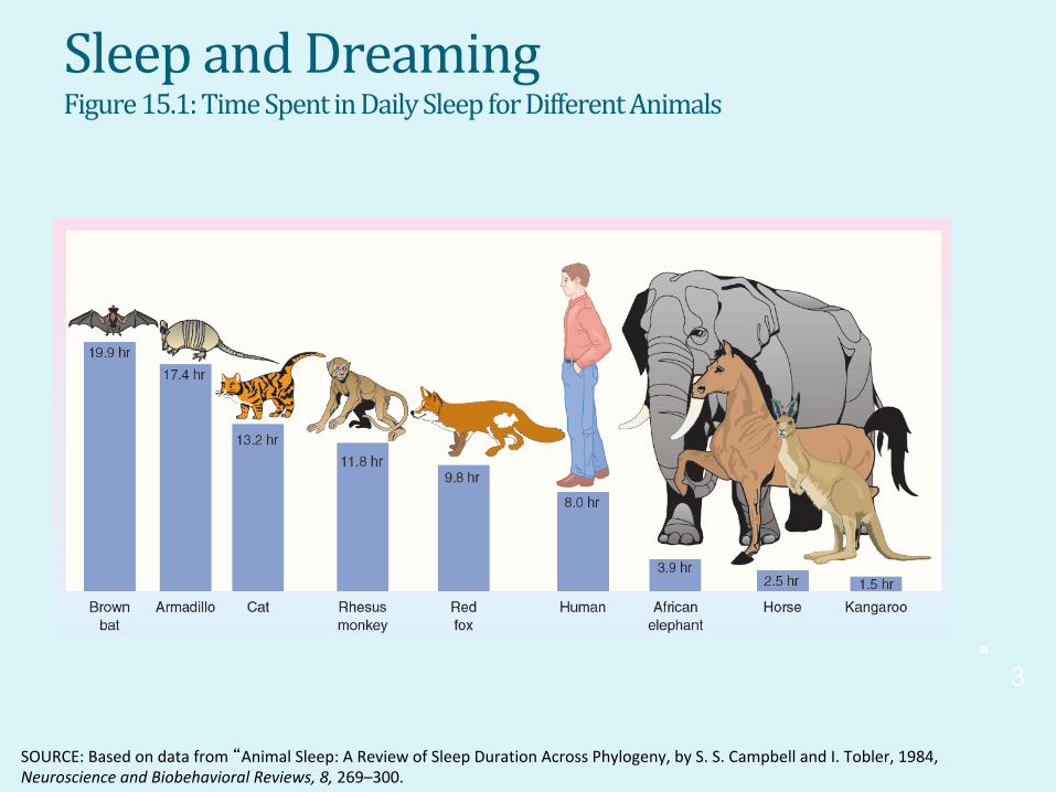
SleepandDreamingFigure 15.1: Time Spent in Daily Sleep for Different Animals
Gar
rett:
Bra
in &
Beh
avio
r 4e
SOURCE: Based on data from “Animal Sleep: A Review of Sleep Duration Across Phylogeny, by S. S. Campbell and I. Tobler, 1984, Neuroscience and Biobehavioral Reviews, 8, 269–300.
3
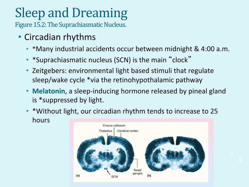
SleepandDreamingFigure 15.2: The Suprachiasmatic Nucleus.
• Circadian rhythms • *Many industrial accidents occur between midnight & 4:00 a.m. • *Suprachiasmatic nucleus (SCN) is the main “clock”
• Zeitgebers: environmental light based stimuli that regulate sleep/wake cycle *via the retinohypothalamic pathway
• Melatonin, a sleep-inducing hormone released by pineal gland is *suppressed by light.
• *Without light, our circadian rhythm tends to increase to 25 hours
Gar
rett:
Bra
in &
Beh
avio
r 4e
4
7/29
/19

Gar
rett:
Bra
in&
Beh
avio
r 4e
SleepandDreamingFigure 15.3: Sleep and Wake Periods During Isolation From Time Cues
• Rhythms During Waking and Sleeping • Ultradianrhythms are cycles that are shorter than a day
• The basic rest and activity cycle is *90-100 minutes long
• *The ‘after lunch’ siesta or break coincides with a natural ultradian rhythm rest period.
SOURCE: From Introduction to Psychology, Gateways to Mind and Behavior (with InfoTrac) 9th edition, by Coon, 2001. Reprinted with permission of Wadsworth, a division of Thomson Learning.
5
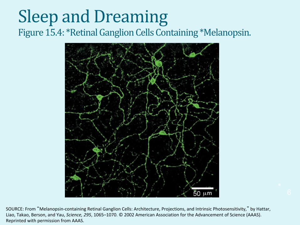
SleepandDreamingFigure 15.4: *Retinal Ganglion Cells Containing *Melanopsin.
6
Gar
rett:
Bra
in &
Beh
avio
r 4e
SOURCE: From “Melanopsin-containing Retinal Ganglion Cells: Architecture, Projections, and Intrinsic Photosensitivity,” by Hattar, Liao, Takao, Berson, and Yau, Science, 295, 1065–1070. © 2002 American Association for the Advancement of Science (AAAS). Reprinted with permission from AAAS.
Gar
rett:
Bra
in&
Beh
avio
r 4e
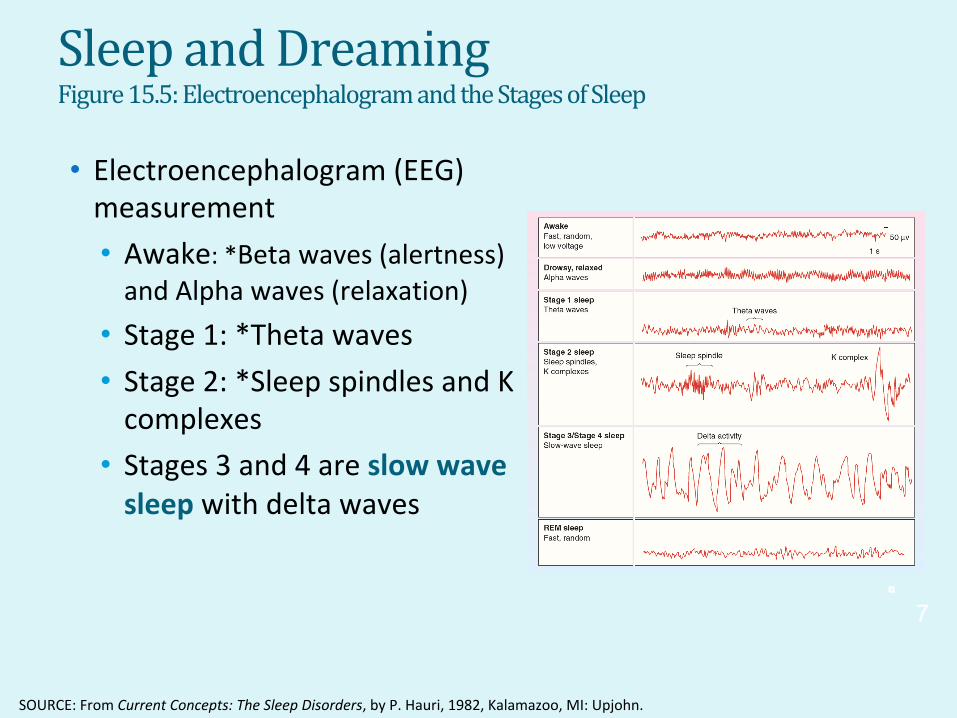
SleepandDreamingFigure 15.5: Electroencephalogram and the Stages of Sleep
• Electroencephalogram (EEG) measurement • Awake: *Beta waves (alertness) and Alpha waves (relaxation)
• Stage 1: *Theta waves • Stage 2: *Sleep spindles and K complexes
• Stages 3 and 4 are slowwave sleep with delta waves
7/29
/19
SOURCE: From Current Concepts: The Sleep Disorders, by P. Hauri, 1982, Kalamazoo, MI: Upjohn.
7

Figure 15.6: Time Spent in VariousSleepStagesDuringtheNight • The sleeper returns through the stages in reverse order, and then heads into REM sleep for the first time • Thereafter the percentage of SWS declines with each subsequent cycle
• *Cycling through each series of stages takes about 90 minutes.
Gar
rett:
Bra
in &
Beh
avio
r 4e
8
Gar
rett:
Bra
in&
Beh
avio
r 4e
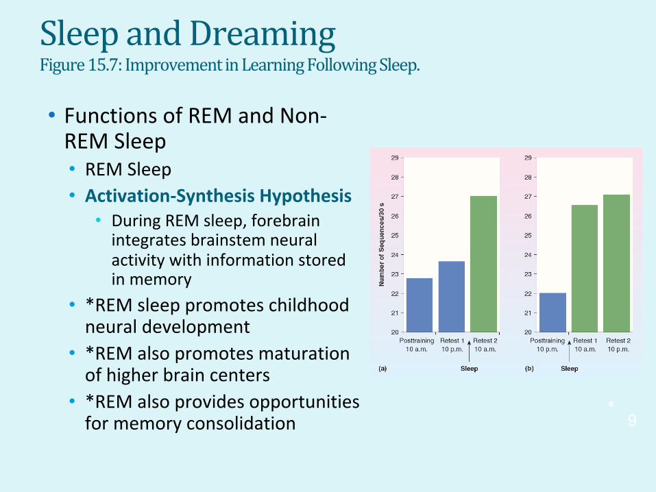
SleepandDreamingFigure 15.7: Improvement in Learning Following Sleep.
• Functions of REM and Non-REM Sleep • REM Sleep • Activation-Synthesis Hypothesis
• During REM sleep, forebrainintegrates brainstem neural activity with information storedinmemory
• *REM sleep promotes childhood neural development
• *REM also promotes maturation of higher brain centers
• *REM also provides opportunities for memory consolidation 9

SleepandDreaming
• Functions of REM and Non-REM Sleep • Non-REM Sleep
• Slow wave sleep responds to temperature
• *Slow wave sleep may promote cerebral recovery G
arre
tt: B
rain
& B
ehav
ior 4
e
10
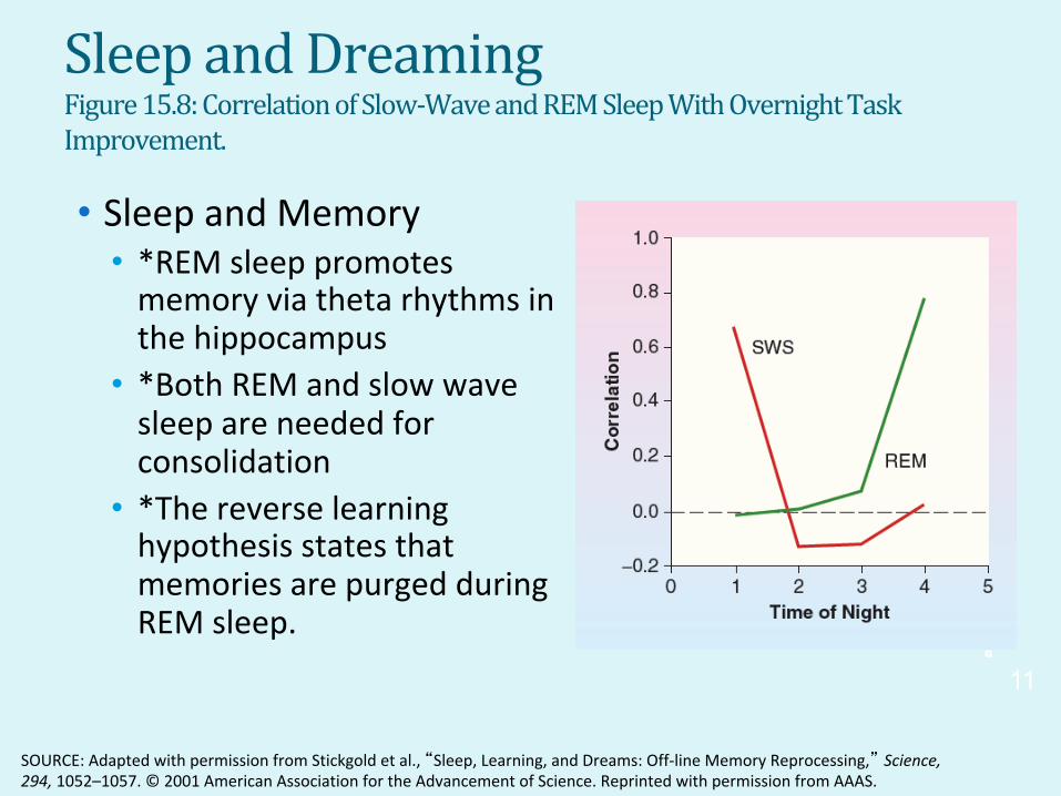
SleepandDreamingFigure 15.8: Correlation of Slow-Wave and REM Sleep With Overnight TaskImprovement.
• Sleep and Memory • *REM sleep promotesmemory via theta rhythms in the hippocampus
• *Both REM and slow wave sleep are needed for consolidation
• *The reverse learninghypothesis states that memories are purged during REM sleep.
Gar
rett:
Bra
in &
Beh
avio
r 4e
SOURCE: Adapted with permission from Stickgold et al., “Sleep, Learning, and Dreams: Off-line Memory Reprocessing,” Science, 294, 1052–1057. © 2001 American Association for the Advancement of Science. Reprinted with permission from AAAS.
11
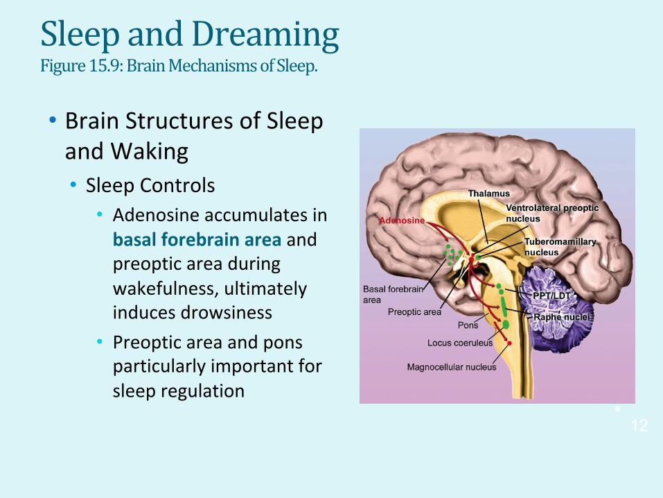
SleepandDreamingFigure 15.9: Brain Mechanisms of Sleep.
• Brain Structures of Sleep and Waking • Sleep Controls
• Adenosine accumulates in basal forebrain area and preoptic area during wakefulness, ultimately inducesdrowsiness
• Preoptic area and pons particularly important for sleep regulation
Gar
rett:
Bra
in &
Beh
avio
r 4e
12

Figure 15.10: Arousal Structures ofSleepandWaking • Basal forebrain area
• Inhibits arousal-producing neurons, inducing drowsiness and reduces EEG.
• Waking and Arousal • *Major pathway 1: PPT/LDT
Gar
rett:
Bra
in &
Beh
avio
r 4e
13

SleepandDreamingFigure 15.11: Firing Rates in Brain Stem Arousal Centers During Waking andSleep
• Waking and Arousal • *The Ventromedial POA inhibits activity in Major Pathway 2: which includes the Tuberomammillary nucleus of the Hypothalamus, Locus coeruleus (NE) and raphé nucleus (S)
• These areas are active while awake, quiet during non-REM, silent during REM.
Gar
rett:
Bra
in &
Beh
avio
r 4e
14

SleepandDreamingFigure 15.12: Locations of Orexin Receptors in the Rat Brain
• Waking and Arousal • Arousing pathway
• Lateral hypothalamus releases orexin (hypocretin) to prevent the brain from switching into sleep.
Gar
rett:
Bra
in &
Beh
avio
r 4e
15
SOURCE: From “Mice Lacking the M3 Muscarinic Acetylcholine Receptor Are Hypophagic and Lean,” by Yamada et al., Nature, 410, 207–212, © 2001. Used with permission.

SleepandDreamingFigure 15.13: PGO Waves, EEG Desynchrony, and Muscle Atonia
• Waking and Arousal • Pons: the source of PGO waves
• Excitation travels from pons through lateral geniculate to occipital area
• PGO waves trigger EEG desynchrony of REM • *The pons sends impulses to the magnocellular nucleus in the medulla to produce REM atonia (paralysis)
• Disordered atonia is seen in cataplexy, a form of narcolepsy
Gar
rett:
Bra
in &
Beh
avio
r 4e
16
SOURCE: Copyright 1989 by the Society for Neuroscience.

SleepDisordersFigure 15.14: Effects of Disrupted Circadian Rhythm on Sleep.
• Insomnia • Inability to sleep or obtain qualitysleep
• Can shorten the lifespan and maycontribute to obesity
• *Triggers include stress,depression and using sleepingpills. It is more common in peoplewith mental health issues.
• Drugs used in treatment can beaddictive
• Circadian phase delay or advance • Desynchrony between bodytemperature and sleep period
Gar
rett:
Bra
in &
Beh
avio
r 4e
17
SleepDisorders
• Sleepwalking • Occurs during slow wave sleep
• Can be triggered by stress, alcohol and sleep deprivation
• Individual may engage in complex behavior while sleepwalking
• REM Sleep Behavior Disorder • Characterized by physical activity during REM sleep and can lead to injury
• Often associated with a neurological disorder or a
Gar
rett:
Bra
in &
Beh
avio
r 4e
7/29
/19
tumor 18
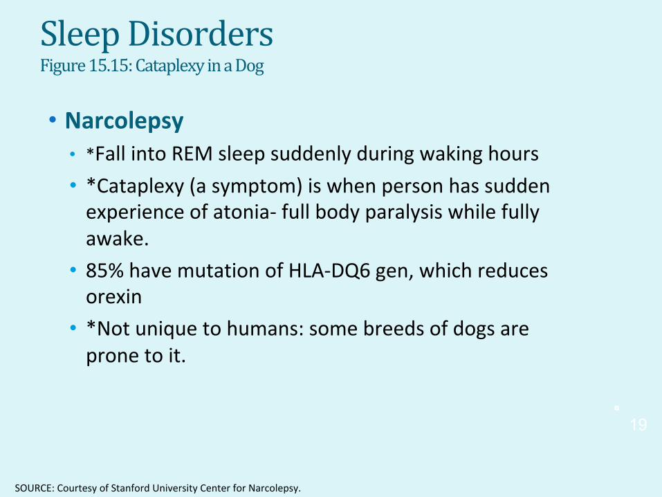
SleepDisordersFigure 15.15: Cataplexy in a Dog
• Narcolepsy • *Fall into REM sleep suddenly during waking hours • *Cataplexy (a symptom) is when person has sudden experience of atonia- full body paralysis while fully awake.
• 85% have mutation of HLA-DQ6 gen, which reduces orexin
• *Not unique to humans: some breeds of dogs are prone to it.
Gar
rett:
Bra
in &
Beh
avio
r 4e
SOURCE: Courtesy of Stanford University Center for Narcolepsy.
19

SleepDisorders
• Sleep as a Form of Consciousness • Lucid dreamers are aware of when they are dreaming and in some cases can control the nature of the dream
• The gradations of sleep lead us to confront the question of what defines consciousness
Gar
rett:
Bra
in &
Beh
avio
r 4e
7/29
/19
20

TheNeural BasisofConsciousness Figure 15.17: Synchronized Activity Among Areas Involved in Learning.
• Awareness • Awareness of something specific is easier to study than pure awareness
• Binding problem: how the brain combines information about an object • Synchronized 40-Hz activity between V1 and V5 in cats.
Gar
rett:
Bra
in &
Beh
avio
r 4e
21
SOURCE: Adapted from “Coherence of Gamma-Band EEG Activity as a Basis for Associative Learning,” by Miltner et al., Nature 397, 434–436. © 1999 Nature Publishing. Reprinted by permission.

TheNeural BasisofConsciousness Figure 15.18: Setup for Demonstrating the Cheshire Cat Effect
• Attention • How the brain allocates limited resources to focus on some inputswhile excluding others.
• Cheshirecat effect: Binocular rivalry example
• Physiological process • Changes in attention matched withchanges in neural activity
• Thalamus is a critical region • *Dorsal Attention network allows us to direct our attention (toward a goalor object)
• Also requires working memory andother brain areas
Gar
rett:
Bra
in &
Beh
avio
r 4e
22

TheNeural BasisofConsciousness
• Sense ofSelf • Self recognition, sense of agency
• *Body Image (my tongue, my hand, etc) • *Mirror neurons for social ‘understanding’
• *Memory
• A sense of self would likely be severely impaired by the loss of long term, but not necessarily short-term, memory.
• Confabulation suggests the importance of memory to self identity
Gar
rett:
Bra
in &
Beh
avio
r 4e
7/29
/19
23

TheNeural BasisofConsciousness Figure 15.20: (a) The Anterior Cingulate and (b) the Insula
• Sense ofSelf • Self recognition, sense of agency
• Body image
• Anterior cingulate and Insula involved in sense of body image
Gar
rett:
Bra
in &
Beh
avio
r 4e
24
SOURCE: (a) Courtesy of Heal Collection, University of Utah. (b) Reproduced with the permission of the Museum of neuroanatomy Tomas A Mascitti; Institute of Cognitive Neurology (INECO)

TheNeural BasisofConsciousness Figure 15.21: Brain Areas Involved in the Sense of Agency
• Sense ofSelf • Self recognition, sense of agency
• Anterior cingulate and Insula involved in sense of body image
Gar
rett:
Bra
in &
Beh
avio
r 4e
25
SOURCE: From “Experiencing Oneself Vs. Another Person as Being the Cause of an Action: The Neural Correlates of the Experience of Agency,” by C. Farrer et al., NeuroImage, 15, 596–603, fig. 2 and fig. 3, p. 598. © 2002 with permission from Elsevier, Ltd.

TheNeural BasisofConsciousness Figure 15.23: Different Intentions Distinguished by Mirror Neurons
• Sense ofSelf • Self, Theory of Mind, and Mirror Neurons
• Mirror neurons (Ch14) responsible or social understanding
• Understanding intentions of others (figure below)
Gar
rett:
Bra
in &
Beh
avio
r 4e
26
SOURCE: From “Grasping the Intentions of Others With One’s Own Mirror Neuron System,” by M. Iacoboni, 2005, PLoS Biology, 3, pp. 529–535, fig. 1 upper right and lower right, p. 530.
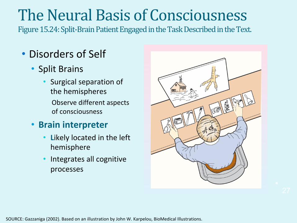
TheNeural BasisofConsciousness Figure 15.24: Split-Brain Patient Engaged in the Task Described in the Text.
• DisordersofSelf • Split Brains
• Surgical separation of the hemispheres Observe different aspects ofconsciousness
• Brain interpreter • Likely located in the left hemisphere
• Integrates all cognitive processes G
arre
tt: B
rain
& B
ehav
ior 4
e
27
SOURCE: Gazzaniga (2002). Based on an illustration by John W. Karpelou, BioMedical Illustrations.
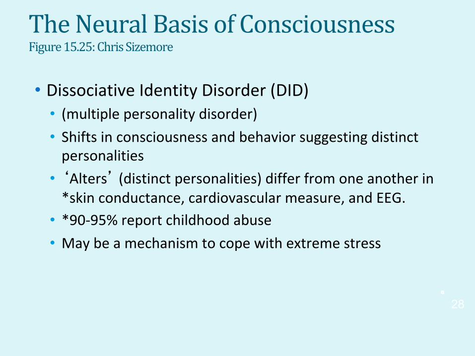
TheNeural BasisofConsciousness Figure 15.25: Chris Sizemore
• Dissociative Identity Disorder (DID) • (multiple personality disorder) • Shifts in consciousness and behavior suggesting distinct personalities
• ‘Alters’ (distinct personalities) differ from one another in *skin conductance, cardiovascular measure, and EEG.
• *90-95% report childhood abuse
• May be a mechanism to cope with extreme stress
Gar
rett:
Bra
in &
Beh
avio
r 4e
28

TheNeural BasisofConsciousness Figure 15.26: Hippocampal Activity During the Switch Between MultiplePersonalities
• Dissociative Identity Disorder (DID) • Increasing incidence raised questions of how many cases are
“real”
• Amnesia associated with DID may be state-dependent learning
• MRI data suggest learning mechanisms may be involved
Gar
rett:
Bra
in &
Beh
avio
r 4e
29
SOURCE: From “Functional Magnetic Resonance Imaging of Personality Switches in a Woman With Dissociative Identity Disorder,” by Tsai et al., Harvard Review of Psychiatry, 7(15), pp. 119–122. © 1999. Reprinted by permission of Taylor & Francis.

TheNeural BasisofConsciousness Visually Presented Words.
• Network Explanations • Theories require a widely distributed neuronal network
• Theorized to be coordination of this network
• Crick suggested claustrum is executive center or director of consciousness
Figure 15.27: Map of Event Related Potentials to Masked and Unmasked
Gar
rett:
Bra
in &
Beh
avio
r 4e
30
SOURCE: From “Cerebral Mechanisms of Word Masking and Unconscious Repetition Priming,” by S. Dehaene et al., Nature Neuroscience, 4, 752–758, fig. 3, p. 755. © 2001 Macmillan Publishing. Used with permission.

TheNeural BasisofConsciousness IN THENEWS: Consciousness and theDyingBrain
• Near Death Experiences • 3% have experienced this state
• Out of bodyexperience
• Brain mechanisms • Widespread, synchronized brain activity
• Similar to aroused brain
Gar
rett:
Bra
in &
Beh
avio
r 4e
31!
SOURCE: ScienceDaily, http://www.sciencedaily.com/releases/2013/08/130812153553.htm
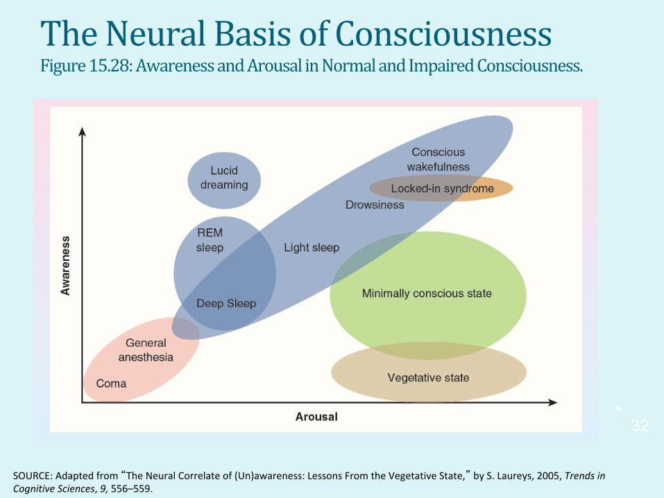
TheNeural BasisofConsciousness Figure 15.28: Awareness and Arousal in Normal and Impaired Consciousness.
32
Gar
rett:
Bra
in &
Beh
avio
r 4e
SOURCE: Adapted from “The Neural Correlate of (Un)awareness: Lessons From the Vegetative State,” by S. Laureys, 2005, Trendsin Cognitive Sciences, 9, 556–559.

TheNeural BasisofConsciousness APPLICATION:DeterminingConsciousnessWhenItCounts
33
Gar
rett:
Bra
in &
Beh
avio
r 4e
SOURCE: From “Brain Function in Coma, Vegetative State, and Related Disorders,” by S. Laureys, A. M. Owen, and N. D. Schiff, 2004, Lancet Neurology, 3, 537–546.