skeletons as forensic evidence - tucson unified school...
TRANSCRIPT

Bellwork: Objectives:
Identify/name the bones
1, 2, and 3.
I will:
Finish the
“Anthropological Crime
Scene” Lab and
Questions
Explain how age can be
determined by
examining a skeleton.
1.
3.
2.
4.14.10
Clavicle
radius
Tibia

SKELETONS AS FORENSIC
EVIDENCE
Video:
What did
they do with
the remains
they
received?

AFTER REMAINS ARE RECOVERED
Coffin or remains transported to morgue or other
facility
Careful opening – protect evidence for court

ANTHROPOLOGICAL EXAMINATION
Excavated remains
cleaned in forensic lab
Arranged in correct
anatomical order

BIOLOGICAL PROFILES:
Depending upon bones present, forensic
anthropologist may be able to determine
Sex
Race
Age
Stature

AGE: IMMATURE VS MATURE SKELETON
Before puberty biological identification of
remains can be difficult
Bones are mostly cartilage
Growth plates not fused
Sexual differences not as pronounced
Best determinant – skull with dentition

AGE DETERMINATION -
Video:
https://www.youtube.com
/watch?v=NgbrqzgyWRo
Write down ways to tell the age. (6:26)

WHAT CAN BE LEARNED FROM THE
SKULL? In infants, we can determine the age, in weeks,
fairly accurately by examining the development
of the skull:
1. difference in size,
2. how the fontanels (the soft spots that ultimately
become the sutures, or fixed joints between the
bones in the skull) change over time, gradually
becoming smaller.

Take the following two
images, and try to figure
out the approximate age of
the second image (on the
right), after examining the
first image (above).
Aged 31 weeks, 32 weeks, and 40 weeks chimpanzee skull

Examine the image below to see how the
fontanels, grow and fuse with the other
bones to form the sutures.
Although these
sutures are as
unique as
fingerprints,
as they cannot
be seen
antemortem
(not without
surgery, or a
violent
accident), they
are not useful
in terms of
identification

SUTURES
Sutures are the junctions
between adjacent bones in
the skull
The sutures are useful in
terms of determining the
approximate age of a skull.
As we grow older, the sutures
ultimately completely fill in.
The less filled in, the younger
the person.
We also often tend to lose teeth as
we age, and if we really do a bad
job flossing, we lose BONE as
well, as in the geriatric (elderly)
skull pictured:

SUTURES
In cases where
cultures have
bound the skull, the
sutures have had to
move as the bones
take on the new
shape forced by the
binding of the
skull. Note the
change in the
coronal suture in
the skull pictured:

Sutures and Age
Coronal Suture closed
about age 50
Sagittal
Suture
closed
by ~ 32
Lambdoidal
suture close
between 21-
30 yrs old

Newborn Skull

Newborn Skull

Age?
15 to 16
months. It is
not currently
possible to
reliably
determine sex
or major racial
group with
child remains.

1-year-old
Human Child
Skull

DETERMINING AGE Age can also be
determined, in some cases, by the degree to which the bones show arthritis.
Arthritis can be broken down to the prefix arthr- = joint, and -itis = inflammation.
Arthritis is therefore the swelling of the joints.
One of the effects of arthritis is to change the shape of the bones at the point where the joints are swollen, as you can see in the pictures
Arthritic on the Left, and Normal on
the Right

AGE
Arthritic on the Top, and Normal on
the Bottom

AGE
Another aspect of aging involves the fusion of certain bones with one another, often as a result as osteoarthritis, in which the cartilage in the joints ossifies (turns to bone)
As there is cartilage between bones (Cartilage that connects bone to bone are called ligaments; cartilage connecting muscle to bone are called tendons.), this process, in essence, creates a larger bone made of the fusion between two bones.
Top: a vertebra fused with the
sacrum
Bottom: the manubrium (top of
the sternum) fused with the
clavicle (shown cut here)

Age
X-rays can be used to show
comparative age, simply by
looking at the end of the long
bones, in an area called the
epiphysis (\i-pi-fə-səs\ ).
In a child, the area of growth
is made of cartilage, and is
called the epiphyseal plate.
In the x-ray, it appears as a
clear space running
approximately parallel to the
end of the bone:

Age
In an adult hand (i.e., by the
early to mid twenties), however,
the growth plate has completely
ossified (turned to bone). At
that point, the bones stop
growing.
On the x-ray, these epiphyseal
lines will appear as white lines
in the same location as the
plates were in the child's x-ray:
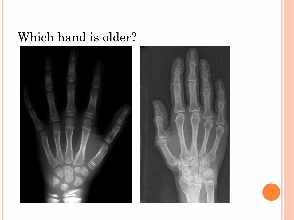
Which hand is older?

CLOSING QUESTION
What are 3 differences between an immature and
mature skeletons?