sixth nerve palsy+ipsilateral horner’s syndrome...
TRANSCRIPT
Saudi Journal of Ophthalmology (2015) 29, 63–66
Original Article
Sixth nerve palsy + ipsilateral Horner’s Syndrome = Parkinson’sSyndrome
Peer review under responsibilityof Saudi Ophthalmological Society,King Saud University Production and hosting by Elsevier
Access this article onlinwww.saudiophthaljournwww.sciencedirect.com
Received 18 September 2014; accepted 18 September 2014; available online 5 October 2014.
Neuro-ophthalmology Unit, Dept. of Ophthalmology, Buenos Aires British Hospital, Argentina
⇑ Corresponding author at: Av. Coronel Diaz 2277, 5-D, Buenos Aires 1425, Argentina. Tel./fax: +54 11 4824 2222.e-mail address: [email protected] (R.N. Ebner).
Roberto N. Ebner ⇑, Dolores Ribero Ayerza, Fernando Aghetoni
Abstract
Purpose: To present five patients with VIth nerve palsy and ipsilateral Horner’s Syndrome (HS), as a result of cavernous sinusalteration.Study design: Consecutive case series.Material and methods: Five patients presented abducens palsy with horizontal diplopia (3 in primary position and 2 in lateral gazeonly) and ipsilateral HS.Apraclonidine 0.5% drops evidenced sympathetic denervation in all patients 40–60 min after instillation. All 5 cases hadneuroimages (MRI in 3 cases, Computerized Tomography – CT in one case and Magnetic Resonance Angiography – MRA inone case) demonstrating cavernous sinus lesions; 2 meningiomas, 1 carotid-cavernous aneurism, 1 foreign body (bullet) and1 squamous cell carcinoma.Conclusion: Lesions on the cavernous sinus need to be considered in cases of abducens nerve palsy and ipsilateral Horner’sSyndrome.
Keywords: Sixth nerve palsy, Horner’s Syndrome, Cavernous sinus, Apraclonidine
� 2014 Production and hosting by Elsevier B.V. on behalf of Saudi Ophthalmological Society, King Saud University.http://dx.doi.org/10.1016/j.sjopt.2014.09.010
Introduction
Disfunction of the sixth (abducens) cranial nerve may resultfrom lesions occurring anywhere along its pathway betweenthe sixth nerve nucleus in the dorsal pons and the lateral rec-tus muscle within the orbit.
Horner Syndrome can be caused by damage to thesympathetic pathway at any location of its route from thehypothalamus to the eye.
The association of the sixth nerve palsy with ipsilateralHorner’s Syndrome has a localizing value in the posterior cav-ernous sinus, also known as Parkinson’s Syndrome.
The syndrome has been described mainly in aneurisms inand around the posterior cavernous sinus presenting acutely
with variable pain or anesthesia–hypoesthesia in the side ofthe lesion.
We present five patients with VIth nerve palsy and ipsilat-eral Horner’s Syndrome (HS), an association orienting diag-nosis toward the cavernous sinus.
Material and methods
Consecutive case series of five patients with horizontaldiplopia secondary to VIth nerve palsy and HS with ipsilateralcavernous sinus lesion has been registered with theinstitutional review board and followed the tenets of theDeclaration of Helsinki. After clinical diagnosis all patientswere asked for neuroimaging. The diagnosis of Horner
e:al.com
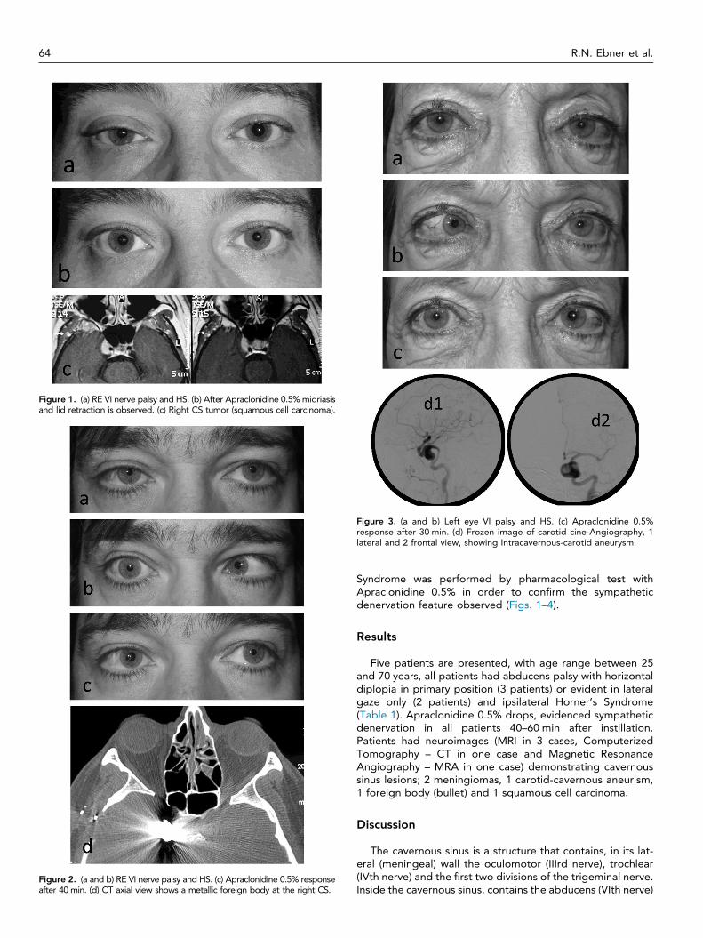
Figure 1. (a) RE VI nerve palsy and HS. (b) After Apraclonidine 0.5% midriasisand lid retraction is observed. (c) Right CS tumor (squamous cell carcinoma).
Figure 2. (a and b) RE VI nerve palsy and HS. (c) Apraclonidine 0.5% responseafter 40 min. (d) CT axial view shows a metallic foreign body at the right CS.
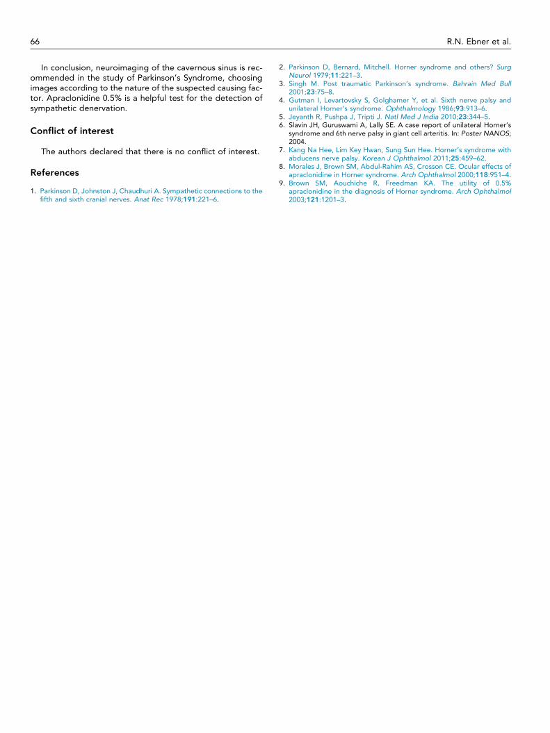
igure 3. (a and b) Left eye VI palsy and HS. (c) Apraclonidine 0.5%sponse after 30 min. (d) Frozen image of carotid cine-Angiography, 1teral and 2 frontal view, showing Intracavernous-carotid aneurysm.
64 R.N. Ebner et al.
Frela
Syndrome was performed by pharmacological test withApraclonidine 0.5% in order to confirm the sympatheticdenervation feature observed (Figs. 1–4).
Results
Five patients are presented, with age range between 25and 70 years, all patients had abducens palsy with horizontaldiplopia in primary position (3 patients) or evident in lateralgaze only (2 patients) and ipsilateral Horner’s Syndrome(Table 1). Apraclonidine 0.5% drops, evidenced sympatheticdenervation in all patients 40–60 min after instillation.Patients had neuroimages (MRI in 3 cases, ComputerizedTomography – CT in one case and Magnetic ResonanceAngiography – MRA in one case) demonstrating cavernoussinus lesions; 2 meningiomas, 1 carotid-cavernous aneurism,1 foreign body (bullet) and 1 squamous cell carcinoma.
Discussion
The cavernous sinus is a structure that contains, in its lat-eral (meningeal) wall the oculomotor (IIIrd nerve), trochlear(IVth nerve) and the first two divisions of the trigeminal nerve.Inside the cavernous sinus, contains the abducens (VIth nerve)

Figure 4. (a) Ptosis and miosis on LE (HS). MRI; (b) axial and (c) coronal views showing images compatible with cavernous sinus meningioma.
Figure 5. (a) Carotid artery, (b) sympathetic plexus, (c) III nerve (superiordivision), (d) IV nerve, (e) VI nerve, (f) III nerve (inferior division),(g) Maxillary nerve.
Parkinson’s syndrome 65
and the oculosympathetic nerve plexus around the internalcarotid artery.
These anatomical distributions are the cause of eithersolitary or combined diverse impairments of cranial nervesand eventual postganglionic Horner’s Syndrome.
Isolated VIth nerve palsy or Horner Syndrome alone, hasno localizing value per se, neuroimaging or pharmacologicaltests are necessary to determine the injury site.
The association of VIth nerve palsy with ipsilateral HS,described by Parkinson and named after him (PS),1,2 has agreat localizing value, being the cavernous sinus the site ofthe lesion.
The sympathetic plexus and the VIth nerve attempt can beexplained by the close anatomical relationship betweenthese two elements inside the cavernous sinus (Fig. 5).A single lesion may produce both deficits simultaneously.
The most frequent etiologies reported for PS are;aneurism, invasive tumors, trauma, meningioma and giantcell arteritis.3–7
In our small series of five cases we found two meningio-mas, one carotid-cavernous aneurism, 1 foreign body (bullet)and a squamous cell carcinoma as responsible for PS.Neuroimaging (MRI, CT and Angiography) confirmed the
Table 1. Clinical and ancillary findings in case series.
Patient Diplopia Apraclonidine 0
WC – 32 yrs (Fig. 3) Abd only +CA – 27 yrs (Fig. 1) PPG +JA – 67 yrs (Fig. 2) PPG +MA – 58 yrs Abd only +MC – 43 yrs PPG +
Abbreviations: PPG, primary position of gaze; abd, abduction; MR, Magnetic Resonance; CTsinus; CC, carotid-cavernous.
initial clinical diagnostic showing cavernous sinus lesions inall our patients.
The use of Apraclonidine 0.5% has a great diagnosticvalue for HS but does not provide information about the levelof the sympathetic pathway lesion. In our five cases helped toconfirm the presence of sympathetic denervation (ipsilateralto the VI nerve deficit).8,9
.5% CS imaging Findings
MR Squamous cell carcinomaCT Metallic foreign bodyMRA CC aneurysmMR CC meningiomaMR CC meningioma
, Computerized Tomography; MRA, Magnetic Resonance Angiography; CS, cavernous
66 R.N. Ebner et al.
In conclusion, neuroimaging of the cavernous sinus is rec-ommended in the study of Parkinson’s Syndrome, choosingimages according to the nature of the suspected causing fac-tor. Apraclonidine 0.5% is a helpful test for the detection ofsympathetic denervation.
Conflict of interest
The authors declared that there is no conflict of interest.
References
1. Parkinson D, Johnston J, Chaudhuri A. Sympathetic connections to thefifth and sixth cranial nerves. Anat Rec 1978;191:221–6.
2. Parkinson D, Bernard, Mitchell. Horner syndrome and others? SurgNeurol 1979;11:221–3.
3. Singh M. Post traumatic Parkinson’s syndrome. Bahrain Med Bull2001;23:75–8.
4. Gutman I, Levartovsky S, Golghamer Y, et al. Sixth nerve palsy andunilateral Horner’s syndrome. Ophthalmology 1986;93:913–6.
5. Jeyanth R, Pushpa J, Tripti J. Natl Med J India 2010;23:344–5.6. Slavin JH, Guruswami A, Lally SE. A case report of unilateral Horner’s
syndrome and 6th nerve palsy in giant cell arteritis. In: Poster NANOS;2004.
7. Kang Na Hee, Lim Key Hwan, Sung Sun Hee. Horner’s syndrome withabducens nerve palsy. Korean J Ophthalmol 2011;25:459–62.
8. Morales J, Brown SM, Abdul-Rahim AS, Crosson CE. Ocular effects ofapraclonidine in Horner syndrome. Arch Ophthalmol 2000;118:951–4.
9. Brown SM, Aouchiche R, Freedman KA. The utility of 0.5%apraclonidine in the diagnosis of Horner syndrome. Arch Ophthalmol2003;121:1201–3.