site-specific proteolytic cleavage of the amino terminus of herpes
TRANSCRIPT

JOURNAL OF VIROLOGY, Dec. 2011, p. 12910–12918 Vol. 85, No. 240022-538X/11/$12.00 doi:10.1128/JVI.06268-11Copyright © 2011, American Society for Microbiology. All Rights Reserved.
Site-Specific Proteolytic Cleavage of the Amino Terminus ofHerpes Simplex Virus Glycoprotein K on Virion
Particles Inhibits Virus Entry�
Nithya Jambunathan, Sona Chowdhury, Ramesh Subramanian, Vladimir N. Chouljenko,Jason D. Walker, and Konstantin G. Kousoulas*
Division of Biotechnology and Molecular Medicine and Department of Pathobiological Sciences,School of Veterinary Medicine, Louisiana State University, Baton Rouge, Louisiana
Received 8 September 2011/Accepted 29 September 2011
Herpes simplex virus 1 (HSV-1) glycoprotein K (gK) is expressed on virions and functions in entry,inasmuch as HSV-1(KOS) virions devoid of gK enter cells substantially slower than is the case for the parentalKOS virus (T. P. Foster, G. V. Rybachuk, and K. G. Kousoulas, J. Virol. 75:12431–12438, 2001). Deletion of theamino-terminal 68-amino-acid (aa) portion of gK caused a reduction in efficiency and kinetics of virus entrysimilar to that of the gK-null virus in comparison to the HSV-1(F) parental virus. The UL20 membrane proteinand gK were readily detected on double-gradient-purified virion preparations. Immuno-electron microscopyconfirmed the presence of gK and UL20 on purified virions. Coimmunoprecipitation experiments usingpurified virions revealed that gK interacted with UL20, as has been shown in virus-infected cells (T. P. Foster,V. N. Chouljenko, and K. G. Kousoulas, J. Virol. 82:6310–6323, 2008). Scanning of the HSV-1(F) viral genomerevealed the presence of a single putative tobacco etch virus (TEV) protease site within gD, while additionalTEV predicted sites were found within the UL5 (helicase-primase helicase subunit), UL23 (thymidine kinase),UL25 (DNA packaging tegument protein), and UL52 (helicase-primase primase subunit) proteins. The re-combinant virus gD�TEV was engineered to eliminate the single predicted gD TEV protease site withoutappreciably affecting its replication characteristics. The mutant virus gK-V5-TEV was subsequently con-structed by insertion of a gene sequence encoding a V5 epitope tag in frame with the TEV protease siteimmediately after gK amino acid 68. The gK-V5-TEV, R-gK-V5-TEV (revertant virus), and gD�TEV virusesexhibited similar plaque morphologies and replication characteristics. Treatment of the gK-V5-TEV virionswith TEV protease caused approximately 32 to 34% reduction of virus entry, while treatment of gD�TEVvirions caused slightly increased virus entry. These results provide direct evidence that the gK and UL20proteins, which are genetically and functionally linked to gB-mediated virus-induced cell fusion, are structuralcomponents of virions and function in virus entry. Site-specific cleavage of viral glycoproteins on mature andfully infectious virions utilizing unique protease sites may serve as a generalizable method of uncoupling theroles of viral glycoproteins in virus entry and virion assembly.
Membrane fusion phenomena are of paramount importancein the infectious life cycle of herpes simplex virus 1 (HSV-1).HSV-1 enters cells predominantly via fusion of its viral enve-lope with cellular plasma membranes in a pH-independentmanner. An alternative pathway involves receptor-mediatedendocytosis and fusion of the viral envelope with endocyticmembranes facilitated by the low-pH environment of endo-somes (30). The virus can spread from infected to uninfectedcells by causing virus-induced cell fusion, allowing virions toenter uninfected cells without being exposed to extracellularspaces. These membrane fusion phenomena are known to bemediated by viral glycoproteins embedded in the viral enve-lope and expressed on infected cellular plasma membranes(reviewed in reference 33).
Virus entry into susceptible cells involves the coordinatedfunctions of glycoproteins gD, gB, gH, and gL (3, 8, 21, 25),
while a fifth glycoprotein, gC, is known to enhance initialbinding of the virus to cellular membranes (20). The virionglycoproteins gB and gC bind to glycosaminoglycan (GAG)moieties of cell surface proteoglycans (20, 36). This initialattachment of virions to cellular membranes is thought to fa-cilitate subsequent interaction of gD with one or more of itsspecific receptors, including the herpesvirus entry mediator(HVEM, or HveA), nectin-1 (HveC), or 3-O-sulfated heparansulfate (17, 27, 35). Apparently, gB can also bind to additionalreceptors, including paired immunoglobulin-like type 2 recep-tor alpha (PILR-�), nonmuscle myosin heavy chain IIA(NMHC-IIA), and myelin-associated glycoprotein (MAG),that function in virion attachment and virus entry (1, 34, 38).Recent data support the hypothesis that gB is the sole fusionglycoprotein, since it is the only glycoprotein that possessesfeatures of other known viral fusion glycoproteins, such as thewell-characterized vesicular stomatitis virus (VSV) G glyco-protein (19, 32). Virus-induced cell fusion is thought to bemediated by a mechanism very similar to that occurring duringfusion of the viral envelope with cellular membranes, inasmuchas the viral glycoproteins gD, gB, gH, and gL and the presenceof viral receptors are also required for virus-induced cell fusion
* Corresponding author. Mailing address: Division of Biotechnologyand Molecular Medicine and Department of Pathobiological Sciences,Louisiana State University, Skip Bertman Drive, Baton Rouge, LA70803. Phone: (225) 578-9682. Fax: (225) 578-9701. E-mail: [email protected].
� Published ahead of print on 12 October 2011.
12910
on April 12, 2018 by guest
http://jvi.asm.org/
Dow
nloaded from

(2, 31, 40, 41). However, virus-induced cell fusion requires thepresence of additional viral glycoproteins and membrane pro-teins, including gE, gI, gM, gK, and the UL20 and UL45proteins (5, 7, 12, 18, 26, 43).
We reported that gK is a structural component of virions asa Golgi complex-dependent glycosylated species and functionsin virus entry, inasmuch as virions lacking gK enter susceptiblecells in cell culture substantially slower (15). Furthermore, weshowed that HSV-1 gK and UL20 functionally and physicallyinteract and that these interactions are absolutely necessary fortheir coordinate intracellular transport, cell surface expression,and functions in the HSV-1 life cycle (10, 14). Recently, weshowed that a peptide composed of the amino-terminal 82amino acids (aa) (including the predicted 30-aa signal se-quence) of gK (gKa) complemented in trans for gB-mediatedcell fusion (5). In addition, we demonstrated that UL20 andthe gKa peptide physically interacted with gB and gH in in-fected cells, suggesting that gB-mediated virus-induced cell fusionis regulated via direct interactions with gK and UL20 (6).
In this work, we sought to confirm the presence of gK onpurified virion particles and to assess whether the UL20 pro-tein is also a structural component of virion particles in asso-ciation with gK and function in virus entry. A substantial dif-ficulty in assessing the role of gK in virus entry is the fact thata lack of gK leads to drastic defects in cytoplasmic virionenvelopment and infectious virus production (11, 22, 23). Wepresent here a generalizable strategy to modify/inactivate gKor potentially any other viral glycoprotein on mature virionparticles, effectively uncoupling the roles of gK in cytoplasmicvirion envelopment and virus entry.
MATERIALS AND METHODS
Cell culture. African green monkey kidney (Vero) cells were obtained fromthe American Type Culture Collection (Rockville, MD). VK302 cells perma-nently expressing gK were originally obtained from David Johnson (OregonHealth Sciences University, Portland, OR). Cells were maintained in Dulbecco’smodified Eagle’s medium (DMEM) (Gibco-BRL, Grand Island, NY) supple-mented with 10% fetal calf serum and antibiotics.
Bioinformatics analysis of predicted protease sites within HSV-1 proteins.The ExPASy PeptideCutter software tool (16), available at the ExPASy Bioin-formatics Resource portal (http://web.expasy.org/peptide_cutter/), was utilizedto predict potential tobacco etch virus (TEV) cleavage sites within each pre-dicted amino acid sequence of the HSV-1(F) genome (GenBank accession num-ber GU734771). The PeptideCutter algorithm predicted the presence of a singleputative TEV site within gD at amino acid 235, while additional TEV predictedsites were found within the UL5 (helicase-primase helicase subunit; single TEVpredicted site at amino acid 466), UL23 (thymidine kinase; two TEV predictedsites at amino acid 250 and 331), UL25 (DNA packaging tegument protein; oneTEV site at amino acid 533), and UL52 (helicase-primase primase subunit; oneTEV site at amino acid 184). Treatment of purified virions will access onlystructural envelope proteins, such as gD, and not any of the other nonstructuralviral proteins containing TEV sites.
Construction of HSV-1 recombinant mutants. The recombinant mutantYE102-VC1 was engineered to express gK and UL20 containing the V5 and3�FLAG antigenic epitopes, respectively. Specifically, a DNA sequence encod-ing the V5 epitope and the enterokinase recognition site, Asp-Asp-Asp-Asp-Lys,was inserted in frame immediately after amino acid 68 of gK (including thepredicted 30-aa signal sequence). The 3�FLAG epitope was inserted in frame atthe amino terminal of UL20, as described earlier (24). All recombinant viruseswere constructed using the markerless two-step red-mediated recombinationmutagenesis system (42) on the bacterial artificial chromosome (BAC) plasmidpYEbac102 carrying the HSV-1(F) genome or the BAC plasmid pYK608 carry-ing the HSV-1(F) genome modified to express three fluorescent proteins asfusion proteins with gB, VP22, and VP26 (gifts from Yasushi Kawaguchi, Uni-versity of Tokyo, Tokyo, Japan). The mutant virus gD�TEV was constructed by
altering the DNA sequence within the gD gene predicted to specify a TEVprotease recognition site on the bacterial artificial chromosome YK608 genome.Specifically, gD amino acid 235 was changed from glutamine to asparagine (Fig.1). The gD�TEV virus was subsequently used to generate the gK-V5-TEVrecombinant virus, which expressed gK containing the V5 epitope and the TEVprotease recognition site ENLYFQG inserted in frame immediately after aminoacid 68 of gK.
Virus entry assay. The construction and characterization of the gK mutantvirus gK�31-68 lacking 68 aa from its amino terminus (including the 30-aa signalsequence) was recently described (5). The relative entry efficiencies of the wild-type HSV-1(F) YE102, gK-null (�gK), and gK�31-68 viruses were tested onVero cells by determining the number of cells expressing viral antigens afterentry was allowed to occur for 1 h at 34°C. Specifically, viruses at a multiplicityof infection (MOI) of 1 were allowed to attach to Vero cell monolayers at 4°C for1 h. The virus inoculum was subsequently removed, warm medium (34°C) wasadded, and the cultures were shifted to 34°C to allow virus penetration. After 1 hat 34°C, extracellular virions were inactivated by treatment with low-pH buffer(0.1 M glycine, pH 3.0), and cells were washed three times with phosphate-buffered saline (PBS). Twelve hours postinfection (hpi), the cells were fixed andstained with anti-ICP4 antibody (Virusys, Inc., Taneytown, MD). The relativeefficiency of virus entry was calculated as the percentage of cells expressing ICP4after low-pH treatment relative to results for PBS-treated cells by flow cytometry.Mean values and standard deviations of three independent experiments werecalculated. Similarly, kinetics of virus entry were determined at 34°C by measur-ing the number of ICP4-positive Vero cells by flow cytometry at 12 hpi. Thepercent positive cells for each time point (10, 20, 30, and 60 min) was determinedfor a total number of 5,000 Vero cells infected at an MOI of 1.
Growth kinetics and plaque morphology of wild-type and mutant viruses.Growth kinetics studies were performed essentially as we have described earlier(11, 13, 15). Briefly, viruses were adsorbed onto a 90%-confluent monolayer ofVero cells in a 12-well plate at 4°C for 1 h at a low multiplicity of infection(MOI � 0.2). Subsequently, cells were incubated at 37°C and 5% CO2 for 1 h toallow penetration of the virus. Any remaining extracellular virions were inacti-vated by briefly treating the cells with PBS at low pH (pH 3.0). Virus stocks wereprepared from infected cells at 6, 12, 24, 36, and 48 hpi, and the virus titerswere obtained by endpoint titration on Vero cell in triplicate. Viral plaques werestained using anti-HSV-1 polyclonal antibody, as described previously (5). Viralplaques were also visualized by fluorescence microscopy.
Virus entry kinetics of TEV protease-treated virions. Partially purified virions(generated by concentrating viruses by ultracentrifugation of infected cell super-natants) were used for enzyme digestion studies. Equal PFU of recombinantmutant viruses gD�TEV and gK-V5-TEV were treated as follows: (i) PBS alone(all PBS used for these experiments was at pH 6.8), (ii) PBS plus 1 unit ofAcTEV protease (Invitrogen, Inc., Carlsbad, CA), and (iii) PBS plus 0.01%SDS-inactivated AcTEV protease. SDS-inactivated enzyme was passed throughZeba spin desalting columns (Thermo Fisher Scientific, Waltham, MA) to elim-inate SDS. All treatments were kept at 30°C for 1 h, mixed with serum-freemedium, and adsorbed to a 90%-confluent monolayer of Vero cells on a 12-wellplate. The plates were rocked gently for 1 h at room temperature. The virusinoculum was removed, and the wells were overlaid with 1 ml of DMEM con-taining 1% methylcellulose and 2% fetal bovine serum (FBS). Cells were fixed at48 hpi with ice-cold methanol for 10 min, and immunocytochemistry was per-formed as described above. The effect of TEV protease treatment on the kineticsof virus entry was performed essentially as described earlier, with the exceptionthat viruses were treated with TEV protease or PBS and that the number ofinfectious virions was determined via limited dilution on Vero cells, as describedpreviously (15).
TEV digestion of infected cell extracts and immunoblot assays. Nearly con-fluent monolayers of Vero cells in a T-75 flask were infected with gK-V5-TEVvirus. The cells were collected 48 hpi and lysed with mammalian protein extrac-tion reagent (M-PER; Pierce, Rockford, IL) lysis buffer containing proteaseinhibitors and briefly sonicated. The gK-V5-TEV protein was immunoprecipi-tated with protein G magnetic beads bound to V5 antibody (Invitrogen, Carls-bad, CA). The eluted protein was treated with the AcTEV protease as suggestedby the manufacturer (Invitrogen). Sample buffer containing 5% �-mercaptoeth-anol was added to the digested protein and heated at 55°C for 15 min. Proteinswere resolved on 4 to 20% SDS-PAGE gel and immobilized on nitrocellulosemembranes. Immunoblot assays were carried out using mouse anti-V5 antibodyand goat anti-mouse horseradish peroxidase (HRP) light-chain secondary anti-body (Abcam, Inc., Cambridge, MA).
Virus purification. Wild-type HSV-1(F) pYEbac102 and double-labeledYE102-VC1 (gK-EK-V5; UL20-3�FLAG) virions were purified by two serialpassages using iodixanol gradients. Briefly, supernatants and cells from 24 T-150
VOL. 85, 2011 HSV-1 GLYCOPROTEIN K FUNCTIONS IN VIRUS ENTRY 12911
on April 12, 2018 by guest
http://jvi.asm.org/
Dow
nloaded from

flasks of Vero cells infected with either pYEbac102 or YE102-VC1 were col-lected at 24 and 36 hpi, respectively, and treated with TNE (10 mM Tris; 500 mMNaCl; 1 mM EDTA; pH 7.5) buffer for 15 min at 4°C. Subsequently, the cellpellet was separated by centrifugation at 6,000 � g for 45 min at 4°C. Polyeth-ylene glycol (PEG) (molecular weight, 8,000; 7% [wt/vol]) was added to thesupernatant and kept on a stirrer overnight at 4°C. The next day, this solution wasspun at 6,000 � g for 30 min at 4°C. The resulting pellet was resuspended in 10ml of TNE buffer, loaded onto a 50 to 20% discontinuous iodixanol gradient, andspun at 141,000 � g for 3 h. The viral band from the 30 to 40% interface wascollected, loaded onto a second 50 to 20% iodixanol step gradient, and centri-fuged again at 141,000 � g for 3 h at 4°C. The resulting optically dense band wascentrifuged through a 20% iodixanol cushion at 141,000 � g for 1 h at 4°C. Theresulting pellet was resuspended in 250 �l of NP-40 lysis buffer (Invitrogen) andused for immunoprecipitation and immunoblot assays.
Immunoprecipitation and immunoblot assays. The gK-V5-EK (gK taggedwith V5 and enterokinase amino acid sequence) protein was immunoprecipitatedwith protein G magnetic Dynabeads bound to V5 antibody according to themanufacturer’s instructions (Invitrogen). The protein was eluted from the mag-netic beads in 40 �l of elution buffer and used for immunoblot assays. Immu-noblot assays were carried out using anti-glyceraldehyde-3-phosphate-dehydro-genase (anti-GAPDH) (Chemicon, International Inc.), anti-gB, anti-gD, anti-gC(Virusys), anti-FLAG (Sigma-Aldrich, Inc., St. Louis, MO), anti-V5 antibody(Invitrogen), goat anti-mouse HRP against light-chain (Fab), heavy chain (Fc)whole mouse IgG molecule (Abcam), and anti-ICP8 monoclonal antibody (Ab-cam).
Immunogold labeling for TEM. Semipurified virions immobilized on 400-meshButvar/carbon-coated nickel grids (Electron Microscopy Sciences, Inc., Hatfield,PA) were used to detect the presence of gK, gB, and UL20 on the virions usingimmunogold labeling, as has been described previously for bovine herpesvirus 1(37). Briefly, 5 �l of semipurified virions suspension was adsorbed onto thecoated surface of the grids for 30 min. The grids were then blocked using 1%
bovine serum albumin (BSA) in Tris-buffered saline (TBS) for 5 min. Excessblocking buffer was removed using the tip of a filter paper, followed by incuba-tion of the grids with 5 �l of mouse anti-V5 antibody (Invitrogen) at a dilutionof 1:10,000 in 1% BSA in TBS for 30 min. This sample preparation step wasfollowed by washing the grids with 1% BSA in TBS for 5 min. A 5-�l droplet ofgoat anti-mouse IgG (whole molecule)-Gold 10-nm colloidal gold (Sigma-Al-drich) at 1:80 dilution in 1% BSA in TBS was added on top of each grid andincubated for 30 min. The grids were then washed with 1% BSA in TBS byadding 5 �l on each grid, and excess liquid was removed with the tip of a filterpaper. A 2% solution of sodium phosphotungstate, pH 6.8, was added as a finalstep for contrast purposes. Grids were desiccated and visualized by transmissionelectron microscopy (TEM).
RESULTS
The UL20 membrane protein and gK are structural compo-nents of HSV-1 virions. To assess whether both gK and UL20are present and interact as structural components of HSV-1virions, the recombinant virus HSV-1(F)-YE102-VC1 was con-structed by modifying the gK and UL20 amino acid sequencesto contain V5 and 3�FLAG antigenic epitopes, respectively(Fig. 1A). The V5 epitope tag was inserted immediately aftergK amino acid 68 (this amino acid numbering includes thepredicted signal sequence), while the 3�FLAG epitope wasinserted at the N terminus of the UL20 protein, as describedpreviously for similar gene constructions (9, 10, 14) and de-tailed in Materials and Methods. The YE102-VC1 virus repli-
FIG. 1. Schematic representation of mutant viral genomes. (A) HSV-1(F)-YE102-VC1 recombinant virus generated from pYEbac102, carryingan in-frame insertion of the V5 epitope and enterokinase recognition site inserted immediately after amino acid 68 of gK, as well as an in-frameinsertion of the 3�FLAG epitope at the amino terminus of UL20. (B) Schematic representation of recombinant viruses constructed forTEV-specific proteolytic activity. The V5 epitope tag and the TEV protease site (ENLYFQG) were inserted immediately after amino acid 68 ofgK to generate mutant virus gK-V5-TEV.
12912 JAMBUNATHAN ET AL. J. VIROL.
on April 12, 2018 by guest
http://jvi.asm.org/
Dow
nloaded from

cated efficiently in Vero cells; however, average plaque sizeswere consistently smaller, by approximately 30%, than those ofthe parental HSV-1(F)-YE102 virus (Fig. 2A). Similarly,YE102-VC1 replicated slower than the YE102 parental virus,approaching viral titers that were approximately half a loglower than YE102 at 24 hpi (Fig. 2B). However, at longer timespostinfection (36 to 48 hpi), titers of both viruses were similar(not shown).
YE102-VC1 virions were purified by sequential isopycnicdensity sedimentation in iodixanol discontinuous gradients asdescribed in Materials and Methods. The cellular proteinGAPDH was absent from gradient fractions containing infec-tious virions, indicating the relative absence of contaminatingcellular proteins, while GAPDH was readily detected in con-trol cellular extracts (Fig. 3, panel labeled �-GAPDH). Simi-larly, the nonstructural, immediate-early protein ICP8 was notdetected in purified virion samples, while it was detected read-ily in samples obtained from infected cells (Fig. 3, panel la-beled �-ICP8). Purified virions were tested for the presence ofthe viral glycoproteins gB, gD, gC, and gK and the membraneprotein UL20 using monoclonal antibodies against gB, gD, gC,anti-V5 (gK), and anti-3�FLAG (UL20) in Western immuno-blots. Monoclonal antibody to gB detected putative mature gBprotein species migrating with an apparent molecular mass ofapproximately 120 kDa, while a relatively prominent gB-re-lated species of approximately 45 kDa was also detected (Fig.3, panel labeled �-gB). Monoclonal antibody to gD detected asingle protein species migrating with an apparent molecular
mass of approximately 55 kDa (Fig. 3, panel labeled �-gD),while antibody to gC detected a protein with an apparentmolecular mass of 125 kDa. Anti-3�FLAG antibody readilydetected the presence of the UL20 protein migrating with acharacteristic ladder pattern, with the most prominent proteinspecies migrating with an apparent molecular mass of approx-imately 27 kDa, in agreement with our previous findings (6), aswell as UL20-associated protein species migrating with appar-ent molecular masses of approximately 35 and 45 kDa (Fig. 3,panel �-FLAG). The anti-V5 antibody detected the presenceof the V5-tagged gK-associated protein species migrating withan apparent molecular mass of approximately 65 to 75 kDa,which was more than twice the apparent molecular mass ofglycosylated species of gK previously detected in infected cells(5, 6). The anti-V5 (gK) antibody immunoprecipitated gK mi-grating with an apparent molecular mass of 125 kDa and co-immunoprecipitated UL20 migrating at approximately 27 kDa,which was detected with the anti-3�FLAG antibody (Fig. 3,panel labeled IP:V5Ab).
The presence of gK on virion particles was further assessedusing colloidal gold immuno-electron microscopy (Fig. 4).More than 100 virion particles for each virus were visuallyscanned for the presence of gold particles. V5-tagged gK wasreadily detected on most virion envelopes (�90 out of 100
FIG. 2. Replication kinetics and plaque morphology of theYE102-VC1 virus in comparison to those of HSV-1(F)-YE102.(A) Representative viral plaques of HSV-1(F)-YE102 and HSV-1(F)-YE102-VC1 on Vero cells at 48 hpi. Viral plaques were visu-alized by phase-contrast microscopy after immunostaining withanti-HSV-1 polyclonal antibody. (B) One-step replication kineticsof HSV-1(F)-YE102 and HSV-1(F)-YE102-VC1 at an MOI of 0.2on Vero cells. The x axis shows hours postinfection, and the y axisshows PFU/ml. All experiments were performed in triplicate. Errorbars represent standard errors of the means.
FIG. 3. Detection of gK and UL20 on gradient-purified virions.Western immunoblot analysis of double-gradient-purified virions wasperformed using anti-V5 (gK) and anti-FLAG (UL20) antibodies.Individual lanes are labeled as follows: C, positive control for GAPDH;W, lysates from purified wild-type virus [HSV-1(F)-YE102]; M, lysatesfrom purified mutant virus YE102-VC1; L, lysates from infected Verocells; V, samples derived from purified virions. Individual blots wereprobed with anti-GAPDH, anti-ICP8, anti-gB, anti-gD, anti-gC, anti-FLAG (UL20), or anti-V5 (gK) antibody as indicated. The panellabeled at the top as IP:V5Ab shows immunoblots of immunoprecipi-tations with anti-V5 (gK) antibody probed with either anti-FLAG(UL20) or anti-V5 (gK) antibodies. In this panel, lanes are immuno-precipitates from mock-infected Vero cells (U), HSV-1(F)-YE102 pu-rified virions (W), or YE102-VC1 purified virions (M). Molecular massstandards are shown with dots on each panel (250, 150, 100, 75, 50, 37,25, 20, 15, and 10 kDa; Precision Plus protein standards; Bio-Rad).HRP-conjugated goat anti-mouse (HRP-GAb; IgG) was used for alldata, except data shown on the panel labeled IP:V5Ab, wherein theF(ab)2- and Fc-purified portions of the HRP-GAb IgG were used forthe �-V5 and �-FLAG panels, respectively.
VOL. 85, 2011 HSV-1 GLYCOPROTEIN K FUNCTIONS IN VIRUS ENTRY 12913
on April 12, 2018 by guest
http://jvi.asm.org/
Dow
nloaded from

visualized virions contained gold particles) of recombinant vi-ruses VC1 and gK-V5-TEV, both of which contained the V5epitope tag inserted after gK amino acid 68 (Fig. 4B and C),while the antibody did not react with the VC1 parental virusYE102 (none of 100 visualized virions contained gold parti-cles) (Fig. 4A). Similarly, anti-gB monoclonal antibody readilydetected gB on most visualized virions (not shown).
The amino terminus of gK functions in virus entry. Previ-ously, we reported that deletion of 68 aa from the aminoterminus of gK (including the 30-aa signal sequence; actualdeletion of 38 aa after cleavage of the predicted signal se-quence) allowed efficient virus envelopment and infectious vi-rus production, while gB-mediated virus-induced cell fusionwas abolished (5). The entry efficiency of the recombinant virusgK�31-68, which lacks the first 68 aa of gK, was compared tothose of HSV-1(F; YE102) and �gK viruses. The constructionand characterization of these viruses have been described pre-viously (5). Flow cytometry was utilized to determine the rel-ative efficiencies and kinetics of entry of these viruses intoVero cells (see Materials and Methods). Both �gK andgK�31-68 entered Vero cells less efficiently than their parentalvirus, YE102 (Fig. 5A and B).
To further confirm that deletion of the amino-terminal 68 aaof gK caused defects in virus entry, we utilized incorporation ofa unique TEV protease site within gK to selectively delete theamino terminus of gK from mature virions. Scanning of theHSV-1(F) viral genome revealed the presence of a single pu-tative TEV site within gD, while additional TEV predictedsites were found within the UL5 (helicase-primase helicasesubunit), UL23 (thymidine kinase), UL25 (DNA packagingtegument protein), and UL52 (helicase-primase primase sub-unit) (see Materials and Methods). The recombinant virusgD�TEV was constructed via site-directed mutagenesis of theYK608 genome, changing the single putative gDTEV site fromPQAYQQG to PQAYQNG (Q235 to N235; bold and under-lined). Subsequently, the recombinant virus gK-V5-TEV wasconstructed by inserting a 19-aa gene segment coding for theV5 epitope tag fused in frame with the predicted TEV proteasesite (ENLYFQG), inserted immediately after gK amino acid68. Both gD�TEV and gK-V5-TEV viruses produced average-size plaques that were approximately 30% smaller than thoseof the YK608 parental virus at 48 hpi, while all viruses ex-
FIG. 4. Transmission immunogold electron microscopy to detect gK and gB on virion particles. (A) HSV-1(F)-YE102 virions stained withanti-gK (anti-V5) antibody; (B) YE102-VC1 stained with anti-gK (anti-V5) antibody; (C) gK-V5-TEV stained with anti-gK (anti-V5) antibody.
FIG. 5. Assessment of the efficiencies of gK-null and gK�31–68 virusentry into Vero cells. (A) The relative efficiency of entry is shown as thepercentage of cells expressing ICP4 at 12 hpi. Mean values and standarddeviations for three independent experiments were calculated as shown.(B) Representative kinetics of virus HSV-1(F), gK-null, and gK�31-68entry into Vero cells. Vero cell monolayers were infected with an MOI of1. At different times postentry at 34°C (10, 20, 30, and 60 min), viruses thathad not entered yet were inactivated with low pH, and the kinetics of virusentry were determined by monitoring ICP4 expression at 12 hpi using flowcytometry. For each sample, the percent ICP4-positive cells in a popula-tion of 5,000 Vero cells was measured.
12914 JAMBUNATHAN ET AL. J. VIROL.
on April 12, 2018 by guest
http://jvi.asm.org/
Dow
nloaded from

pressed the characteristic fluorescence pattern of green, red,and cyan due to fluorescent proteins fused in frame to theVP26, VP22, and gB viral proteins, respectively (Fig. 6A) (39).The gD�TEV and gK-V5-TEV viruses replicated efficiently,although slower in Vero cells than was the case for their pa-rental wild-type virus, YK608, while the two mutant virusesapproached similar viral titers at 48 hpi (Fig. 6B). A gK-V5-TEV revertant (R-gK-V5-TEV) virus was generated by replac-ing the gK-V5-TEV gene with the wild-type HSV-1(F) gK geneusing two-step red-mediated recombination mutagenesis. TheR-gK-V5-TEV virus exhibited plaque morphology and growthcharacteristics similar to those of the gD�TEV virus, suggest-ing that gK-V5-TEV did not contain any secondary site muta-tions (not shown).
To ascertain whether the TEV protease was active againstthe gK-V5-TEV tagged protein, immunoprecipitates of gKprepared from gK-V5-TEV virus-infected cells were treatedwith the TEV protease as described in Materials and Methods.Treatment of cellular extracts with the TEV protease caused asubstantial reduction in the amount of mature gK migratingwith the expected molecular mass of approximately 40 kDa incomparison to results for phosphate-buffered saline (PBS)-
treated samples. In contrast, inactivated TEV protease failedto reduce the amount of mature gK (Fig. 7A and B).
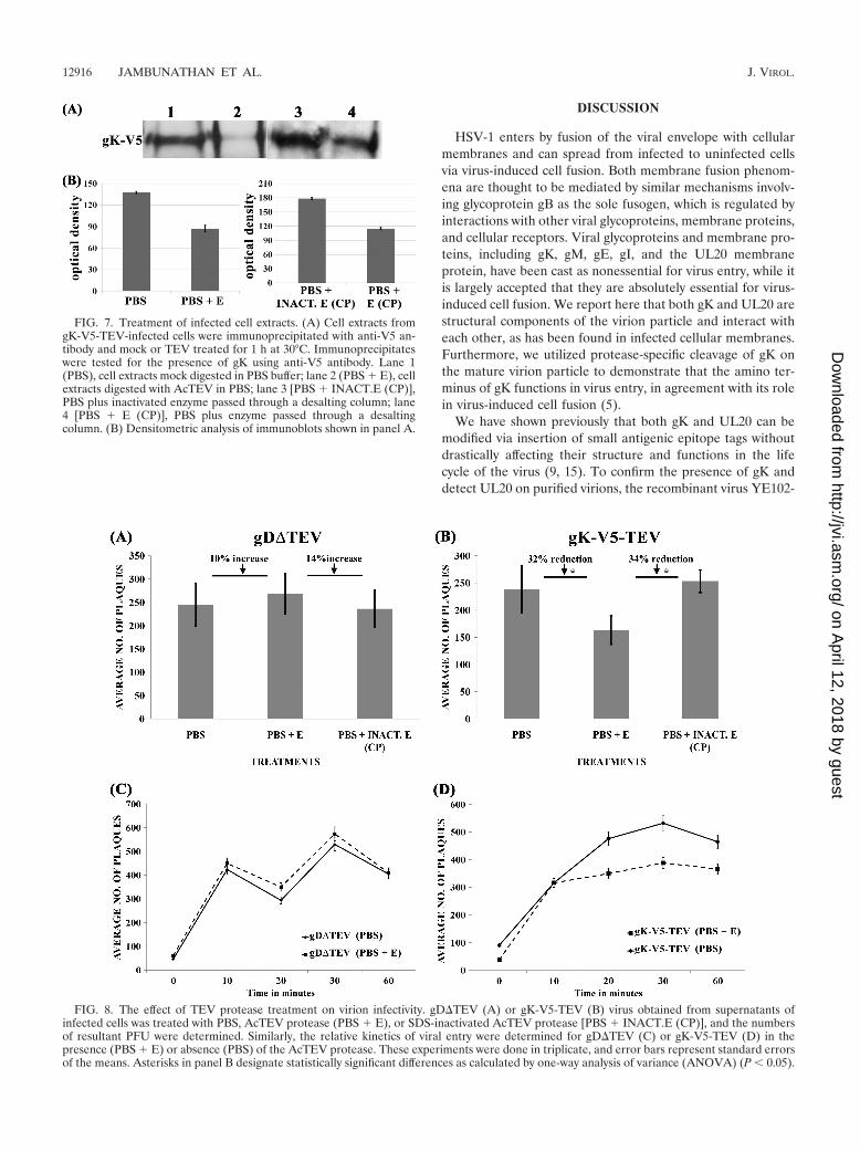
To assess whether TEV treatment of recombinant virusesaffected virus entry, partially purified and concentrated virionstocks were treated with the TEV protease and viral infectivitywas assessed by determining the number of PFU producedafter TEV treatment on Vero cells, as described in Materialsand Methods. TEV treatment of gK-V5-TEV virions resultedin a 34% or 32% reduction in virus infectivity compared toresults for treatment with inactivated TEV protease or PBS(respectively) (Fig. 8A and B). This reduction in virus entry canbe appreciated even more when compared to results for TEVtreatment of gD�TEV, which appeared to be modestly pro-tected after TEV treatment, exhibiting a 14% or 10% increasein viral infectivity compared to inactivated TEV protease orPBS treatment, respectively. Similar results were obtainedwhen kinetics of viral entry were obtained by inactivating virionremaining on the cell surface at different times using thelow-pH inactivation protocol (see Materials and Methods).TEV treatment resulted in a consistent reduction of gK-V5-TEV virion entry at different time points, while it did not haveany appreciable effect on gD�TEV virion entry (Fig. 8C and D).
FIG. 6. Construction and characterization of recombinant viruses containing TEV modifications. (A) Representative plaque morphology of thegD�TEV and gK-V5-TEV viruses in comparison to those of their parental HSV-1(F)-YK608 virus at 48 hpi. Viral plaques were visualized afterimmunohistochemical staining using phase-contrast microscopy and by fluorescence microscopy. (B) Replication kinetics of HSV-1(F) YK608,gD�TEV, and gK-V5-TEV viruses on Vero cells infected with an MOI of 0.2. The x axis shows hours postinfection, and the y axis shows PFU/ml.All experiments were performed in triplicate. Error bars represent standard errors of the means.
VOL. 85, 2011 HSV-1 GLYCOPROTEIN K FUNCTIONS IN VIRUS ENTRY 12915
on April 12, 2018 by guest
http://jvi.asm.org/
Dow
nloaded from
DISCUSSION
HSV-1 enters by fusion of the viral envelope with cellularmembranes and can spread from infected to uninfected cellsvia virus-induced cell fusion. Both membrane fusion phenom-ena are thought to be mediated by similar mechanisms involv-ing glycoprotein gB as the sole fusogen, which is regulated byinteractions with other viral glycoproteins, membrane proteins,and cellular receptors. Viral glycoproteins and membrane pro-teins, including gK, gM, gE, gI, and the UL20 membraneprotein, have been cast as nonessential for virus entry, while itis largely accepted that they are absolutely essential for virus-induced cell fusion. We report here that both gK and UL20 arestructural components of the virion particle and interact witheach other, as has been found in infected cellular membranes.Furthermore, we utilized protease-specific cleavage of gK onthe mature virion particle to demonstrate that the amino ter-minus of gK functions in virus entry, in agreement with its rolein virus-induced cell fusion (5).
We have shown previously that both gK and UL20 can bemodified via insertion of small antigenic epitope tags withoutdrastically affecting their structure and functions in the lifecycle of the virus (9, 15). To confirm the presence of gK anddetect UL20 on purified virions, the recombinant virus YE102-
FIG. 7. Treatment of infected cell extracts. (A) Cell extracts fromgK-V5-TEV-infected cells were immunoprecipitated with anti-V5 an-tibody and mock or TEV treated for 1 h at 30°C. Immunoprecipitateswere tested for the presence of gK using anti-V5 antibody. Lane 1(PBS), cell extracts mock digested in PBS buffer; lane 2 (PBS � E), cellextracts digested with AcTEV in PBS; lane 3 [PBS � INACT.E (CP)],PBS plus inactivated enzyme passed through a desalting column; lane4 [PBS � E (CP)], PBS plus enzyme passed through a desaltingcolumn. (B) Densitometric analysis of immunoblots shown in panel A.
FIG. 8. The effect of TEV protease treatment on virion infectivity. gD�TEV (A) or gK-V5-TEV (B) virus obtained from supernatants ofinfected cells was treated with PBS, AcTEV protease (PBS � E), or SDS-inactivated AcTEV protease [PBS � INACT.E (CP)], and the numbersof resultant PFU were determined. Similarly, the relative kinetics of viral entry were determined for gD�TEV (C) or gK-V5-TEV (D) in thepresence (PBS � E) or absence (PBS) of the AcTEV protease. These experiments were done in triplicate, and error bars represent standard errorsof the means. Asterisks in panel B designate statistically significant differences as calculated by one-way analysis of variance (ANOVA) (P 0.05).
12916 JAMBUNATHAN ET AL. J. VIROL.
on April 12, 2018 by guest
http://jvi.asm.org/
Dow
nloaded from

VC1 was constructed to express gK tagged with the V5 aminoacid sequence immediately followed by a 5-aa sequence pre-dicted to be a cleavage site of the enterokinase protease. This19-aa sequence was inserted in frame after gK amino acid 68(threonine), while the previously reported V5-alone insertionwas immediately after amino acid 69 (histidine) (9). TheYE102-VC1 virus was also modified to include a 3�FLAGepitope tag at the amino terminus of UL20, as described pre-viously (10, 14). The VC-1 virus replicated less efficiently inVero cells and formed smaller plaques than its parental virus,YE102, suggesting that the presence of both gK and UL20modifications only mildly inhibited virus growth and spread.VC-1 replication and plaque morphology were partially com-plemented on either gK-expressing VK302 cells or UL20-ex-pressing FRT cells, suggesting that both gK and UL20 modi-fications were responsible for the mild reduction in infectiousvirus production and virus spread exhibited by the YE102-VC1virus (not shown).
UL20 and gK were detected on highly purified virions, asevidenced by the absence of the cellular protein GAPDH andthe nonstructural ICP8 viral protein. V5 (gK) antibody immu-noprecipitates probed with either anti-V5 antibody or anti-FLAG (UL20) antibodies revealed the presence of UL20 andgK of approximately 27 kDa and 125 kDa, respectively. This125-kDa apparent molecular mass of gK was much larger thanthe monomeric form of gK (37 kDa), although potentiallydimeric gK protein species of approximately 65 kDa have beendetected previously in infected cells (10). These results suggestthat UL20 interacted predominantly with multimeric (tetra-meric) forms of gK in the virion particle. These multimericforms of gK were probably not readily detected in infected celllysates because they could not enter gels in the presence ofother cellular proteins. Both gK and UL20 are very sensitive totemperature-dependent aggregation, and samples containinggK and/or UL20 cannot be boiled because they form aggre-gates that do not enter separating gels. The larger amount oftotal protein present in gK and UL20 samples obtained frominfected cells exacerbates this phenomenon (unpublished ob-servations). The presence of gK on purified virions was con-firmed using transmission immuno-electron microscopy. TheUL20 protein was also readily detected on virions by TEM,although the anti-UL20 antibody was directed against theamino terminus of UL20, predicted to be located on the innerside of the viral envelope. This staining was probably due topartial breakage of virion envelopes that exposed the UL20epitope to the antibody (not shown). Collectively, these datasupport our hypothesis that gK is present in the viral envelopeas an interactive complex with UL20, in agreement with ourprevious findings in infected cells (10, 14).
We showed previously that HSV-1(KOS) gK-null virus en-tered Vero cells substantially slower than its parental KOSstrain (15). Because gK is required for efficient cytoplasmicvirion envelopment and infectious virus production, viral entryexperiments with gK-null viruses reflect the ability of only avery small portion of potentially aberrantly enveloped virionsto enter cells, substantially complicating interpretation of viralentry results. Virus entry experiments with the gK�31-68 virus,which carries a deletion of the first 68 aa (38-aa deletion afterthe signal sequence of 30 aa) of gK, revealed that it enteredVero cells substantially less efficiently than the parental virus,
YE102, confirming that the amino terminus of gK is involvedin virus entry, as has been found for virus-induced cell fusion(5, 6).
The purpose for engineering the enterokinase site within theamino terminus of gK was to assess whether treatment withenterokinase affected virus entry. Unfortunately, enterokinasetreatment of virions did not affect virus entry. Optimum en-terokinase protease activity required prolonged incubation at37°C in the presence of detergent, which was incompatible withthe use of this enzyme in virus entry experiments (not shown).As an alternative strategy, we chose to construct recombinantviruses containing TEV protease recognition sites. TEV pro-tease is highly stable at temperatures lower than 37°C and awide range of pH (6.0 to 8.0) conditions (29). Scanning of theHSV-1(F) genome for potential TEV protease sites revealedthe presence of a single putative TEV site within gD among allviral glycoproteins, while additional sites were located withinthe UL5, UL23, UL25, and UL52 genes. The putative TEVprotease site in gD of HSV-1(F) YK608 virus was inactivatedby a single amino acid change, glutamine to asparagine, atamino acid position 235. The gD amino acid sequence extend-ing from amino acid 234 to 244 was shown to be highly impor-tant for infectious virus production (28). Furthermore, inser-tion of the amino acid segment REDLP at gD amino acid 235abrogated viral infectivity and resulted in the loss of binding ofmonoclonal antibody AP7, which is known to target the dis-continuous neutralizing epitope on gD (4). However, the sin-gle-amino-acid replacement (glutamine to asparagine) in gDspecified by the gD�TEV virus was apparently well tolerated,since it caused only a modest reduction in the average plaquesize and infectious virus production, suggesting that this aminoacid position does not drastically affect gD functions.
TEV protease treatment of gK-V5-TEV virions resulted in amore than 30% reduction in virus infectivity compared to re-sults for treatment with either inactivated TEV protease orPBS. This reduction in virus entry can be appreciated evenmore when compared to TEV protease treatment of the pa-rental gD�TEV virions, which appeared to slightly enhancevirus entry. The inhibition level of gK-V5-TEV entry afterTEV protease is in agreement with the observed reduction ofgK�31-68 virus entry, suggesting that the amino terminus ofgK is contributing to virus entry. We have shown previouslythat the amino-terminal 82-aa region of gK binds to the aminoterminus of gB and that this interaction appeared to be im-portant in infectious virus production and virus-induced cellfusion. Therefore, the observed reduction in virus entry aftercleavage of the amino-terminal 68 aa of gK is consistent withthe hypothesis that this gK peptide may bind gB within thevirion particle and modify its ability to mediate viral-envelope-to-cellular-membrane fusion during virus entry.
The results presented herein provide additional evidencethat gK and its interacting partner UL20 are structural com-ponents of the virion particle and function in virus entry. In thisregard, these results support the hypothesis that viral-enve-lope-to-cell-membrane fusion is similar to virus-induced cellfusion. It is highly likely that functional interactions amongviral glycoproteins necessary for virus-induced cell fusion areconserved within the viral envelope, suggesting that deletion ofviral glycoproteins other than gB, gD, gH, and gL may affectviral entry, as has been shown here for gK. UL20, gK, and
VOL. 85, 2011 HSV-1 GLYCOPROTEIN K FUNCTIONS IN VIRUS ENTRY 12917
on April 12, 2018 by guest
http://jvi.asm.org/
Dow
nloaded from
other membrane proteins embedded in the viral envelope mayfunction to optimize gB’s fusogenicity for yet-unknown rea-sons, one of which may be the optimal recognition of variousreceptors and entry into different types of cells.
The use of specific protease cleavage sites, such as the TEVprotease site described here, provides a methodology to inves-tigate the role of viral glycoproteins in virus entry indepen-dently of their role in virion assembly. In addition, site-specificcleavage of viral glycoproteins may provide a means of probingdynamic conformational changes of viral glycoproteins duringvirus entry and virus-induced cell fusion.
ACKNOWLEDGMENTS
This work was supported by grant AI43000 from the National Insti-tutes of Health (NIH), National Institute of Allergy and InfectiousDiseases (to K.G.K.), and Core Laboratories supported by the NIHNational Center for Research Resources, grant P20 RR020159.
We acknowledge Greg McCormick for assistance with the TEMexperiments.
REFERENCES
1. Arii, J., et al. 2010. Non-muscle myosin IIA is a functional entry receptor forherpes simplex virus-1. Nature 467:859–862.
2. Atanasiu, D., W. T. Saw, G. H. Cohen, and R. J. Eisenberg. 2010. Cascade ofevents governing cell-cell fusion induced by herpes simplex virus glycopro-teins gD, gH/gL, and gB. J. Virol. 84:12292–12299.
3. Cai, W. H., B. Gu, and S. Person. 1988. Role of glycoprotein B of herpessimplex virus type 1 in viral entry and cell fusion. J. Virol. 62:2596–2604.
4. Chiang, H. Y., G. H. Cohen, and R. J. Eisenberg. 1994. Identification offunctional regions of herpes simplex virus glycoprotein gD by using linker-insertion mutagenesis. J. Virol. 68:2529–2543.
5. Chouljenko, V. N., A. V. Iyer, S. Chowdhury, D. V. Chouljenko, and K. G.Kousoulas. 2009. The amino terminus of herpes simplex virus type 1 glyco-protein K (gK) modulates gB-mediated virus-induced cell fusion and virionegress. J. Virol. 83:12301–12313.
6. Chouljenko, V. N., A. V. Iyer, S. Chowdhury, J. Kim, and K. G. Kousoulas.2010. The herpes simplex virus type 1 UL20 protein and the amino terminusof glycoprotein K (gK) physically interact with gB. J. Virol. 84:8596–8606.
7. Davis-Poynter, N., S. Bell, T. Minson, and H. Browne. 1994. Analysis of thecontributions of herpes simplex virus type 1 membrane proteins to theinduction of cell-cell fusion. J. Virol. 68:7586–7590.
8. Desai, P. J., P. A. Schaffer, and A. C. Minson. 1988. Excretion of non-infectious virus particles lacking glycoprotein H by a temperature-sensitivemutant of herpes simplex virus type 1: evidence that gH is essential for virioninfectivity. J. Gen. Virol. 69:1147–1156.
9. Foster, T. P., X. Alvarez, and K. G. Kousoulas. 2003. Plasma membranetopology of syncytial domains of herpes simplex virus type 1 glycoprotein K(gK): the UL20 protein enables cell surface localization of gK but notgK-mediated cell-to-cell fusion. J. Virol. 77:499–510.
10. Foster, T. P., V. N. Chouljenko, and K. G. Kousoulas. 2008. Functional andphysical interactions of the herpes simplex virus type 1 UL20 membraneprotein with glycoprotein K. J. Virol. 82:6310–6323.
11. Foster, T. P., and K. G. Kousoulas. 1999. Genetic analysis of the role ofherpes simplex virus type 1 glycoprotein K in infectious virus production andegress. J. Virol. 73:8457–8468.
12. Foster, T. P., J. M. Melancon, J. D. Baines, and K. G. Kousoulas. 2004. Theherpes simplex virus type 1 UL20 protein modulates membrane fusion eventsduring cytoplasmic virion morphogenesis and virus-induced cell fusion. J. Vi-rol. 78:5347–5357.
13. Foster, T. P., J. M. Melancon, and K. G. Kousoulas. 2001. An alpha-helicaldomain within the carboxyl terminus of herpes simplex virus type 1 (HSV-1)glycoprotein B (gB) is associated with cell fusion and resistance to heparininhibition of cell fusion. Virology 287:18–29.
14. Foster, T. P., J. M. Melancon, T. L. Olivier, and K. G. Kousoulas. 2004.Herpes simplex virus type 1 glycoprotein K and the UL20 protein are inter-dependent for intracellular trafficking and trans-Golgi network localization.J. Virol. 78:13262–13277.
15. Foster, T. P., G. V. Rybachuk, and K. G. Kousoulas. 2001. Glycoprotein Kspecified by herpes simplex virus type 1 is expressed on virions as a Golgicomplex-dependent glycosylated species and functions in virion entry. J. Vi-rol. 75:12431–12438.
16. Gasteiger, E., et al. 2005. Identification and analysis tools on the ExPASyserver, p. 571–607. In J. M. Walker (ed.), The proteomics protocols hand-book. Humana Press, Totowa, NJ.
17. Geraghty, R. J., C. Krummenacher, G. H. Cohen, R. J. Eisenberg, and P. G.
Spear. 1998. Entry of alphaherpesviruses mediated by poliovirus receptor-related protein 1 and poliovirus receptor. Science 280:1618–1620.
18. Haanes, E. J., C. M. Nelson, C. L. Soule, and J. L. Goodman. 1994. TheUL45 gene product is required for herpes simplex virus type 1 glycoproteinB-induced fusion. J. Virol. 68:5825–5834.
19. Heldwein, E. E., et al. 2006. Crystal structure of glycoprotein B from herpessimplex virus 1. Science 313:217–220.
20. Herold, B. C., D. WuDunn, N. Soltys, and P. G. Spear. 1991. Glycoprotein Cof herpes simplex virus type 1 plays a principal role in the adsorption of virusto cells and in infectivity. J. Virol. 65:1090–1098.
21. Hutchinson, L., et al. 1992. A novel herpes simplex virus glycoprotein, gL,forms a complex with glycoprotein H (gH) and affects normal folding andsurface expression of gH. J. Virol. 66:2240–2250.
22. Hutchinson, L., and D. C. Johnson. 1995. Herpes simplex virus glycoproteinK promotes egress of virus particles. J. Virol. 69:5401–5413.
23. Jayachandra, S., A. Baghian, and K. G. Kousoulas. 1997. Herpes simplexvirus type 1 glycoprotein K is not essential for infectious virus production inactively replicating cells but is required for efficient envelopment and trans-location of infectious virions from the cytoplasm to the extracellular space.J. Virol. 71:5012–5024.
24. Lee, H. C., V. N. Chouljenko, D. V. Chouljenko, M. J. Boudreaux, and K. G.Kousoulas. 2009. The herpes simplex virus type 1 glycoprotein D (gD)cytoplasmic terminus and full-length gE are not essential and do not functionin a redundant manner for cytoplasmic virion envelopment and egress.J. Virol. 83:6115–6124.
25. Ligas, M. W., and D. C. Johnson. 1988. A herpes simplex virus mutantin which glycoprotein D sequences are replaced by beta-galactosidase se-quences binds to but is unable to penetrate into cells. J. Virol. 62:1486–1494.
26. Melancon, J. M., P. A. Fulmer, and K. G. Kousoulas. 2007. The herpessimplex virus UL20 protein functions in glycoprotein K (gK) intracellulartransport and virus-induced cell fusion are independent of UL20 functions incytoplasmic virion envelopment. Virol. J. 4:120.
27. Montgomery, R. I., M. S. Warner, B. J. Lum, and P. G. Spear. 1996. Herpessimplex virus-1 entry into cells mediated by a novel member of the TNF/NGF receptor family. Cell 87:427–436.
28. Muggeridge, M. I., W. C. Wilcox, G. H. Cohen, and R. J. Eisenberg. 1990.Identification of a site on herpes simplex virus type 1 glycoprotein D that isessential for infectivity. J. Virol. 64:3617–3626.
29. Nallamsetty, S., et al. 2004. Efficient site-specific processing of fusion pro-teins by tobacco vein mottling virus protease in vivo and in vitro. ProteinExpr. Purif. 38:108–115.
30. Nicola, A. V., A. M. McEvoy, and S. E. Straus. 2003. Roles for endocytosisand low pH in herpes simplex virus entry into HeLa and Chinese hamsterovary cells. J. Virol. 77:5324–5332.
31. Pertel, P. E., A. Fridberg, M. L. Parish, and P. G. Spear. 2001. Cell fusioninduced by herpes simplex virus glycoproteins gB, gD, and gH-gL requires agD receptor but not necessarily heparan sulfate. Virology 279:313–324.
32. Roche, S., S. Bressanelli, F. A. Rey, and Y. Gaudin. 2006. Crystal structureof the low-pH form of the vesicular stomatitis virus glycoprotein G. Science313:187–191.
33. Roizman, B., and D. M. Knipe. 2001. Herpes simplex viruses and theirreplication, p. 2399–2459. In D. M. Knipe and P. M. Howley (ed.), Fieldsvirology, 3rd ed., vol. 2. Lippincott-Williams & Wilkins, Philadelphia, PA.
34. Satoh, T., et al. 2008. PILRalpha is a herpes simplex virus-1 entry coreceptorthat associates with glycoprotein B. Cell 132:935–944.
35. Shukla, D., et al. 1999. A novel role for 3-O-sulfated heparan sulfate inherpes simplex virus 1 entry. Cell 99:13–22.
36. Shukla, D., and P. G. Spear. 2001. Herpesviruses and heparan sulfate: anintimate relationship in aid of viral entry. J. Clin. Invest. 108:503–510.
37. Simeonov, K., M. Pollianova, P. Jordanova, and R. Roussev. 2007. ProteinA-gold immunoelectron microscope analysis of the antigen activity of thestructural components of bovine herpesvirus-4 (BHV-4) particles and sur-face antigen expression of cells infected with BHV-4. Bull. Vet. Inst. Pulawy51:203–206.
38. Suenaga, T., et al. 2010. Myelin-associated glycoprotein mediates membranefusion and entry of neurotropic herpesviruses. Proc. Natl. Acad. Sci. U. S. A.107:866–871.
39. Sugimoto, K., et al. 2008. Simultaneous tracking of capsid, tegument, andenvelope protein localization in living cells infected with triply fluorescentherpes simplex virus 1. J. Virol. 82:5198–5211.
40. Terry-Allison, T., R. I. Montgomery, M. S. Warner, R. J. Geraghty, and P. G.Spear. 2001. Contributions of gD receptors and glycosaminoglycan sulfationto cell fusion mediated by herpes simplex virus 1. Virus Res. 74:39–45.
41. Terry-Allison, T., et al. 1998. HveA (herpesvirus entry mediator A), a core-ceptor for herpes simplex virus entry, also participates in virus-induced cellfusion. J. Virol. 72:5802–5810.
42. Tischer, B. K., J. von Einem, B. Kaufer, and N. Osterrieder. 2006. Two-stepred-mediated recombination for versatile high-efficiency markerless DNAmanipulation in Escherichia coli. Biotechniques 40:191–197.
43. Visalli, R. J., and C. R. Brandt. 1991. The HSV-1 UL45 gene product is notrequired for growth in Vero cells. Virology 185:419–423.
12918 JAMBUNATHAN ET AL. J. VIROL.
on April 12, 2018 by guest
http://jvi.asm.org/
Dow
nloaded from