sinusitis of odontogenic origin: implications of clinical ... · central annals of otolaryngology...
TRANSCRIPT
Central Annals of Otolaryngology and Rhinology
Cite this article: Dobele I, Kragis G (2016) Sinusitis of Odontogenic Origin: Implications of Clinical and Radiological Diagnostics. Ann Otolaryngol Rhinol 3(4): 1099.
*Corresponding author
Ilze Dobele, Department of Otolaryngology, Riga Stradins University, Pilsonu 13, LV- 1002, Riga, Latvia, Email:
Submitted: 08 March 2016
Accepted: 29 March 2016
Published: 01 April 2016
ISSN: 2379-948X
Copyright© 2016 Dobele et al.
OPEN ACCESS
Keywords•Dental sinusitis•Maxillary sinusitis•Computed tomography
Case Report
Sinusitis of Odontogenic Origin: Implications of Clinical and Radiological DiagnosticsIlze Dobele1* and GintsKragis2
1Department of Otolaryngology, Riga Stradins University, Latvia2Department of Prosthetic Dentistry, Riga Stradins University, Latvia
Abstract
Objectives: The purpose of this case report was to describe the intra orbital complication of maxillary sinusitis caused by tooth 2.6.periapical abscess. Cone beam tomography (CBCT) has diagnostic value and interdisciplinary collaboration of dentists and otolaryngologist is crucial in the effective treatment of odontogenic sinusitis.
Aim and Introduction: Recent publications highlight that 40% cases of inflammation in unsuccessfully treated maxillary sinuses are caused by odontogenic infections, this proportion being significantly higher than historic data with only 10-12% cases [1-6]. Identification of odontogenic infections as a cause of maxillary sinusitis is difficult if evaluation is performed by intraoral radiograph and dental evaluation, whereas CBCT provides more detailed picture of the association of maxillary posterior teeth pathology and maxillary sinusitis [7-10].
CASE PRESENTATIONA 29-year-old female patient presented to clinic of
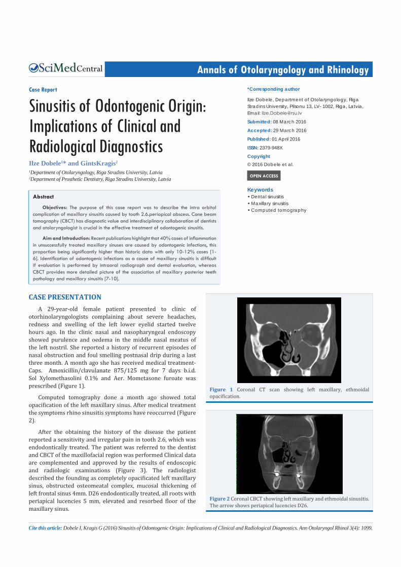
otorhinolaryngologists complaining about severe headaches, redness and swelling of the left lower eyelid started twelve hours ago. In the clinic nasal and nasopharyngeal endoscopy showed purulence and oedema in the middle nasal meatus of the left nostril. She reported a history of recurrent episodes of nasal obstruction and foul smelling postnasal drip during a last three month. A month ago she has received medical treatment-Caps. Amoxicillin/clavulanate 875/125 mg for 7 days b.i.d. Sol Xylomethasolini 0.1% and Aer. Mometasone furoate was prescribed (Figure 1).
Computed tomography done a month ago showed total opacification of the left maxillary sinus. After medical treatment the symptoms rhino sinusitis symptoms have reoccurred (Figure 2).
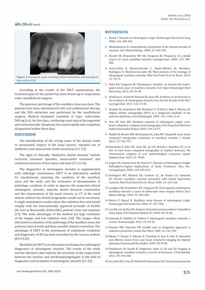
After the obtaining the history of the disease the patient reported a sensitivity and irregular pain in tooth 2.6, which was endodontically treated. The patient was referred to the dentist and CBCT of the maxillofacial region was performed Clinical data are complemented and approved by the results of endoscopic and radiologic examinations (Figure 3). The radiologist described the founding as completely opacificated left maxillary sinus, obstructed osteomeatal complex, mucosal thickening of left frontal sinus 4mm. D26 endodontically treated, all roots with periapical lucencies 5 mm, elevated and resorbed floor of the maxillary sinus.
Figure 1 Coronal CT scan showing left maxillary, ethmoidal opacification.
Figure 2 Coronal CBCT showing left maxillary and ethmoidal sinusitis. The arrow shows periapical lucencies D26.
Central
Dobele et al. (2016)Email:
Ann Otolaryngol Rhinol 3(4): 1099 (2016) 2/3
According to the results of the CBCT examinations, the treatment plan for the patient has been drawn up in cooperation with a maxillofacial surgeon.
The puncture and lavage of the maxillary sinus was done. The patients have been administered with oral antibacterial therapy and the D26 extraction was performed by the maxillofacial surgeon. Medical treatment consisted of Caps. Cefuroxime 500 mg b.i.d. for five days, continuing nasal topical decongestant and corticosteroids. Symptoms decreased rapidly and completely disappeared within three days.
DISCUSSIONThe identification of the wrong cause of the disease leads
to unreasoned surgery of the nasal sinuses, repeated use of antibiotics and unnecessary tooth extractions [11-13].
The signs of clinically “distinctive dental sinusitis” include recurrent sinonasal episodes, unsuccessful treatment and unilateral sensation of foul odour and taste [11,14-16].
The diagnostics of odontogenic sinusitis is supplemented with radiologic examination; CBCT is an informative method for simultaneous assessing the condition of the maxillary sinus and the teeth, and the evaluation of dissemination of pathologic condition. In order to approve the suspected clinical odontogenic sinusitis, separate dental intraoral examination and the examinations of the nasal sinuses, or CT of the nasal sinuses without the dental programme would not be necessary. A single examination would reduce the radiation dose and would comply with the internationally approved principle of ALARA (As Low as Reasonably Achievable), patient’s time and expenses [17]. The main advantages of the method are high resolution of the images and low radiation dose [18]. The images allow informative evaluation of the pathology of the maxillary sinus and posterior lateral teeth and their possible mutual connection. The advantage of CBCT in the assessment of endodontic treatment and diagnostics of RS has been described by the various authors [8,9,19,20].
Maxillofacial CBCT is an informative technique for radiological diagnostics of odontogenic sinusitis. The results of the study and the literature data indicate the necessity of the cooperation between the dentists and otorhinolaryngologists in the field of diagnostics and treatment of odontogenic sinusitis [21-25].
REFERENCES1. Brook I. Sinusitis of odontogenic origin. Otolaryngol Head Neck Surg.
2006; 135: 349-355.
2. Bhattacharyya N. Contemporary assessment of the disease burden of sinusitis. Am J Rhinol Allergy. 2009; 23: 392-395.
3. Bomeli SR, Branstetter BF 4th, Ferguson BJ. Frequency of a dental source for acute maxillary sinusitis. Laryngoscope. 2009; 119: 580-584.
4. Arias-Irimia O, Barona-Dorado C, Santos-Marino JA, Martínez-Rodriguez N, Martínez-González JM. Meta-analysis of the etiology of odontogenic maxillary sinusitis. Med Oral Patol Oral Cir Bucal. 2010; 15: 70-73.
5. Patel NA, Ferguson BJ. Odontogenic sinusitis: an ancient but under-appreciated cause of maxillary sinusitis. Curr Opin Otolaryngol Head Neck Surg. 2012; 20: 24-28.
6. Hoskison E, Daniel M, Rowson JE, Jones NS. Evidence of an increase in the incidence of odontogenic sinusitis over the last decade in the UK. J Laryngol Otol. 2012; 126: 43-46.
7. Bremke M, Sesterhenn AM, Murthum T, Al Hail A, Bien S, Werner JA. Digital volume tomography (DVT) as a diagnostic modality of the anterior skull base. Acta Otolaryngol. 2009; 129: 1106-1114.
8. Nair UP, Nair MK. Maxillary sinusitis of odontogenic origin: cone-beam volumetric computerized tomography. Oral Surg Oral Med Oral Pathol Oral Radiol Endod. 2010; 110: 53-57.
9. Maillet M, Bowles WR, McClanahan SL, John MT, Ahmad M. Cone-beam computed tomography evaluation of maxillary sinusitis. J Endod. 2011; 37: 753-757.
10. Benavides E, Rios HF, Ganz SD, An CH, Resnik L, Reardon GT, et al. Use of cone beam computed tomography in implant dentistry: the international congress of oral implantologists consensus report. Implant Dent. 2012; 21: 78-86.
11. Legert KG, Zimmerman M, Stierna P. Sinusitis of Odontogenic Origin: Pathophysio-logical Implications of Early Treatment. Acta Oto-Laryngologica. 2004; 124: 655-663.
12. Rodrigues MT, Munhoz ED, Cardoso CL, de Freitas CA, Damante JH. Chronic maxillary sinusitis associated with dental impression material. Med Oral Patol Oral Cir Bucal. 2009; 14: 163-166.
13. Longhini AB, Branstetter BF, Ferguson BJ. Unrecognized odontogenic maxillary sinusitis: a cause of endoscopic sinus surgery failure. Am J Rhinol Allergy. 2010; 24: 296-300.
14. Mehra P, Murad H. Maxillary sinus disease of odontogenic origin. Otolaryngol Clin North Am. 2004; 37: 347-364.
15. Lee WJ, Lee SJ, Kim HS. Analysis of location and prevalence of maxillary sinus septa. J Periodontal Implant Sci. 2010; 40: 56-60.
16. Simuntis R, Kubilius R, Vaitkus S. Odontogenic maxillary sinusitis: a review. Stomatologija. 2014; 16: 39-43.
17. Hendee WR, Edwards FM. ALARA and an integrated approach to radiation protection. Semin Nucl Med. 1986; 16: 142-150.
18. Okano T, Harata Y, Sakaino R, Tsuchida R, Iwai K, Seki K. Absorbed and effective doses from cone beam volumetric imaging for implant planning. Dentomaxillofac Radiol. 2009; 38:79-85.
19. Shahbazian M, Jacobs R. Diagnostic value of 2D and 3D imaging in odontogenic maxillary sinusitis: a review of literature. J Oral Rehabil. 2012; 39: 294-300.
20. Xu J, Reh DD, Carey JP, Mahesh M, Siewerdsen JH. Technical assessment
Figure 3 Periapical scans showing failed endodontia and periapical destruction D26.

Central
Dobele et al. (2016)Email:
Ann Otolaryngol Rhinol 3(4): 1099 (2016) 3/3
Dobele I, Kragis G (2016) Sinusitis of Odontogenic Origin: Implications of Clinical and Radiological Diagnostics. Ann Otolaryngol Rhinol 3(4): 1099.
Cite this article
of a cone– beam CT scanner for otolaryngology imaging: image quality, dose, and technique protocols. Med Phys. 2012; 39: 4932-4942.
21. Bagain ZH, Neuman L, Hyde N. How serious are dental infections? J Laryngol Otol. 2004; 118: 561-565.
22. Bauer WH. Maxillary sinusitis of dental origin. Am J Oral Surg. 1943; 29:133-151.
23. Longhini AB, Branstetter BF, Ferguson BJ. Otolaryngologists’ perceptions of odontogenic maxillary sinusitis. Laryngoscope. 2012;
122: 1910-1914.
24. Mylonas AI, Tzerbos FH, Mihalaki M, Rologis D, Boutsikakis I. Cerebral abscess of odontogenic origin. J Craniomaxillofac Surg. 2007; 35: 63-67.
25. Sakamoto T, Aoki T, Kise Y, Watanabe D, Sasaki J. Descending necrotizing media-stinitis due to odontogenic infections. Oral and Maxillofacial Surgery. 2000; 89; 412-419.