single-trial decoding of auditory novelty responses ... dehaene single-trial decoding of... ·...
TRANSCRIPT

NeuroImage 83 (2013) 726–738
Contents lists available at ScienceDirect
NeuroImage
j ourna l homepage: www.e lsev ie r .com/ locate /yn img
Single-trial decoding of auditory novelty responses facilitates thedetection of residual consciousness
J.R. King a,b,c,⁎, F. Faugeras c,d, A. Gramfort b,e,f, A. Schurger a,b, I. El Karoui c, J.D. Sitt a,b,c, B. Rohaut c,g,C. Wacongne a,b, E. Labyt a,b, T. Bekinschtein h, L. Cohen c,d,i, L. Naccache c,d,h, S. Dehaene a,b,j,k
a Cognitive Neuroimaging Unit, Institut National de la Santé et de la Recherche Médicale, U992, F-91191 Gif/Yvette, Franceb NeuroSpin Center, Institute of BioImaging Commissariat à l'Energie Atomique, F-91191 Gif/Yvette, Francec Institut du Cerveau et de la Moelle Épinière Research Center, Institut National de la Santé et de la Recherche Médicale, U975 Paris, Franced AP-HP, Groupe hospitalier Pitié-Salpêtrière, Department of Neurophysiology, Paris, Francee Institut Mines-Télécom, Télécom ParisTech, CNRS LTCI, F-75014 Paris, Francef INRIA Saclay —Ile de France, PARIETAL INRIA, Neurospin, CEA Saclay, Bâtiment 145, 91191 Gif-sur-Yvette Cedex, Franceg AP-HP, Groupe hospitalier Pitié-Salpêtrière, Department of Neurology, Intensive Care Unit, Paris, Franceh Cognition and Brain Sciences Unit, Medical Research Council, Cambridge, UKi Faculté de Médecine Pitié-Salpêtrière, Université Paris 6, Paris, Francej Université Paris 11, Orsay, Francek Collège de France, F-75005 Paris, France
⁎ Corresponding author at: Cognitive NeuroimagingUnide la Recherche Médicale, U992, F-91191 Gif/Yvette, Fran
E-mail address: [email protected] (J.R. King
1053-8119/$ – see front matter © 2013 Elsevier Inc. Allhttp://dx.doi.org/10.1016/j.neuroimage.2013.07.013
a b s t r a c t
a r t i c l e i n f oArticle history:Accepted 4 July 2013Available online 13 July 2013
Keywords:Vegetative stateUnresponsive wakefulness syndromeMinimally conscious stateMultivariate pattern classifierDecodingEEGMEG
Detecting residual consciousness in unresponsive patients is a major clinical concern and a challenge fortheoretical neuroscience. To tackle this issue, we recently designed a paradigm that dissociates twoelectro-encephalographic (EEG) responses to auditory novelty. Whereas a local change in pitch automaticallyelicits a mismatch negativity (MMN), a change in global sound sequence leads to a late P300b response. Thelatter component is thought to be present only when subjects consciously perceive the global novelty. Unfor-tunately, it can be difficult to detect because individual variability is high, especially in clinical recordings.Here, we show that multivariate pattern classifiers can extract subject-specific EEG patterns and predictsingle-trial local or global novelty responses. We first validate our method with 38 high-density EEG, MEGand intracranial EEG recordings. We empirically demonstrate that our approach circumvents the issues asso-ciated with multiple comparisons and individual variability while improving the statistics. Moreover, we con-firm in control subjects that local responses are robust to distraction whereas global responses depend onattention. We then investigate 104 vegetative state (VS), minimally conscious state (MCS) and consciousstate (CS) patients recorded with high-density EEG. For the local response, the proportion of significantdecoding scores (M = 60%) does not vary with the state of consciousness. By contrast, for the global re-sponse, only 14% of the VS patients' EEG recordings presented a significant effect, compared to 31% in MCSpatients' and 52% in CS patients'. In conclusion, single-trial multivariate decoding of novelty responses pro-vides valuable information in non-communicating patients and paves the way towards real-time monitoringof the state of consciousness.
© 2013 Elsevier Inc. All rights reserved.
Introduction
Despite a recent flurry of experimental discoveries, the neuronalmechanisms that support conscious perception remain a major chal-lenge for neuroscience. Numerous studies have associated consciousperception with macroscopic neurophysiological phenomena such assynchronous activity across distant cortical regions and sustainedfronto-parietal activations (Fahrenfort et al., 2007, 2012; Gaillard etal., 2009; Melloni et al., 2007; Sergent et al., 2005) which are thought
t, Institut National de la Santé etce.).
rights reserved.
to reflect global information integration (Dehaene and Changeux,2011; Dehaene and Naccache, 2001; Fisch et al., 2009; Lamme, 2006;Melloni et al., 2011; Rees et al., 2002; Seth et al., 2011; Tononi andEdelman, 1998; Tononi and Sporns, 2003). However, identifying theneuronal signatures of conscious processing is not just a theoretical ex-ercise. Every year, severe brain injuries lead thousands of patients tolose their communication abilities and fall into a variety of clinicalconditions ranging from coma, to vegetative state (VS), minimally con-scious state (MCS) or conscious but paralyzed patients (locked-in syn-drome). Clinically, separating disorders of consciousness (DOC) fromcommunication deficits can be difficult. In particular, VS (also knownas “unresponsive wakefulness syndrome”, Laureys et al. 2010) presentmoments of arousal, during which they open their eyes and produce

727J.R. King et al. / NeuroImage 83 (2013) 726–738
complex behavioral reflexes. Yet, they showno clear signs of intentionalbehavior, even after careful clinical examination performed by experi-enced teams (Bruno et al., 2011a). “Minimally conscious state” (MCS)patients, present some intentional behaviors but seem unable to estab-lish any long-lasting functional communication (Giacino et al., 2002).
To facilitate clinical diagnosis, brain imaging techniquesmay play animportant role (Laureys and Schiff, 2011; Laureys et al., 2004; Monti etal., 2010; Owen, 2008; Owen et al., 2006). By directly detecting the neu-ral activity associated with conscious processing, they could be used tocircumvent communication deficits and thus provide crucialinformation for the diagnosis of these patients. The Local–Globalparadigm (Bekinschtein et al., 2009) was designed for this purpose(Fig. 1). This experimental setup allows the isolation of two event relat-ed potentials (ERPs) elicited by two types of auditory novelty. First, achange in pitch within a five-sound sequence (hereafter referred toas local deviancy) typically leads to a frontal mismatch negativity(MMN) ~150 ms after stimulus onset. Second, a change in auditory se-quence in a fixed global context generates a late P300b response overcentro-posterior electrodes. Crucially, these auditory changes can bearranged to create a 2 × 2 design in which local deviancy and globaldeviancy are orthogonally manipulated (Bekinschtein et al., 2009).
There is now growing evidence that the MMN reflects a predictionerror signal elicited whenever the incoming sound does not fit with aprediction constructed on the basis of previous local auditory regular-ities (Garrido et al., 2007, 2008; Näätänen et al., 1978, 2010;Wacongne et al., 2011, 2012; Winkler, 2007). Moreover, manipula-tions of attention, sleep and anesthesia show that the MMN may per-sist even in unconscious states (Atienza and Cantero, 1997;Bekinschtein et al., 2009; Brázdil et al., 2001; Garrido et al., 2008;Heinke et al., 2004; Muller-Gass et al., 2007; Näätänen et al., 2010;Rohaut et al., 2009; Tzovara et al., 2013). By contrast, the P300b isthought to reflect a higher-order violation of subjects' expectationsof a given rule, constructed over a longer time period and has thusbeen closely linked to working memory (Goldstein et al., 2002;Polich, 2007) and conscious access (Dehaene and Changeux, 2011;Dehaene et al., 2006). Converging lines of evidence suggest that thedissociation between these two neural signatures could discriminatepatients in vegetative state (VS) from those in conscious (CS) or min-imally conscious states (MCS) (Faugeras et al., 2011, 2012; Fischeret al., 2010; Naccache et al., 2005; Rohaut et al., 2009; Wijnen et al.,2007).
However, isolating these event-related responses in single patientscan unfortunately be difficult for several reasons. First, clinical recordingsoften present a low signal-to-noise ratio (SNR) because of numerousphysiological (movements, eye blinks…) and environmental artifacts(presence of auditory noise, no Faraday cage…). Moreover, patientsoften present severe brain and even skull damages which can alter the
Fig. 1. The Local–Global paradigm. The Local–Global paradigm (Bekinschtein et al.,2009) is an auditory odd-ball experimental setup that implicitly tests subjects ontheir ability to detect two orthogonal types of auditory novelty. Each trial is composedof five successive tones (SOA = 150 ms). The first four sounds are always identical.Local-deviant (LD) trials differ from local-standard trials (LS) because their fifthsound deviates in pitch. Global-deviant (GD) trials correspond to the presentation ofa sequence of five sounds which is rare in a given block, compared to the frequent“global standard” sequence (GS). Both local and global novelties depend entirely onthe fifth sound, which therefore serves as the origin of time scales in all subsequentgraphs.
scalp electrical projections of their cortical activity. This topographicalvariability can be made worse by temporal delays and inter-trial vari-ability caused by processing impairments or white matter damage(Newcombe et al., 2010; Tshibanda et al., 2009). In other words, unlikecontrol recordings, a patient's MMN and P300b may not be optimallyobserved over the frontal and parietal channels at ~150 and ~350 msrespectively. While more liberal analyses testing a greater number ofEEG channels and time samples could be implemented, correction formultiple comparisons would largely diminish either sensitivity or con-fidence in the presence of a given brain response.
Toovercome these commonelectrophysiological issues, we evaluatein the present research the potential of a single-trial multivariate pat-tern (MVP) analysis. We implemented, separately for each subject, aMVP classifier that aims at maximally extracting information fromeach trial by combining evidence frommultiple EEG channels and mul-tiple time-samples. After training on an independent dataset, the classi-fier estimates the probability that each trial contains a local or a globalresponse to auditory novelty. This prediction can be compared to trials'effective classes. Classification scores can thus indicate whether a givensubject is able to detect the corresponding type of novelty.
To optimize the detection of single-trial local and global novelty re-sponses, the present research followed a strict logic. First, to optimizeour methods, we tested them with EEG, MEG and intracranial EEG re-cordings acquired from control subjects. Second, we then applied it to158 high-density EEG recordings from 104 distinct patients whosestate of consciousness (VS, MCS, or CS) was assessed immediately priorthe experiment. We consider three successive questions: (1) Whatlevel of accuracy can be achieved from each type of recordings anddoes our method present genuine improvements as compared to tradi-tional analyses? (2) Can decoders be formed to generalize the detectionof novelty from one experimental context to another? (3) Is our methodsensitive enough to be applied to the detection of residual noveltyprocessing in non- or poorly communicating patients?
Methods
Procedure, material & apparatus
The data analyzed here come from four different experimental set-tings, using either scalp EEG, MEG, or intracranial EEG, which togetherenables the direct comparison of the utility of each approach with re-gard to single trial decoding. Events Related Potentials (ERPs) andEvents Related Fields (ERFs) have been partially reported elsewhere(Bekinschtein et al., 2009; Faugeras et al., 2012; Wacongne et al.,2011). All experiments were approved by the relevant regional ethicalcommittees (Comité pour la Protection des Personnes Pitié-Salpêtrièreand Bicêtre hospitals). Healthy volunteers received a financial compen-sation for their participation. Unless specified otherwise, the procedureused in our experiments exactly followed the Local–Global paradigm(Bekinschtein et al., 2009) which enables the comparison of effects en-gendered by physically identical but contextually different auditorystimuli. In the standard Local–Global design, subjects are asked tocount the global deviant trials and report this number at the end ofeach block, in order to ensure that they pay attention to the task.
The auditory stimuli were 50 ms-duration sounds composed of 3sinusoidal tones (either 350, 700, and 1400 Hz, hereafter sound A;or 500 Hz, 1000 Hz, and 2000 Hz, hereafter sound B), with 7-msrise and 7-ms fall times. Sequences were composed of five stimulipresented at a Stimulus Onset Asynchrony (SOA) of 150 ms, andwere separated by a variable silent interval of 1350 to 1650 ms(50 ms steps). The sequences could comprise five identical tones(xxxxx) or four identical tones followed by a distinct one (xxxxY,where x can be sound A or sound B and Y the other sound). Followingthe original design, in a given block, 80% of trials consisted in one typeof sequence (e.g. aaaaB) and 20% of trials were global deviants (aaaaain this example), pseudo randomly distributed at least one and at

728 J.R. King et al. / NeuroImage 83 (2013) 726–738
most six global-standard trials apart (Fig. 1). In all experiments, trialsimmediately following a global deviant were removed from the anal-yses. Each block started with a 30 s habituation phase during whichthe frequent sound sequences were repeatedly presented to establishthe global regularity, before the first infrequent stimulus was heard.In Experiments 1, 3–5, sounds were presented via headphones withan intensity of 70 dB, using E-prime v1.2 (Psychology SoftwareTools Inc.). Trials from the habituation phase were not included inthe analyses. In Experiment 2, sounds were directly presented fromthe computer's speakers because the intracranial recording apparatuswas incompatible with headphones.
Experiment 1: Counting task (EEG)In the first experiment (partially reported in (Faugeras et al., 2012)),
ten healthy adults (Age: M = 23.0 years old, SD = 0.67 years, 3 fe-males) performed the standard Local–Global paradigm, while EEGwas continuously recorded using a 256-channel EEG geodesic net(EGI) sampled at 250 Hz.
Each subject was recorded for approximately 45 min, comprising8 blocks of 3–4 min duration. Each block was interleaved with restingperiods of a few minutes. In each block, subjects were instructed tomentally count the global deviant trials. This experiment will there-fore be referred to as ‘counting EEG’.
Bad sensors, defined as those showing no signal at all, constantwhite noise, or presenting intermittent signals,were interpolated. Trialsduring which more than 20% of the sensors were bad, the EEG voltagesexceeded ±150 μV, EEG transients exceeded ±100 μV or electro-oculogram activity exceeded±80 μVwere excluded from the analyses.All signalswere digitally low-passfiltered at 40 Hz, and referencedwitha common average. Trials were then segmented from −800 ms to736 ms after the onset of the fifth sound, and were baseline correctedover a 200 ms window before the onset of the first of the five sounds.All EEG processing stages were performed in the EGI Waveform ToolsPackage and with the Fieldtrip toolbox (Oostenveld et al., 2011) andMATLAB 2009b.
Experiment 2: Counting task (iEEG)In the second experiment, nine patients (Age: M = 33 years old,
SD = 11 years, 5 women) suffering from drug-resistant epilepsy,and who had consequently undergone electrode implantation forpre-surgical purposes, performed a standard Local–Global paradigm(two patients were already reported in (Bekinschtein et al., 2009))in 8 consecutive blocks. It should be noted that, although epileptic pa-tients cannot be considered as healthy controls, their attention andtheir state of consciousness were relatively comparable to those ofhealthy subjects. Therefore, their intracranial EEG recordings mayshed additional light on the EEG and MEG responses observed inhealthy subjects. On average, patients had 56 intracranial electrodes(IEEG) sampled at 400 Hz or 1024 Hz (depending on the system)placed in various cortical areas including the temporal, the occipital,and the frontal lobes. Electrode locations are reported in Supplemen-tary Fig. 2. The task was identical to Experiment 1s.
After removing channels showing inter-ictal activity, analyses wereperformedbypooling over the 401 channels recorded across all subjects.All signals were digitally low-pass filtered at 40 Hz and down-sampledto 256 Hz. Trials were then segmented from −800 ms to 700 ms afterthe critical stimulus onset, and were corrected for baseline over a200 ms window before the onset of the first of the five sounds. All EEGprocessing stageswere performedwithMATLAB2009b and the Fieldtriptoolbox (Oostenveld et al., 2011).
Experiment 3: Attentive task (MEG)In the third experiment (partially reported in Wacongne et al.,
2011), ten healthy adults (Age: M = 25 years old, SD = 4.7 years, 5females) performed a modified version of the Local–Global paradigm(see below) while their brain activity was measured with MEG (Elekta
Neuromag®MEG system, Helsinki, Finland, comprising 204 planar gra-diometers and 102 magnetometers in a helmet-shaped array) and EEG(built-in 64 electrodes system). Scalp EEG electrodeswere not analyzedin the present study. Data were sampled at 1 KHz with on-line analoglow-pass filtering at 330 Hz, and on-line analog high-pass filtering at0.1 Hz. The head position with respect to the sensor array was deter-mined by four head position indicator coils attached to the scalp. The lo-cations of the coils and EEG electrode positions were digitized withrespect to three anatomical landmarks (nasion and preauricular points)with a 3D digitizer (Polhemus Isotrak system®). Then, head positionwith respect to the device origin was acquired before each block ofMEG/EEG recording.
Each recording session lasted 1 h, comprising 14 blocks of 3–4 minduration with resting periods between each block. Subjects wereasked to keep their eyes opened and to avoid eye movements by focus-ing on a fixation cross displayed in the center of the screen. Subjectswere instructed to pay attention to the auditory stimuli.
The MEG task differed from Experiment 1 in the following respects.First, although the Local Standard and Local Deviant sequences weremainly identical, 10% of the trials were omission trials composed ofonly four sounds. Furthermore, in two block blocks, the frequent audito-ry sequence was also made of only four sounds. These conditions wereapplied in order to test the brain responses to expected and unexpectedomissions. All trials made of only four sounds, or in a block where thefrequent sequences were composed of only four sounds were excludedfrom the present analyses, but are reported in detail in Wacongne et al.(2011). Second, subjects performedmore trials than in the previous ex-periment (780 trials instead of 500). Third, subjects were not asked tocount the number of globally deviant trials, but were only required topay attention to the auditory stimuli. This ‘attentive MEG’ experimentaimed at demonstrating that the previously identified neurophysiolog-ical signatures associatedwith local and global deviant trials were inde-pendent of the counting task. This control can thus validate theapplicability of the Local–Global paradigm in a clinical setup, in whichpatients may not be able to perform complex instructions such ascounting global deviant trials. At the end of the recording, a list of ques-tions was submitted to the subject to check that they had detected thevarious auditory regularities.
Signal space separation (SSS, Taulu et al., 2004) was applied tosuppress unwantedmagnetic interferences (e.g. outside disturbances,limb movements), to interpolate noisy MEG sensors and to realignMEG data into a subject-specific head position. This reference headposition was determined from all head position measurements doneat the beginning of each recording session. This data transformationhelps the direct comparisons of MEG data between conditions andblocks.
Except if explained otherwise, eye blinks and cardiac artifact werecorrected separately for each type of channel (gradiometer and mag-netometers) by decomposing the average artifacts into principal com-ponents, and regressing out those principal components from thecontinuous recording— a technique known as signal space projection(SSP). Noisy MEG sensors were removed with Maxfilter in the SSSpreprocessing step. All signals were digitally low-pass filtered at40 Hz and down-sampled to 256 Hz. Trials were then segmentedfrom −800 ms to 700 ms after the critical stimulus onset, and werecorrected for baseline over a 200 ms window before the onset ofthe first of the five sounds. Segmentation was done with Fieldtrip(Oostenveld et al., 2011). Trials with more than 20% of bad sensorswere rejected.
Experiment 4: Distracting task (EEG)In the fourth experiment (partially reported in Bekinschtein et al.,
2009), 9 subjects (6 females aged between 21 and 33 years old) wereactively distracted by a continuous speeded visual detection task si-multaneous with the presentation of the Local–Global auditory stim-uli, while EEG was continuously recorded at 250 Hz using a

729J.R. King et al. / NeuroImage 83 (2013) 726–738
256-channel EEG geodesic net (EGI) referenced to the vertex. Partic-ipants were instructed to detect a visual target in a rapid streamof successive letters presented at the fovea and were explicitlyasked to neglect the unrelated auditory stimuli. The visual stimuliwere twelve 1° × 1° colored upper or lower case letters, and weremaximally presented for 1000 ms, using E-prime v1.1 (PsychologySoftware Tools Inc.). To check that subjects did not consciously per-ceived the global structure of the auditory stimuli, they were asked atthe end of the experiment whether they had perceived any globalregularity or novelty. EEG preprocessing was identical to Experiment 1.
Experiment 5: Levels of consciousness in patients (EEG)One hundred and fifty eight (158) EEG recordings were acquired
from 104 distinct patients (Age = 48 years old, SD = 17 years, 35females) while they performed the Local–Global paradigm. The first65 recordings were partially reported in Faugeras et al. (2012). Imme-diately before the EEG recording, each patient was carefully examinedby a trained neurologist (FF, BR, LN) and was assessed with theFrench version of the Coma Recovery Scale — Revised (CRS-R,Schnakers et al., 2008). This scale allows a clinical categorization ofeach patient into one of three states of consciousness. The vegetativestate (VS) refers to awake patients who fail to perform very simpletasks such as visual fixation and localization to noxious stimulations.The minimally conscious state (MCS) refers to responsive butnon-communicating patients (Giacino et al., 2002). Recently, Brunoet al. (2011b) proposed to sub-categorize this category into MCS+and MCS− classes to further separate MCS patients who can followcommands from those who cannot. As explained in the supplementa-ry materials, MCS− can for instance track a visual stimulus whileremaining unable to demonstrate intentional communication. Finally,conscious state (CS) refers to patients who present a functional com-munication or a functional capacity to intentionally use objects. Withthe exception of one recording (#140, see Supplementary Table 1),the formal classification of consciousness states is formally based onthe CRS-R. In total, 70 EEG recordings were acquired from VS patients,65 from MCS patients (in which 25 were acquired from MCS− pa-tients) and 23 from CS patients. As can be seen in SupplementaryTable 1, some patients (n = 29) were recorded several times for clin-ical purposes. Most of them (n = 18/29) were recorded in at leasttwo distinct state of consciousness. Patients' etiologies was heteroge-neous: 30 had suffered from an anoxia, 33 from a stroke, 20 from atraumatic brain injury (TBI) and 21 suffered from another etiology.On average, EEG recordings were acquired 178 days after patients' in-jury. All patients had been without sedation for at least 24 h prior tothe recording session. All clinical details are presented in Supplemen-tary Table 1.
After clinical examination, each participant was asked to performa task identical to Experiment 1 (count the global deviants). Patientswere verbally stimulated between each block (~4 min) to ensure sta-ble arousal, and instructions were explicitly repeated by the experi-menter before each run. Although we present results demonstratingthat global effects are independent of the instruction to count, we rea-soned that such instructions could help patients pay attention to theauditory stimulations.
Recording high-density scalp ERPs from non-communicating pa-tients in the intensive care unit or a similar environment is very chal-lenging for technical reasons. First, the electro-magnetic environmentis noisy, and patients were not recorded in a shielded room but atbedside. Second, many patients presented physiological artifactssuch as EMG, eye-movements and blinks, or other involuntary move-ments. Therefore, it is particularly important to systematically evalu-ate the technical quality of data before statistical analysis. Recordingsincluding at least one block with more than 50% of rejected trialswere discarded from further analyses in order to avoid possible biasesacross experimental conditions. EEG preprocessing was identical toExperiment 1.
Analyses
ClassificationIn the following experiments, we aimed at evaluating, for each
subject, the number of trials that could be accurately classified intwo distinct and orthogonal types of classes: (1) as local standard(LS, i.e. an xxxxx sequence) versus local deviant (LD, i.e. an xxxxY se-quence, ‘local classification’); (2) as global standard (GS, frequent se-quence) versus global deviant (GD, rare sequence in a given block,‘global classification’, see Fig. 1). The Local Standard class correspondsto the union of LSGS and LSGD trials, and the Local Deviant class cor-responds to the union of LDGS and LDGD trials. Similarly, the GlobalStandard class corresponds to LSGS and LDGS trials, and the GlobalDeviant class corresponds to LSGD and LDGD trials. These analysescontrast trials evenly distributed across blocks and are therefore lesslikely to be contaminated by block-design pitfalls (Goldfine et al.,2012; Lemm et al., 2011).
The decoding steps are summarized in Supplementary Fig. S6. Aten-fold stratified cross-validation was implemented for each within-subject analysis. Stratified cross-validation balances the proportion ofeach class across K folds (K = 10) in order to maximize the classifiers'ability to generalize to unknown data. Stratified cross-validation is thusparticularly relevant for problem with unbalanced class frequencies.Cross-validation consists in repeatedly applying a series of computationsfitted to a fraction of the dataset (1 − 1/K percent of the trials, calledthe training set), and then evaluate the results on the remaining fraction(1/Kpercent of the trials, called the test set).Within each fold,we estimat-ed the mean (μtrain) and the standard deviation (σtrain) of each sample ofeach sensor across all training trials. The training set Xtrain and the test set(Xtest) were then normalized (Xtrain = (Xtrain − μtrain) / σtrain; Xtest =(Xtest − μtrain) / σtrain). As detailed below, in some analyses a univariatefeature selection was fitted on the training set and subsequently appliedto both the training and the test set. Finally, a support vector classifier(SVC) with a linear kernel (Chang and Lin, 2001) was supplementedwith a continuous output method providing, for each trial, an estimateof the probability of belonging to a given class (Platt, 1999). Amongstthe several advantages of this continuous method, we note that it allowsacross-trial rank statistics and hence avoids the computationally expen-sive permutation analysis generally required with discrete and/or imbal-anced classifiers (see Fig. S5). After fitting the SVC on the training set,classification scores were estimated on the independent test set. Allpreprocessing steps posterior to trials segmentation (feature selection,normalization, parameter selection etc) were fitted, within each cross-validation loop, on the training set only, and are thus immune to circularanalysis issues (Lemm et al., 2011). Cross-validation was constructedfrom chronologically shuffled data which minimizes effects of non-stationarity that could have been observed between the beginning andthe end of the recording session for instance (Lemm et al., 2011). Tofurther minimize the possibility of a trial ordering confound, the cross-validation was applied four times for each patient. Each of these cross-validations was based on a different chronologically shuffled datasetthus implying different folds too. This step did not however fundamental-ly change the obtained results.
Classification scores were estimated with an empirical receiver-operative curve (ROC) analysis applied on trials' predicted probabili-ties. The ROC analysis is a standard 2-class (positive and negative)non-parametric statistical method which allows the estimation ofthe effect size of a Wilcoxon/Mann–Whitney U Test (Mason andGraham, 2002). It is based on the plotting of true positive rate as afunction of false positive rate. The result of this function can be sum-marized by the area under its curve (AUC). An AUC of 50% impliesthat true positive predictions (e.g. trial predicted deviant and is devi-ant) and false positive predictions (e.g. trial predicted as deviant butis standard) are, on average, equally probable; an AUC of 100%indicates a perfect positive prediction with no false positive. Amongstthe advantages of the ROC analysis, we note that, unlike mean accuracy,

730 J.R. King et al. / NeuroImage 83 (2013) 726–738
it is robust to imbalanced problems and, as a non-parametric analysis,does not make any hypothesis about the distribution underlying thedata. The reported AUCs correspond to the averaged within-subjecteffect size: the ability of the classifier to discriminate standard from de-viant trials in a given subject. Note that the average within-subject AUCcan be relatively low and yet remains significant across subjects. Suchcases suggest that while the analysis robustly identifies a significanteffect across an entire EEG session, it is unlikely to be useful to onlinedecoding of single-trials.
Generalization across contextWe implemented a set of generalization analyses which aimed at
testing the invariance of the neurophysiological signatures of localand global novelty detection. These generalization analyses consistedin training the classifier using data from a fixed context, and subse-quently testing it in a different context. Classifiers were for instancetrained to discriminate local standard trials from local deviant trialsusing only global standard trials (LSGS versus LDGS). The discrimi-native hyper-plane (w) found was subsequently applied to trialsfrom a different global context, in this case the global deviants(LSGD versus LDGD). This approach was also adapted to the contex-tual generalization of global effects. In this case, the classifier wasfirst trained, for instance, to discriminate global effects in a fixedlocal context (LSGS versus LSGD), and then tested on a differentlocal context (LDGS versus LDGD, see Figs. 1 & 4). This procedurewas systematically applied in a symmetrical manner: a classifierwas either trained in standard contexts and tested in deviantones, or trained in deviant contexts and tested in standard ones,and the results were then averaged over both directions of general-ization. This procedure thus aimed to identify the neural signaturesof novelty processing that are robust and generic to different con-texts, with the added difficulty of being based on only half of thetraining trials.
Time-windows of interestWe applied several multivariate pattern classifiers on different
temporal regions of interest (Fig. S5). First, we repeatedly tested aclassifier using all channels on a sliding window of a single timesample (~4 ms). Second, we implemented a set of classifiers usingmultiple time samples. These classifiers combined either all or asub-selection of the time samples following the onset of the lastsound (t = [0,736] ms): the early time-window refers to t = [0,367] ms, and the late time window refers to t = [367, 700] ms. Asa control, supplementary analyses also tested the ability of classifiersto extract the electrophysiological information on a pre-stimuluswindow ([−367, 0] ms relative the onset of the fifth sound). Finally,the last approach consisted in averaging brain signals within a giventime windows (Fig. S3). The method, rationale and results arepresented in the supplementary materials.
Support Vector Machine (SVM)Each trial was transformed into a p-dimensional vector x, in which
each coordinate (“attribute”) corresponds to a single data sample at agiven sensor (p = nsensors ∙ nsamples). The entire dataset can hence berepresented as a matrix X in which each row i corresponds to one trialxi, and each column corresponds to an attribute. Moreover, trial classes(standard or deviant) can also be represented as a binary vector y, inwhich standard trials are labeled as 1, and deviant trials as −1. As forall linear classification analyses, the aim is to find a hyperplane (materi-alized by a p-dimensional vector of weights w) that discriminates thetwo classes (ystandard and ydeviant). For a new trial xnew, the sign of thedot product xnew. w is then generally used to predict its class. In thepresent case, a cumulative probability distribution function was fittedto the training set using Platt's method (Platt, 1999). The signeddistance computed from the dot product xnew. w could thus betransformed into a continuous value bounded between 0 and 1. This
value is directly representative of the probability of belonging to oneof the two classes. Note that rank statistics are not affected by the trans-formation of the signed distance between a given trial and w by a mo-notonous function, such as a cumulative distribution function. Whenthe number of trials is small compared to the number of attributes(here up to 56,000), there are infinitely manyw that can fit the trainingdata equally well. To limit this issue, we implemented within thecross-validation, a dimensionality reduction based on a univariatefeature-selection step that selects a subset of attributes prior to the clas-sification analysis. Feature selection was performed with an ANOVAnested within the cross-validation step. Various levels of the ANOVA pvalue threshold were explored in Experiment 1 (1%, 5%, 10%, 30%, 50%,100%), and on this basis a fixed value of 10%was chosen for all other ex-periments. No feature selection was used for the decoding of each timepoint as dimensionality was relatively small (e.g. from 40 channels to306 channels depending on the type of recording). SVC's regularizationparameter was calibrated by nested cross-validation in Experiment 1(.01, .1, .3, .5, 1, 2) and remained arbitrarily fixed (1) for all other exper-iments as its values did not dramatically affect classification scores. Fi-nally, sample weights were applied in proportion of the trial classes(LSGS, LSGD, LDGS, LDGD) so that each of the category would equallycontribute to the definition of w, and this independently of thecontrast of interest (local or global). This step is known to contributeto theminimization of imbalanced dataset artifacts and thusmaximizesthe number of trials that can be considered in the training process. Inpractice this step did not lead to a strong improvement of classificationperformance. All multivariate analyses were performed with the Scikit-learn toolbox (Pedregosa et al., 2011).
As a control, in supplementary analyses, we applied the same ap-proach after randomly shuffling the label (y) of each trial and subse-quently training the classifier with this incorrect class labeling.Ideally, this method would be applied 10,000 times per subject toallow the estimation of the null distribution of each subject. However,this approach would be too computationally expensive with thepresent dataset, because the classification of a single EEG recordingsession takes approximately 30 min of computer time. We thus ap-plied shuffling only once per EEG recording, and thus only reportacross-subjects statistics.
StatisticsROC analyses and AUCs are methods to estimate the size of a given
effect, and in our case, the classifiers' ability to discriminate two typesof trials. To test for statistical significance within subjects, weperformed Mann–Whitney U tests across trials. Because the classifierattributes a continuous estimate to each trial (its predicted probabil-ity of belonging to a given class C), we can indeed efficiently comparethose predicted probabilities across the two levels of the trials' trueclasses. For instance, for each trial, the classifier outputs a predictedprobability of belonging to the standard class (S). Note that the prob-ability of belonging to the other, deviant class (D) can be calculated asP(D) = 1 − P(S). We can then compare the predicted probability ofbelonging to S depending on whether the trials truly are standard(y = S) or not (y = D) and thus apply a traditional Mann–WhitneyU Test between P(S|y = S) and P(S|y = D).
Similarly, across-subjects statistics were performed using WilcoxonSigned Rank Tests based on the mean predicted probability conditionalof trials' true classes. For each subject, the predicted probability of belong-ing to the standard class (S) is averaged across standard trials (y = S)and, separately, over deviant trials (y = D), yielding P(S|y = S) andP(S|y = D). For each subject i, we can thus compute the sum of positiveranks and perform a Wilcoxon test. A correction for multiple compari-sons, either across classifiers or across subjects, was performed usingthe standard False Discovery Rate (FDR) correction, and is hereafter re-ferred to as pFDR. Statistical analyses were performed with R andMATLAB 2009b.

731J.R. King et al. / NeuroImage 83 (2013) 726–738
Results
The Local–Global paradigm enables the isolation of two types ofneurophysiological activity: either the response to a change in pitchwithin a five-sound sequence (local effect), or the response to a rareauditory sequence within a block dominated by another frequentlyrepeated sequence (global effect, Fig. 1).
Topographical analyses
Traditional ERP analyses revealed topographies and time coursessimilar to the ones observed in previous studies (Bekinschtein et al.,2009; Faugeras et al., 2012; King et al., 2011; Wacongne et al., 2011).EEG results from Experiment 1, summarized in Fig. 2, showed that localeffects arose between approximately 130 ms and 350 ms and mainlyevoked a frontal negativity (MMN) followed by a central positivity(P300a). In contrast, global effects were mainly observed from 200 msonwards, and were characterized by a sustained centro-posterior posi-tivity (P300b) peaking between 300 ms and 500 ms. These two effectsreplicate the EEG components previously reported in this type of para-digms. Interestingly, and as can be seen on Fig. 2, a vast amount ofinter-individual variability can be observed in the single-subject data.This variability thus highlights the potential usefulness of tailoring theanalyses to each subject.
Decoding across time circumvents individual variabilities
To maximize the detection of the neurophysiological responseselicited by local and global novelties, we applied, to each subjectseparately, a multivariate pattern (MVP) classifier. The classificationscores of each classifier reported below refer to the area under thecurve (AUC) estimated separately for each subject from a receiveroperative curve analysis (see Methods).
We first aimed at characterizing the dynamics of classificationscores across time, and thus trained a different classifier for eachtime sample. As expected, during the time period preceding theonset of the last sound, both local and global decoding remained atchance level (AUC did not differ from 50%). Accuracy of the local de-coder exceeded chance level earlier in the epoch than did the
Fig. 2. Inter-individual variability in the topography of EEG effects in Experiment 1. In the twthe global effect (GD N GS) are plotted as a function of time following the onset of the fifth sostandard and deviant conditions, corrected for multiple comparisons with FDR. The top andglobal effects in their respective time windows of interest. Local deviant trials elicit significfollowed by a short central positivity between 200 and 300 ms. Global deviant trials elicitstatistics replicate the traditional MMN and P3b associated with the Local–Global paradigm (ity in healthy subjects, and thus highlight the usefulness of tailoring the analyses to each su
accuracy of the global decoder. As can be seen on Fig. 3a, two peaksof local decoding were observed in each experiment between150 ms and 300 ms after stimulus onset. Mean single-trial local clas-sification scores across subjects varied from 62.0% to 77.8% of trialsdepending on the experimental apparatus (all pFDR b .05).
The dynamics of global effects were more variable across experi-ments. Experiment 1, in which subjects were instructed to countrare global deviant trials, revealed a quick rise of EEG-based classifica-tion scores around ~130 ms, followed by a sustained period duringwhich global classification remained above chance almost throughoutthe epoch (between 300 ms and 730 ms). However, in intracranialEEG (Experiment 2), despite similar instructions, only a transient pe-riod of global decoding was seen, peaking at ~260 ms, then droppingback to chance level. Finally, Experiment 3, in which subjects wererecorded with MEG and were instructed to merely pay attention tothe sounds, demonstrated sustained global classification scores from200 ms onwards (AUC = 66%). These results suggest that the neuro-physiological signatures of novelty detection seem relatively inde-pendent from task instructions.
Interestingly, a sharp increase in global classification scores wasobserved as early as 150 ms in the attentive MEG condition (Experi-ment 3). Although much smaller (AUC = 55.0%, pFDR b .05) a similartrend was also apparent in the counting EEG condition (Experiment1). Finally, intracranial recordings, mainly taken from the temporalcortices (Supplementary Fig. 2) presented significant global classifica-tion performance between 100 ms and 300 ms (max AUC = 66.1%,pFDR b .05). Taken together, these results suggest, contrary to whatwas previously suggested (Bekinschtein et al., 2009), that global nov-elty can affect early processes too.
Finally, subjects who were distracted by a concurrent visual task(Experiment 4) presented lower but still significant local classifica-tion scores (AUC = 63.7%, pFDR b .05). Moreover, distraction dramat-ically impaired global classification scores, which consequently failedto reach significance across subjects (AUC = 54.0%, pFDR N .05).
Overall decoding local and global effects across time showed thatmultivariate pattern classifiers could extract qualitatively similar ef-fects as the ones observed from ERPs (for ERFs see Wacongne et al.,2011) but could also reveal subtle effects such as an early global effectunreported in previous studies.
o central rows, control EEG topographies of the mean local effect (LD N LS) and of meanund. Black dots correspond to EEG channels presenting significant differences betweenbottom rows show the topography in individual subjects, separately for the local andant differences over anterior regions with an initial negativity peaking around 150 msa centro-posterior sustained positivity mainly from 300 ms onwards. While the groupBekinschtein et al., 2009), individual analyses reveal substantial topographical variabil-bject.

Fig. 3. Local and global multivariate decoding scores. a. Decoding of the local effect (red) and of the global effect (blue) was applied successively to each time sample of the scalpEEG, MEG and intracranial EEG recordings obtained under different experimental conditions: subjects either counted the global deviant trials (Experiments 1–2), were attentive tothe sounds (Experiment 3), or were distracted by a visual task (Experiment 4). Results demonstrate a non-sustained decoding of the local effect (~130–400 ms), with above-chanceperformance regardless of recording apparatus and experimental condition. Decoding of the global effect appeared later and was more sustained in time, but was absent in thedistracted condition. b. Comparison of the decoding scores based on the best time point (left dot in each graph) or the full trial dynamics (right dot). Each dot represents thedecoding score of an individual subject. All subjects but one presented significant local decoding scores. All experiments led to significant global decoding scores, except for thedistracted condition. Local and global decoding scores based on the dynamics of the electrophysiological signal across multiple time samples (tall) led to a significant improvementof decoding scores in most scalp recordings.
732 J.R. King et al. / NeuroImage 83 (2013) 726–738
Using signal dynamics facilitates decoding
Decoding a spatial topography at each time sample, as was done inthe previous section, can overcome individual topographical variabil-ity but remains dependent on the precise timing of a given effect. Tomaximize the extraction of local and global effects, we thus trainedanother decoder using the full dynamics of brain signals on a giventrial. For each subject separately, and for local and global effects,this decoder was trained to distinguish the standard and deviant trialsbased on all the information available on a given trial (all channels × alltime samples). Results are presented in Fig. 3b.
Overall, this dynamic approach provided better classification scoresthan in the previous section. To prove this, we compared it to the perfor-mance of the best time sample of the previous section. Note that this is a
very conservative test. Indeed, the a posteriori selection of the best timesample is not cross-validated, and thus likely overestimates the actualscores one could hope to obtain if an independent data sample wasavailable (e.g. Vul et al., 2009). Still, an ANOVA across subjects, contrasts(local and global) and type of classifier (dynamic versus best single timepoint) revealed a significantmain effect of classifier type, F(1,37) =47.9,p b 10−10, indicating better classification overall with the dynamic ap-proach (mean AUC = 72.0%) than with the previous approach (meanAUC = 63.7%). This improvementwas robust across recordingmethods(all p b .05) except for intracranial EEG (p N .33), and therefore suggeststhat the classifier manages to exploit the time course of brain signals toprovide information at the single-trial level (See Supplementary Fig. 1for a depiction of single trials' local and global predictions in Experiment3.). Supplementary analyses confirmed that the observed improvement

733J.R. King et al. / NeuroImage 83 (2013) 726–738
of decoding scores reflects an ability of the classifier to extract the dy-namics of the brain signals (Fig. S3).
In more detail, using the dynamic approach, the local classificationscores reached an average AUC of 77.8% (p b .01) in the counting EEGcondition (Experiment 1), 73.2% (p b .01) in the counting intracranialcondition (Experiment 2), 73.5% (p b .01) in the distracted EEG con-dition (Experiment 4) and 89.8% (p b .01) in the attentive MEG con-dition (Experiment 3). The latter performance, interestingly, washigher than the one obtained from high-density EEG (all p b .01)and even than intracranial recordings (p b .05). Importantly, acrossfour experiments, all but one subject (from the distracted group)presented significantly above chance local classification scores.
Similar results were obtained for global classification scores acrossExperiments 1–3. The counting EEG condition (Experiment 1) led toan average AUC of 67.9% (p b .01), the counting intracranial condition(Experiment 2) led to an average AUC of 66.1% (p b .01) and the at-tentive MEG condition (Experiment 3) led to an average AUC of72.3% (p b .01). Amongst the 28 subjects who were paying attentionto the sounds, only one subject failed (from Experiment 1) to presenta significant global decoding score.
Crucially, global classification scores were dramatically reduced indistracted subjects (p b .01 as compared to EEG and MEG recordings —Experiments 1 and 3; p b .05 as compared to intracranial recordings —Experiment2). Not only was the global decoding score not-significantlydifferent from chance for distracted subjects (AUC = 55.0%, p N .05),but 8 out of 10 subjects did not present any significant global decoding.
In summary, by maximizing the extraction of information from in-dividual recordings, these results demonstrate that the human brainresponse to auditory deviancy, both local and global, can be detectedin single trials and a fortiori in individual subjects. It also confirm thatthis information is not entirely dependent on the fact that subjects areasked to count the rare global deviant trials, but remains presentunder the instruction to merely attend to the sounds. Finally, thecomparison between the active and the distracted conditions con-firms the automaticity of processes underlying local novelty detec-tion, and the necessity of attention in the detection of global novelty.
Generalization across contexts isolates rule-specific effects
The above analysis, while sensitive, could be partially affected by acontextual modulation of the local mismatch effects (MMN). Indeed,the MMN elicited by an xxxxY sequence is stronger when this se-quence is rare than when it is frequent (Wacongne et al., 2012).This effect could be used by the global decoder to provide earlyabove-chance classification of global deviants from global standards,even though it does not reflect a genuine response to global novelty.To address this issue, we reasoned that a cross-context generalizationwould provide a stricter criterion for a physiological signature of thebrain's response to global novelty. For instance, a neuronal processresponding to rare sequences, if generic, should be found whenevera rare global deviant sequence is presented, whether this sequenceis xxxxY (in blocks where xxxxx is the frequent stimulus) or xxxxx(in blocks when xxxxY is the frequent stimulus). Such generalizationanalysis could sort out the genuine signatures of global effects fromthe modulations of local novelty effects. With this idea in mind, weinvestigated the generalization of global classification across differentlocal contexts (and vice-versa see Methods), and restricted the localand the global analyses to the early (0–367 ms) and to the late(367–700 ms) time windows respectively. As intracranial classifica-tion scores were not significant in the late time window (Fig. 3), wetested this analysis on Experiments 1, 3 and 4. Results are presentedin Fig. 4.
An ANOVA across subjects, contrasts (local or global) and type ofclassifier (using both contexts or within-context decoding) showedthat classification scores resulting from within-context decoders weresmaller than those obtained from decoders using all trials (Introduction
section): F(2,28) = 26.56, p b 10−6. This is expected as restricting thedecoder to trials performed in a given context automatically reducesthe number of trials available for the training set. Note however thatthe within-context classification scores remained significantly abovechance in all experimental conditions: Local: Experiment 1: 69.1%(p b .01), Experiment 3: (71.2% p b .01), and Experiment 4: 68.8%(p b .01); Global: Experiment 1: 66.0% (p b .01), Experiment 3: 63.2%(p b .01), and Experiment 4: 56.7% (p b .05).
Cross-context generalization scores appeared significantly worsethan within-context classification scores (Local: Experiment 1: p b .01,Experiments 3–4: p b .05; Global: Experiment 1: p b .01, Experiments3–4: p b .10). As can be seen in Fig. 4a presenting the MEG generaliza-tion scores across time, the differences between within-context andacross-context scores were mainly visible i) in early time windows;and ii) in the global contrast. This result suggests that part of the earlyglobal effect previously described was due to an early modulation ofnovelty responses that did not generalize across contexts. However,the overall reduction of cross-context generalizations suggests thatthis conceptually more suitable analysis may, in practice, be less appro-priate when the signal-to-noise ratio is low.
Crucially, whereas all local generalization scores remained signifi-cantly above chance (Experiment 1: 57.1%, Experiment 3: MEG: 66.3%,Experiment 4: 61.5%, all p b .01), global generalization scores werenow exclusively significant in subjects who were paying attention tothe sounds (Experiment 1: 57.9%, Experiment 3: 60.2%, both p b .01).Distracted subjects did not present any significant global generalizationscores (48.1%, p N .10), and the latter were significantly smaller thanthe ones obtained in the counting (Experiment 1: p b .001) and theattentive (Experiment 3: p b .0001) conditions.
Finally, it is interesting to note that global generalization scoressteadily increased from 180 ms after stimulus onset, until they eventu-ally reached, at the end of the epoch, scores that resembled thoseobtained with the within-context decoding analysis in MEG (Fig. 4a).This pattern of results suggests that distinct neurophysiological signa-tures were initially elicited by the two forms of global deviancy testedhere. Most likely, the rare xxxxY sequence was easily detected in anxxxxx context, whereas, conversely, the rare xxxxx sequence took alonger time to be detected in an xxxxY context. Late in the epoch, how-ever, the neuronal activity that they evoked eventually converges to thesame state, thus permitting similar levels of within-block classificationand cross-block generalization.
Patients: Decoding across time and dynamics
The above results demonstrate that multivariate classifiers effi-ciently capture the dynamics of the brain responses to local and glob-al deviancy. In particular, we confirmed that the processing of localnovelty was relatively independent of attention, while the late effectselicited by global novelty only emerged and generalized across con-texts in attentive subjects. These analyses allowed us to formulate asimple hypothesis: in non-communicating patients, cerebral activityspecific to local and to global novelty may provide useful markers ofautomatic versus conscious processing of auditory regularities. Wethus applied the present method to 158 EEG recordings acquiredfrom patients diagnosed in vegetative state (VS), minimally consciousstate (MCS) or conscious state (CS). Individual and group results aresummarized in Fig. 5.
First, it appeared that the decoding of the EEG dynamics of the pa-tients was qualitatively similar to what was observed in healthy con-trols: local classification scores were maximal between 150 ms and300 ms after stimulus onset, whereas global effects, if present, appearedlater and were relatively sustained between 250 ms and 700 ms (seeFig. 5a). Classifiers based on the full dynamics of the EEG signals (seeMethods) were again superior to selecting the best time point obtainedfrom healthy controls (local: p b .10−5, global: p b .01) or directly from
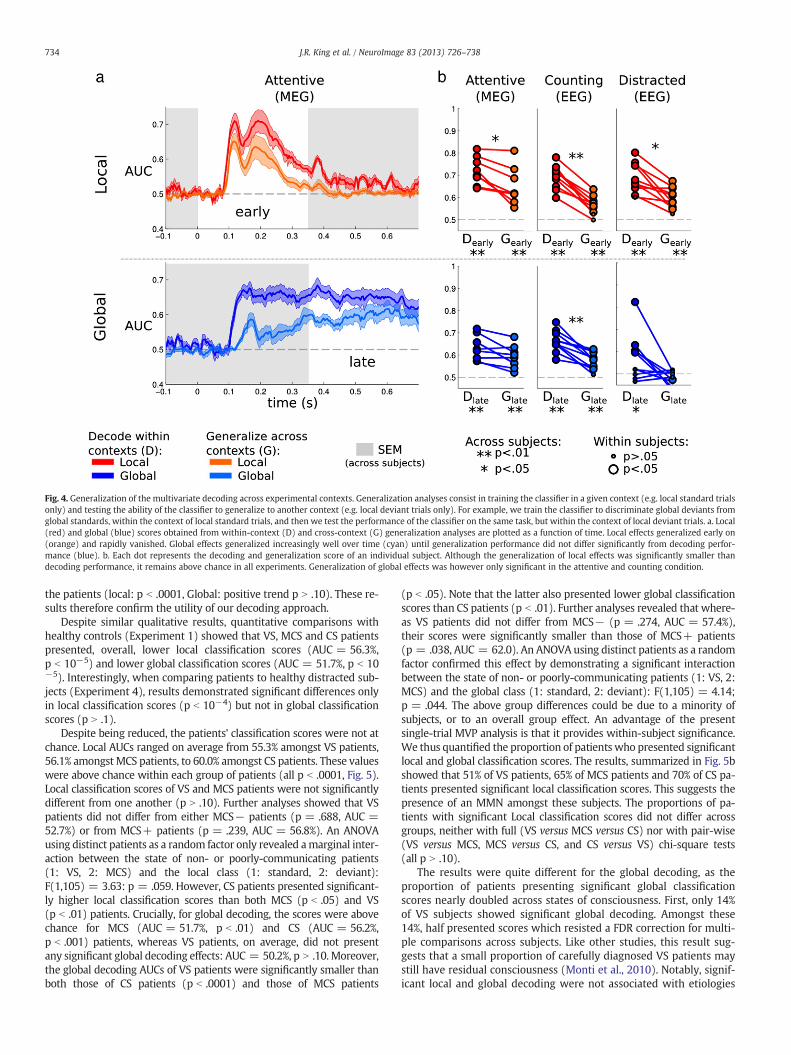
Fig. 4. Generalization of the multivariate decoding across experimental contexts. Generalization analyses consist in training the classifier in a given context (e.g. local standard trialsonly) and testing the ability of the classifier to generalize to another context (e.g. local deviant trials only). For example, we train the classifier to discriminate global deviants fromglobal standards, within the context of local standard trials, and then we test the performance of the classifier on the same task, but within the context of local deviant trials. a. Local(red) and global (blue) scores obtained from within-context (D) and cross-context (G) generalization analyses are plotted as a function of time. Local effects generalized early on(orange) and rapidly vanished. Global effects generalized increasingly well over time (cyan) until generalization performance did not differ significantly from decoding perfor-mance (blue). b. Each dot represents the decoding and generalization score of an individual subject. Although the generalization of local effects was significantly smaller thandecoding performance, it remains above chance in all experiments. Generalization of global effects was however only significant in the attentive and counting condition.
734 J.R. King et al. / NeuroImage 83 (2013) 726–738
the patients (local: p b .0001, Global: positive trend p N .10). These re-sults therefore confirm the utility of our decoding approach.
Despite similar qualitative results, quantitative comparisons withhealthy controls (Experiment 1) showed that VS, MCS and CS patientspresented, overall, lower local classification scores (AUC = 56.3%,p b 10−5) and lower global classification scores (AUC = 51.7%, p b 10−5). Interestingly, when comparing patients to healthy distracted sub-jects (Experiment 4), results demonstrated significant differences onlyin local classification scores (p b 10−4) but not in global classificationscores (p N .1).
Despite being reduced, the patients' classification scores were not atchance. Local AUCs ranged on average from 55.3% amongst VS patients,56.1% amongst MCS patients, to 60.0% amongst CS patients. These valueswere above chance within each group of patients (all p b .0001, Fig. 5).Local classification scores of VS and MCS patients were not significantlydifferent from one another (p N .10). Further analyses showed that VSpatients did not differ from either MCS− patients (p = .688, AUC =52.7%) or from MCS+ patients (p = .239, AUC = 56.8%). An ANOVAusing distinct patients as a random factor only revealed amarginal inter-action between the state of non- or poorly-communicating patients(1: VS, 2: MCS) and the local class (1: standard, 2: deviant):F(1,105) = 3.63: p = .059. However, CS patients presented significant-ly higher local classification scores than both MCS (p b .05) and VS(p b .01) patients. Crucially, for global decoding, the scores were abovechance for MCS (AUC = 51.7%, p b .01) and CS (AUC = 56.2%,p b .001) patients, whereas VS patients, on average, did not presentany significant global decoding effects: AUC = 50.2%, p N .10. Moreover,the global decoding AUCs of VS patients were significantly smaller thanboth those of CS patients (p b .0001) and those of MCS patients
(p b .05). Note that the latter also presented lower global classificationscores than CS patients (p b .01). Further analyses revealed that where-as VS patients did not differ from MCS− (p = .274, AUC = 57.4%),their scores were significantly smaller than those of MCS+ patients(p = .038, AUC = 62.0). An ANOVA using distinct patients as a randomfactor confirmed this effect by demonstrating a significant interactionbetween the state of non- or poorly-communicating patients (1: VS, 2:MCS) and the global class (1: standard, 2: deviant): F(1,105) = 4.14;p = .044. The above group differences could be due to a minority ofsubjects, or to an overall group effect. An advantage of the presentsingle-trial MVP analysis is that it provides within-subject significance.We thus quantified the proportion of patients who presented significantlocal and global classification scores. The results, summarized in Fig. 5bshowed that 51% of VS patients, 65% of MCS patients and 70% of CS pa-tients presented significant local classification scores. This suggests thepresence of an MMN amongst these subjects. The proportions of pa-tients with significant Local classification scores did not differ acrossgroups, neither with full (VS versus MCS versus CS) nor with pair-wise(VS versus MCS, MCS versus CS, and CS versus VS) chi-square tests(all p N .10).
The results were quite different for the global decoding, as theproportion of patients presenting significant global classificationscores nearly doubled across states of consciousness. First, only 14%of VS subjects showed significant global decoding. Amongst these14%, half presented scores which resisted a FDR correction for multi-ple comparisons across subjects. Like other studies, this result sug-gests that a small proportion of carefully diagnosed VS patients maystill have residual consciousness (Monti et al., 2010). Notably, signif-icant local and global decoding were not associated with etiologies

Fig. 5. Local and global decoding in patients whose state was diagnosed as vegetative (VS), minimally conscious (MCS) and conscious (CS). a. Local (top) and global (bottom)decoding scores are plotted as a function of time. Overall, local and global decoding scores follow the same qualitative trends as the ones observed in healthy subjects (Fig. 2).CS patients (blue) presented higher local and global decoding scores over time than either MCS (green) or VS (red) patients. b. Decoding scores obtained from the EEG dynamicsof each trial are summarized for each state of consciousness. The graphs give the mean and standard error of the decoding scores in each group, its significance relative to chancelevel, and the significance of pair-wise group comparisons. Pie charts summarize the proportion of patients who presented significant decoding scores.
735J.R. King et al. / NeuroImage 83 (2013) 726–738
(local: χ2(3, N = 70) = 2.30, p = .513; global: χ2(3, N = 70) = .66,p =.882) nor with the delay separating the insult from the EEG re-cording (both p N .669). Second, in contrast, 31% of the MCS patientsand 52% of the conscious patients presented significant global scores.The proportions of significant global scores across the three states ofconsciousness were significantly different from one another: χ2(2,N = 158) = 13.73, p b .001. Pair-wise chi-square tests confirmedthis finding by showing a smaller proportion of patients with globaldecoding in VS relative to MCS (χ2(1, N = 135) = 4.39, p = .036)and to CS (χ2(1, N = 93) = 11.74, p = .0001). No differences wereobserved between the proportions of CS and MCS patients with sig-nificant global decoding (χ2(1, N = 88) = 2.50, p = .113).
To increase the specificity of the present results, we also applied thegeneralization method detailed above. Scores obtained from within-context classification were similar to but lower than the classificationscores obtained from the joint analysis of both contexts (unsurprisinglygiven that the number of training trials was halved). For the local effect,while not differing from one another (VS–MCS: p = .252), local scoresfrom VS (AUC = 56%, p b .0001) and MCS (AUC = 57%, p b .0001)were significantly smaller than those obtained than CS patients(AUC = 61%, p b .0001, CS–VS: p = .005, CS-MCS: p = .039). Globalwithin-context classification scores presented a similar pattern: VS(AUC = 54%, p b .0001) andMCS (54%, p b .0001) scores did not differfrom each other (p = .969), but were significantly smaller from CS(59%; both p = .001). Finally, cross-context generalization scoreswere low (all below 53%). Local cross-context generalization perfor-mance was lower for VS patients than for MCS (p = .038) and CS(p = .003) patients, but none of the global cross-context generalizationscores differed from one another (all p N .287).
In summary, by quantifying the proportion of patients with signifi-cant local and global effects, we confirmed the earlier finding thatDOC patients may still exhibit similar local mismatch effects, relatively
independently of their conditions (Bekinschtein et al., 2009; Faugeraset al., 2012; Fischer et al., 1999; Naccache et al., 2005; Tzovara et al.,2013), and found that the capacity to show global effects was modulat-ed by the state of consciousness. A classifier based on the whole set oftrials and their temporal dynamics proved themost sensitive tool to de-tect these local and global effects. Cross context generalization was un-able to dissociate the three types of patients, presumably because of ahalving of the number of training trials.
Discussion
In this study, we investigated, at the single trial level, the neuronalresponse following the violation of two embedded auditory regularitiesstructured across two different time ranges (“local” and “global”). Weimplemented, for this purpose, a series of multivariate pattern (MVP)analyses extracting this information from the temporal dynamics ofthe neurophysiological activity recordedwith high-density EEG (Exper-iment 1 and 4, n = 20), intracranial EEG (Experiment 2, n = 9) orMEG(Experiment 3, n = 9). Analyses were performed on attentive and dis-tracted healthy control subjects, as well as on three types of patients,namely patients in vegetative state (VS, n = 70), minimally consciousstate (MCS, n = 65) and conscious state (CS, n = 23), all recordedwith high-density EEG for clinical purposes.
Results from control experiments revealed that when subjectswere attentive, single-trial classification could lead to AUCs between73% and 90% for local novelty, and between 66% and 72% for globalnovelty, depending on the recording apparatus (Experiments 1–3).Although EEG, MEG and IEEG record different types of signals andnoise and are therefore not easily comparable, it is remarkable tonote that MEG recordings achieved classification scores comparableto, or even higher than intracranial EEG data. Moreover, we showedthat providing the decoder with multiple time samples systematically

736 J.R. King et al. / NeuroImage 83 (2013) 726–738
improved classification as compared to a decoder trained on the sin-gle best time sample. This result therefore demonstrates the utility ofMVP classifiers in the present context, and confirms that this methodcan reliably and automatically extract neuronal dynamics specific toeach subject in order to efficiently classify each trial.
It should be noted that our method, although different in technicaldetails, follows a similar approach to that taken in a recent study(Tzovara et al., 2013). Tzovara and collaborators decoded, from EEG re-cordings of coma patients, the difference between regular and irregulartrials, that is, the equivalent of the local effect in the present study. Toclassify each trial, the authors used a different method which modeledthe ERPs with a mixture of Gaussians. Advantages and disadvantagescan be identified in each of these classification methods. Our approachdoes not make any Gaussian assumptions, can make use of imbalancedtraining dataset and can extract a large number of different types of to-pographies by searching across all channels — which therefore are notrestricted to EEG. In contrast, Tzovara and collaborators first transformthe ERPs into components. This computational step reduces the dimen-sionality of the data, and thus likely improves the efficiency of the ensu-ing classification process. However, their classifier is unable tooptimally use imbalanced datasets, which are necessarily encounteredin this type of novelty paradigm. Taken together, these differencesmay explain why our approach provided slightly better classificationscores (AUC = 77.8%) than theirs (AUC = 71%) in similar control sub-jects recorded with EEG. Future efforts should capitalize on a combina-tion of these methodological technicalities.
Crucially, our MVP classifiers replicated and extended previousobservations on the key electrophysiological properties of local andglobal effects (Bekinschtein et al., 2009). As expected, local noveltymainly affected the early part of the neural signal (b300 ms) andremained unaffected by visual distraction. In sharp contrast, globalnovelty elicited late and stable effects in both counting and attentivesubjects, but most distracted subjects presented dramatically re-duced, and in fact, non significant global classification scores.
The present decoding approach allowed us to investigate whetherthe neuronal activities elicited by local and global novelties are specif-ic and reproducible enough to generalize from one context to theother. Specifically, we asked which aspects of the neuronal responseto global novelty could generalize from a context in which thexxxxY sequence was rare (in xxxxx blocks) to another in which thexxxxx sequence was rare (in xxxxY blocks), and vice-versa. General-izations of the global effects confirmed a progressive increase in thesimilarity of these two types of global novelty responses from180 ms onwards, until they eventually fully resembled each other atthe end of the trial. Conversely, local novelty detection showed a sig-nificant generalization across contexts only in the early part of theevent-related response. Taken together, these results reveal a doubledissociation. Local effects are attention independent, context depen-dent and are not maintained across time. By contrast, late global ef-fects are attention-dependent, context-independent and stable for aprolonged temporal duration.
One of the major motivations of the Local–Global paradigm is toprovide a minimal design that dissociates two types of auditory nov-elty processing — one generating the MMN followed by a P300a, bothlikely being automatic, and the other generating the P3b which de-pends on working memory and conscious access. This goal is of par-ticular importance for DOC patients. Despite an increasing interestfor this clinical population (Laureys et al., 2004; Owen et al., 2009),unambiguously distinguishing patients suffering from communica-tion disorders from those with a genuine loss of conscious processingremains a challenging task (Laureys and Schiff, 2011; Owen et al.,2006). In this context, our present capacity to dissociate, from EEGalone, an automatic process of local novelty detection from a laterprocess that depends on conscious processing thus opens up thepossibility of detecting states of consciousness independently of thepatient's ability to communicate. Remarkably, Bekinschtein et al.
(2009) have applied the Local–Global paradigm to four MCS andfour VS patients, and shown that the latter were less likely to presentlate global effects than the former. More recent studies testing largergroups of patients however failed to identify significant global effectsin these two groups of patients (Faugeras et al., 2012).
We here applied our MVP classifier to 158 EEG recordings ac-quired at bedside from awake but non- or poorly communicating pa-tients, whose state of consciousness (VS, MCS or CS) was assessedimmediately before the experiment. The state of consciousness ishere determined clinically based on the CRS-R (Giacino et al., 2004;Schnakers et al., 2008). This immediate assessment differs from clin-ical practice in which several assessments are often conducted tomaximize the chance of detecting residual consciousness. Here, weaim at identifying a neural marker of consciousness state. It is there-fore crucial to determine the patients' state of consciousness at thetime of their EEG recording.
The results demonstrated that, at the group level, our method couldaccurately distinguish neuronal responses elicited by local deviantsrelative to local standard sounds: 51% of VS, 65% of MCS and 70% of CSpatients presented significant local classification scores. Moreover,whereas CS patients presented significantly higher classification scores,VS and MCS patients did not differ from one another, suggesting thatthe cerebral processes that detect local novelty are not unique to con-scious processing and can remain functional in all states of conscious-ness. These results are in line with a series of studies demonstratingthe presence of the MMN in many DOC patients (Bekinschtein et al.,2009; Faugeras et al., 2012; Fischer et al., 2010; Wijnen et al., 2007), in-cluding coma patients (Fischer et al., 1999; Kane et al., 1996, 2000;Naccache et al., 2005; Tzovara et al., 2013). Indeed, they confirm thatthe effects elicited by local changes in pitch, as observed in the auditoryodd-ball paradigm, are poor predictors of the state of consciousness(Bekinschtein et al., 2009; Tzovara et al., 2013). The lack of a detectablelocal effect in some patients could be due to a variety of causes, includ-ing poor signal-to-noise, excessive number of artifacted trials, or an im-pairment to auditory pathways.
The situation was quite different for the global effect. Crucially, thedetection of global deviants was considerably reduced in patients ascompared to healthy controls, but as predicted it remained weakly butsignificantly above chance amongst MCS and CS patients. These resultsdemonstrate that the present approachoutperforms traditionalmethodswhich have, so far, failed to identify a significant global effect in thesetwo groups of patients (Faugeras et al., 2012). Importantly, as a group,VS patients did not present any significant global effects, and their classi-fication scores were smaller than those of MCS and CS patients. Takingadvantage of the fact that the present analyses allow for single-subjectpredictions, we showed that only a small proportion (14%) of VS patientsexhibited a significant global decoding score, and that this proportionwas significantly smaller than the proportions of MCS (31%) and CS(52%) patients in whom a global effect could be significantly detected.This finding confirms that global effects provide a reliable, although par-tial, index of the state of consciousness.
Further studies will need to assess the sensitivity and specificity ofthe global effect as a test of consciousness. Although a small fractionof VS patients still showed a significant global effect, this needs notnecessarily imply a failure of our test, but rather may indicate thatthe clinical label of VS may not be fully reliable. Indeed, fMRI studiesalso reveal that a fraction of VS patients, continue to present complexcortical responses that suggest preserved consciousness (Monti et al.,2010). We have previously reported that two VS patients with a sig-nificant global effect moved to the MCS category within the nextfew days (Faugeras et al., 2011).
In the converse direction, unfortunately, our test clearly lacks sen-sitivity since about half of CS patients and two-third of MCS patientsdid not present any significant global classification scores. Further re-search should determine i) if this problem can be remedied, for in-stance, by using a larger number of trials, different days of testing,
737J.R. King et al. / NeuroImage 83 (2013) 726–738
or better noise reduction techniques, or ii) if it reflects a genuine cog-nitive limit, whereby patients are conscious but too cognitively im-paired to successfully detect global novelties. Another importantissue is that the global effect is known to vanish under conditions ofinattention (Bekinschtein et al., 2009). Yet, our auditory stimuli aremonotonous and devoid of interest. Some patients are thus likely tolose focus during the 40 min recording session. In the future, specialefforts should thus be dedicated to enhance the patients' motivationand attention towards the stimuli, as well as to improve the qualityof EEG recordings.
These difficulties are not unique to our test. Most other tests ofconsciousness currently require patients to understand and maintain,for several minutes, a complex instruction such as imagining playingtennis (Owen et al., 2006), or retrieving the answer to a spoken ques-tion (Monti et al., 2010). These tests, like ours, are therefore asym-metrical: when positive, they are highly indicative of preservedconsciousness, but they may also fail to detect residual consciousnessif the patient suffers from hearing, linguistic, attentional or workingmemory deficits. For instance, in the Monti et al. (2010)'s study, 30out of 31 MCS patients, who therefore gave occasional behavioralsigns of consciousness upon clinical examination, showed no sign ofresponse to command via imagined tennis playing or spatial naviga-tion. Relative to these fMRI studies, the current approach presentsat least two advantages. First, it relies on a method (EEG) which iseasy to implement, available in all clinics and applicable at bedside.Second, it only depends on subjects' attention to the stimuli anddoes not seem to require complex task instructions.
While the ability of the classifiers to discriminate single trialsremained significant at the group level – as well as within several pa-tients – the single trials were often poorly discriminated. Consequent-ly, while the present method may be useful for an overall detection ofconsciousness, it is unlikely to be an efficient tool for monitoring ofconsciousness at bedside. Some patients however presented relative-ly high decoding accuracies (up to 83.1% in the local classification andup to 76.5% in the global classification) and could thus be potentialsubjects for these types of online paradigms.
Alternative ways of investigating consciousness, which bypass en-tirely the need to attend to external stimuli, are also being developed.For instance, Massimini and collaborators have developed a TMS–EEGapparatus that tests the complexity and the functional connectivity ofbrain responses to TMS pulses. The results demonstrate that this arti-ficial probe differentiates well the states of consciousness in sleep(Massimini et al., 2010), anesthesia (Ferrarelli et al., 2010) and DOCpatients (Rosanova et al., 2012). Our own research, again using onlyhigh-density EEG, also suggests that the intrinsic complexity of theEEG and, especially, the amount of information shared across distantelectrode sites, provides an index that usefully complements thepresent approach. Ultimately, a combination of simple experimentalparadigms with sophisticated signal post-processing, as attemptedhere, may prove crucial for the automatic detection of conscious pro-cessing in non-communicating subjects.
Acknowledgments
We thankGael Varoquaux for useful discussions, and the Neurospin,the ICM and the hospital infrastructure for their generous help and ad-ministrative support. This work was supported by a Direction Généralede l'Armement (DGA) grant to JRK, by an ‘Equipe FRM 2010’ grant ofFondation pour la RechercheMédicale (FRM) to LN, by Institut Nationalde la Santé et de la Recherche Médicale (INSERM), Commissariat àl'Energie Atomique (CEA), and a European Research Council (ERC) se-nior grant “NeuroConsc” to SD, by Journées de Neurologie de LangueFrançaise and FRM (Fondation pour la Recherche Médicale) to FF,by INSERM to BR and by AXA Research Fund to IEK. The NeurospinMEG facility was sponsored by grants from INSERM, CEA, FRM, theBettencourt-Schueller Foundation, and the Région île-de-France.
Appendix A. Supplementary data
Supplementary data to this article can be found online at http://dx.doi.org/10.1016/j.neuroimage.2013.07.013.
Conflict of interest
The authors declare no conflict of interest.
References
Atienza, M., Cantero, L., 1997. The mismatch negativity component reveals the sensorymemory during REM sleep in humans. Neurosci. Lett. 237, 21–24.
Bekinschtein, T. a, Dehaene, S., Rohaut, B., Tadel, F., Cohen, L., Naccache, L., 2009. Neuralsignature of the conscious processing of auditory regularities. Proc. Natl. Acad. Sci.U. S. A. 106, 1672–1677.
Brázdil, M., Rektor, I., Daniel, P., Dufek, M., Jurák, P., 2001. Intracerebral event-relatedpotentials to subthreshold target stimuli. Clin. Neurophysiol. 112, 650–661.
Bruno, M.-A., Gosseries, O., Ledoux, D., Hustinx, R., Laureys, S., 2011a. Assessment ofconsciousness with electrophysiological and neurological imaging techniques.Curr. Opin. Crit. Care 17, 146–151.
Bruno, M.-A., Vanhaudenhuyse, A., Thibaut, A., Moonen, G., Laureys, S., 2011b. Fromunresponsive wakefulness to minimally conscious PLUS and functional locked-insyndromes: recent advances in our understanding of disorders of consciousness.J. Neurol. 258, 1373–1384.
Chang, C., Lin, C., 2001. LIBSVM: a library for support vector machines. Computer 2,1–30.
Dehaene, S., Changeux, J.-P., 2011. Experimental and theoretical approaches to con-scious processing. Neuron 70, 200–227.
Dehaene, S., Naccache, L., 2001. Towards a cognitive neuroscience of consciousness:basic evidence and a workspace framework. Cogn. Neurosci. Conscious. 1–37.
Dehaene, S., Changeux, J.P., Naccache, L., Sackur, J., Sergent, C., 2006. Conscious, precon-scious, and subliminal processing: a testable taxonomy. Trends Cogn. Sci. 10,204–211.
Fahrenfort, J.J., Scholte, H.S., Lamme, V. a F., 2007. Masking disrupts reentrant process-ing in human visual cortex. J. Cogn. Neurosci. 19, 1488–1497.
Fahrenfort, J.J., Snijders, T.M., Heinen, K., Van Gaal, S., Scholte, H.S., Lamme, V.A.F., 2012.Neuronal integration in visual cortex elevates face category tuning to consciousface perception. Proc. Natl. Acad. Sci. 109, 21504–21509.
Faugeras, F., Rohaut, B., Weiss, N., Bekinschtein, T. a, Galanaud, D., Puybasset, L., Bolgert,F., Sergent, C., Cohen, L., Dehaene, S., Naccache, L., 2011. Probing consciousnesswith event-related potentials in the vegetative state. Neurology 77, 264–268.
Faugeras, F., Rohaut, B., Weiss, N., Bekinschtein, T., Galanaud, D., Puybasset, L., Bolgert,F., Sergent, C., Cohen, L., Dehaene, S., Naccache, L., 2012. Event related potentialselicited by violations of auditory regularities in patients with impaired conscious-ness. Neuropsychologia 50, 403–418.
Ferrarelli, F., Massimini, M., Sarasso, S., Casali, A., Riedner, B. a, Angelini, G., Tononi, G.,Pearce, R. a, 2010. Breakdown in cortical effective connectivity during midazolam-induced loss of consciousness. Proc. Natl. Acad. Sci. U. S. A. 107, 2681–2686.
Fisch, L., Privman, E., Ramot, M., Harel, M., Nir, Y., Kipervasser, S., Andelman, F., Neufeld,M.Y., Kramer, U., Fried, I., Malach, R., 2009. Neural “ignition”: enhanced activationlinked to perceptual awareness in human ventral stream visual cortex. Neuron 64,562–574.
Fischer, C., Morlet, D., Bouchet, P., Luaute, J., Jourdan, C., Salord, F., 1999. Mismatch nega-tivity and late auditory evoked potentials in comatose patients. Clin. Neurophysiol.110, 1601–1610.
Fischer, C., Luaute, J., Morlet, D., 2010. Event-related potentials (MMN and novelty P3)in permanent vegetative or minimally conscious states. Clin. Neurophysiol. 121,1032–1042.
Gaillard, R., Dehaene, S., Adam, C., Clémenceau, S., Hasboun, D., Baulac, M., Cohen, L.,Naccache, L., 2009. Converging intracranial markers of conscious access. PLoSBiol. 7, 1–21.
Garrido, M.I., Kilner, J.M., Kiebel, S.J., Stephan, K.E., Friston, K.J., 2007. Dynamic causalmodelling of evoked potentials: a reproducibility study. Neuroimage 36, 571–580.
Garrido, M.I., Friston, K.J., Kiebel, S.J., Stephan, K.E., Baldeweg, T., Kilner, J.M., 2008. Thefunctional anatomy of the MMN: a DCM study of the roving paradigm. Neuroimage42, 936–944.
Giacino, J.T., Ashwal, S., Childs, N., Cranford, R., Jennett, B., Katz, D.I., Kelly, J.P.,Rosenberg, J.H., Whyte, J., Zafonte, R.D., 2002. The minimally conscious state: def-inition and diagnostic criteria. Neurology 58, 349.
Giacino, J.T., Kalmar, K., Whyte, J., 2004. The JFK Coma Recovery Scale—Revised: mea-surement characteristics and diagnostic utility. Arch. Phys. Med. Rehabil. 85, 2020.
Goldfine, A.M., Victor, J.D., Conte, M.M., Bardin, J.C., Schiff, N.D., 2012. Authors' replyLancet 379, 1702.
Goldstein, A., Spencer, K.M., Donchin, E., 2002. The influence of stimulus deviance andnovelty on the P300 and novelty P3. Psychophysiology 39, 781–790.
Heinke, W., Kenntner, R., Gunter, T.C., Sammler, D., Olthoff, D., Koelsch, S., 2004. Se-quential effects of increasing propofol sedation on frontal and temporal corticesas indexed by auditory event-related potentials. Anesthesiology 100, 617.
Kane, N.M., Curry, S.H., Rowlands, C.A., Manara, A.R., Lewis, T., Moss, T., Cummins, B.H.,Butler, S.R., 1996. Event-related potentials — neurophysiological tools for predictingemergence and early outcome from traumatic coma. Intensive Care Med. 22, 39–46.

738 J.R. King et al. / NeuroImage 83 (2013) 726–738
Kane, N.M., Butler, S.R., Simpson, T., 2000. Coma outcome prediction using event-related potentials: P(3) and mismatch negativity. Audiol. Neurootol. 5, 186–191.
King, J.-R., Bekinschtein, T., Dehaene, S., 2011. Comment on “Preserved feedforward butimpaired top-down processes in the vegetative state”. Science (N. Y., N. Y.) 334,1203.
Lamme, V.A.F., 2006. Towards a true neural stance on consciousness. Trends Cogn. Sci.10, 494–501.
Laureys, S., Schiff, N.D., 2011. Coma and consciousness: paradigms (re)framed by neu-roimaging. Neuroimage 61, 478–491.
Laureys, S., Owen, A.M., Schiff, N.D., 2004. Brain function in coma, vegetative state, andrelated disorders. Lancet Neurol. 3, 537–546.
Laureys, S., Celesia, G.G., Cohadon, F., Lavrijsen, J., León-Carrión, J., Sannita, W.G., Sazbon, L.,Schmutzhard, E., VonWild, K.R., Zeman, A., Dolce, G., 2010. Unresponsive wakefulnesssyndrome: a new name for the vegetative state or apallic syndrome. BMC Med. 8, 68.
Lemm, S., Blankertz, B., Dickhaus, T., Müller, K.-R., 2011. Introduction to machine learningfor brain imaging. Neuroimage 56, 387–399.
Mason, S.J.J., Graham, N.E.E., 2002. Areas beneath the relative operating characteristics(ROC) and relative operating levels (ROL) curves: statistical significance and inter-pretation. Q. J. R. Meteorol. Soc. 128, 2145–2166.
Massimini, M., Ferrarelli, F., Murphy, M., Huber, R., Riedner, B., Casarotto, S., Tononi, G.,2010. Cortical reactivity and effective connectivity during REM sleep in humans.Cogn. Neurosci. 1, 176–183.
Melloni, L., Molina, C., Pena, M., Torres, D., Singer, W., Rodriguez, E., 2007. Synchronizationof neural activity across cortical areas correlates with conscious perception.J. Neurosci. 27, 2858–2865.
Melloni, L., Schwiedrzik, C.M., Müller, N., Rodriguez, E., Singer, W., 2011. Expectationschange the signatures and timing of electrophysiological correlates of perceptualawareness. J. Neurosci. 31, 1386–1396.
Monti, M.M., Vanhaudenhuyse, A., Coleman, M.R., Boly, M., Pickard, J.D., Tshibanda, L.,Owen, A.M., Laureys, S., 2010. Willful modulation of brain activity in disorders ofconsciousness. N. Engl. J. Med. 362, 579–589.
Muller-Gass, A., Macdonald, M., Schröger, E., Sculthorpe, L., Campbell, K., 2007. Evi-dence for the auditory P3a reflecting an automatic process: elicitation duringhighly-focused continuous visual attention. Brain Res. 1170, 71–78.
Näätänen, R., Gaillard, A.W.K., Mäntysalo, S., 1978. Early selective-attention effect onevoked potential reinterpreted. Acta Psychol. 42, 313–329.
Näätänen, R., Astikainen, P., Ruusuvirta, T., Huotilainen, M., 2010. Automatic auditoryintelligence: an expression of the sensory-cognitive core of cognitive processes.Brain Res. Rev. 64, 123–136.
Naccache, L., Puybasset, L., Gaillard, R., Serve, E., Willer, J.C., 2005. Auditory mismatchnegativity is a good predictor of awakening in comatose patients: a fast and reli-able procedure. Clin. Neurophysiol. 116, 988.
Newcombe, V.F.J., Williams, G.B., Scoffings, D., Cross, J., Carpenter, T.A., Pickard, J.D.,Menon, D.K., 2010. Aetiological differences in neuroanatomy of the vegetativestate: insights from diffusion tensor imaging and functional implications. J. Neurol.Neurosurg. Psychiatry 81, 552–561.
Oostenveld, R., Fries, P., Maris, E., Schoffelen, J.-M., 2011. FieldTrip: open source soft-ware for advanced analysis of MEG, EEG, and invasive electrophysiological data.Comput. Intell. Neurosci. 2011, 156869.
Owen, A.M., 2008. Disorders of consciousness. Ann. N. Y. Acad. Sci. 1124, 225–238.
Owen, A.M., Coleman, M.R., Boly, M., Davis, M.H., Laureys, S., Pickard, J.D., 2006.Detecting awareness in the vegetative state. Science (N. Y., N. Y.) 313, 1402.
Owen, A.M., Schiff, N.D., Laureys, S., 2009. A New Era of Coma and ConsciousnessScience. Elsevier, Progress in Brain Research.
Pedregosa, F., Weiss, R., Brucher, M., 2011. Scikit-learn: machine learning in Python.J. Mach. Learn. Res. 12, 2825–2830.
Platt, J.C., 1999. Probabilistic outputs for support vector machines and comparisons toregularized likelihood methods. Adv. Large Margin Classif. 10, 61–74.
Polich, J., 2007. Updating P300: an integrative theory of P3a and P3b. Clin. Neurophysiol.118, 2128–2148.
Rees, G., Kreiman, G., Koch, C., 2002. Neural correlates of consciousness in humans. Nat.Rev. Neurosci. 3, 261–270.
Rohaut, B., Faugeras, F., Bekinschtein, T.a., Wassouf, a., Chausson, N., Dehaene, S.,Naccache, L., 2009. Prédiction du réveil et détection de la conscience: intérêt despotentiels évoqués cognitifs. Reanimation 18, 659–663.
Rosanova, M., Gosseries, O., Casarotto, S., Boly, M., Casali, A.G., Bruno, M.-A., Mariotti,M., Boveroux, P., Tononi, G., Laureys, S., Massimini, M., 2012. Recovery of corticaleffective connectivity and recovery of consciousness in vegetative patients. Brain135, 1308–1320.
Schnakers, C., Majerus, S., Giacino, J., Vanhaudenhuyse, A., Bruno, M.-A., Boly, M.,Moonen, G., Damas, P., Lambermont, B., Lamy, M., Damas, F., Ventura, M.,Laureys, S., 2008. A French validation study of the Coma Recovery Scale-Revised(CRS-R). Brain Inj. 22, 786–792.
Sergent, C., Baillet, S., Dehaene, S., 2005. Timing of the brain events underlying access toconsciousness during the attentional blink. Nat. Neurosci. 8, 1391–1400.
Seth, A.K., Barrett, A.B., Barnett, L., 2011. Causal density and integrated information asmeasures of conscious level. Philos. Transact. A Math. Phys. Eng. Sci. 369,3748–3767.
Taulu, S., Kajola, M., Simola, J., 2004. Suppression of interference and artifacts by theSignal Space Separation Method. Brain topogr. 16 (4), 269–275. http://www.ncbi.nlm.nih.gov/pubmed/15379226.
Tononi, G., Edelman, G.M., 1998. Consciousness and complexity. Science (N. Y., N. Y.)282, 1846.
Tononi, G., Sporns, O., 2003. Measuring information integration. BMC Neurosci. 4, 31.Tshibanda, L., Vanhaudenhuyse, A., Galanaud, D., Boly, M., Laureys, S., Puybasset, L.,
2009. Magnetic resonance spectroscopy and diffusion tensor imaging in coma sur-vivors: promises and pitfalls. Prog. Brain Res. 177, 215–229.
Tzovara, A., Rossetti, A.O., Spierer, L., Grivel, J., Murray, M.M., Oddo, M., De Lucia, M.,2013. Progression of auditory discrimination based on neural decoding predictsawakening from coma. Brain 136 (1), 81–89.
Vul, E., Harris, C., Winkielman, P., Pashler, H., 2009. Puzzlingly high correlations in fMRIstudies of emotion, personality, and social cognition. Perspect. Psychol. Sci. 4, 274–290.
Wacongne, C., Labyt, E., Van Wassenhove, V., Bekinschtein, T., Naccache, L., Dehaene, S.,2011. Evidence for a hierarchy of predictions and prediction errors in human cor-tex. Proc. Natl. Acad. Sci. U. S. A. 108, 20754–20759.
Wacongne, C., Changeux, J.-P., Dehaene, S., 2012. A neuronal model of predictivecoding accounting for the mismatch negativity. J. Neurosci. 32, 3665–3678.
Wijnen, V.J.M., Van Boxtel, G.J.M., Eilander, H.J., De Gelder, B., 2007. Mismatch negativ-ity predicts recovery from the vegetative state. Clin. Neurophysiol. 118, 597–605.
Winkler, I., 2007. Interpreting the mismatch negativity. 21, 147–163.