simultaneous iliacus and psoas muscle variations · the psoas major muscle to lie between psoas...
TRANSCRIPT
CASE REPORT
Int J Anat Var Vol 10 No 2 June 2017 30
Rehabilitation Services, 800 N. First Street, Springfield, IL 62702, USA
Correspondence: Dr. Philip A Fabrizio, Rehabilitation Services, 800 N. First Street, Springfield, IL 62702, USA. Telephone 404-769-8706, e-mail [email protected]
This open-access article is distributed under the terms of the Creative Commons Attribution Non-Commercial License (CC BY-NC) (http://creativecommons.org/licenses/by-nc/4.0/), which permits reuse, distribution and reproduction of the article, provided that the original work is properly cited and the reuse is restricted to noncommercial purposes. For commercial reuse, contact [email protected]
Simultaneous iliacus and psoas muscle variationsFabrizio PA*, Duncan AE, Proctor B
INTRODUCTION
Dissection in the Physical Therapy Anatomy Laboratory identified a bilateral iliacus muscle variation and two psoas muscle variations on the
left side in a single cadaver. The typical arrangement of the muscles of the iliac region has been described as consisting of the psoas major and minor and the iliacus muscles. The psoas major muscle typically has a proximal attachment from the anterior surfaces of the transverse processes, bodies and intervertebral discs of all of the lumbar vertebra and the twelfth thoracic vertebra. The iliacus muscle has a typical proximal attachment from the upper 2/3 of the iliac fossa, the medial lip of the iliac crest, the iliolumbar and sacroiliac ligaments, and the lateral aspect of the sacrum. The fibers of the iliacus and psoas major muscles typically converge to form a combined tendon which has a distal attachment to the lesser trochanter of the femur. The psoas minor muscle, when present, arises from the sides and bodies of the twelfth thoracic and first lumbar vertebra and intervening disc and has its distal attachment as the pectin of the pubis and iliopubic ramus. The lumbar plexus typically lies within the substance of the psoas major. The femoral nerve is formed by the dorsal divisions of lumbar rami two, three and four. The femoral nerve typically emerges from the inferior portion of the psoas major muscle to lie between psoas major and iliacus muscles (1,2).
Variations with the iliacus and psoas muscles have been previously reported in the literature; however, to the authors’ knowledge, none have been reported identical to the current case. The literature describes variations of the iliacus muscle (3-6). Bergman et al, described a small detached portion of iliacus, the iliacus minor or iliocapsularis, has been described as having a proximal attachment from the anterior inferior iliac spine and distal attachment into the lower part of the intertrochanteric line of the femur or into the iliofemoral ligament (3). The “iliacus minor” muscle has been further proposed by Spratt et al, who described two similar cases of iliacus muscle variations (4). The variations described by these authors included unilateral slips of iliacus muscle with proximal attachments at the iliolumbar ligament in one specimen and the ala of the sacrum in the second and distal attachments to the lesser trochanter of the femur. Macalister describes variations of the iliacus muscle as being divided into two separate parts, with a slip from the psoas muscle crossing the femoral nerve, with a separate slip arising from the ilio-pectineal line, or with slips arising from the quadratus lumborum muscle (5). Fabrizio described a variant iliacus muscle belly originating from the superior lateral aspect of the iliac fossa, inserting into the psoas major muscle forming a proximally blended iliacus-psoas muscle that altered the course of the femoral nerve (6).
Few variations of the psoas major muscle have been reported in literature (5,7,8). Macalister reported variations in the psoas major muscle including having proximal slips from the psoas minor tendon, with a lateral head crossing the femoral nerve, a second psoas minor lying between psoas major and the iliacus muscles, with a complete separation from the iliacus
muscle, and with a band of fibers arising from the medial arcuate ligament of diaphragm and right crus (5). Tubbs described the psoas quartus variation as lateral to psoas major muscle and arising from the transverse process of L3 and the medial aspect of the quadratus lumborum muscle (7). A case of bilateral iliacus and psoas variations was described by Jelev et al demonstrated a “split” femoral nerve in a case with an accessory psoas major muscle. Further, an accessory iliacus was also noted on the same side of the specimen. However, these accessory muscles were demonstrated as distinct and separate muscle bellies that were separate from the typical iliacus and psoas major muscles (8).
CASE REPORT
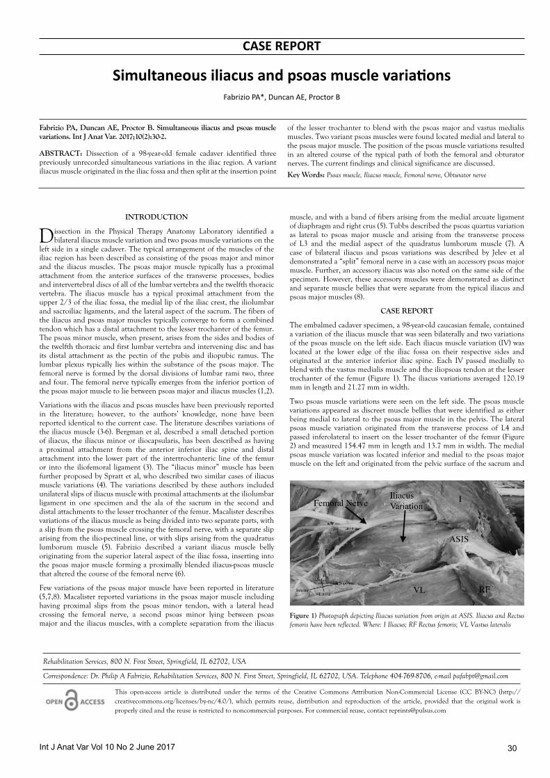
The embalmed cadaver specimen, a 98-year-old caucasian female, contained a variation of the iliacus muscle that was seen bilaterally and two variations of the psoas muscle on the left side. Each iliacus muscle variation (IV) was located at the lower edge of the iliac fossa on their respective sides and originated at the anterior inferior iliac spine. Each IV passed medially to blend with the vastus medialis muscle and the iliopsoas tendon at the lesser trochanter of the femur (Figure 1). The iliacus variations averaged 120.19 mm in length and 21.27 mm in width.
Two psoas muscle variations were seen on the left side. The psoas muscle variations appeared as discreet muscle bellies that were identified as either being medial to lateral to the psoas major muscle in the pelvis. The lateral psoas muscle variation originated from the transverse process of L4 and passed inferolateral to insert on the lesser trochanter of the femur (Figure 2) and measured 154.47 mm in length and 13.7 mm in width. The medial psoas muscle variation was located inferior and medial to the psoas major muscle on the left and originated from the pelvic surface of the sacrum and
Fabrizio PA, Duncan AE, Proctor B. Simultaneous iliacus and psoas muscle variations. Int J Anat Var. 2017;10(2):30-2.
ABSTRACT: Dissection of a 98-year-old female cadaver identified three previously unrecorded simultaneous variations in the iliac region. A variant iliacus muscle originated in the iliac fossa and then split at the insertion point
of the lesser trochanter to blend with the psoas major and vastus medialis muscles. Two variant psoas muscles were found located medial and lateral to the psoas major muscle. The position of the psoas muscle variations resulted in an altered course of the typical path of both the femoral and obturator nerves. The current findings and clinical significance are discussed.
Key Words: Psoas muscle, Iliacus muscle, Femoral nerve, Obturator nerve
Figure 1) Photograph depicting Iliacus variation from origin at ASIS. Iliacus and Rectus femoris have been reflected. Where: I Iliacus; RF Rectus femoris; VL Vastus lateralis

Fabrizio et al
Int J Anat Var Vol 10 No 2 June 201731
passed inferolateral to insert on the lesser trochanter of the femur (Figure 3) and measured 144.77 mm in length and 9.37 mm in width. The two psoas muscle variations combined in a “V” shaped connection just anterior to the psoas major muscle (Figure 4). A space was formed between the psoas major muscle and the medial psoas muscle (Figure 3).
The space created a tunnel which was occupied by the obturator nerve as the nerve coursed from the lumbar plexus medially towards the obturator foramen. The femoral nerve traversed the psoas major muscle and emerged between psoas major muscle and the lateral psoas muscle (Figure 2). The femoral nerve then followed a typical path in the anterior compartment of the thigh.
thoracic vertebra and courses on the superficial surface of psoas major to insert on the ilio-pectineal eminence (2,9).
Previous literature discusses very little about accessory iliacus muscles and no research has been found that describes a variation located inferior to iliacus. Spratt et al, discussed an iliacus variation, anterior to the iliacus, that arose from the iliolumbar ligament and then divided into two tendons at the insertion. One tendon inserted into the lesser trochanter and the other insertion was unable to be found but coursed into the medial thigh (4). Jelev et al, reported an iliacus muscle variation, located anterior to iliacus muscle, which originated in the middle portion of the iliac crest and inserted into an accessory psoas major muscle (8). The previous cases presented an accessory muscle that was distinct from the iliacus muscles, as consistent with the present case, but was located anterior to the iliacus instead of posterior. Fabrizio describes a case of an iliacus muscle variation that attached proximally to the psoas and unilateral in contrast to the present case (6).
Few variations of psoas major muscles have been reported in literature. Tubbs et al, reported a psoas variation referred to as the “psoas quartus muscle”. This muscle, located lateral to psoas major muscle, arose from the transverse process of L3 and the medial aspect of the quadratus lumborum muscle (7). In between this variation and psoas major, the femoral nerve coursed from the lumbar plexus into the anterior thigh by passing between the psoas quartus and psoas major muscles (7). The present case presented a variant muscle lateral to psoas major muscle with the femoral nerve traveling between the psoas major and the lateral psoas muscles however, the lateral psoas muscle in the present case did not originate from the quadratus lumborum muscle. A case of bilateral iliacus and psoas variations as described by Jelev et al demonstrated a “split” femoral nerve in a case involving an accessory psoas major muscle. Further, an accessory iliacus was also noted on the same side of the specimen. However, these accessory muscles were demonstrated as distinct and separate muscle bellies that were separate from the typical iliacus and psoas major muscles and were not consistent with the present case (8). Embryologically, during the fourth and fifth week of development of the lower extremity, the nerves “grow out” distally toward the extremity followed by differentiation of the skeletal muscles (10). Perhaps during the fourth or fifth week of development, aggressive femoral nerve in- growth may be the cause of disturbance of the undifferentiated iliacus-psoas muscles which may have led to “re-routed” development of the typical iliopsoas muscles structure into the multiple variations seen in the present case (8,11).
Psoas and iliacus muscle variations have been shown to alter the course of the femoral nerve (6,12). Vazquez et al, reported a muscular slip of iliacus and psoas, which resulted in an altered femoral nerve course (12). The authors reported the variant muscle splitting the femoral nerve, resulting in the femoral nerve surrounding the muscle variation and thus creating a possible site of compression (12). Fabrizio reported a femoral nerve that changed course to travel laterally behind a variant muscle before coursing over the anterior aspect of the muscle variation. Further an hourglass compression indentation was found on the femoral nerve where it changed expected course (6). In the present case, the femoral nerve neither split nor passed posterior to the muscle variant, rather it traveled in-between psoas major and the lateral psoas muscles. Variation in the course of the obturator nerve
Figure 2) Photograph depicting lateral Psoas muscle variation (Psoas Variation #1) coursing from origin to insertion and femoral nerve bisecting psoas variation and psoas major. Inguinal ligament has been removed. Where: P Mi Psoas minor; P Ma Psoas major; I Iliacus
Figure 3) Photograph showing media Psoas muscle (Psoas Variation #2) at origin and coursing to insertion. (*) denotes space created between Variation 2 and Psoas Major. Where: P Mi Psoas minor; P Ma Psoas major; I Iliacus
DISCUSSION
The muscles that arise in the iliac region include psoas major, psoas minor and the iliacus. Psoas major is typically described as originating from the transverse processes and intervertebral discs of the twelfth thoracic vertebra thru the fifth lumbar vertebra (1,2). Iliacus is described as originating in the iliac fossa (1,2). These two muscles then course medially to converge together forming a tendon to insert into the lesser trochanter on the femur (1,2). Psoas minor, present in about 50% of the population, arises from the twelfth
Figure 4) Photograph depicting Psoas Variations 1 and 2 coursing over rim of pelvis to come together over Psoas Major. Where: P Mi Psoas minor; P Ma Psoas major; I Iliacus; VL Vastus lateralis

Int J Anat Var Vol 10 No 2 June 2017 32
Simultaneous iliacus and psoas muscle variations
has also been reported by Kirchmair et al, who reported the obturator nerve located completely behind the psoas major, as opposed to emerging from the posterior surface (13). In the present case, the obturator nerve was found to be traveling through the space posterior to psoas major and anterior to medial psoas muscles.
The variations found in the present case could have several clinical implications. Variations of the iliopsoas muscle are shown to have a possible effect on nerve course. This disruption in the course of major nerves can lead to compression of the nerves in-between variant muscular slips. When the nerve is compressed due to variant muscles, locating the cause of pain could become more difficult and result in mistaking the pain for pathology. Also, the medial psoas muscle present in this case created a space that could result in misinterpretation during pelvic cavity imaging. Knowledge of these muscle variations and how they have altered course of nerves can help clinicians look into other anatomical possibilities that can result in iliopsoas pain as well as be aware of muscle variations when reading images.
REFERENCES
1. Standring S, Borley NR, Healy JC, et al. Gray’s anatomy: the anatomical basis of clinical practice. Elsevier. 2008;40:1367-8.
2. Moore KL, Dalley AF, Agur AMR. Clinically oriented anatomy. Baltimore, Lippincott Williams & Wilkins. 2010;6:545-6.
3. Bergman RA, Thompson SA, Afifi AK. Catalog of human variations. Baltimore, Urban & Schwartzenberg. 1984;51-2.
4. Spratt JD, Logan BM, Abrahams PH. Variant slips of psoas and iliacus muscles, with splitting of the femoral nerve. Clin Anat. 1996;9:401-4.
5. Macalister A. Observations on muscular anomalies in the human anatomy. Third series with a catalogue of the principal muscular variations hitherto published. Trans Roy Irish Acad Sci. 1875;25:1-130.
6. Fabrizio PA. Anatomic variation of the iliacus and psoas major muscles. Int J Anat Var.2011;4:28-30.
7. Tubbs RS, Oakes WJ, Salter EG. The psoas quartus muscle. Clin Anat. 2006;19:678-80.
8. Jelev L, Shirarov V, Surchev L. Bilateral variations of the psoas major and the iliacus muscles and the presence of an undescribed variant muscle- accessory iliopsoas muscle. Ann Anat. 2005;187:281-6.
9. Cronin CG, Lohan DG, Meehan CP, et al. Anatomy, pathology, imaging and intervention of the iliopsoas muscle revisited. Emerg Radiol. 2008;15:295-310.
10. Bardeen CR. Development and variation of the nerves and the musculature of the inferior extremity and of the neighboring regions of the trunk in man. Am J Anat. 1907;6:259-390.
11. Saadeh FA, Bergman RA. An aberrant psoas major muscle fascicle. Anat Anz. 1985;160:367-8.
12. Vazquez MT, Murillo J, Maranillo E, et al. Femoral nerve entrapment: A new insight. Clin Anat. 2007;20:175-9.
13. Kirchmair L, Lirk P, Colvin J, et al. Lumbar plexus and psoas major muscle: not always as expected. Reg Anesth Pain M. 2008;33:109-14.