simultaneous determination of sulfonamides, penicillins and
TRANSCRIPT

Simultaneous Determination of Sulfonamides, Penicillins and Coccidiostats in Porkby High-Performance Liquid Chromatography–Tandem Mass Spectrometry
C. Nebot*, P. Regal, J. Miranda, A. Cepeda and C. Fente
Department of Analytical Chemistry, Nutrition and Bromatology, Faculty of Veterinary Medicine,
University of Santiago de Compostela, 27002, Lugo, Spain
*Author to whom correspondence should be addressed. Email: [email protected]
Received 31 August 2010; revised 31 May 2011
Veterinary drugs are widely and legally used to treat and preventdisease in livestock. However, drugs are also used illegally asgrowth-promoting agents. To protect the health of consumers,maximum residue limits (MRL) in food of animal origin have beenestablished and are listed in Regulation 37/2010. According to thisregulation, more than 300 drugs need to be controlled regularly inlaboratories for residues of veterinary drugs. A cost-effective ana-lytical method is very important and explains why the developmentof multi-residual methods is becoming popular in laboratories.
The aim of this work is to describe a simple, rapid and economicalhigh-performance liquid chromatography–tandem mass spectrometrymethod for the simultaneous identification and quantification of 21veterinary drugs in pork muscle samples. The sample clean-up pro-cedure is performed with acidified dichloromethane and does notrequire solid phase extraction. The method is applicable to nine sulfo-namides and seven coccidiostats identified within 36 min. Calculatedrelevant validation parameters such as recoveries (from 72.to 126%), intra-precision and intermediate precision (relative standard devi-ation below 40 %) and decision limits (below 7mg Kg21) were withinacceptable range and in compliance with the requirements ofCommission Decision 2002/657/EC.
Introduction
Veterinary drugs are widely and legally used to treat and prevent
disease in livestock, but some are also used illegally as growth-
promoting agents (1). Regardless of the use of these substances,
they can remain as unwanted residues in food of animal origin,
endangering the health of consumers. The European Parliament
has set up maximum residue limits (MRLs) and maximum levels
(ML) for residues of veterinary drugs in food of animal origin to
protect consumer health. Evaluated drugs are described in
Regulation 470/2009 (2) and their limits are listed in Table I of
the Annex of Regulation 37/2010 (3). These regulations repeal
Regulation 2377/90 and amend Directive 2001/82 and
Regulation 726/2004. Food samples containing residues of veter-
inary drugs at concentrations below MRL are considered safe and
designated as “compliant.” In contrast, samples with concentra-
tions of drugs above the MRL are considered non-safe and desig-
nated as “non-compliant.”
Regulation 37/2010 sets MRL levels for more than 300
pharmacologically active substances in food matrices such as
muscle, fat, liver, kidney and milk. To detect these residues,
microbiological or bioassay techniques are widely used as screen-
ing methods. These methods either do not distinguish between
members of a class of antibiotics or cannot quantify them.
Development of a new analytical technique such as atmos-
pheric pressure ionization (API) permits high-performance
liquid chromatography (HPLC) to be coupled with mass spec-
trometry (MS), which opened a new era in qualitative and quan-
titative analysis of veterinary drug residues. The combination of
HPLC with MS or MS-MS techniques allows identification of vet-
erinary drugs in classes that could not be chromatographically
resolved with HPLC. In addition to the high level of specificity,
these techniques are sensitive and sometimes require less than
1 pg of material. The specificity and sensitivity of these instru-
ments enable the development of multi-residual methods for
veterinary drugs at concentrations below MRLs.
The applications of HPLC–MS-MS to veterinary drug residue
analysis and residue control have increased in the past five
years. HPLC–MS-MS methods have been reported for the ana-
lysis of sulfonamides in salmon muscle (4), kidney (5, 6), egg
(7) and milk (8). They have also been employed for the analysis
of other veterinary drugs residues such as tetracycline (9),
chloramphenicol (10), cyproheptadine (11) and coccidiostats
(12, 13). Griseofulvine is widely used in veterinary medicine
for the treatment of fungal infections, but it causes birth
defects in laboratory animals (14) and its concentrations in
food samples need to be controlled. Similarly, sulfonamide resi-
dues in food are of concern because of their potential carcino-
genic nature and the possible development of antibiotic
resistance in humans (15). Enrofloxacin, a fluoroquinolone, is a
synthetic antibiotic with broad-spectrum antibacterial activity
developed exclusively for use in animals (16). It is well known
that continuous use of fluoroquinolons in poultry has led to
the development of fluoroquinolone-resistant Campylobacter
species in poultry, which is transferrable to humans.
Anti-coccidial drugs (coccidiostats) are licensed to be used as
feed additive and their presence in food samples is forbidden.
However, because the unavoidable carryover of these sub-
stances in non-target feed and consequently in food of animal
origin, MRLs for these substances were established in 2009 and
regulated in the Commission Regulation (17). Toxicological
studies indicate that some coccidiostats produce mutagens
(18) and potential carcinogens. Poisoning in animals and
humans caused by coccidiostats (particularly polyether antibio-
tics) are widely described in the literature (19, 20, 21, 22).
Each year sees an increase in the number of veterinary drug
residues to be monitored; consequently, food laboratories are
hastening the search for high-throughput techniques that may
be more effective in tackling large amounts of samples in a
limited time (23). New screening methods are still being
# The Author [2012]. Published by Oxford University Press. All rights reserved. For Permissions, please email: [email protected]
Journal of Chromatographic Science 2012;50:414–425
doi:10.1093/chromsci/bms021 Article
Dow
nloaded from https://academ
ic.oup.com/chrom
sci/article/50/5/414/352726 by guest on 13 January 2022

reported (23, 24), and offered on the market. Screening
methods permit rapid analysis of numerous drugs at mg/kg and
even at pg/g. However, they also have numerous disadvantages:
no specificity, unavailability for all active compounds, high cost
and many false positives. It is well known that a suspect posi-
tive requires further analysis with a confirmatory method.
However, it is actually more practical to directly analyze
samples with a multi-class confirmation than to first use a
screening method followed a confirmatory method (twice),
than to use a screening method once and an HPLC–MS-MS
twice. Nowadays, the tendency is to develop multi-class HPLC–
MS-MS methods. These methods have been developed in matri-
ces such as pig liver, kidney and muscle where 16 b-agonists
have been detected (25); 12 coccidiostats have been detected
in chicken liver (12) and 42 antibiotics have been detected in
honey (26, 27), egg (28) and, more recently, in milk (29, 30,
31, 32). Multi-class methods are an effective way to monitor a
wide variety of drugs in a single analysis, maximizing laboratory
resources and sample throughput. However, only a few multi-
class confirmatory methods for analysis of residues of veterin-
ary drugs in muscle samples have been reported (23, 25).
HPLC–MS-MS methods, multi-class or not, for the analysis of
residues of drugs in muscle involve long and tedious extraction
protocols. Generally, two extractions are employed: one with
water and a one with solid-phase extraction cartridges. The
combination of these two extraction steps is sometimes time-
consuming, and in many cases, low recoveries are attributable
to losses during these steps.
This paper describes a rapid, straightforward, reliable and
economical HPLC–MS-MS method for the identification and
quantification of 21 drugs in pork muscle samples. The drugs
belong to different therapeutic classes, including sulfonamides,
penicillins, quinolones and coccidiostats, commonly analyzed
by European laboratories dedicated to residue control. The ex-
traction protocol is simple and fast and does not require solid-
phase extraction. After evaporation and reconstitution of
the extracts, the presence of 21 analytes was confirmed by
HPLC–MS-MS within 36min. The whole method has been
validated according to the guidelines of Commission Decision
657/2002 (23) and the decision limits (CCa) achieved
were ,2mg/kg for most coccidiostats, , 4 mg/kg for sulfona-
mides, 4 mg/kg for enrofloxacin and 6 mg/kg for trimethoprim.
Experimental
Reagents and stock solutions
Sulfachloropyridazine, sulfadimethoxine, sulfamethazine, sul-
fathiazole, sulfamethoxazole, sulfamethoxypyridazine, sulfapyri-
dine, sulfaquinoxaline, sulfamethizole, trimethoprim, penicillin
G, penicillin V, decoquinate, lasalocid, maduramicin, monensin,
narasin, robenidine, salinomycin, enrofloxacin, griseofulvin
(purity higher than 98 %) and the two internal standards (IS),
sulfadoxine-d3 and robenidine-d8, were obtained from
Sigma-Aldrich (St. Louis, MO). Formic acid (purity higher than
99% for analysis) was purchased from Acros Organics (Geel,
Belgium) and acetonitrile, methanol and dichloromethane were
purchased from Scharlau Chemie (Barcelona, Spain). Ultrapure
water was made in-house with a Milli-Q water system
(Millipore, Bedford, MA) and the nitrogen was generated by an
in-house nitrogen generator from Peak Scientific Instruments
Ltd. (Chicago, IL)
Stock solutions of individual analytes were prepared by dilut-
ing 50mg of drug with 50mL of 0.1% formic acid in methanol.
A standard solution mixture of drugs was prepared by appropri-
ate dilution of the stock solution of individual analytes, and the
final concentrations of drugs were: 5,000 ng/mL of decoqui-
nate; 3,750 ng/mL of trimethoprim; 2,500 ng/mL of sulfachlor-
pyridazine, sulfadimethoxine, sulfamethazine, sulfathiazole,
sulfamethoxazole, sulfamethoxypyridazine, sulfapyridine, sulfa-
quinoxaline, sulfamethizole, penicillin G, penicillin V, enroflox-
acin and griseofulvin; 1,250 ng/mL of lasalocid, narasin and
robenidine; 500/ng mL of maduramicin, monensin and salino-
mycin. This solution was diluted several times with 0.1% of
formic acid in methanol to obtain a serial standard solution
containing 5, 10, 15, 25, 37.5, 50, 125, 200, 500 and 750 ng/mL
Table IFormula, Maximum Residue Limit, Maximum Levels, Validation Level and SRM Parameters of the LC–MS-MS Analysis of the Selected Veterinary Drugs
Formula MRL (mg/kg) VL (mg/Kg) Rt Precursor ion Product ion 1 Product ion 2 DP (V) EP (V) CEP (V) CE (V) CXP (V)
Sulfachlorpyridazine C10H9ClN4O2S 100 10 13.9 284 156 92 56 9 14 17 4Sulfadimethoxine C12H14N4O4S 100 10 14.9 310 156 92 66 10 14 25 4Sulfamethazine C12H14N4O2S 100 10 12.7 278 186 92 56 10 16 19 4Sulfamethizole C9H10N4O2S2 100 10 13.0 270 156 92 56 9 14 17 4Sulfamethoxazole C10H11N3O3S 100 10 14.1 253 108 92 66 10 12 37 4Sulfamethoxypyridazine C11H12N4O3S 100 10 13.2 280 156 92 76 10 14 39 4Sulfapyridine C11H11N3O2S 100 10 13.1 249 92 108 61 10 12 35 4Sulfaquinoxaline C14H12N4O2S 100 10 15.1 300 156 92 81 10 16 19 4Sulfathiazole C9H9N3O2S2 100 10 12.1 255 156 92 61 10 12 17 4Penicilin G C16H18N2O4S 25 10 14.8 335 217 160 46 10 24 17 4Penicilin V C16H18N2O5S 25 10 15.6 351 257 229 56 56 56 56 56Trimethoprim C14H18N4O3 50 15 12.2 291 230 123 50 10 13 10 3Enrofloxacin C19H22FN3O3 100 10 13.6 360 316 245 51 9 12 23 6Griseofulvin C17H17ClO6 N/A 10 17.1 352 165 214 76 10 14 27 4Decoquinate C24H35NO5 20 20 20.2 418 204 148 56 9 14 57 4Lasalocid C34H54O8 5 3 21.8 613 377 559 71 10 24 35 6Maduramicin C47H83NO17 2 1 22.1 939 877.5 859 46 12 24 41 34Monensin C36H62O11 2 1 21.6 693 461.3 479 693 81 8 80 49Narasin C43H72O11 5 3 22.4 787 430.9 279 126 11 32 69 4Robenidine 25875-51-8 5 3 19.0 336 140.2 111 41 9 30 57 4Salinomycin C42H70O11 2 1 21.8 773 431.3 531 151 8 18 55 20
Simultaneous Determination of Sulfonamides, Penicillins and Coccidiostats in Pork by High-Performance Liquid Chromatography–Tandem Mass Spectrometry 415
Dow
nloaded from https://academ
ic.oup.com/chrom
sci/article/50/5/414/352726 by guest on 13 January 2022

of sulfachlorpyridazine (Table II). These standard solutions
were employed to build instrument calibration curves for each
analyte.
Stock solutions containing individual IS were prepared at
1 mg/mL in methanol containing 0.1% of formic acid. This solu-
tion was then diluted to a final concentration of 1mg/mL of
each IS.
Pork muscle samples
The development of the extraction procedure and validation of
the method were conducted with commercial meat. Pork
muscle samples bought from local supermarkets were minced,
stored in plastic bags and frozen. The applicability of the
method was investigated in pork muscle samples obtained
from different Spanish slaughterhouses that collaborated with
the laboratory in a national control program of residue of vete-
rinary drugs.
Sample preparation
Sample homogenization and extraction
Defrosted pork sample was homogenized with a hand food
blender Mini Chopper (Moulinex Ind. Ltd, France). The homo-
genized sample was weighted (500mg+10) in a 2-mL
Eppendorf tube (Hamburg, Germany) and 1mL of 0.1% of
formic acid in dichloromethane, 10 mL of a mixture of IS and
200mL of water were added to the tube. The mixture was
vortex mixed (10 s), sonicated (10 min), vortex mixed again
(10 s) and centrifuged at approximately 1,500 � g (15min).
The organic phase (lower layer) was transferred into a gradu-
ated 10-mL PYREX conical centrifuge tube and the entire ex-
traction procedure was repeated with a subsequent 1 mL of
0.1% of formic acid in dichloromethane. The final mixture of
extracts (approximately 2mL of extract) was evaporated to
dryness with a stream of nitrogen at 37 8C on a Turbo Vap II
from Zyrmark (Hopkinton, MA).
The dried extract was reconstituted with 0.2 mL 0.1% of
formic acid in methanol and transferred into an Ultrafree-MC
centrifugal filter (Millipore, Bedford, MA). After filtration, the
extract was transferred into an amber HPLC vial containing a
200mL insert and the vial was kept at –18 8C until sample ana-
lysis by HPLC–MS-MS. The concentration of the analyte in the
sample was calculated with the instrument calibration curve of
the day and correction was applied for recoveries and the IS.
The applied correction was the one calculated for the fortified
sample.
Quality control samples
During the routine analysis, the complete analytical procedure
was applied to four quality control samples. These quality
control samples were processed together with the samples to
be confirmed: a blank sample (a sample from which the analyte
is absent, negative for the analytes that are going to be ana-
lyzed), a fortified sample (fortified pork muscle sample con-
taining known amounts of analyte), a reagent blank (reagents
only, no muscle) and fortified reagents (reagents spiked to a
known concentration of analytes). The fortified sample and for-
tified reagents were prepared by adding appropriate aliquots of
the mixed standard, vortexing and allowing the sample to
settle for 30min in the dark.
HPLC–MS-MS system
The HPLC system consists of an HPLC 1100 separation module,
a vacuum degasser and an auto-sampler from Agilent
Technologies (Waldbronn, Germany) and connected to a Qtrap
2000 from Applied Biosystems, MSD Sciex (Toronto, Canada)
equipped with a TurboIonSpray source. The whole system was
controlled with the software Analyst 1.4.1 from Applied
Biosystems, MSD Sciex (Toronto, Canada).
Ultra-filtrated extract was injected (10 mL) into a Synergi
2.5mm Polar-RP 100A 50 � 2.00 mm column connected to a
Polar-RP 4.0 � 2.0 mm security guard cartridge, both obtained
from Phenomenex (Macclesfield, UK). The mobile phase con-
sisted of a mixture of 0.1% formic acid in water and 0.1%
Table IIConcentration (ng/mL) Drugs in the Standard Solutions Employed to Build Instrument Calibration Curves
Sulfachlorpyridazine 2,500 1,000 750 500 200 20 50 37.5 25 15 10 5Sulfadimethoxine 2,500 1,000 750 500 200 20 50 37.5 25 15 10 5Sulfamethazine 2,500 1,000 750 500 200 20 50 37.5 25 15 10 5Sulfamethizole 2,500 1,000 750 500 200 20 50 37.5 25 15 10 5Sulfamethoxazole 2,500 1,000 750 500 200 20 50 37.5 25 15 10 5Sulfamethoxypyridazine 2,500 1,000 750 500 200 20 50 37.5 25 15 10 5Sulfapyridine 2,500 1,000 750 500 200 20 50 37.5 25 15 10 5Sulfaquinoxaline 2,500 1,000 750 500 200 20 50 37.5 25 15 10 5Sulfathiazole 2,500 1,000 750 500 200 20 50 37.5 25 15 10 5Penicilin G 2,500 1,000 750 500 200 20 50 37.5 25 15 10 5Penicilin V 2,500 1,000 750 500 200 20 50 37.5 25 15 10 5Trimethoprim 3,750 1,500 1,125 750 300 30 75 56.25 37.5 22.5 15 7.5Enrofloxacin 2,500 1,000 750 500 200 20 50 37.5 25 15 10 5Griseofulvin 2,500 1,000 750 500 200 20 50 37.5 25 15 10 5Decoquinate 5,000 2,000 1,500 1000 400 40 100 75 50 30 20 10Lasalocid 1,250 500 375 250 100 10 25 18.75 12.5 7.5 5 2.5Maduramicin 500 200 150 100 40 4 10 7.5 5 3 2 1Monensin 500 200 150 100 40 4 10 7.5 5 3 2 1Narasin 1,250 500 375 250 100 10 25 18.75 12.5 7.5 5 2.5Robenidine 1,250 500 375 250 100 10 25 18.75 12.5 7.5 5 2.5Salinomycin 500 200 150 100 40 4 10 7.5 5 3 2 1
416 Nebot et al.
Dow
nloaded from https://academ
ic.oup.com/chrom
sci/article/50/5/414/352726 by guest on 13 January 2022
formic acid in acetonitrile. Elution of the analytes was per-
formed on a gradient mode (Table III). The flow rate was held
at 0.150mL/min throughout the analysis.
The entire effluent from the HPLC column was directed into
the electrospray source of the MS, which was working in posi-
tive ion-mode. The optimum signal for [M þ H]þ ions was
obtained with the source temperature set to 4008C, vacuumgauge to 2.2 atm, ion spray to 5.500 V, curtain gas to 25 psi, ion
source 1–55 psi and ion source 2–50 psi.
In this work, the MS was used in selected reaction monitor-
ing (SRM) mode. This mode allows the transition between a
particular precursor ion and its respective product ion to be
monitored, which provides excellent selectivity. The transition
can be written as parent m/z . fragment (product ion) m/z.According to the Commission Decision 2002/657/EC (33),
four identification points are required to identify a residue in
food of animal product. These four identification points are
earned by monitoring the transition from a precursor ion to
two product ions. Therefore, for this particular research, two
SRM transitions were employed to monitor each veterinary
drug. Table I summarizes precursor and product ions and the
declustering potential (DP), entrance potential (EP), collision
cell entrance potential (CEP), collision energy (CE) and
cell exit potential (CXP) employed for their detection. The
transitions were monitored with a dwell time of 5 ms.
Veterinary drugs were identified by their retention times (Rt)
in two SRM.
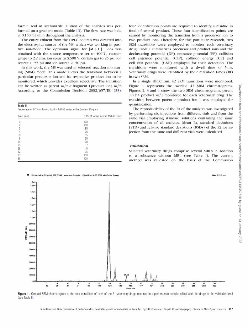
In a single HPLC run, 42 SRM transitions were monitored.
Figure 1 represents the overlaid 42 SRM chromatograms.
Figures 2, 3 and 4 show the two SRM chromatograms, parent
m/z . product m/z monitored for each veterinary drug. The
transition between parent . product ion 1 was employed for
quantification.
The reproducibility of the Rt of the analytes was investigated
by performing six injections from different vials and from the
same vial employing standard solutions containing the same
concentration of all analytes. Mean Rt, standard deviations
(STD) and relative standard deviations (RSDs) of the Rt for in-
jection from the same and different vials were calculated.
Validation
Selected veterinary drugs comprise several MRLs in addition
to a substance without MRL (see Table I). The current
method was validated on the basis of the Commission
Table IIIPercentage of 0.1% of Formic Acid in Milli-Q water in the Gradient Program
Time (min) 0.1% of formic acid in Milli-Q water
0 1002 1003 854 758 559 50
14 3025 728 032 7533 10036 100
Figure 1. Overlaid SRM chromatogram of the two transitions of each of the 21 veterinary drugs obtained in a pork muscle sample spiked with the drugs at the validation level(see Table II).
Simultaneous Determination of Sulfonamides, Penicillins and Coccidiostats in Pork by High-Performance Liquid Chromatography–Tandem Mass Spectrometry 417
Dow
nloaded from https://academ
ic.oup.com/chrom
sci/article/50/5/414/352726 by guest on 13 January 2022

Decision 2002/657/EC (33), this regulation requires a valid-
ation around the MRL for authorized drugs and at concentra-
tions as low as possible for substances without MRL. The
validation of coccidiostats was accomplished at the ML set up
in the Commission Regulation 124/2009/EC. Although sulfo-
namides have MRL of 100mg/Kg in muscle, they were vali-
dated at 10mg/Kg because the Commission Regulation 37/2010 states that “the combined total residues of all substances
within the sulfonamide group should not exceed 100mg/Kg.”Similarly, trimethoprim, enrofloxacin, the penicillins and
griseofulvin were validated at 15, 10, 25 and 10 mg/Kg, re-
spectively. Following recommendations of the Commission
Decision 2002/657/EC (33), the validation process was con-
ducted with six levels of concentrations: 0, 0.5, 1, 1.5, 2 and
5 � MRL or validation level (VL).
Intra-day and inter-day repeatability, within-laboratory repro-
ducibility, sensitivity and specificity of the method were inves-
tigated. All calculation was performed on the program ResVal
(version 2.2) from the Community Reference Laboratory (CRL)
for hormones (RIVM, Bilthoven, Netherlands).
Figure 2. SRM chromatograms, parent ion m/z . (product ion) m/z, monitored to identify each sulfonamide in a pork muscle sample containing 21 veterinary drugs at thevalidation level (see Table II).
418 Nebot et al.
Dow
nloaded from https://academ
ic.oup.com/chrom
sci/article/50/5/414/352726 by guest on 13 January 2022

Validation process
Four experiments (Exps. 1, 2, 3 and 4), conducted on four dif-
ferent days, were carried out for validation of the method. Each
day, different concentrations, (15, 25, 37.5, 50 and 125, 200,
500 and 750 ng/mL of Sulfachloropyridazine) of standard solu-
tions containing a mixture of drugs spiked with the ISs were
analyzed to obtain instrument calibration curves (ICC). ICCs
were built by representing the ratio of the area of the selected
ion of the analyte and the IS against concentration of the cocci-
diostats in the standard.
To conduct Exps. 1, 2 and 3, a homogeneous muscle
sample was divided into 63 sub-samples. Twenty-one samples
were fortified and analyzed each day of the validation. The
samples were fortified as follows: one sample was not spiked
with drugs (blank), six samples were spiked at 0.5 � VL, six
spiked at 1 � VL, six spiked 1.5 � VL, one spiked at 2 � VL
and one spiked at 5 � VL. These samples were used to con-
struct the sample calibration curves (SCCs) for each analyte
on each day. SCCs were constructed by calculating the area
of the selected ion of the analyte and the IS and their ratio
was used as the response variable. ICC was constructed by
linear curve fitting using linear regression. ICC were
described by the equation y ¼ bx þ a, where b is the slope
and a is the intercept.
Exp. 4 consisted of 20 muscle samples collected from differ-
ent slaughterhouses. Ten were not spiked with drugs and 10
were spiked to a concentration of 1 � VL, and ten were only
spiked with the IS. The samples were shaken vigorously for
30min after being spiked with the coccidiostats to
homogenize.
Exps. 1, 2 and 3 allowed precision, accuracy, decision limit
(CCa) and detection capability (CCb) to be estimated.
According to European Commission Decision 2002/657/EC(33), CCa and CCb can be defined for substances for which
no permitted limit has been established and for substances
with established permitted limit. Depending on the case, dif-
ferent a errors for CCa shall be applied (1 or 5%). During
separation for this particular study, to simplify calculation,
the same a error, 1%, was applied for all drugs. Similarly, to
calculate CCa and CCb, the same formula was applied to all
analytes:
CCa ¼ ðYa þ 2:33STDEV YaÞ � Ya
bð1Þ
CCb ¼ ðYa þ 2:33STDEV Ya þ 1:64STDEVYaÞ � YaÞb
ð2Þ
where Ya ¼ concentration corresponding to the Y-intercept;
STDEVYa ¼ standard error at the intercept; b ¼ slope of the
calibration curve.
The IS was used to calculate a correction factor (RF) for
each analyte. The calculation of an RF for each coccidiostats
was as follows:
RF � ðAx X CsÞ=ðAs X CxÞ
Figure 3. SRM chromatograms, parent ion m/z . (product ion) m/z, monitored to identify each penicillin, trimethoprim, enrofloxacin and griseofulvin in a pork muscle samplecontaining 21 veterinary drugs at the validation level (see Table II).
Simultaneous Determination of Sulfonamides, Penicillins and Coccidiostats in Pork by High-Performance Liquid Chromatography–Tandem Mass Spectrometry 419
Dow
nloaded from https://academ
ic.oup.com/chrom
sci/article/50/5/414/352726 by guest on 13 January 2022

where Ax ¼ area of the target analyte to be measured; As ¼
area of the appropriate IS; Cs¼ concentration of the IS in the
extract; Cx ¼ concentration of the analyte (coccidiostat) in the
extract.
A mean RF was obtained from the analysis of the 73
pork muscle samples employed for the validation procedure.
The mean RF was then employed to calculate the
concentration of the analyte in the sample and the recovery
of the analyte.
While sulfadoxine-d3 was used as IS to quantify sulfona-
mides, penicillins, trimethoprim, enrofloxacin and griseoful-
vin; robenidine-d8 was employed to quantify decoquinate,
lasalocid, maduramicin, monensin, narasin, robenidine and
salinomycin.
Figure 4. SRM chromatograms, parent ion m/z . (product ion) m/z, monitored to identify each coccidiostat in a pork muscle sample containing 21 veterinary drugs at thevalidation level (see Table II).
420 Nebot et al.
Dow
nloaded from https://academ
ic.oup.com/chrom
sci/article/50/5/414/352726 by guest on 13 January 2022

Result and Discussion
Method development
To identify adequate precursor and product ions for each
analyte, standard solutions containing individual drugs at
1.0 mg/mL (0.1% formic acid in methanol) were infused direct-
ly into the electrospray ion source at 20 mL/min. The MS
system employed in this study could alternate between positive
and negative ionization mode, but the use of this switching
mode results in a decrease in sensitivity and lifetime of the MS
capillary (34). Both ionization modes were tested, and only
drugs that ionize with positive electrospray ionization were
selected. The same ionization mode was reported for coccidio-
stats (35, 36), sulfonamides (37) and penicillins (24). Selected
precursor and product ions were those that gave a higher
signal response; proton adducts [M þ H]þ were employed in
most cases.
After selecting precursor and product ion and MS parameters
for detection, 10mL of a stock solution mixture of drugs at
100 ng/mL was injected directly into the mobile phase (50:50
of phase A:B). The mixture was introduced into the MS capil-
lary at a flow rate of 0.15mL/min. Under these conditions,
other MS parameters were optimized to obtain the best signal
response for all analytes; tested conditions include: source tem-
perature (350, 400 and 450 8C), ion spray (4,500, 5,000 and
5,500 V) and curtain gas (20, 25 and 30 psi).
Based on supplier recommendations and satisfactory results
obtained for sulfonamide analysis, an ether-linked phenyl
column was selected as a suitable HPLC column. To obtain
good resolution of the analyte, different combinations of
solvents were tested, always on a gradient mode: a mixture
of methanol–water containing 0.1% of formic acid, aceto-
nitrile–water containing 0.1% formic acid and the same condi-
tions with another buffer (1% of ammonium acetate). Because
ammonium acetate signal suppression has been shown to be
lower than other buffers (28, 38) it was included on the
mobile phase components to maintain mobile phase pH.
However, the use of ammonium acetate produced a decrease
of the signal intensity (5 times) and was discarded.
The use of methanol as mobile phase components for ana-
lysis of coccidiostats has been reported previously (39), as has
the use of acetonitrile (40). Methanol has also been used in sul-
fonamide analysis (41), but most published methods implement
acetonitrile in the mobile phase (42). No significant differences
were observed on peak shape, signal intensity or resolution;
however, peak shapes were more symmetric with acetonitrile
than with methanol. Therefore, acetonitrile was preferred.
The primary problem encountered during drug identification
was the formation of ions by the sulfonamides; three product
ions are generated in sulfonamide fragmentation: [M-RNH2]þ
196 (m/z 156), [M-RNH2-SO]þ (m/z 108) and [M-RNH2-SO2]þ
197 (m/z 92). The first attempt was to elute the sulfonamides
as much as possible to avoid signal contamination. This signal
contamination could occur with high concentrations of a par-
ticular ion monitored in two SRM transitions. However, no
signal contaminations were observed for sulfonamides or any
other drugs (Figures 2, 3 and 4) with the gradient selected, al-
though some sulfonamides coeluted. The HPLC method
resulted in reproducible Rt for all selected drugs (Table IV)
and RSDs for replicated injections from the same vial and
different vials resulted below 0.5% for all concentrations tested.
RSDs of mean Rt were below 1 for all drugs and at all concen-
trations. The developed method allows the selected drugs to
be identified by their Rt and two SRM (Table I).
The aim of this work was to develop a simple, rapid, reliable
multi-residue method applicable in pork muscle for the simul-
taneous extraction, detection and confirmation of the most fre-
quently controlled veterinary drugs in laboratories dedicated to
residue analysis. During the method development, different ex-
traction solvents were tested to improve the extraction of the
drugs and to decrease interferences (data not shown). Drug re-
coveries obtained with methanol, acetonitrile and dichloro-
methane acidified with 0.1% of formic acid and non-acidified
were compared. A higher number of drugs and higher recover-
ies were achieved with 0.1% of formic acid in dichloromethane,
probably because of dichloromethane polarity.
To avoid contamination, the complete extraction procedure
was conducted in disposable plastic materials and the glassware
was rinsed with acetone before and after its use. Matrix effects
were reduced by reconstitution of the extract in 0.2 mL instead
of lower volumes and applying ultra-filtration to the reconsti-
tuted extract.
Method performances and validation results
Specificity/selectivity
The specificity and selectivity of the method were evaluated on
Day 4. Twenty pork muscle samples collected from different
animals were divided into two sets of samples; one set was
spiked with the drugs at the VL and the other set was not
spiked. The method was considered selective and sensitive due
to the absence of any interference peaks around the Rt of the
analytes in their SRM chromatograms. The method was able to
detect the drugs spiked in the samples without obtaining any
Table IVMean Rt, Standard Deviation and RSD (%) Obtained from Replicated Injections (n ¼ 6) from the
Same Vial and Different Vials
Replicate injections (1 vial) Replicate injections (6 vials)
Mean STD RSD Mean STD RSD
Sulfachlorpyridazine 14.1 0.1 0.4 13.9 0.1 0.5Sulfadimethoxine 15.1 0.0 0.1 15.0 0.0 0.3Sulfamethazine 12.7 0.0 0.4 12.7 0.0 0.3Sulfamethizole 12.9 0.7 0.1 13.1 0.1 0.4Sulfamethoxazole 14.3 0.1 0.4 14.1 0.1 0.5Sulfamethoxypyridazine 12.9 0.7 0.3 12.8 0.7 0.4Sulfapyridine 12.0 0.0 0.4 11.8 0.1 0.8Sulfaquinoxaline 13.9 0.4 0.6 15.1 0.1 0.4Sulfathiazole 12.3 0.0 0.3 12.1 0.1 0.5Penicillin G 14.9 0.0 0.2 14.5 0.8 6.3Penicillin V 15.4 0.0 0.2 15.6 0.0 0.1Trimethoprim 12.3 0.0 0.2 12.2 0.0 0.3Enrofloxacin 13.2 0 0.1 13.1 0.0 0.3Griseofulvin 17.1 0.0 0.0 17.0 0.1 0.3Decoquinate 20.3 0.0 0.1 20.2 0.0 0.1Lasalocid 21.8 0.0 0.0 21.8 0.0 0.2Maduramicin 22.1 0.0 0.1 22.1 0.0 0.1Monensin 21.6 0.0 0.0 21.6 0.1 0.2Narasin 22.4 0.0 0.0 22.4 0.0 0.2Robenidine 19.2 0.0 0.2 19.0 0.0 0.1Salinomycin 21.7 0.0 0.1 21.8 0.0 0.2
Simultaneous Determination of Sulfonamides, Penicillins and Coccidiostats in Pork by High-Performance Liquid Chromatography–Tandem Mass Spectrometry 421
Dow
nloaded from https://academ
ic.oup.com/chrom
sci/article/50/5/414/352726 by guest on 13 January 2022

false negatives. In addition, the method analyzed non-spiked
milk samples without false positives, demonstrating its easy
applicability in raw milk samples.
Linearity
For each veterinary drug, a quadratic regression mode was
fitted to ICC and SCC. The regression coefficients (R2) of ICC
were higher than 0.98 during the four days of validation for all
the analytes over the whole concentration range from 15 to
500 ng/mL (Table V). The linearity of the method was also
observed in the SCC (Table V). R2 of SCCs were slightly lower
than those achieved for ICC, probably due to matrix effects.
Overall, R2 of the SCCs were higher than 95 over the whole
concentration range (0.5, 1. 1.5, 2 and 5 � VL), as expected for
the case of salinomycin (0.95).
Repeatability, reproducibility and recoveries
Inter-day and intra-day assays were performed to evaluate pre-
cision (RSD) and accuracy (% deviation). Inter-day precision
and accuracy were determined by analyzing nine batches of six
muscle samples spiked at three concentrations, 0.5, 1 and
1.5 � VL, and run within three days (Exps. 1, 2 and 3).
Intra-day precision and accuracy were obtained by analyzing
one batch of 10 muscle samples (Exp. 4) spiked at the VL.
Accuracy represents the closeness of agreement between a test
result and the acceptable value. Accuracy has to be estimated
by the determination of trueness and precision. However, true-
ness can only be established by analyzing certificated reference
materials (CRM). When no CRMs are available, recovery was
calculated instead of trueness (33). Recoveries of the analytes
were determined as the percentage of the drugs recovered
during the analytical procedure, employing the correction
factor. Inter-day recovery values obtained at the three levels
were acceptable, at mean recoveries within 70 and 126%
(Table IV). Intra-day recovery values were also acceptable,
ranging from 70 to 120% (Table IV), but they were approxi-
mately 15% lower than those achieved for the intra-day study.
Repeatability, expressed as RSD of the recoveries, was
between 3 and 20, with the exception of sulfathiazole (31%).
This is an acceptable range, because the Commission Decision
657/2002/EC states that RSD of the mean shall not exceed
20% for concentrations between 10 and 100 mg/Kg. Inter-dayreproducibility, expressed as RSD of the recoveries obtained in
three different days, ranged between 5 and 39%, with the ex-
ception of robenidine (59%). The Commission Decision 657/2002/EC states that mass fractions lower than 100 mg/Kgshould be as low as possible, and for mass fraction of 100, the
RSD should be lower than 23. Therefore, taking into account
the fact that the VLs employed in the validation were below
21mg/Kg, RSD below 39% could be considered acceptable.
CCa and CCb
The calculated mean CCa and CCb are shown in Table IV. CCa
and CCb were calculated with Exps. 1, 2 and 3. Table IV sum-
marizes the mean CCa and CCb of three days. CCa values
were below the VL selected for each analyte: sulfonamides
(1–2.3mg/Kg), coccidiostats (0.9–4mg/Kg), penicillins (2–
3.4mg/Kg), trimethoprim (6.9mg/Kg), enrofloxacin (4.3mg/Kg) and griseofulvin (2.9mg/Kg). Likewise, CCb values were
below the VL (Table IV).
Not many HPLC–MS-MS methods have reported CCa for the
selected analytes in pork muscle samples, so comparison was
difficult. Sergi et al. (2007) presented an LC–electrospray ion-
ization tandem MS confirmatory procedure for monitoring 13
SAs in animal tissues with CCa between 104.9–108.2 mg/Kg(43), which are lower than those presented. Dubois et al.
(2004) reported a method for simultaneously determining nine
coccidiostats in muscle from chicken at very low levels
(� 1 mg/Kg), lower than those reported for the same coccidio-
stats in liver (12). The fact that Dubois’s method (35) is applic-
able only to coccidiostats could explain lower CCa, because
Table VValidation levels, Regression Coefficients of Instrument Calibration Curves and Sample Calibration Curves, Decision Limits, Detection Capabilities and Relative Standard Deviation Achieved in the
Validation Study
Intra-day Inter-day
VL R2 of ICC R2 of SCC Recovery RSD Recovery RSD CCa CCb
Sulfachlorpyridazine 10 0.993 0.993 80 7 84 8 1.1 3.1Sulfadimethoxine 10 0.971 0.991 95 15 110 16 2.3 4.3Sulfamethazine 10 0.982 0.992 85 5 72 23 1.7 3.7Sulfamethizole 10 0.987 0.976 83 9 79 10 3 5Sulfamethoxazole 10 0.999 0.983 79 8 76 39 3 6Sulfamethoxypyridazine 10 0.994 0.986 80 3 82 6 1 3Sulfapyridine 10 0.989 0.968 85 12 82 39 3 5Sulfaquinoxaline 10 0.993 0.978 78 15 88 19 4 7Sulfathiazole 10 0.996 0.995 81 31 81 31 2.3 4.1Penicilin G 25 0.993 0.973 89 10 84 14 3.4 6.1Penicilin V 25 0.988 0.976 70 20 72 32 2 4Trimethoprim 15 0.996 0.974 84 7 80 10 6.9 12Enrofloxacin 10 0.993 0.974 111 9 114 7 4.3 7.3Griseofulvin 10 0.992 0.980 120 12 126 14 2.9 4.8Decoquinate 20 0.991 0.975 80 4 82 5 4 7Lasalocid 5 0.993 0.983 91 9 91 12 1.9 4.3Maduramicin 2 0.992 0.959 73 13 61 17 1.2 1.8Monensin 2 0.988 0.975 75 13 73 16 0.9 2.1Narasin 5 0.991 0.973 79 10 75 25 3 3.8Robenidine 5 0.985 0.970 72 20 70 29 2 3Salinomycin 2 0.987 0.963 74 10 71 31 1.2 2
422 Nebot et al.
Dow
nloaded from https://academ
ic.oup.com/chrom
sci/article/50/5/414/352726 by guest on 13 January 2022

the method is more specific than the method presented.
Granelli et al. (2009) reported a multi-class method for enro-
floxacin and sulfonamides, in addition to other antibiotics, in
muscle with CCa for the same antibiotic of 100 ng/g. CCa of
enrofloxacin achieved in this research was 25 times lower than
those reported previously (44, 45, 46). A multi-class method
that employs different extraction protocols, based on
QuEChERS, reported higher CCa for maduramicin (9.31mg/Kg), lasalocid (22.6mg/Kg), narasin (8.04mg/Kg) and salinomy-
cin (6.45 mg/Kg) than those achieved with a simple extraction.
Therefore, the presented method is able to detect the selected
analyte with some of the lowest CCa reported.
Applicability of the method
The applicability of the presented method was demonstrated in
real pork-muscle samples obtained from a quality control
program in which 25 slaughterhouses participated. A total of
100 pork muscle samples were analyzed within four days for
the presence of the selected drugs. All samples were found to
be compliant with the exception of one, which was non-
compliant for monensin (525mg/Kg, Figure 5). After revising
the results, 20 compliant and the non-compliant samples were
re-analyzed with other procedures, set up in the laboratory.
These procedures involved solid-phase extraction and HPLC–
diode array detection analysis; these procedures could analyze
sulfonamides, penicillins and monensin with limits of detection
of approximately 50mg/Kg. The results were the same than
those obtained with the HPLC–MS-MS method; compliant
samples were equally compliant and the non-compliant
samples also resulted positive to monensin at the same range
of concentration. These results demonstrate the practicability
of the method.
Conclusion
There is an increasing interest in multi-residual methods, espe-
cially for the analysis of residues of veterinary drugs in food
samples. The method presented in this article is able to extract
and analyze 21 veterinary drugs using a single extraction pro-
cedure. The CCa and CCb were found to be sufficiently low to
determine the residues of the drugs in muscle below the MRL
and VL set up by European legislations. The method saves cost
and time and was validated in conformity with the primary
revised EU requirements for detecting residues of veterinary
drug substances in animal products; the method also demon-
strated good linearity, accuracy and precision. Therefore, the
method could be easily implemented in residue control labora-
tories in Europe where these drugs are constantly monitored.
Figure 5. SRM chromatogram of pork muscle samples positive for monensin (525 mg/Kg).
Simultaneous Determination of Sulfonamides, Penicillins and Coccidiostats in Pork by High-Performance Liquid Chromatography–Tandem Mass Spectrometry 423
Dow
nloaded from https://academ
ic.oup.com/chrom
sci/article/50/5/414/352726 by guest on 13 January 2022

Acknowledgments
The authors wish to thank the European Rural Development
Fund (Fondo Europeo Agrıcola de Desarrollo Rural; FEADER)
and the Consellerıa de Medio Rural for founding this study
through the project FMR331A.
References
1. Serratosa, J., Blass, A., Rigau, B., Mongrell, B., Rigau, T., Tortades, M.,
et al.; Residues from veterinary medicinal products growth promo-
ters, performance enhancers in food-producing animals: A
European Union perspective; Rev. Sci. Tech. Off. Int. Epiz.; (2006);
25: 637–653.
2. 470/2010/EU. EU, The European parliament and the Council of
the European Union. Regulation (EC) 470/2009 of the European
Parliament and of the Council of 6 May 2009 laying down
Community procedures for the establishment of residue limits of
pharmacologically active substances in foodstuffs of animal origin,
repealing Council Regulation (EEC) No 2377/90 and amending
Directive 2001/82/EC of the European Parliament and of the
Council and Regulation (EC) No 726/2004 of the European
Parliament and of the Council. Official Journal of the European
Union, (2009); L 152.
3. 37/2010/EU. EU, The European parliament and the Council of the
European Union. Commission Regulation (EU) No 37/2010 of 22
December 2009 on pharmacologically active substances and their
classification regarding maximum residue limits in foodstuffs of
animal origin. Official Journal of the European Union, (2010); L 15.
4. Potter, R.A., Burns, B.G., van de Riet, J.M., North, D.H., Darvesh, R.;
Simultaneous determination of 17 sulfonamides, the potentiators
ormetoprim, trimethoprim in salmon muscle by liquid chromatog-
raphy with tandem mass spectrometry detection; Journal of AOAC
International, (2007); 90: 343–348.
5. Shao, B., Dong, D., Wu, Y., Hu, J., Meng, J., Tu, X., et al.;
Simultaneous determination of 17 sulfonamide residues in porcine
meat kidney, liver by solid-phase extraction, liquid chromatog-
raphy–tandem mass spectrometry; Analytica Chimica Acta,
(2005); 546: 174–181.
6. Bogialli, S., Curini, R., Di Corcia, A., Nazzari, M., Sergi, M.;
Confirmatory analysis of sulfonamide antibacterials in bovine liver,
kidney: Extraction with hot water, liquid chromatography coupled
to a single- or triple-quadrupole mass spectrometer; Rapid
Communications in Mass Spectrometry, (2003); 17: 1146–1156.
7. Tamosiunas, V., Padarauskas, A., Babiciene, D., Petrenas, T.;
High-performance liquid chromatography–tandem mass spectrom-
etry for the determination of sulfonamides in eggs; Chemija,
(2007); 18: 20–24.
8. Volmer, D.A.; Multiresidue determination of sulfonamide antibiotics
in milk by short-column liquid chromatography coupled with
electrospray ionization tandem mass spectrometry; Rapid
Communications in Mass Spectrometry, (1996); 10: 1615–1620.
9. Cinquina, A.L., Longo, F., Anastasi, G., Giannetti, L., Cozzani, R.;
Validation of a high-performance liquid chromatography method
for the determination of oxytetracycline, tetracycline, chlortetra-
cycline and doxycycline in bovine milk and muscle; Journal of
Chromatography A, (2003); 987: 227–233.
10. Nicolich, R.S., Werneck-Barroso, E., Marques, M.A.S.; Food safety
evaluation: Detection, confirmation of chloramphenicol in milk by
high performance liquid chromatography-tandem mass spectrom-
etry; Analytica Chimica Acta, (2006); 565: 97–102.
11. Fente, C.A., Regal, P., Vazquez, B.I., Feas, X., Franco, C.M., Cepeda,
A.; Development, validation of an LC-MS/MS confirmatory method
for residue analysis of cyproheptadine in urine of food-producing
animals; Journal of Agricultural and Food Chemistry, (2009); 57:
2595–2598.
12. Olejnik, M., Szprengier-Juszkiewicz, T., Jedziniak, P.; Multi-residue
confirmatory method for the determination of twelve coccidiostats
in chicken liver using liquid chromatography tandem mass spec-
trometry; Journal of Chromatography B, (2009); 1216:
8141–8148.
13. Mortier, L., Daeseleire, E., Van Peteghem, C.; Determination of the
ionophoric coccidiostats narasin monensin, lasalocid, salinomycin
in eggs by liquid chromatography/tandem mass spectrometry;
Rapid Communications in Mass Spectrometry, (2005); 19:
533–539.
14. Woodward, K.N.; Veterinary pharmacovigilance. Part 3. Adverse
effects of veterinary medicinal products in animals and on the en-
vironment; Journal of Veterinary Pharmacology and
Therapeutics, (2005); 28: 171–184.
15. Balizs, G., Hewitt, A.; Determination of veterinary drug residues by
liquid chromatography, tandem mass spectrometry; Analytica
Chimica Acta, (2003); 492: 105–131.
16. Mitchell, M.A.; Enrofloxacin; J. Ext. Pet Med., (2006); 15: 66–69.
17. 124/2009/EC. EU, The European parliament and the Council of
the European Union. Commission Regulation N. 124/2009 of 10
February 2009 setting maximum levels for the presence of cocci-
diostats or histomonostats in food resulting from the unavoidable
carry-over of these substances in non-target feed. Official Journal of
the European Union, (2009); L 40.
18. Celik, A., Aras, A.N.; The frequency of sister chromatid exchanges in
cultured human peripheral blood lymphocyte treated with metro-
nidazole in vitro; Drug and Chemical Toxicology, (2006); 29:
85–94.
19. Story, P., Doube, A.; A case of human poisoning by salinomycin an
agricultural antibiotic; N.Z. Med. J., (2004); 117: U799.
20. Safran, N., Aizenberg, I., Bark, H.; Paralytic syndrome attributed to
lasalocid residues in a commercial ration fed to dogs; Journal of
the American Veterinary Medical Association, (1993); 202:
1273–1275.
21. Nicpon, J., Czerw, P., Harps, O., Deegen, E.; Salinomycin poisoning
in a Polish stud horse; Tierarztl. Prax. Ausg. G. Grosstiere
Nutztiere., (1997); 25: 438–441.
22. Franca, T.N., Nogueira, V.A., Yamasaki, E.M., Caldas, S.A., Tokarnia,
C.H., Peixoto, P.V.; Intoxicacao acidental por monensina em ovinos
no Estado do Rio de Janeiro; Pesq. Vet. Bras., (2009); 29: 743–746.
23. Tang, H.P., Ho, C., Lai, S.S.; High-throughput screening for multi-
class veterinary drug residues in animal muscle using liquid chro-
matography/tandem mass spectrometry with on-line solid-phase
extraction; Rapid Communications in Mass Spectrometry, (2006);
20: 2565–2572.
24. Granelli, K., Elgerud, C., Lundstrom, A., Ohlsson, A., Sjoberg, P.;
Rapid multi-residue analysis of antibiotics in muscle by liquid chro-
matography–tandem mass spectrometry; Analytica Chimica Acta,
(2009); 637: 87–91.
25. Shao, B., Jia, X., Zhang, J., Meng, J., Wu, Y., Duan, H., et al.;
Multi-residual analysis of 16 b-agonists in pig liver kidney, muscle
by ultra performance liquid chromatography tandem mass spec-
trometry; Food Chemistry, (2009); 114: 1115–1121.
26. Hammel, Y., Mohamed, R., Gremaud, E., LeBreton, M.-H., Guy, P.A.;
Multi-screening approach to monitor, quantify 42 antibiotic resi-
dues in honey by liquid chromatography–tandem mass spectrom-
etry; Journal of Chromatography A, (2008); 1177: 58–76.
27. Debayle, D., Dessalces, G., Grenier-Loustalot, M.F.; Multi-residue
analysis of traces of pesticides, antibiotics in honey by
HPLC-MS-MS; Analytical and Bioanalytical Chemistry, (2008);
391: 1011–1020.
28. Heller, D.N., Nochetto, C.B., Rummel, N.G., Thomas, M.H.;
Development of multiclass methods for drug residues in eggs:
Hydrophilic solid-phase extraction cleanup, liquid chromatog-
raphy/tandem mass spectrometry analysis of tetracycline fluoro-
quinolone, sulfonamide,, beta-lactam residues; Journal of
Agricultural and Food Chemistry, (2006); 54: 5267–5278.
29. Bohm, D.A., Stachel, C.S., Gowik, P.; Multi-method for the determin-
ation of antibiotics of different substance groups in milk, validation
in accordance with Commission Decision 2002/657/EC; Journalof Chromatography A, (2009); 1216: 8217–8223.
424 Nebot et al.
Dow
nloaded from https://academ
ic.oup.com/chrom
sci/article/50/5/414/352726 by guest on 13 January 2022

30. Stolker, A., Rutgers, P., Oosterink, E., Lasaroms, J., Peters, R., van
Rhijn, J., et al.; Comprehensive screening, quantification of veterin-
ary drugs in milk using UPLC–ToF-MS; Analytical and
Bioanalytical Chemistry, (2008); 391: 2309–2322.
31. Marazuela, M.D., Moreno-Bondi, M.C.; Multiresidue determination of
fluoroquinolones in milk by column liquid chromatography with
fluorescence, ultraviolet absorbance detection; Journal of
Chromatography A, (2004); 1034: 25–32.
32. Koesukwiwat, U., Jayanta, S., Leepipatpiboon, N.; Validation of a
liquid chromatography–mass spectrometry multi-residue method
for the simultaneous determination of sulfonamides, pyrimethamine
in milk; Journal of Chromatography A, (2007); 1140: 147–156.
33. 2002/657/EC. EU, The European parliament and the Council of
the European Union. Commission Decision of 12 August 2002
implementing Council Directive 96/23/EC concerning the per-
formance of analytical methods and the interpretation of results.
Official Journal of the European Communities, (2002); L 221.
34. Nebot, C., Gibb, S.W., Boyd, K.G.; Quantification of human pharma-
ceuticals in water samples by high performance liquid
chromatography-tandem mass spectrometry; Analytica Chimica
Acta, (2007); 598: 87–94.
35. Dubois, M., Pierret, G., Delahaut, Ph.; Efficient, sensitive detection
of residues of nine coccidiostats in egg, muscle by liquid chroma-
tography–electrospray tandem mass spectrometry; Journal of
Chromatography B, (2004); 813: 181–189.
36. Mortier, L., Daeseleire, E., Van Peteghem, C.; Liquid chromatograph-
ic tandem mass spectrometric determination of five coccidiostats
in poultry eggs and feed,; Journal of Chromatography B, (2005);
820: 261–270.
37. Chang, H., Hu, J., Asami, M., Kunikane, S.; Simultaneous analysis of
16 sulfonamide, trimethoprim antibiotics in environmental waters
by liquid chromatography–electrospray tandem mass spectrometry;
Journal of Chromatography A, (2008); 1190: 390–393.
38. Ortelli, D., Cognard, E., Jan, P., Edder, P.; Comprehensive fast multi-
residue screening of 150 veterinary drugs in milk by ultra-
performance liquid chromatography coupled to time of flight mass
spectrometry; Journal of Chromatography B, (2009); 877:
2363–2374.
39. Weigel, S., Kallenborn, R., Huhnerfuss, H.; Simultaneous solid-phase
extraction of acidic neutral, basic pharmaceuticals from aqueous
samples at ambient (neutral) pH, their determination by gas
chromatography-mass spectrometry; Journal of Chromatography
B, (2004); 1023: 183–195.
40. Vincent, U., Chedin, M., Yasar, S., von Holst, C.; Determination of
ionophore coccidiostats in feedingstuffs by liquid chromatog-
raphy–tandem mass spectrometry: Part I. Application to targeted
feed; Journal of Pharmaceutical and Biomedical Analysis,
(2008); 47: 750–757.
41. Ramaswamy, J., Prasher, S.O., Patel, R.M., Hussain, S.A., Barrington,
S.F.; The effect of composting on the degradation of a
veterinary pharmaceutical; Bioresource Technology, (2010); 101:
2294–2299.
42. Ye, Z., Weinberg, H.S., Meyer, M.T.; Trace analysis of trimethoprim,
sulfonamide macrolide, quinolone,, tetracycline antibiotics in
chlorinated drinking water using liquid chromatography electro-
spray tandem mass spectrometry; Analytical Chemistry Symposia
Series, (2007); 79: 1135–1144.
43. Sergi, M., Gentili, A., Perret, D., Marchese, S., Materazzi, S., Curini, R.;
MSPD extraction of sulphonamides from meat followed by LC
tandem MS determination; Chromatographia, (2007); 65: 757–761.
44. Clemente, M., Hermo, M.P., Barron, D., Barbosa, J.; Confirmatory,
quantitative analysis using experimental design for the extraction,
liquid chromatography–UV liquid chromatography–mass spec-
trometry, liquid chromatography–mass spectrometry/mass spec-
trometry determination of quinolones in turkey muscle; Journal of
Chromatography A, (2006); 1135: 170–178.
45. Hoof, N.V., De Wasch, K., Okerman, L., Reybroeck, W., Poelmans, S.,
Noppe, H., et al.; Validation of a liquid chromatography–tandem
mass spectrometric method for the quantification of eight quino-
lones in bovine muscle milk, aquacultured products; Analytica
Chimica Acta, (2005); 529: 265–272.
46. Rubies, A., Vaquerizo, R., Centrich, F., Compano, R., Granados, M.,
Prat, M.D.; Validation of a method for the analysis of quinolones
residues in bovine muscle by liquid chromatography with electro-
spray ionisation tandem mass spectrometry detection; Talanta,
(2007); 72: 269–276.
Simultaneous Determination of Sulfonamides, Penicillins and Coccidiostats in Pork by High-Performance Liquid Chromatography–Tandem Mass Spectrometry 425
Dow
nloaded from https://academ
ic.oup.com/chrom
sci/article/50/5/414/352726 by guest on 13 January 2022