simultaneous bilateral posterior ischemic optic neuropathy
TRANSCRIPT
CASE REPORT Open Access
Simultaneous bilateral posterior ischemicoptic neuropathy secondary to giant cellarteritis: a case presentation and review ofthe literatureAnas Mohammad Albarrak1* , Yousef Mohammad2, Sajjad Hussain3, Sufia Husain4 and Taim Muayqil5
Abstract
Background: This report highlights a rare case of simultaneous bilateral blindness due to posterior ischemic opticneuropathy. Typically, ophthalmic involvement in giant cell arteritis is monocular or sequential ischemia of theanterior portion of the optic nerve, and less frequently simultaneous.
Case presentation: An 80-year-old Saudi male came with a history of simultaneous bilateral vision loss 5 days priorto presentation. The exam showed dilated non-reactive pupils, no light perception in both eyes, and normal fundusexam. C-reactive protein and erythrocyte sedimentation rate levels were high Magnetic resonance imaging andmagnetic resonance angiography of the brain showed a right posterior optic nerve lesion and absence of flow inboth ophthalmic arteries respectively. A left temporal artery biopsy confirmed giant cell arteritis.
Conclusion: The presentation of GCA can be atypical and patients may present with simultaneous blindness. Bilateralsimultaneous PION does not exclusively occur in a post surgical setting, emphasizing the importance of decreasing thethreshold of suspicion of similar cases to avoid further neurological complications.
Keywords: Giant cell arteritis, Blindness, Headache, Posterior ischemic optic neuropathy, Neuro-ophthalmology
BackgroundGiant cell (temporal) arteritis (GCA) is a chronic vascu-litis of large and medium-sized vessels, which can affectindividuals over 50 years of age [1] and is the most com-mon systemic vasculitis [2]. Typically, patients maypresent with a new headache, jaw claudication and/ormonocular vision loss [3]. In comparison to anterior is-chemic optic neuropathy (AION), posterior ischemicoptic neuropathy (PION) is an uncommon form of opticnerve ischemia that can occur secondarily to GCA [4].Here, we describe an elderly patient who presented withsimultaneous bilateral loss of vision due to posterior is-chemic optic neuropathies and biopsy proven GCA. Thiscase outlines the importance of rapid recognition of
GCA in the setting of an uncommon presentation asearly management can prevent devastating and irreversiblecomplications.
Case reportWe report a case of an 80-year old hypertensive and dia-betic Saudi male referred to our center after developingsudden bilateral painless visual loss five days earlier.There was a history of a bilateral temporal headache thathad started a year earlier. The headache was more prom-inent on the left side, mild to moderate in severity, andstabbing in nature. It used to occur on average once aweek and would spontaneously resolve over several sec-onds. However, the frequency had increased in themonths preceding visual loss, occurring almost daily.There was no report of any previous episodes of diplo-pia, transient visual loss, jaw claudication, myalgia, con-stitutional symptoms, motor or sensory symptoms.
* Correspondence: [email protected] of Internal Medicine, College of Medicine, Prince Sattam BinAbdulaziz University, Alkharj, Saudi ArabiaFull list of author information is available at the end of the article
© The Author(s). 2018 Open Access This article is distributed under the terms of the Creative Commons Attribution 4.0International License (http://creativecommons.org/licenses/by/4.0/), which permits unrestricted use, distribution, andreproduction in any medium, provided you give appropriate credit to the original author(s) and the source, provide a link tothe Creative Commons license, and indicate if changes were made. The Creative Commons Public Domain Dedication waiver(http://creativecommons.org/publicdomain/zero/1.0/) applies to the data made available in this article, unless otherwise stated.
Albarrak et al. BMC Ophthalmology (2018) 18:317 https://doi.org/10.1186/s12886-018-0994-9

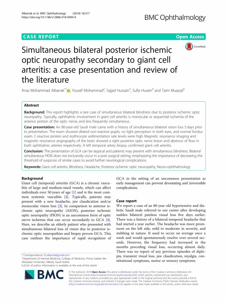
Examination showed normal blood pressure, heart rate,and temperature. He was unable to perceive light in botheyes, and the pupils were bilaterally dilated, seven millime-ters each, with no reaction to light. Fundoscopy showednormal appearing discs and retina. Ocular movementswere full. The motor, sensory and coordination examin-ation was normal. The C-reactive protein (CRP) upon ad-mission was 132mg/L and the erythrocyte sedimentationrate (ESR) was 40mm/hr. A magnetic resonance imaging(MRI) of the brain was done (Fig. 1) and it showed a lesionin right optic nerve suggesting acute ischemia. The oph-thalmic arteries were not visualized bilaterally by contrastmagnetic resonance angiography (MRA) (Fig. 2). The clin-ical impression was of a bilateral Posterior Ischemic OpticNeuropathy (PION) due to giant cell arteritis (GCA).The patient was started on intravenous Methylpredniso-
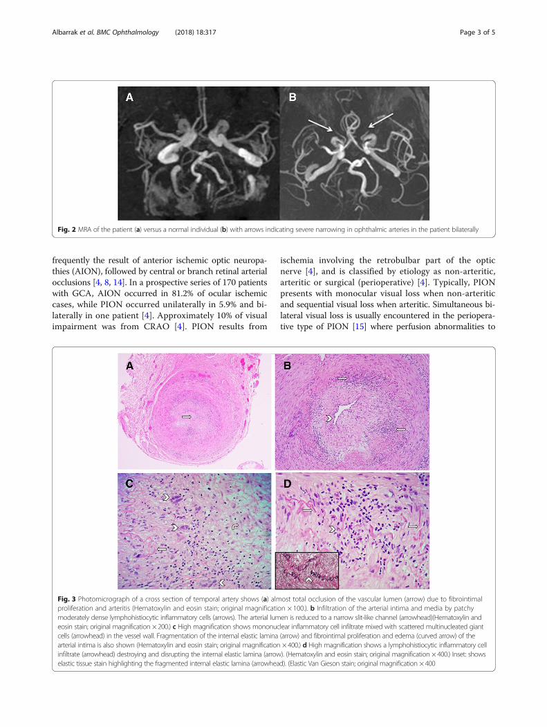
lone 1000mg for five days then shifted to daily prednisol-one 60mg orally. The vision did not improve but theheadache improved significantly after a few days. A 2 cmsegment of the left temporal artery was biopsied and thepathological findings confirmed giant cell arteritis (Fig. 3).
DiscussionHere we present a patient of Arab ethnicity who devel-oped bilateral blindness after suffering from a simultan-eous onset bilateral PION and a biopsy proven GCA.GCA is a granulomatous, medium to large vessel, in-flammatory disease that can have devastating ischemicconsequences to the eye [5, 6]. It is considered a relativelyunusual occurrence in Arabs [7] and is more commonlyencountered in Caucasian ethnicity [8]. Its incidence ismost marked among white individuals of Scandinaviandescent [9, 10], with one large Norwegian study finding anincreasing incidence between 1972 and 1992 that subse-quently plateaued. Women are more likely to be affectedthan men and the typical patient is in the 8th decade oflife [5, 9]. The typical symptoms of GCA include head-ache, scalp tenderness, jaw claudication, and loss of vision[3]. Headache is a common symptom in GCA and occursin more than two-thirds of patients [11]. Visual impair-ments of varying degrees occur in one or both eyes inabout 25 to 50% of GCA patients [4, 12, 13]. PION fromGCA is uncommon (Table 1), the visual loss is most
Fig. 1 a Axial diffusion weighted images. b ADC maps, c high resolution T2 weighted images and d coronal T2 weighted images. Arrowsshowing true diffusion restriction in the right optic nerve with T2 signal hyperintensity
Albarrak et al. BMC Ophthalmology (2018) 18:317 Page 2 of 5
frequently the result of anterior ischemic optic neuropa-thies (AION), followed by central or branch retinal arterialocclusions [4, 8, 14]. In a prospective series of 170 patientswith GCA, AION occurred in 81.2% of ocular ischemiccases, while PION occurred unilaterally in 5.9% and bi-laterally in one patient [4]. Approximately 10% of visualimpairment was from CRAO [4]. PION results from
ischemia involving the retrobulbar part of the opticnerve [4], and is classified by etiology as non-arteritic,arteritic or surgical (perioperative) [4]. Typically, PIONpresents with monocular visual loss when non-arteriticand sequential visual loss when arteritic. Simultaneous bi-lateral visual loss is usually encountered in the periopera-tive type of PION [15] where perfusion abnormalities to
Fig. 2 MRA of the patient (a) versus a normal individual (b) with arrows indicating severe narrowing in ophthalmic arteries in the patient bilaterally
Fig. 3 Photomicrograph of a cross section of temporal artery shows (a) almost total occlusion of the vascular lumen (arrow) due to fibrointimalproliferation and arteritis (Hematoxylin and eosin stain; original magnification × 100.). b Infiltration of the arterial intima and media by patchymoderately dense lymphohistiocytic inflammatory cells (arrows). The arterial lumen is reduced to a narrow slit-like channel (arrowhead)(Hematoxylin andeosin stain; original magnification × 200.) c High magnification shows mononuclear inflammatory cell infiltrate mixed with scattered multinucleated giantcells (arrowhead) in the vessel wall. Fragmentation of the internal elastic lamina (arrow) and fibrointimal proliferation and edema (curved arrow) of thearterial intima is also shown (Hematoxylin and eosin stain; original magnification × 400.) d High magnification shows a lymphohistiocytic inflammatory cellinfiltrate (arrowhead) destroying and disrupting the internal elastic lamina (arrow). (Hematoxylin and eosin stain; original magnification × 400.) Inset: showselastic tissue stain highlighting the fragmented internal elastic lamina (arrowhead). (Elastic Van Gieson stain; original magnification × 400
Albarrak et al. BMC Ophthalmology (2018) 18:317 Page 3 of 5
the eyes occur [16]. In contrast to AION, the optic discexamination is normal in the acute setting of PION, withthe development of optic atrophy on follow up assess-ments appearing about 6 weeks after the event [17]. Find-ings on MRI are either normal or show hyper-intensesignals on diffusion restricted images, while enhancementof the optic nerve with contrast can be seen in arteritic is-chemic optic neuropathies [18].The current patient presented with a history of head-
ache for one year that culminated in bilateral visual lossfrom optic nerve ischemia. The differential diagnosis fora presentation of acute optic neuropathy with headacheincludes arteritic ischemic optic neuropathy, infections(cat-scratch, syphilis), inflammatory (para-infectious, mul-tiple sclerosis, systemic autoimmune, paraneoplastic, andsarcoidosis). GCA in the current scenario presented fairlytypically, readily distinguishing it from other conditions.The ESR and CRP were high which suggested an inflam-matory process and abnormal signals were seen on theMRI suggesting infarction.Simultaneous and sudden complete blindness of both
eyes due to PION is an unusual presentation in GCA.Bilateral visual loss is usually due to the sequential onsetof AION, CRAO, or PION in any combination [4, 15].One cohort that looked at a small subgroup of simultan-eous onset bilateral ischemic optic neuropathies foundexamination findings of different ages in each eye, rais-ing the possibility that patients may not become awareof visual loss until it involves the other eye [4]. Thus, animportant consideration in the current case is defininghow simultaneous was the onset. Our patient witnessedthe visual loss occur while he was awake, and was foundto have symmetrical exam findings when assessed at thereferring center on the day of onset, and again at our
center 5 days later with no clear disc abnormalities.There was diffusion restriction involving the right opticnerve with absence of both ophthalmic arteries by MRA.It was not surprising to find asymmetrical findings onMRI as PION changes may not always seen on MRI[19], and our patient did not have imaging done untilone week after the event reducing the possibility of wit-nessing acute changes. Asymmetry in the observed MRIsignals of bilateral simultaneous onset post surgicalPION have also been described [20].
ConclusionsIn this report we presented a patient who was clinicallyand histologically diagnosed as GCA. IV steroids werestarted upon his arrival five days from symptom onset,with dramatic improvement in headache, however, noimprovement in vision occurred as is the case with opticnerve ischemia secondary to GCA [21]. Given the year-long history of headache, this case emphasizes the im-portance of early recognition of headache disordersamong elderly; the threshold of suspecting GCA in eld-erly patients should be very low. This case highlights ararely identified ocular presentation of GCA, it is im-portant to identify this early in order to start treatmentto avoid devastating and potentially permanent ocular orsystemic complication. When bilateral PION occurs sim-ultaneously in a non-surgical setting in an older individ-ual, then the possibility of GCA as an etiology should beconsidered high.
AbbreviationsAION: Anterior ischemic optic neuropathy; CRP: C-reactive protein;ESR: Erythrocyte sedimentation rate; GCA: Giant cell arteritis; MRI: Magneticresonance imaging; MRA: Magnetic resonance angiography; PION: Posteriorischemic optic neuropathy
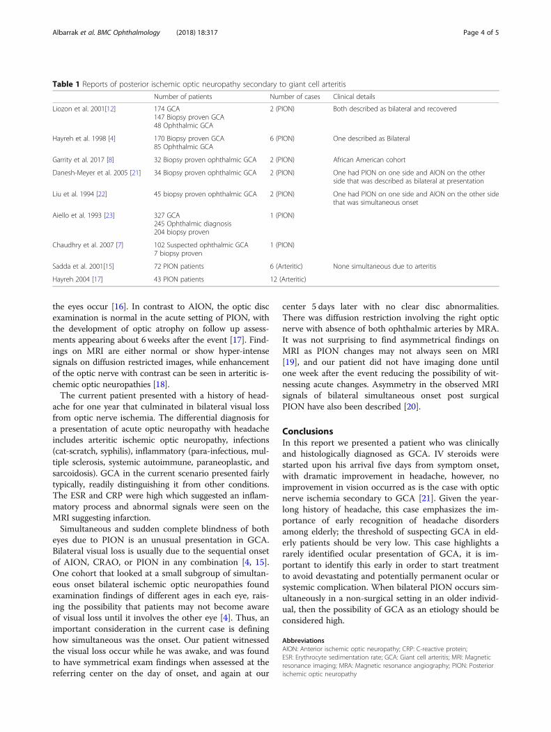
Table 1 Reports of posterior ischemic optic neuropathy secondary to giant cell arteritis
Number of patients Number of cases Clinical details
Liozon et al. 2001[12] 174 GCA147 Biopsy proven GCA48 Ophthalmic GCA
2 (PION) Both described as bilateral and recovered
Hayreh et al. 1998 [4] 170 Biopsy proven GCA85 Ophthalmic GCA
6 (PION) One described as Bilateral
Garrity et al. 2017 [8] 32 Biopsy proven ophthalmic GCA 2 (PION) African American cohort
Danesh-Meyer et al. 2005 [21] 34 Biopsy proven ophthalmic GCA 2 (PION) One had PION on one side and AION on the otherside that was described as bilateral at presentation
Liu et al. 1994 [22] 45 biopsy proven ophthalmic GCA 2 (PION) One had PION on one side and AION on the other sidethat was simultaneous onset
Aiello et al. 1993 [23] 327 GCA245 Ophthalmic diagnosis204 biopsy proven
1 (PION)
Chaudhry et al. 2007 [7] 102 Suspected ophthalmic GCA7 biopsy proven
1 (PION)
Sadda et al. 2001[15] 72 PION patients 6 (Arteritic) None simultaneous due to arteritis
Hayreh 2004 [17] 43 PION patients 12 (Arteritic)
Albarrak et al. BMC Ophthalmology (2018) 18:317 Page 4 of 5

AcknowledgementsThe authors thank the patient and his son for their collaboration.
FundingNo targeted funding reported.
Availability of data and materialsThe datasets used and/or analysed during the current study are availablefrom the corresponding author on reasonable request.
Authors’ contributionsAA and TM acquisition of data, analysis and interpretation, manuscriptconcept and design; YM critical revision of the manuscript for importantintellectual content and study supervision. SH and FH analysis andinterpretation of data. All authors read and approved the final manuscript.
Ethics approval and consent to participateNot applicable
Consent for publicationWritten informed consent was obtained from the patient for publication ofthis case report and any accompanying images. A copy of the consent formis available for review by the Editor of this journal.
Competing interestsThe authors declare that they have no competing interests.
Publisher’s NoteSpringer Nature remains neutral with regard to jurisdictional claims inpublished maps and institutional affiliations.
Author details1Department of Internal Medicine, College of Medicine, Prince Sattam BinAbdulaziz University, Alkharj, Saudi Arabia. 2Department of Internal Medicine,Neurology division, King Khalid University Hospital, Riyadh, Saudi Arabia.3Department of Medical Imaging, King Saud University / King KhalidUniversity Hospital, Riyadh, Saudi Arabia. 4Department of Pathology andLaboratory Medicine, King Khalid University Hospital, College of Medicine,King Saud University, Riyadh, Saudi Arabia. 5Neurology division, Departmentof Medicine, College of Medicine, King Saud University, Riyadh, Saudi Arabia.
Received: 1 August 2018 Accepted: 4 December 2018
References1. Dejaco C, Brouwer E, Mason JC, Buttgereit F, Matteson EL, Dasgupta B. Giant
cell arteritis and polymyalgia rheumatica: current challenges andopportunities. Nat Rev. Rheumatol. 2017;13(10):578–92.
2. Gonzalez-Gay MA, Garcia-Porrua C. Systemic vasculitis in adults innorthwestern Spain, 1988–1997. Clinical and epidemiologic aspects.Medicine (Baltimore). 1999;78(5):292–308.
3. Salvarani C, Pipitone N, Versari A, Hunder GG. Clinical features of polymyalgiarheumatica and giant cell arteritis. Nat Rev. Rheumatol. 2012;8(9):509–21.
4. Hayreh SS, Podhajsky PA, Zimmerman B. Ocular manifestations of giant cellarteritis. Am J Ophthalmol. 1998;125(4):509–20.
5. De Smit E, O’Sullivan E, Mackey DA, Hewitt AW. Giant cell arteritis: ophthalmicmanifestations of a systemic disease. Graefe’s Arch Clin Exp Ophthalmol. 2016;254(12):2291–306 Available from: https://doi.org/10.1007/s00417-016-3434-7.
6. Vodopivec I, Rizzo JF. Ophthalmic manifestations of giant cell arteritis.Rheumatol (United Kingdom). 2018;57(May):ii63-ii72.
7. Chaudhry IA, Shamsi FA, Elzaridi E, Arat YO, Bosley TM, Riley FC.Epidemiology of giant-cell arteritis in an Arab population: a 22-year study.Br J Ophthalmol. 2007;91(6):715–8.
8. Garrity ST, Pistilli M, Vaphiades MS, Richards NQ, Subramanian PS, Rosa PR,et al. Ophthalmic presentation of giant cell arteritis in African-Americans.Eye 2017;31(1):113–8. Available from: https://doi.org/10.1038/eye.2016.199
9. Brekke LK, Diamantopoulos AP, Fevang B-T, Aβmus J, Esperø E, Gjesdal CG.Incidence of giant cell arteritis in Western Norway 1972–2012: a retrospectivecohort study. Arthritis Res Ther. 2017;19(1):278 Available from: https://arthritis-research.biomedcentral.com/articles/10.1186/s13075-017-1479-6.
10. Gonzalez-Gay MA, Vazquez-Rodriguez TR, Lopez-Diaz MJ, Miranda-Filloy JA,Gonzalez-Juanatey C, Martin J, et al. Epidemiology of giant cell arteritis andpolymyalgia rheumatica. Arthritis Rheum. 2009;61(10):1454–61.
11. Gonzalez-Gay MA, Barros S, Lopez-Diaz MJ, Garcia-Porrua C, Sanchez-Andrade A, Llorca J. Giant cell arteritis: disease patterns of clinicalpresentation in a series of 240 patients. Medicine (Baltimore). 2005;84(5):269–76.
12. Liozon E, Herrmann F, Ly K, Robert PY, Loustaud V, Soria P, et al. Risk factorsfor visual loss in giant cell (temporal) arteritis: a prospective study of 174patients. Am J Med. 2001;111(3):211–7.
13. Singh AG, Kermani TA, Crowson CS, Weyand CM, Matteson EL, WarringtonKJ. Visual manifestations in giant cell arteritis: trend over 5 decades in apopulation-based cohort. J Rheumatol. 2015;42(2):309–15.
14. Miller NR. Visual manifestations of temporal arteritis. Rheum Dis Clin NorthAm. 2001;27(4):781–97 vi.
15. Sadda SR, Nee M, Miller NR, Biousse V, Newman NJ, Kouzis A. Clinicalspectrum of posterior ischemic optic neuropathy. Am J Ophthalmol. 2001;132(5):743–50.
16. Biousse V, Newman NJ. Ischemic Optic Neuropathies. N Engl J Med[Internet]. 2015;372(25):2428–36 Available from: http://www.nejm.org/doi/10.1056/NEJMra1413352.
17. Hayreh SS. Posterior ischaemic optic neuropathy: clinical features,pathogenesis. and management. Eye (Lond). 2004;18(11):1188–206.
18. Lee AG, Eggenberger ER, Kaufman DI, Manrique C. Optic nerveenhancement on magnetic resonance imaging in arteritic ischemic opticneuropathy. J Neuroophthalmol. 1999;19(4):235–7.
19. He M, Cestari D, Cunnane MB, Rizzo JF. The use of diffusion MRI in ischemicoptic neuropathy and optic neuritis. Semin Ophthalmol. 2010;25(5–6):225–32.
20. Bhatt NP, Morales RE. Mathews MK. MRI findings in Post-operative BilateralPosterior Ischemic Optic Neuropathy. Open J Ophthalmol. 2013;3:51–3.
21. Danesh-Meyer H, Savino PJ, Gamble GG. Poor prognosis of visual outcomeafter visual loss from giant cell arteritis. Ophthalmology. 2005;112(6):1098–103.
22. Liu GT, Glaser JS, Schatz NJ, Smith JL. Visual morbidity in giant cell arteritis.Clinical characteristics and prognosis for vision. Ophthalmology. 1994;101(11):1779–85.
23. Aiello PD, Trautmann JC, McPhee TJ, Kunselman AR, Hunder GG. VisualPrognosis in Giant Cell Arteritis. Ophthalmology [Internet]. 1993;100(4):550–555.Available from: https://doi.org/10.1016/S0161-6420(93)31608-8.
Albarrak et al. BMC Ophthalmology (2018) 18:317 Page 5 of 5