simplified approach to identification actinomycetes by
TRANSCRIPT

APPLIED MICROBIOLOGY, Aug. 1974, p. 226-231 Vol. 28, No. 2Copyright i 1974 American Society for Microbiology Printed in U.S.A.
Simplified Approach to Identification of AerobicActinomycetes by Thin-Layer Chromatography
JOSEPH L. STANECK AND GLENN D. ROBERTSSection of Clinical Microbiology, Mayo Clinic and Mayo Foundation, Rochester, Minnesota 55901
Received for publication 11 March 1974
A system has been developed for the identification of aerobicactinomycetes in the clinical laboratory based on analysis of whole cells fordiaminopimelic acid and carbohydrates and on the ability of the organism todecompose casein, tyrosine, and xanthine media. The whole-cell analyses wereperformed by a simple thin-layer chromatographic procedure that is described.Eighteen reference cultures were correctly identified and, subsequently, 35isolates from clinical material were grouped by using this system. The method iswell suited for use in routine clinical laboratories.
Infections due to the aerobic actinomycetes,particularly of the genus Nocardia, are beingreported with greater frequency in situationsthat favor the opportunistic invasion and multi-plication of microorganisms. Nocardiosis hasbeen associated with malignancies (13, 16),pulmonary alveolar proteinosis (5), im-munosuppressive therapy (1, 2, 14), and steroidtreatment (15), and it will be a problem inpatients compromised by either disease ortherapy. Bacteriology, mycobacteriology, andmycology laboratories should be capable ofproviding rapid detection, accurate identifica-tion, and, if necessary, susceptibility data onorganisms such as species of Nocardia, Strep-tomyces, and Actinomadura.A recent article by Goodfellow (7) suggested
that chemotaxonomic markers should be impor-tant in the classification of microorganisms.The chemical composition of cell walls has beenaccepted by Lechevalier and Lechevalier (12) asa criterion for the classification of aerobic ac-tinomycetes. Becker et al. (3) introduced asystem of paper chromatography able to sepa-rate the taxonomically important stereoisomersof diaminopimelic acid (DAP). Lechevalier (11)described a system to identify carbohydrates fordiagnostic purposes. Chromatographic analysisof cell walls was recently used by Berd (4) in anextensive study of the classification of aerobicactinomycetes.Our paper presents an adaption of the paper
chromatographic system to thin-layer chroma-tography (TLC). The use of TLC simplifies thetechniques, materials, and time necessary foranalysis and encourages the adoption of chro-
matographic systems for diagnostic purposes bythe routine clinical laboratory.
MATERIALS AND METHODSCultures. Reference cultures (Table 1) were ob-
tained from Center for Disease Control laboratorysurveys and from Ruth Gordon (Rutgers University).Unknown cultures were obtained from clinical speci-mens submitted to our laboratory between December1972 and December 1973 and from frozen stockcultures of strains originally isolated from clinicalmaterial. Working stock cultures were maintained onbrain heart infusion (BHI; Difco) agar slants. Incuba-tion of all cultures was at 30 C. Primary isolationsfrom clinical specimens were made on either BHIagar, Lowenstein-Jensen medium (Difco), or sheepblood agar plates, depending on the nature of thespecimen. The modified cold acid-fast stainingmethod of Kinyoun (6), with 1% sulfuric acid substi-tuted for acid-alcohol in the decolorizing step, wasused to determine acid fastness.
Decomposition media. The medium for determin-ing casein decomposition was made by dissolving 10 gof dehydrated skim milk in 100 ml of distilled waterand autoclaving. A separate 100-ml solution of 2%agar in distilled water was also autoclaved. Aftercooling to 45 C, these two solutions were mixed andpoured into plates.The tyrosine or xanthine medium consisted of 23 g
of nutrient agar, 5 g of tyrosine (or 4 g of xanthine),and 1 liter of distilled water. The ingredients weremixed, adjusted to pH 7.0, autoclaved, and pouredinto plates; the plates were gently swirled while themedium cooled to obtain a smooth suspension of theamino acid.A heavy inoculum of the test organism was streaked
onto a section of each type of plate and incubated.The plates were observed for 14 days for areas of
226
on April 29, 2016 by WAYN
E STATE UN
IVERSITY
http://aem.asm
.org/D
ownloaded from

TLC IDENTIFICATION OF AEROBIC ACTINOMYCETES
clearing around the bacterial growth, which wouldindicate decomposition of the substrate.
Identification procedure. The following routineconsists of variations of the work of Gordon and Mihm(9, 10), Lechevalier and Lechevalier (11, 12), Beckeret al. (3), and Berd (4).Any slowly growing aerobic colony consisting of
small, branched cells that often, but not necessarily,were partially acid fast was suspected of belonging tothe aerobic actinomycetes group. Strains not wellisolated on primary media were restreaked on BHIagar plates to obtain several isolated colonies thatwere picked and suspended in 2 ml of BlJI broth toform a heavy inoculating suspension. Decompositionmedia were streaked with inoculum, incubated, andobserved for 14 days.The remaining suspension was added to a 250-ml
flask containing 100 ml of BHI broth and incubatedwith constant shaking at 30 C until the broth becameturbid. These cells were killed with formalin (finalconcentration, 1%) for 24 h at room temperature andcollected by centrifugation. The cells were washedonce in distilled water and once in 95% ethanol andthen dried by suitable means (vacuum desiccation,
TABLE 1. Reference cultures and source
Strain No. Source,
'Nocardia asteroides 3 CDC laboratory surveyN. brasiliensis 2 CDC laboratory surveyN. caviae 1 CDC laboratory surveyStreptomyces species 1 CDC laboratory surveyActinomadura dassonvillei 2 Ruth Gordon (NCTC#434,
#711)A. madurae 1 Ruth Gordon (#1092)Micromonospora species 2 Ruth Gordon (#3450,
#3640)"Rhodochrous" group 5 Ruth Gordon (#1240,
W#3408, #1346, #382,and #1256)
S. somaliensis 1 Ruth Gordon (#1448)a CDC, Center for Disease Control.
forced air, or overnight drying at 45 C). The driedcells were analyzed for carbohydrates and DAP asdescribed below.The identification is made as shown in Table 2.Hydrolysis and chromatography. The procedure
of Becker et al. (3) was followed for the hydrolysis ofwhole cells preceding DAP analysis. Approximately 3mg (dry weight) of cells was placed into a smallampoule with 1 ml of 6 N hydrochloric acid. Thesealed ampoule was kept at 100 C in an oven for 18 h.After cooling, the hydrolysate was filtered throughWhatman no. 1 paper. The filtrate was evaporated todryness in a boiling water bath, redissolved in 1 ml ofdistilled water, and taken to dryness again. Thisresidue was dissolved in 0.3 ml of distilled water, and2 ,liters was applied at the base line of the TLC sheet(Chromagram-Eastman Kodak no. 6064 cellulosewithout fluorescent indicator). Ascending TLC wasperformed with the solvent system methanol-distilledwater-6 N HCl-pyridine (80:26:4: 10, vol/vol) forapproximately 3.5 h. After the chromatogram was airdried, spots were visualized by spraying with 0.2%ninhydrin in acetone and heating at 100 C for 3 min.As a DAP standard, 1 gliter of 0.01 M DL-DAP (SigmaChemical Co.), which contains both meso- and L-DAPisomers, was used. The DAP spots were seen asgray-green fading to yellow, with the L isomer movingahead of the meso isomer (Fig. 1). With hydrolysates,amino acid spots appeared purple or red and migratedahead of the DAP spot. The easy visualization, afterdevelopment, of 0.5 ghliters of the 0.01 M standardsolution of DL-DAP indicated the ability of the systemto detect as little as 1 Ag of a DAP isomer. Sampleapplication, development, and identification requiredless than 4 h.The carbohydrate analysis was based on the work of
Lechevalier (11). Approximately 25 mg (dry weight)of cells was placed into an ampoule with 1.5 ml of 1 Nsulfuric acid. The sealed ampoule was heated for 2 hin a boiling water bath. After cooling, the hydrolysatewas transferred to a 15-ml conical centrifuge tube,and saturated barium hydroxide was added dropwise
TABLE 2. Identification of aerobic actinomycetes
DAP Carbohydrate Decomposition mediaisomer productso Casein Tyrosine Xanthine
L-DAP + + + Streptomyces sp.+ + _ Streptomyces somaliensis
meso-DAP Ara + Gal _ _ _ Nocardia asteroides+ + _ N. brasiliensis_ _ + N. caviae_ -_ "Rhodochrous" groupb
Gal or none + + + Actinomadura dassonvilleiGal + Mad + A. madurae or pelletieriAra + XylC + - Micromonospora sp.
a Ara, Arabinose; Gal, galactose; Mad, madurose; Xyl, xylose.b Strains of the "rhodochrous" group that fail to decompose tyrosine are difficult to distinguish from
Nocardia by these or other biochemical tests; differentiation depends on colonial and microscopic morphology(4, 8).
c With or without galactose.
227VOL. 119, 1974
on April 29, 2016 by WAYN
E STATE UN
IVERSITY
http://aem.asm
.org/D
ownloaded from
STANECK AND ROBERTS
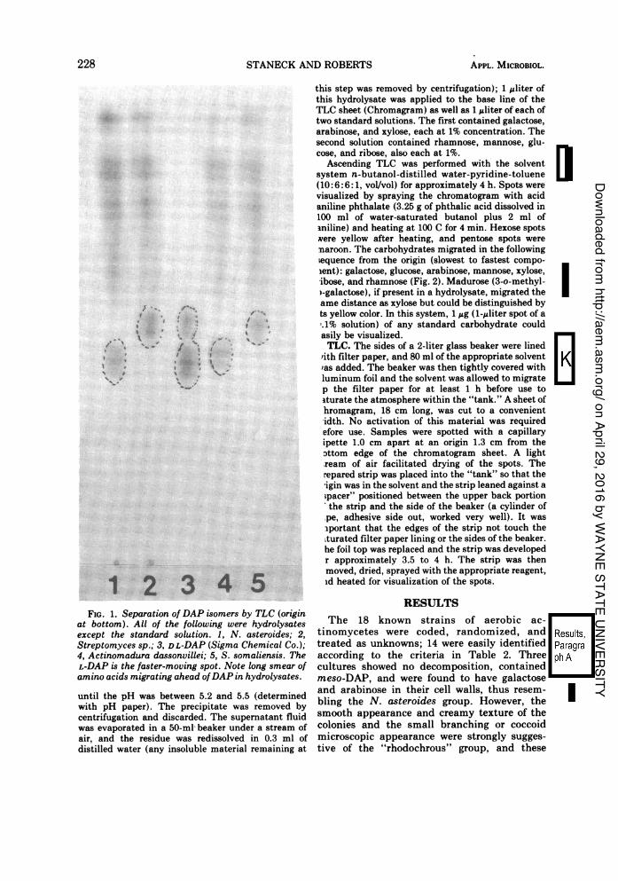
FIG. 1. Separation of DAP isomers by TLC (originat bottom). All of the following were hydrolysatesexcept the standard solution. 1, N. asteroides; 2,Streptomyces sp.; 3, DL-DAP (Sigma Chemical Co.);4, Actinomadura dassonvillei; 5, S. somaliensis. TheL-DAP is the faster-moving spot. Note long smear ofamino acids migrating ahead ofDAP in hydrolysates.
until the pH was between 5.2 and 5.5 (determinedwith pH paper). The precipitate was removed bycentrifugation and discarded. The supernatant fluidwas evaporated in a 50-ml-beaker under a stream ofair, and the residue was redissolved in 0.3 ml ofdistilled water (any insoluble material remaining at
this step was removed by centrifugation); 1 uliter ofthis hydrolysate was applied to the base line of theTLC sheet (Chromagram) as well as 1 gliter of each oftwo standard solutions. The first contained galactose,arabinose, and xylose, each at 1% concentration. Thesecond solution contained rhamnose, mannose, glu-cose, and ribose, also each at 1%.
Ascending TLC was performed with the solventsystem n-butanol-distilled water-pyridine-toluene(10:6:6: 1, vol/vol) for approximately 4 h. Spots werevisualized by spraying the chromatogram with acidaniline phthalate (3.25 g of phthalic acid dissolved in100 ml of water-saturated butanol plus 2 ml ofiniline) and heating at 100 C for 4 min. Hexose spotsvere yellow after heating, and pentose spots wereimaroon. The carbohydrates migrated in the following,equence from the origin (slowest to fastest compo-ient): galactose, glucose, arabinose, mannose, xylose,ibose, and rhamnose (Fig. 2). Madurose (3-o-methyl-)-galactose), if present in a hydrolysate, migrated theame distance as xylose but could be distinguished byts yellow color. In this system, 1 ;&g (1-Aliter spot of a'.1% solution) of any standard carbohydrate couldasily be visualized.TLC. The sides of a 2-liter glass beaker were lined
rith filter paper, and 80 ml of the appropriate solvent'as added. The beaker was then tightly covered withluminum foil and the solvent was allowed to migratep the filter paper for at least 1 h before use toturate the atmosphere within the "tank." A sheet ofhromagram, 18 cm long, was cut to a convenientidth. No activation of this material was requiredefore use. Samples were spotted with a capillaryipette 1.0 cm apart at an origin 1.3 cm from thefltom edge of the chromatogram sheet. A lightream of air facilitated drying of the spots. Therepared strip was placed into the "tank" so that theigin was in the solvent and the strip leaned against a;pacer" positioned between the upper back portionthe strip and the side of the beaker (a cylinder of
.pe, adhesive side out, worked very well). It wasTiportant that the edges of the strip not touch theLturated filter paper lining or the sides of the beaker.he foil top was replaced and the strip was developedr approximately 3.5 to 4 h. The strip was thenmoved, dried, sprayed with the appropriate reagent,id heated for visualization of the spots.
RESULTSThe 18 known strains of aerobic ac-
tinomycetes were coded, randomized, andtreated as unknowns; 14 were easily identifiedaccording to the criteria in Table 2. Threecultures showed no decomposition, containedmeso-DAP, and were found to have galactoseand arabinose in their cell walls, thus resem-bling the N. asteroides group. However, thesmooth appearance and creamy texture of thecolonies and the small branching or coccoidmicroscopic appearance were strongly sugges-tive of the "rhodochrous" group, and these
228 APPL. MICROBIOL.
on April 29, 2016 by WAYN
E STATE UN
IVERSITY
http://aem.asm
.org/D
ownloaded from
TLC IDENTIFICATION OF AEROBIC ACTINOMYCETES
strains therefore were classified as such. Onestrain demonstrated casein and tyrosine decom-position and contained meso-DAP but producedonly galactose. This strain was suspected ofbeing either Actinomadura madurae or A. pel-letieri; in a repeat test with application of 5pliters rather than the usual 1 jliter of hydroly-sate to the TLC plate, a faint madurose spotwas detected, confirming the identification. In aclinical situation, further tests, such as car-bohydrate fermentations as described by Berd(4), would have to be used to differentiatebetween A. madurae and A. pelletieri. Acid-faststaining at times was difficult to interpret andwas therefore given little consideration in theidentification.
Subsequently, 35 strains, collected from re-cent clinical isolations and from stock culturesof previous clinical isolations, were subjected toanalysis by this system. All could be classifiedeasily and included the following isolates: 23 N.asteroides, one N. brasiliensis, one N. caviae,eight Streptomyces sp., and two A. dassonvillei.One of the two A. dassonvillei isolates was sentto the Center for Disease Control and ouridentification was confirmed; the other has beententatively identified as A. dassonvillei by RuthGordon.Most of our identifications were made within
approximately 8 days after initial isolation ofthe organism. Although decomposition mediawere always held for 14 days, we have foundthat, when the inoculum is adequate, changesrarely occur in the pattern beyond the 8th day.Thus, a tentative identification at approxi-mately 8 days was often possible based on thedecomposition pattern and supported by thechromatographic analyses.
Identification was delayed in the case ofslowly growing strains. Among our referencestrains, Micromonospora sp. and A. maduraerequired relatively long incubation periods inbroth cultures to reach densities suitable forchemical analyses, and two strains of A. pel-letieri failed to grow to sufficient densities foranalyses and were therefore not included in thisstudy.
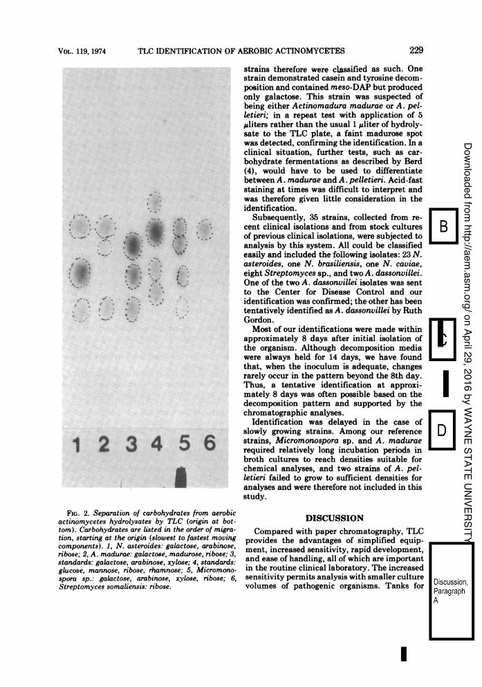
FIG. 2. Separation of carbohydrates from aerobicactinomycetes hydrolysates by TLC (origin at bot-tom). Carbohydrates are listed in the order of migra-tion, starting at the origin (slowest to fastest movingcomponents). 1, N. asteroides: galactose, arabinose,ribose; 2, A. madurae: galactose, madurose, ribose; 3,standards: galactose, arabinose, xylose; 4, standards:glucose, mannose, ribose, rhamnose; 5, Micromono-spora sp.: galactose, arabinose, xylose, ribose; 6,Streptomyces somaliensis: ribose.
DISCUSSIONCompared with paper chromatography, TLC
provides the advantages of simplified equip-ment, increased sensitivity, rapid development,and ease of handling, all of which are importantin the routine clinical laboratory. The increasedsensitivity permits analysis with smaller culturevolumes of pathogenic organisms. Tanks for
229.VOL. 119, 1974
on April 29, 2016 by WAYN
E STATE UN
IVERSITY
http://aem.asm
.org/D
ownloaded from
STANECK AND ROBERTS
descending chromatography and ventilated dry-ing hoods are unnecessary. The decreased devel-opment time hastens identification and thuspermits the laboratory to provide a betterdiagnostic service. In our experience, TLC forDAP and carbohydrate analyses has been moremanageable than the paper system and, in lesstime with less expense, has yielded similarinformation as paper chromatographic methodswith respect to the diagnostically importantcarbohydrates: galactose, arabinose, xylose,and madurose. These factors should encourageother routine clinical laboratories to considerusing the determination of chemotaxonomicmarkers for diagnostic purposes.The modified system for the identification of
aerobic actinomycetes relies heavily on theextensive and thorough characterization ofthese organisms by Berd (4), Lechevalier andLechevalier (12), and Gordon and Mihm (8-10),and we are in no way suggesting that thissystem should be interpreted taxonomicallybeyond its clinical limits; these authors all havereported strains of aerobic actinomycetes thatfit loosely into the identification system that wehave used. Isolates that fail to conform to oursystem should be subjected to further biochemi-cal testing by the primary laboratory or by areference laboratory. However, our modifiedsystem can be most useful in the diagnosis ofinfections caused by this group of organismsand provides an accurate basis for studies todetermine the incidence and significance ofthese infections.The commercially available flexible Chroma-
gram cellulose plates were found to be ex-tremely convenient and represented a consider-able improvement over glass plates for TLC.particularly because they can be cut to size withscissors and are easily handled throughout allsteps of the procedure. No problems were en-countered in the system to identify the isomersof DAP. Occasionally, in carbohydrate analysisa sample applied too heavily produced signifi-cant streaking; however, a repeat test with asmaller application would reveal an interpreta-ble pattern. Conversely, a faint pattern ofsugars could be made more distinctive by in-creasing the size of sample applied. AlthoughLechevalier and Lechevalier (12) report thepresence of glucose, mannose, and ribose inalmost all cultures that they studied, we foundonly ribose consistently in all isolates. Glucoseand mannose, both yellow-staining spots, weredistinguishable, although not completely sepa-rated, from the maroon-staining arabinose spotif the three components were run in a mixturecontaining equal concentrations of each. How-ever, it is conceivable that either glucose or
mannose spots might be masked by the pres-ence of a greater amount of arabinose, sincethese three carbohydrates migrate close to oneanother. The failure of this system to detectglucose or mannose could thus be explainedsince those hydrolysates that contain arabinoseshowed a predominance of arabinose in theircarbohydrate patterns, both in this chromato-graphic system and in the report by Lechevalier(11). This fact does not detract from the clinicalusefulness of the system because the diagnosti-cally important sugars-arabinose, galactose,and xylose-when present dominate the car-bohydrate patterns, and are easily interpreted.This limitation should be borne in mind if thischromatographic system is used outside of thediagnostic context outlined here.The variability of the acid-fast stain can be
misleading. A positive acid-fast reaction sug-gests final identification of a Nocardia sp., butthe lack of acid fastness does not rule out thisgenus. The use of a heavy uniform inoculum forthe decomposition media is preferable for validresults. This system fails to differentiate A.pelletieri from A. madurae and further tests,such as carbohydrate fermentations, are re-quired. Members of the "rhodochrous" group,some species of Mycobacterium, and Coryne-bacterium can present a profile similar to thatof N. asteroides in this system and, in theseinstances, the gross and microscopic morpholo-gies must be considered in an attempt to assesstrue branching (4).
In the past 9 months, the modified system ofidentification of aerobic actinomycetes has ena-bled us to decrease significantly the time ofreporting results, so that the laboratory data areof active rather than historical interest to theclinician. From December 1972 to December1973, we recovered 17 different isolates of N.asteroides. The use of chromatography enabledus to identify two strains of A. dassonvillei, onefrom a blood culture after 16 days of incubationand the other from an ocular exudate; althoughno clinical significance could be assigned tothese isolations, we are encouraged that, withfurther efforts, more members of the aerobicactinomycetes group may be found in clinicalmaterial.
LITERATURE CITED1. Bach, M. C., A. P. Monaco, and M. Finland. 1973.
Pulmonary nocardiosis: therapy with minocycline andwith erythromycin plus ampicillin. J. Amer. Med. Ass.224:1378-1381.
2. Bach, M. C., A. Sahyoun, J. L. Adler, R. M. Schlesinger,J. Breman, P. Madras, F. P'eng, and A. P. Monaco.1973. Influence of rejection therapy on fungal andnocardial infections in renal-transplant recipients.Lancet 1:180-184.
230 APPL. MICROBIOL.
on April 29, 2016 by WAYN
E STATE UN
IVERSITY
http://aem.asm
.org/D
ownloaded from

VOL. 119, 1974 TLC ]IDENTIFICATION OF A
3. Becker, B., M. P. Lechevalier, R. E. Gordon, and H. A.Lechevalier. 1964. Rapid differentiation between No-cardia and Streptomyces by paper chromatography ofwhole-cell hydrolysates. Appl. Microbiol. 12:421-423.
4. Berd, D. 1973. Laboratory identification of clinicallyimportant aerobic actinomycetes. Appl. Microbiol.25:665-681.
5. Carlsen, E. T., R. B. Hill, Jr., and D. T. Rowlands, Jr.1964. Nocardiosis and pulmonary alveolar proteinosis.Ann. Intern. Med. 60:275-281.
6. Georg, L. K., L. Ajello, C. McDurmont, and T. S. Hosty.1961. The identification of Nocardia asteroides andNocardia brasiliensis. Amer. Rev. Resp. Dis.84:337-347.
7. Goodfellow, M. 1973 Characterisation of Mycobacte-rium, Nocardia, Corynebacterium and related taxa.Ann. Soc. Belg. Med. Trop. 53:287-298.
8. Gordon, R E. 1966. Some strains in search of a genus-Corynebacterium, Mycobacterium, Nocardia, orwhat? J. Gen. Microbiol. 43:329-343.
9. Gordon, R. E., and J. M. Mihm. 1959. A comparison ofNocardia asteroides and Nocardia brasiliensis. J. Gen.Microbiol. 20:129-135.
EROBIC ACTINOMYCETES 231
10. Gordon, R. E., and J. M. Mihm. 1962. The type species ofthe genus Nocardia. J. Gen. Microbiol. 27:1-10.
11. Lechevalier, M. P. 1968. Identification of aerobic ac-tinomycetes of clinical importance. J. Lab. Clin. Med.71:934-944.
12. Lechevalier, M. P., and H. Lechevalier. 1970. Chemicalcomposition as a criterion in the classification ofaerobic actinomycetes. Int. J. Syst. Bacteriol.20:435-443.
13. Pinkhas, J., I. Oliver, A. de Vries, S. A. Spitzer, and E.Henig. 1973. Pulmonary nocardiosis complicating ma-lignant lymphoma successfully treated with chemo-therapy. Chest 63:367-370.
14. Riflind, D., T. L. Marchioro, S. A. Schneck, and R. B.Hill, Jr. 1967. Systemic fungal infections complicatingrenal transplantation and immunosuppressive therapy.Amer. J. Med. 43:28-38.
15. Whitmore, D. N., G. A. Gresham, and M. J. Grayson.1961. Nocardiosis in anaemic patients given steroids.J. Clin. Pathol. 14:259-263.
16. Young, L. S., D. Armstrong, A. Blevins, and P. Lieber-man. 1971. Nocardia asteroides infection complicatingneoplastic disease. Amer. J. Med. 50:356-367.
on April 29, 2016 by WAYN
E STATE UN
IVERSITY
http://aem.asm
.org/D
ownloaded from