silica-coated nanocomposites of magnetic nanoparticles and quantum dots
TRANSCRIPT

Silica-Coated Nanocomposites of Magnetic Nanoparticles and Quantum DotsDong Kee Yi,† S. Tamil Selvan,† Su Seong Lee,† Georgia C. Papaefthymiou,‡
Darshan Kundaliya,§ and Jackie Y. Ying*,†
Institute of Bioengineering and Nanotechnology, 31 Biopolis Way, The Nanos, Singapore 138669, Department ofPhysics, VillanoVa UniVersity, VillanoVa, PennsylVania 19085, and Center for SuperconductiVity Research,
Department of Physics, UniVersity of Maryland, College Park, Maryland 20742
Received November 25, 2004; E-mail: [email protected]
In this communication, we report the synthesis of a novel water-soluble hybrid material consisting of quantum dots (QDs) andmagnetic nanoparticles (MPs) encapsulated in a silica shell. QDsor semiconductor nanocrystals have been extensively studied fortheir unique optical and electronic properties,1,2 sharp emission bandwith broad excitation, and strong resistance to photobleaching. Theyare of great interest for their dimensional similarities to bio-macromolecules (e.g., DNA and protein) and for applications inbiological imaging3 and bioconjugation.4 Superparamagnetic nano-particles have been studied for biomedical applications,5 such asMRI contrast enhancement,6 magnetic immobilization, and drugtargeting.7 For many applications, these nanoparticles would benefitfrom having a silica shell to impart wettability and biocompatibility.Silica can also be easily surface modified to link bioconjugators,such as avidin, with interesting biofunctionalities. Herein wedescribe the encapsulation of both MPs and QDs within a silicashell to form a hybrid material denoted as SiO2/MP-QD. AlthoughMP-QD systems have been examined before,8 SiO2/MP-QDnanocomposites have not yet been reported. These materials havepotential in bioimaging, drug targeting, biosensing, and biolabelingapplications; their core-shell asymmetric refractive property haspromise for novel optical communication systems such as photoniccrystals. Silica-capped QDs have been achieved using alkoxysilanewith a silica shell thickness ranging from 2-5 nm9a to 40-80 nm.9b
QDs have also been coated with silica with a reverse microemulsionapproach.10 MP encapsulation within silica has been attainedthrough modified Sto¨ber method11 and reverse microemulsion.12
In situ reverse microemulsion synthesis generally led to poorlycrystalline magnetic cores. Therefore, magnetic cores were preparedseparately, prior to silica capping.12a
A two-step synthesis was used to derive SiO2/MP-QD (seeScheme 1). MPs and QDs were first prepared separately, prior totheir introduction to the reverse microemulsion medium for silicacoating. Specifically, polyoxyethylene(5)nonylphenyl ether (0.544mmol, Igepal CO-520, containing 50 mol % hydrophilic group)was dispersed in cyclohexane (4.5 mL) by sonication. Next, 400µL of γ-Fe2O3 solution13 (0.5 mg/mL of cyclohexane) and 160µLof CdSe QD solution14 (1 mg/mL of cyclohexane) were added. Theresulting mixture was vortexed, and ammonium hydroxide (29.4%,40 µL) was added to form a transparent, brown solution of reversemicroemulsion. Last, tetraethyl orthosilicate (TEOS, 30µL, Aldrich)was added, and the reaction was continued fore48 h. The resultingSiO2/MP-QD nanocomposite particles were collected by magnetor centrifuging, washed, and redispersed in ethanol or deionizedwater.
Transmission electron microscopy (TEM) images showed thatthe MPs were monodispersed (11.8( 1.3 nm in diameter) andsingle crystalline (Figure 1a and inset). X-ray diffraction (XRD)
pattern, as well as X-ray photoelectron Fe(2p3/2) and Fe(2p1/2) peaksat 711.4 and 724.7 eV, respectively, confirmed that the MPs wereγ-Fe2O3 crystallites (see Supporting Information, SI). When onlyMPs were introduced for silica encapsulation, the resulting particlescould include one, two, or three MPs each (Figure 1b). Uniformparticles containing a single MP and tailored SiO2 shell thicknesscould be derived under controlled experimental conditions.15
For SiO2/MP-QD synthesis, MPs (∼11.8 nm) and QDs (∼3.5nm) were shown to form silica-mediated conjugation within 8 h ofreaction, but spherical shells were not achieved at this initial stage(see Figure 1c and inset). Energy-dispersive X-ray (EDX) analysisindicated the presence of CdSe, Fe2O3, and silica (see SI). After48 h of reaction, silica shells were observed to have developedaround the MP-QD cores (Figure 1d). Although spatial discrep-ancies between QDs and MPs within the silica shell caused theMPs to be out of focus, QDs were clearly seen (marked by arrows
† Institute of Bioengineering and Nanotechnology.‡ Villanova University.§ University of Maryland.
Figure 1. TEM micrographs of (a)γ-Fe2O3 MPs, (b) SiO2/MP, (c)interconnected MPs and CdSe QDs (after 8 h of SiO2/MP-QD reaction),(d,e) SiO2/MP-QD nanocomposites (after 48 h of SiO2/MP-QD reaction;note the presence of both Fe2O3 MPs and CdSe QDs (finer crystallitesdenoted by arrows in panel e), and (f) SiO2/MP-QD nanocomposites formedat a lower CdSe concentration (0.5 mg/mL of cyclohexane). (g) High-resolution TEM micrograph of the area marked by the arrow in (f), showingthe presence of CdSe QDs andγ-Fe2O3 MPs.
Scheme 1. Synthesis of SiO2/MP-QD Nanocomposites
Published on Web 03/15/2005
4990 9 J. AM. CHEM. SOC. 2005 , 127, 4990-4991 10.1021/ja0428863 CCC: $30.25 © 2005 American Chemical Society
in Figure 1e). Even at a lower QD concentration of 0.5 mg/mL ofcyclohexane, both QDs and MPs were successfully encapsulatedin the silica shells (Figure 1f,g). EDX analysis of the region shownin Figure 1g confirmed the presence of CdSe, Fe2O3, and silica(see SI). N2 sorption isotherm of the SiO2/MP-QD particlesindicated that the silica shells were nonporous (see SI).
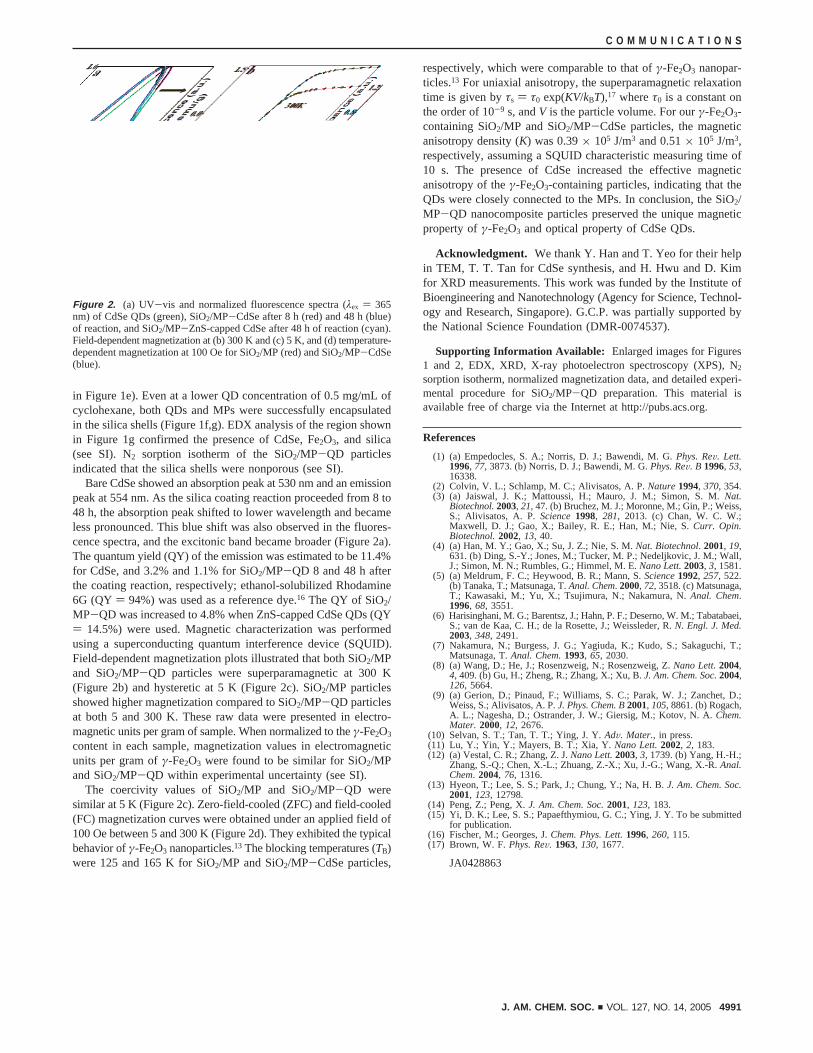
Bare CdSe showed an absorption peak at 530 nm and an emissionpeak at 554 nm. As the silica coating reaction proceeded from 8 to48 h, the absorption peak shifted to lower wavelength and becameless pronounced. This blue shift was also observed in the fluores-cence spectra, and the excitonic band became broader (Figure 2a).The quantum yield (QY) of the emission was estimated to be 11.4%for CdSe, and 3.2% and 1.1% for SiO2/MP-QD 8 and 48 h afterthe coating reaction, respectively; ethanol-solubilized Rhodamine6G (QY ) 94%) was used as a reference dye.16 The QY of SiO2/MP-QD was increased to 4.8% when ZnS-capped CdSe QDs (QY) 14.5%) were used. Magnetic characterization was performedusing a superconducting quantum interference device (SQUID).Field-dependent magnetization plots illustrated that both SiO2/MPand SiO2/MP-QD particles were superparamagnetic at 300 K(Figure 2b) and hysteretic at 5 K (Figure 2c). SiO2/MP particlesshowed higher magnetization compared to SiO2/MP-QD particlesat both 5 and 300 K. These raw data were presented in electro-magnetic units per gram of sample. When normalized to theγ-Fe2O3
content in each sample, magnetization values in electromagneticunits per gram ofγ-Fe2O3 were found to be similar for SiO2/MPand SiO2/MP-QD within experimental uncertainty (see SI).
The coercivity values of SiO2/MP and SiO2/MP-QD weresimilar at 5 K (Figure 2c). Zero-field-cooled (ZFC) and field-cooled(FC) magnetization curves were obtained under an applied field of100 Oe between 5 and 300 K (Figure 2d). They exhibited the typicalbehavior ofγ-Fe2O3 nanoparticles.13 The blocking temperatures (TB)were 125 and 165 K for SiO2/MP and SiO2/MP-CdSe particles,
respectively, which were comparable to that ofγ-Fe2O3 nanopar-ticles.13 For uniaxial anisotropy, the superparamagnetic relaxationtime is given byτs ) τ0 exp(KV/kBT),17 whereτ0 is a constant onthe order of 10-9 s, andV is the particle volume. For ourγ-Fe2O3-containing SiO2/MP and SiO2/MP-CdSe particles, the magneticanisotropy density (K) was 0.39× 10⁵ J/m3 and 0.51× 10⁵ J/m3,respectively, assuming a SQUID characteristic measuring time of10 s. The presence of CdSe increased the effective magneticanisotropy of theγ-Fe2O3-containing particles, indicating that theQDs were closely connected to the MPs. In conclusion, the SiO2/MP-QD nanocomposite particles preserved the unique magneticproperty ofγ-Fe2O3 and optical property of CdSe QDs.
Acknowledgment. We thank Y. Han and T. Yeo for their helpin TEM, T. T. Tan for CdSe synthesis, and H. Hwu and D. Kimfor XRD measurements. This work was funded by the Institute ofBioengineering and Nanotechnology (Agency for Science, Technol-ogy and Research, Singapore). G.C.P. was partially supported bythe National Science Foundation (DMR-0074537).
Supporting Information Available: Enlarged images for Figures1 and 2, EDX, XRD, X-ray photoelectron spectroscopy (XPS), N2
sorption isotherm, normalized magnetization data, and detailed experi-mental procedure for SiO2/MP-QD preparation. This material isavailable free of charge via the Internet at http://pubs.acs.org.
References
(1) (a) Empedocles, S. A.; Norris, D. J.; Bawendi, M. G.Phys. ReV. Lett.1996, 77, 3873. (b) Norris, D. J.; Bawendi, M. G.Phys. ReV. B 1996, 53,16338.
(2) Colvin, V. L.; Schlamp, M. C.; Alivisatos, A. P.Nature1994, 370, 354.(3) (a) Jaiswal, J. K.; Mattoussi, H.; Mauro, J. M.; Simon, S. M.Nat.
Biotechnol.2003, 21, 47. (b) Bruchez, M. J.; Moronne, M.; Gin, P.; Weiss,S.; Alivisatos, A. P.Science1998, 281, 2013. (c) Chan, W. C. W.;Maxwell, D. J.; Gao, X.; Bailey, R. E.; Han, M.; Nie, S.Curr. Opin.Biotechnol.2002, 13, 40.
(4) (a) Han, M. Y.; Gao, X.; Su, J. Z.; Nie, S. M.Nat. Biotechnol. 2001, 19,631. (b) Ding, S.-Y.; Jones, M.; Tucker, M. P.; Nedeljkovic, J. M.; Wall,J.; Simon, M. N.; Rumbles, G.; Himmel, M. E.Nano Lett.2003, 3, 1581.
(5) (a) Meldrum, F. C.; Heywood, B. R.; Mann, S.Science1992, 257, 522.(b) Tanaka, T.; Matsunaga, T.Anal. Chem. 2000, 72, 3518. (c) Matsunaga,T.; Kawasaki, M.; Yu, X.; Tsujimura, N.; Nakamura, N.Anal. Chem.1996, 68, 3551.
(6) Harisinghani, M. G.; Barentsz, J.; Hahn, P. F.; Deserno, W. M.; Tabatabaei,S.; van de Kaa, C. H.; de la Rosette, J.; Weissleder, R.N. Engl. J. Med.2003, 348, 2491.
(7) Nakamura, N.; Burgess, J. G.; Yagiuda, K.; Kudo, S.; Sakaguchi, T.;Matsunaga, T.Anal. Chem.1993, 65, 2030.
(8) (a) Wang, D.; He, J.; Rosenzweig, N.; Rosenzweig, Z.Nano Lett.2004,4, 409. (b) Gu, H.; Zheng, R.; Zhang, X.; Xu, B.J. Am. Chem. Soc.2004,126, 5664.
(9) (a) Gerion, D.; Pinaud, F.; Williams, S. C.; Parak, W. J.; Zanchet, D.;Weiss, S.; Alivisatos, A. P.J. Phys. Chem. B2001, 105, 8861. (b) Rogach,A. L.; Nagesha, D.; Ostrander, J. W.; Giersig, M.; Kotov, N. A.Chem.Mater. 2000, 12, 2676.
(10) Selvan, S. T.; Tan, T. T.; Ying, J. Y.AdV. Mater., in press.(11) Lu, Y.; Yin, Y.; Mayers, B. T.; Xia, Y.Nano Lett.2002, 2, 183.(12) (a) Vestal, C. R.; Zhang, Z. J.Nano Lett.2003, 3, 1739. (b) Yang, H.-H.;
Zhang, S.-Q.; Chen, X.-L.; Zhuang, Z.-X.; Xu, J.-G.; Wang, X.-R.Anal.Chem. 2004, 76, 1316.
(13) Hyeon, T.; Lee, S. S.; Park, J.; Chung, Y.; Na, H. B.J. Am. Chem. Soc.2001, 123, 12798.
(14) Peng, Z.; Peng, X.J. Am. Chem. Soc.2001, 123, 183.(15) Yi, D. K.; Lee, S. S.; Papaefthymiou, G. C.; Ying, J. Y. To be submitted
for publication.(16) Fischer, M.; Georges, J.Chem. Phys. Lett.1996, 260, 115.(17) Brown, W. F.Phys. ReV. 1963, 130, 1677.
JA0428863
Figure 2. (a) UV-vis and normalized fluorescence spectra (λex ) 365nm) of CdSe QDs (green), SiO2/MP-CdSe after 8 h (red) and 48 h (blue)of reaction, and SiO2/MP-ZnS-capped CdSe after 48 h of reaction (cyan).Field-dependent magnetization at (b) 300 K and (c) 5 K, and (d) temperature-dependent magnetization at 100 Oe for SiO2/MP (red) and SiO2/MP-CdSe(blue).
C O M M U N I C A T I O N S
J. AM. CHEM. SOC. 9 VOL. 127, NO. 14, 2005 4991