silencing of hydroxycinnamoyl-coenzyme a …silencing of hydroxycinnamoyl-coenzyme a...
TRANSCRIPT

Silencing of Hydroxycinnamoyl-Coenzyme AShikimate/Quinate HydroxycinnamoyltransferaseAffects Phenylpropanoid Biosynthesis W
Laurent Hoffmann,a Sebastien Besseau,a Pierrette Geoffroy,a Christophe Ritzenthaler,a Denise Meyer,a
Catherine Lapierre,b Brigitte Pollet,b and Michel Legranda,1
a Institut de Biologie Moleculaire des Plantes, Unite Propre de Recherche, 2357 du Centre National de la Recherche Scientifique,
Universite Louis Pasteur, 67000 Strasbourg, Franceb Laboratoire de Chimie Biologique, Unite Mixte de Recherche 206, Institut National de la Recherche Agronomique-Institut
National Agronomique, 78850 Thiverval-Grignon, France
The hydroxyl group in the 3-position of the phenylpropanoid compounds is introduced at the level of coumarate shikimate/
quinate esters, whose synthesis implicates an acyltransferase activity. Specific antibodies raised against the recombinant
tobacco (Nicotiana tabacum) acyltransferase revealed the accumulation of the enzyme in stem vascular tissues of tobacco,
in accordance with a putative role in lignification. For functional analysis, the acyltransferase gene was silenced in
Arabidopsis thaliana and N. benthamiana by RNA-mediated posttranscriptional gene silencing. In Arabidopsis, gene
silencing resulted in a dwarf phenotype and changes in lignin composition as indicated by histochemical staining. An in-
depth study of silenced N. benthamiana plants by immunological, histochemical, and chemical methods revealed the
impact of acyltransferase silencing on soluble phenylpropanoids and lignin content and composition. In particular,
a decrease in syringyl units and an increase in p-hydroxyphenyl units were recorded. Enzyme immunolocalization by
confocal microscopy showed a correlation between enzyme accumulation levels and lignin composition in vascular cells.
These results demonstrate the function of the acyltransferase in phenylpropanoid biosynthesis.
INTRODUCTION
Plants represent an important part of the human diet, mainly as
a source of energy, vitamins, minerals, fibers, and antioxidants.
Among the myriad of plant natural products, compounds issuing
from the phenylpropanoid pathway (Figure 1) have been reported
to have antioxidant effects, estrogen-like and vasodilatation
activities, and anti-inflammatory and anticancer chemopreven-
tive action (Jang et al., 1997; Kahkonen et al., 1999; Burns et al.,
2000; Lekse et al., 2001; Stacewicz-Sapuntzakis et al., 2001;
Bandoniene and Murkovic, 2002; Boveris et al., 2002; Dixon
and Ferreira, 2002). Phenols are ingested in large quantities,
for example, from fruits (Stacewicz-Sapuntzakis et al., 2001;
Bandoniene and Murkovic, 2002), wine (Jang et al., 1997;
Boveris et al., 2002), and coffee (Olthof et al., 2001) and are
thought to provide many of the health benefits associated with
the consumption of plant foods.
In plants, phenylpropanoids fulfil a vast array of important
functions, being involved in development and interactions with
the environment (Croteau et al., 2000). For example, stilbenes,
coumarins, and isoflavonoids are phytoalexins produced by
diseased plants, flavonoids serve as UV irradiation protectants
and signals in interactions with symbionts, and acetosyringone
and salicylic acid are involved in plant–pathogen interactions.
The phenylpropanoid metabolic pathway starts with Phe (Figure
1) and provides, in addition to the products mentioned above, the
precursors of lignin, which is quantitatively the second bio-
polymer on earth after cellulose. Lignin is a major component of
the plant cell wall and provides mechanical strength to tree
trunks and impermeability to vascular tissues (Lewis, 1999;
Humphreys and Chapple, 2002).
Major progress has been made recently in the understanding
of the phenylpropanoid biosynthesis pathway (Schoch et al.,
2001; Franke et al., 2002a, 2002b; Humphreys and Chapple,
2002; Hoffmann et al., 2003). Although free hydroxycinnamic
acids have long been thought to be key intermediates, it is now
clearly established that many enzymatic conversions in fact
occur instead at the level of hydroxycinnamic esters, aldehydes,
and alcohols. Most recent breakthroughs concern the hydrox-
ylation at the 3-position of the aromatic ring, which has been
shown to be catalyzed by a cytochrome P450 enzyme (Schoch
et al., 2001; Franke et al., 2002a, 2002b). The Arabidopsis
thaliana 3-hydroxylase (C3H) accepts the shikimate and quinate
esters of p-coumarate as substrates but not the free acid form or
p-coumaroyl CoA (Schoch et al., 2001). Arabidopsis mutants
targeted in the C3H gene have a reduced epidermal fluores-
cence phenotype because of the inhibition of sinapoylmalate
1 To whom correspondence should be addressed. E-mail [email protected]; fax 33 388 614442.The author responsible for distribution of materials integral to thefindings presented in this article in accordance with the policy describedin the Instructions for Authors (www.plantcell.org) is: Michel Legrand([email protected]).W Online version contains Web-only data.Article, publication date, and citation information can be found atwww.plantcell.org/cgi/doi/10.1105/tpc.020297.
The Plant Cell, Vol. 16, 1446–1465, June 2004, www.plantcell.org ª 2004 American Society of Plant Biologists

accumulation (Figure 1), accumulate p-coumarate esters, and
deposit an unusual lignin (Franke et al., 2002a, 2002b), thus
indicating that p-coumarate esters are probably committed
intermediates in the phenylpropanoid pathway. Although the
3-hydroxylation of shikimate and quinate esters of p-coumarate
by P450 enzymes has been reported earlier (Heller and Kuhnl,
1985; Kuhnl et al., 1987), the acyltransferase that catalyzes
the formation of C3H substrates has been characterized only
recently (Hoffmann et al., 2003). It uses p-coumaroyl CoA as
acyl donor and shikimic acid or quinic acid as acceptor, yielding
the shikimate or quinate ester, respectively. The acyltransferase
has also been shown to catalyze the reverse reaction; that is,
transfer of the caffeoyl moiety of 5-O-caffeoylquinate onto co-
enzyme A leading to caffeoyl CoA, the precursor of guaiacyl and
syringyl units of lignin (Figure 1). Thus, this enzyme, named
hydroxycinnamoyl-CoA shikimate/quinate hydroxycinnamoyl-
transferase (HCT), appears to be potentially implicated in the
pathway both upstream and downstream of the 3-hydroxylation
step.
Here, we report on the immunolocalization of tobacco
(Nicotiana tabacum) HCT and show that it is actively expressed
in vascular tissues, in agreement with a putative role in lignin
biosynthesis. To ascertain the precise function of HCT in planta,
we used RNA-mediated gene silencing to inhibit HCT accumu-
lation in both Arabidopsis andN. benthamiana (Smith et al., 2000;
Ratcliff et al., 2001). In both systems, plants deficient for HCT
function were affected in development and lignin biosynthesis.
Whereas Arabidopsis plants were arrested at an early stage of
development, the availability of large amounts of silenced tissue
from infected N. benthamiana permitted a detailed study of the
impact of HCT silencing on phenylpropanoid metabolism by
immunological, histochemical, and chemical methods. Com-
pared with controls, Klason lignin was decreased by 15% in
silenced stems, and the lignin polymer was enriched in
hydroxyphenyl (H) units, whereas the proportion of dimethoxy-
lated syringyl (S) units was decreased. HCT immunolocalization
and lignin fluorescence were compared in stem sections of
control and HCT-silenced plants by confocal laser scanning
microscopy. Combined with histochemical staining of lignin, the
observations revealed a strict correlation between the level of
HCT accumulation and the nature of the lignin. Moreover, the
pools of caffeoylquinate isomers were differently affected in stem
and leaf tissues of HCT-silenced plants. Taken together, these
results demonstrate that HCT plays a critical role in the
phenylpropanoid biosynthetic pathway.
RESULTS
Obtention of an Antiserum Specific for Tobacco HCT
The tobacco HCT protein (Hoffmann et al., 2003) was expressed
in Escherichia coli cells as a fusion protein with glutathione
S-transferase (GST) (Figure 2A, lane 1). After affinity purification
on glutathione beads, the GST tag was cleaved to yield the puri-
fied HCT protein (Figure 2A, lane 2). The protein preparation was
then injected into rabbits to raise polyclonal antibodies. The anti-
serum was used in immunoblotting experiments to probe pro-
tein extracts from tobacco stems and revealed one major protein
with a molecular mass similar to that of the recombinant protein
(i.e., 51 kD) (Figure 2A, lane 3). When the antibodies were
preincubated in the presence of purified recombinant protein,
subsequent recognition of the 51-kD band was strongly inhibited
(Figure 2A, lane 4), demonstrating that the polyclonal antibodies
specifically recognize the HCT protein in the plant extract. HCT
activity was assayed by incubating the same plant extracts with
(Figure 2D) or without (Figure 2C) p-coumaroyl CoA and
shikimate as substrates and by quantifying the reaction product
by HPLC. After incubation in the presence of the substrates (not
detected in the HPLC profiles of Figure 2D), a major peak
appeared that was identified as p-coumaroylshikimate by its
retention time and UV spectrum (Figure 2D). The minor peak
eluting just before (Figure 2D) displayed the same spectrum and
is another p-coumaroylshikimate isomer. When the antiserum
was added to the incubation medium, enzyme activity was
completely eliminated from the supernatant after centrifugation
in the presence of protein A–Sepharose, whereas the preimmune
serum had no effect in the same conditions (Figure 2B). These
data demonstrate that the protein specifically recognized by the
antibodies (Figure 2A) has HCT activity.
Study of HCT Expression in Tobacco
HCT expression in different tobacco tissues was investigated in
immunoblotting experiments (Figure 3A). The strongest signals
were observed with extracts from internodes of tobacco stems
(Figure 3A, lane 1). Lower levels of expression were detected in
roots and petioles (Figure 3A, lanes 2 and 3), whereas only faint
bands were detected in extracts from different flower tissues
(Figure 3A, lanes 4 to 6). Finally, the level of expression in leaves
was below the detection limits of the immunological method
(Figure 3A, lanes 7 and 8). The same protein extracts were
assayed for acyltransferase activity by incubating plant extracts
in the presence of the two substrates and quantifying the
p-coumaroylshikimate peak after HPLC, as illustrated in Figure
2. The method proved highly sensitive because even the low
activity present in leaves could be accurately measured (Figure
3B). Moreover, these results illustrate that there is a high level of
expression of HCT in lignified tissues, such as stems and roots,
and show a good correlation between the amounts of HCT
protein and the levels of HCT activity.
To further address the putative role of HCT in lignin bio-
synthesis, we investigated the localization of HCT in lignifying
tissues in comparison with that of a typical lignification enzyme,
caffeoyl-CoA O-methyltransferase (CCoAOMT) (Figure 4). The
tissues were observed either by epifluorescence microscopy
(Figures 4A to 4F) at different excitation/emission wavelengths
(see Methods) to reveal Alexa 568 immunolabeling (Figures 4A to
4C) and lignin autofluorescence (Figures 4D to 4F) or under
bright-field conditions to reveal the anatomy of the tissues
(Figures 4G to 4I). All images were acquired using the same
exposure time to allow comparison between the different panels.
The top panels of Figure 4 present serial thin sections of tobacco
stems that have been incubated with antisera directed against
the HCT protein (Figure 4A), the CCoAOMT protein (Figure 4B), or
with a preimmune serum (Figure 4C). Only a very low signal was
HCT Silencing Affects Lignin Synthesis 1447

Figure 1. A Current View of Phenylpropanoid Metabolism.
Route to lignin subunits and major products is shown in bold. The double-headed arrows illustrate coenzyme A (CoASH), quinate, and shikimate
recycling after the 3-hydroxylation step. 4CL, 4-hydroxycinnamoyl-CoA ligase; C3H, p-coumarate 3-hydroxylase; C4H, cinnamate 4-hydroxylase; CAD,
cinnamyl-alcohol dehydrogenase; CCoAOMT, caffeoyl-CoA O-methyltransferase; CCR, cinnamoyl-CoA reductase; COMT I, caffeic/5-hydroxyferulic
acid O-methyltransferase; F5H, ferulate 5-hydroxylase; HCT, hydroxycinnamoyltransferase; PAL, Phe ammonia-lyase; SAD, sinapyl-alcohol
dehydrogenase.
1448 The Plant Cell

observed in the sample treated with the preimmune serum
(Figure 4C) or when the serum was preincubated in the presence
of purified recombinant HCT protein (see supplemental data
online). This low background colocalized with lignified xylem
cells (compare Figure 4C to the intense autofluorescence
generated by the accumulation of phenolic polymers within the
walls of the xylem tracheid elements in Figure 4F) and was also
detected when the secondary antibody was omitted (data not
shown). The background signal is therefore likely to be because
of the weak autofluorescence of lignin at excitation wavelength of
515 to 565 nm and emission wavelength of 582.5 to 627.5 nm. In
contrast with the preimmune control, strong labeling was
observed with specific sera raised against HCT (Figure 4A) or
CCoAOMT (Figure 4B). In the case of HCT, the signal was mainly
localized within the external and inner phloem cells (white
arrowheads) and to a lesser extent in the cambium zone. When
compared with the background detected in xylem with the
preimmune serum (Figure 4C), the signal detected in xylem with
the anti-HCT serum (Figure 4A) was low but significant. This is in
contrast with the CCoAOMT labeling, which was mainly localized
in the young xylem cells that are actively lignifying (Figure 4B,
yellow arrowheads), and to a lesser extent in association with the
older xylem cells situated beneath. This localization of CCoAOMT
is in agreement with the activity of the enzyme, which catalyzes
the synthesis of feruloyl CoA, the precursor of both guaiacyl and
syringyl subunits of lignin that are deposited in xylem tracheids.
As for HCT, CCoAOMT labeling was also detected in internal and
external phloem cells (Figure 4B, white arrowheads), but almost
Figure 2. Specific Antibodies Raised against the Recombinant Protein Inhibit HCT Activity Measured in Plant Extracts by HPLC Analysis of Reaction
Products.
(A) The tobacco HCT clone was expressed in E. coli cells and the GST-HCT fusion protein (arrow in lane 1), affinity-purified, and cleaved to yield the
purified recombinant protein (arrow in lane 2) for injection into rabbits. A crude protein extract (3 mg) from tobacco stems was immunoblotted with the
anti-HCT polyclonal antiserum (lane 3) or with a serum aliquot that had been incubated in the presence of the purified recombinant HCT protein (lane 4;
see Methods). The arrows indicate the position of GST-HCT fusion and HCT proteins. At the right is the position of markers of known molecular mass
(given in kD).
(B) Immunoprecipitation studies. HCT activity was measured in the absence of serum (1) or in the presence of preimmune (2) or anti-HCT serum (3; see
Methods) after centrifugation with protein A–Sepharose.
(C) HPLC analysis of a plant extract after incubation in the absence of substrates.
(D) After incubation in the presence of p-coumaroyl-CoA and shikimate as substrates, the amount of p-coumaroylshikimate was estimated by HPLC
and taken as a measurement of HCT activity.
HCT Silencing Affects Lignin Synthesis 1449

no signal was present within the cambium zone from which the
xylem cells differentiate. No signal was found in cortex and pith
tissues with either serum. Thus, it appears that the distribution of
HCT and CCoAOMT in vascular tissues is not identical, despite
their proximity in the lignin biosynthetic scheme (Figure 1).
Effects of HCT Gene Repression by RNA Silencing
in Arabidopsis
A gene of Arabidopsis (At5g48930) has been cloned previously
and shown to encode an acyltransferase with catalytic activity
similar to that of tobacco HCT (Hoffmann et al., 2003). The
Arabidopsis enzyme, as tobacco HCT, accepts p-coumaroyl
CoA and caffeoyl CoA as substrates and transfers the acyl group
on both shikimate and quinate acceptors (data not shown).
Transgenic Arabidopsis carrying a hairpin repeat of a portion of
the HCT sequence (At5g48930) was produced (see Methods) to
obtain plants in which HCT accumulation was inhibited by RNA-
mediated posttranscriptional gene silencing. Transgenic plants
were selected for their resistance to Basta, and 300 trans-
formants were obtained from ;30,000 seeds. They displayed
severely reduced growth and dark green/purple leaves as
compared with wild-type controls (Figure 5A). Among them,
a large majority (97%) did not develop a floral stem (plant i, Figure
5A), and ;10 had a short stem (plants ii and iii in Figure 5A) but
were also sterile. RNA gel blot analysis performed on pools of 70
plants demonstrated the degradation of HCT mRNA (Figure 5B,
top panel) and the appearance of siRNAs (Figure 5B, middle
panel) in transgenic plants compared with controls. Consistently,
the HCT activity measured in two distinct samples of HCT-
silenced plants was ;1% of the wild-type value (Figure 5C). The
impact of HCT gene repression on lignification of the floral stem
of plant iii (Figure 5A) was evaluated by histochemical methods.
Lignin was revealed by Wiesner staining in interfascicular fibers
and in xylem bundles of wild-type and transgenic stems (Figure
5D). In the latter, some changes in the coloration were revealed,
maybe because of changes in lignin composition, and alterations
in the anatomy of the interfascicular tissues and xylem bundles
were observed. Maule coloration, which is specific for S units of
lignin (dimethoxylated units, Figure 1), stained interfascicular
fibers of wild-type stem section, whereas no staining was
detected in silenced tissues (Figure 5D). This observation
suggests that the Arabidopsis mutant is poor in S lignin because
of HCT gene repression.
Virus-Induced HCT Silencing Affects N. benthamiana
Plant Development
The severe dwarf phenotype and sterility because of HCT
silencing in Arabidopsis prevented an in-depth study of the
impact of low HCT levels on the lignin polymer using chemical
methods that require substantial amounts of material. Therefore,
we used a tobacco rattle virus (TRV)-based virus-induced gene
silencing (VIGS) system to repress HCT gene expression in N.
benthamiana.
We have shown previously that tobacco HCT belongs to the
BADH family of acyltransferases (St-Pierre and De Luca, 2000;
Hoffmann et al., 2003). A partial HCT cDNA from N. benthamiana
was cloned by RT-PCR using primers designed from conserved
sequences in the BADH gene family (see Methods). A 957-bp
fragment was isolated that shared 96% homology with the
tobacco HCT cDNA (data not shown) and was used as a probe to
analyze genomic DNA of N. benthamiana by DNA gel blotting
(Figure 6A). Restriction of the plant DNA with EcoRI (one site in
Figure 3. HCT Expression in Different Tissues of N. tabacum Plants.
(A) Protein extracts prepared from different plant tissues were analyzed
by electrophoresis on SDS-polyacrylamide gels and immunodetected
with polyclonal antibodies raised against the purified recombinant
protein. Expression levels in stems (lane 1), petioles (lane 2), roots (lane
3), stamens (lane 4), pistils (lane 5), petals (lane 6), and young and old
leaves (lanes 7 and 8, respectively) were compared. The arrow indicates
the migration of the HCT protein, and at the right is the position of
markers of known molecular mass (given in kD).
(B) The amount of p-coumaroyl-shikimate formed upon incubation was
used to calculate HCT activity in the different extracts analyzed in (A).
Mean values and standard errors were calculated from three indepen-
dent experiments.
1450 The Plant Cell

the gene coding sequence) or KpnI (no sites) indicated that only
one copy of HCT is present in the N. benthamiana genome
(Figure 6B).
The HCT cDNA fragment was introduced in the TRV vector,
and VIGS was used to silence the N. benthamiana HCT gene in
infected plants. Figure 7 presents the effects of HCT silencing on
plant development. As controls (plants at the left in Figure 7), we
used plants infected with TRV vector containing an unrelated
sequence, the sequence of the green fluorescent protein (GFP).
When the TRV vector harbored the HCT sequence, plant
development was affected to different extents: phenotypes
ranging from no visual phenotype to a severe growth phenotype.
In the example shown in Figure 7 (plants at the right), whole plant
size was severely reduced (Figures 7A and 7B), and root
development was inhibited (Figure 7C). Such phenotypes were
never recorded in control plants, totally excluding the possibility
that the viral infection was at their origin.
The level of HCT inhibition was first evaluated by immuno-
blotting in a set of plants differently affected in their development.
Figure 8 presents HCT expression compared with CCoAOMT
expression in two control plants (C1 and C2) infected with TRV-
GFP and in seven plants infected with TRV-HCT. Expression
levels were evaluated by immunoblotting protein extracts from
stems (Figure 8A) or roots (Figure 8B). In the controls (C1 and C2),
HCT protein (top panels) and the two CCoAOMT isoforms
(bottom panels) (Maury et al., 1999) were readily detected in stem
and root tissues. In tissues of TRV-HCT–infected plants I to VI
(Figures 8A and 8B), the HCT protein was barely detectable,
Figure 4. Comparative Immunolocalization of HCT and CCoAOMT in Tobacco Stem Sections.
(A), (B), and (C) Immunofluorescence labeling obtained with anti-HCT (A), anti-CCoAOMT (B), and HCT preimmune serum (C).
(D), (E), and (F) Autofluorescence corresponding to sections (A), (B), and (C), respectively.
(G), (H), and (I) Anatomy of the same tissues sections as observed under bright-field conditions.
ca, cambium; co, cortex; ip, internal phloem; ep, external phloem; pi, pith; te, tracheary element; xy, xylem. Bars ¼ 200 mm. See additional controls in
supplemental data online.
HCT Silencing Affects Lignin Synthesis 1451

whereas CCoAOMT expression was not affected, in accordance
with the specificity of the VIGS phenomenon (Ratcliff et al., 2001).
In plant VII, HCT expression was much less reduced, thus
indicating that VIGS was less efficient in this particular plant,
which showed attenuated developmental defects (data not
shown). These data demonstrate the efficiency of HCT silencing
in most of the TRV-HCT infected plants and suggest that the
amplitude of the impact on plant development is correlated with
the extent of HCT gene silencing.
Impact of HCT Silencing on Lignin Biosynthesis
in N. benthamiana
Because HCT mainly localizes in lignified tissues (Figures 3,
4, and 8), the effects of HCT silencing on lignification of stem
and root tissues of TRV-infected N. benthamiana plants was
evaluated. Figure 9 presents histochemical staining of stem
and root sections of plants that have been infected with TRV-
GFP or TRV-HCT vectors. In control samples, staining was
strong in xylem vascular tissues, appearing as colored rings in
stem sections (Figures 9A and 9B) and as colored disks in root
tissues (Figures 9C and 9D). Histochemical staining detected
changes in the lignification of both stem and root vascular tissues
of the TRV-HCT–infected plants. Wiesner staining (reflecting
total lignin amount) was significantly affected only in a few cases,
as in the sections shown in Figures 9E, 9G, and 9I (arrows),
whereas Maule staining (specific for lignin S units) proved much
more sensitive and revealed the appearance of unstained white
zones in the xylem tissues of all plants examined (marked by
arrows). The youngest outmost tissues were the most affected in
stem as well as in root tissues. In the Maule-stained sections, the
extent of the white zones depleted in syringyl units varied
between individual plants, as exemplified by plants II, IV, and VII
in Figure 9. It is of note that only minor effects on lignin com-
position were detectable in roots of plant VII (Figure 9P) that
was only moderately silenced for HCT (Figure 8) and had no
growth phenotype. These histochemical data confirm that HCT
repression has a direct effect on lignin synthesis.
The impact of HCT silencing on lignin structure was further
studied by chemical methods. The amount of lignin in 3-month-
old plants was estimated by the gravimetric Klason method on
stem material after solvent extraction (referred to from now on as
extract-free samples). As shown in Table 1, the noninfected and
TRV-GFP–infected controls had similar lignin levels. By contrast,
lignin accumulation was decreased by 15% in silenced plants,
consistent with the relatively small effects observed with the
Wiesner stain (Figure 9). Lignin structure was studied by
Figure 5. RNA Silencing of HCT in Arabidopsis.
(A) Wild-type (one plant per pot) and HCT-silenced (three plants per pot)
Arabidopsis plants at 2 months of age. Bar ¼ 1 cm.
(B) RNA gel blots (8 mg per lane) showing HCT mRNA (top panel) and
HCT siRNA (middle panel). Total RNA in the same samples after staining
with ethidium bromide (EtBr) is shown in the bottom panel. RNA was
extracted from pools of 70 plants. The position of the different RNA
species is indicated at the right.
(C) HCT activity in wild-type and HCT-silenced plants measured on two
independent batches (lanes 1 and 2) of 50 plants each.
(D) Histochemical staining of lignin of wild-type and HCT-silenced
inflorescence stems. Abbreviations are as in Figure 4. f, interfascicular
fibers. Bars ¼ 100 mm.
1452 The Plant Cell

thioacidolysis degradation, which gives rise to H, G, and S
thioethylated lignin-derived monomers from H, G, and S lignin
units involved in labile b-O-4 ether bonds. Therefore, the total
yield of thioacidolysis monomers gives a measure of the
proportion of lignin units involved in such bonds. Conversely,
this yield is reduced when the proportion of resistant interunit
bonds in lignin is high. Table 1 presents the results of thio-
acidolysis analysis of lignin. The yields revealed that lignin of
noninfected and TRV-GFP–infected control samples had
;40% units only involved in b-O-4 bonds. By contrast, this
percentage was substantially lower (27%) in HCT-silenced
plants (Table 1), indicating a more condensed lignin compared
with controls. Together with the enrichment in resistant interunit
lignin bonds, thioacidolysis revealed that the relative proportions
of the main lignin units were markedly altered. As shown in Table
1, when subjected to thioacidolysis the two controls essentially
released the G and S monomers (S/G molar ratio of 2.4 for the
noninfected sample and 2.3 for the TRV-GFP infected one),
whereas the H monomer was recovered as a trace component.
By contrast, the latter nonmethoxylated monomer was obtained
in substantial relative amounts (8% of the lignin-derived
monomers) from the TRV-HCT–infected plants. This relative
increase in H monomer did not affect the proportion of G
monomer, which was 29 to 30% in all cases, although the S
monomer was reduced from 69 to 70% in the controls to 63% in
the HCT-silenced plants (Table 1, molar ratio values). This result
is in agreement with the fact that large areas of some stem
sections did not stain with Maule reagent (Figure 9). These
changes in lignin composition are consistent with the increase of
resistance toward thioacidolysis degradation (i.e., a lower pro-
portion of labile b-O-4 linkages, Table 1) because the C-5
position of the aromatic ring is available for highly resistant
carbon–carbon bonds in the H unit but not in the S unit. Thus, it
appears that changes in the lignin structure of silenced plants
mainly arise from a decrease in dimethoxylated S units and
a concomitant increase in nonmethoxylated H units.
To ascertain that the modifications of the lignin composition
observed in the silenced plants correlated with the extent of
changes in HCT accumulation, the same stem sections from
nonsilenced (TRV-GFP) and silenced (TRV-HCT) N. benthamiana
plants were immunolabeled using anti-HCT antibodies, analyzed
by confocal laser scanning microscopy (CLSM) for labeling and
autofluorescence, and then further processed for Maule staining
as illustrated in Figure 10. In TRV-GFP plants, HCT labeling
(Figure 10A, red signal) was intense and uniformly distributed
within the cambium zone surrounding the strongly autofluoresc-
ing xylem cells (Figure 10B). Some HCT labeling was also
detected in the xylem cells, as is readily visualized on merged
images (Figures 10C and 10M). When observed at higher
Figure 6. One Copy of the HCT Gene Is Present in the N. benthamiana
Genome.
(A) Gel blot analysis of EcoRI- and KpnI-digested genomic DNA (40 mg
per lane) probed with the HCT cDNA. The position of length markers is
given at the left.
(B) Schematic drawing of the relative position of the HCT coding
sequence and EcoRI and KpnI restriction sites as inferred from (A).
HCT Silencing Affects Lignin Synthesis 1453

magnification, HCT labeling was also revealed to radiate within
ray cells (arrowheads, Figure 10D). Using the same CLSM
settings to allow comparison of the data, overall HCT labeling of
silenced plants was faint (Figures 10E and 10G, red signal), and
its distribution within the cambium was much less uniform than
in control plants (cf. red signal in Figures 10M and 10N). At high
magnification, the faint signal within the cambium was better
visualized, and almost no labeling was apparent in the xylem ray
cells (Figure 10H). Contrary to the nonsilenced plants in which the
lignin autofluorescence was rather uniform, silenced plants
showed large patches of intensely autofluorescent xylem cells
(cf. the green signal intensity in Figures 10B and 10F, 10D and
10H, and 10M and 10N, respectively). In the absence of primary
antibody, no red signal was detected at the wavelengths used to
reveal Alexa 568 labeling (Figures 10I, 10K, and 10L); only the
uniform distribution of the autofluorescence of the xylem cells
was visible at the appropriate wavelength (Figures 10J to 10L),
confirming the specificity of the HCT antibody.
To further address the extent and nature of lignification that
occurred in the samples used for HCT immunolabeling (Figures
10A to 10H), the same stem sections were recovered after CLSM
observation and processed for Maule staining. As shown in
Figure 10O and in agreement with previous observations (Figure
9), xylem vascular tissues from nonsilenced plants appeared as
uniformly colored rings, whereas those from silenced plants were
variegated. Interestingly, the white patches of cells depleted
in syringyl units (as indicated by the arrow in Figure 10P)
corresponded to the areas where the lignin autofluorescence
intensity was the highest and HCT labeling was virtually unde-
tectable (Figure 10N, arrow). Therefore, reduced HCT expres-
sion correlated in vivo with reduced lignin syringyl content and
increased lignin autofluorescence. It is concluded that lignin
biosynthesis depends on HCT activity in vivo, and its reduction
by VIGS is most probably at the origin of the altered spectral
characteristics of lignin.
Impact on Cell Wall Degradability
We examined the consequences of the changes in lignin content
and structure upon the resistance of N. benthamiana cell walls to
hydrolase activity. The susceptibility of the tissues to cellulase
activity was studied, and the results are presented in Table 2. The
cell walls of TRV-HCT–infected tissues were degraded to a much
higher extent than controls (noninfected and TRV-GFP) because
weight loss reached 160% of the control values (Table 2). This
was likely the result of facilitated cellulase action in tissues in
which lignification had been altered as a result of HCT silencing.
Changes in the Accumulation of Caffeoylquinate Isomers
in HCT-Silenced Tissues
In view of the upstream position of HCT in the pathway, its
repression may affect not only lignin but also other metabolites,
in particular soluble compounds. Morever, caffeoylquinate has
been shown to be both synthesized and catabolized by HCT
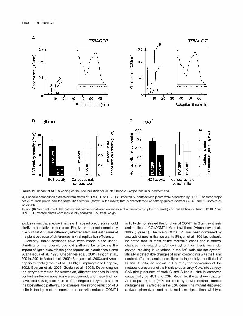
(Hoffmann et al., 2003). Figure 11A presents typical HPLC
profiles of soluble phenolic compounds extracted from stem
tissues of TRV-GFP and TRV-HCT–infected N. benthamiana
Figure 7. Phenotypes of N. benthamiana Plants Subjected to VIGS of
the HCT Gene.
Impacts on the whole plant size ([A] and [B]) and root growth (C) are
shown. At the left in each photograph is a control plant infected with the
TRV-GFP vector, and at the right is a plant infected with the TRV-HCT
vector.
1454 The Plant Cell

plants. In each chromatogram, three major peaks (marked by
arrows in Figure 11A) appeared that displayed identical UV
spectra (insets in Figure 11A), characteristic of caffeoylquinic
acids. From their relative retention times, the three compounds
were identified as 3-, 5-, and 4-caffeoylquinate isomers by
comparison with standards (data not shown) (Strack and Gross,
1990). The amounts of the three caffeoylquinate isomers clearly
increased in stems upon HCT silencing. When leaf content was
examined, important fluctuations in caffeoylquinate amounts
were detected (data not shown). Therefore, HCT activity and
caffeoylquinate content were estimated in stem and leaf extracts
of nine silenced and nonsilenced plants, and the calculated mean
values and deviations are presented in Figures 11B and 11C. In
control (TRV-GFP) plants, a 60-fold higher HCT activity was
measured in stems compared with leaves, similar to what was
observed in tobacco (Figure 3B). Gene silencing resulted in ;70
and 40% reduction of HCT activity in stems and leaves,
respectively. Concerning the impact on soluble phenolic com-
pounds, the results confirm that the reduction of HCT activity has
a differential impact on caffeoylquinate accumulation in the two
tissues. In stems, a threefold increase was measured (Figure
11B), whereas no significant change of caffeoylquinate concen-
tration was observed in silenced leaves (Figure 11C). Inspection
of other metabolites did not reveal any other important alteration
in phenolic profiles as a result of HCT inhibition.
DISCUSSION
The recent characterization of an acyltransferase (HCT)
(Hoffmann et al., 2003) capable of synthesizing p-coumaroyl
shikimate and p-coumaroyl quinate esters and of the cyto-
chrome P450 C3H that uses the p-coumaroyl esters as
substrates (Schoch et al., 2001; Franke et al., 2002a) has
profoundly changed our view of the phenylpropanoid biosyn-
thetic pathway (Humphreys and Chapple, 2002). Here, as a first
step toward the understanding of the role of HCT in lignin
biosynthesis in planta, we have immunolocalized the enzyme in
stem vascular tissues of N. tabacum and N. benthamiana.
Figure 8. HCT Accumulation in Stem and Root Tissues of TRV-Infected N. benthamiana Plants.
Protein extracts (3.5 mg) from stems (A) (internode 5) or roots (B) were immunodetected with specific antibodies raised against tobacco HCT (top
panels) or CCoAOMT (bottom panels) proteins. HCT and CCoAOMT contents of two representative controls (C1 and C2, TRV-GFP) and seven TRV-
HCT–infected plants (lanes I to VII) are presented. Arrows indicate the position of HCT (top panels) or CCoAOMT isoforms (bottom panels). At the right is
the position of markers of known molecular mass (in kD).
HCT Silencing Affects Lignin Synthesis 1455

Figure 9. Histochemical Analysis of the Effects of HCT Silencing on Lignin of N. benthamiana.
Sections of stems ([A], [B], [E], [F], [I], [J], [M], and [N]) and roots ([C], [D], [G], [H], [K], [L], [O], and [P]) were stained using Wiesner (left panels) or
Maule (right panels) methods. Sections of a representative control ([A] to [D]), sections from a plant with a strong phenotype ([E] to [H]), sections from
a plant with an intermediate phenotype ([I] to [L]), and sections of a plant with no visible phenotype ([M] to [P]) are shown. Arrows indicate zones with
attenuated lignin staining. All the pictures in one column are at the same scale (indicated at the top, bars ¼ 1 mm). epd, epidermis; end, endodermis;
other abbreviations are as in Figure 4.
1456 The Plant Cell

Using a specific antiserum raised against the purified
recombinant protein to probe various plant extracts, HCT was
shown to accumulate in lignified tissues of stems and roots
(Figures 3, 4, 8, and 10). Immunocytochemical localization of
HCT in vascular tissues was compared with that of CCoAOMT,
which has previously been associated with lignification in various
plants (Ye, 1997; Inoue et al., 1998; Maury et al., 1999).
Differences in the spatial and temporal enzyme distribution have
been demonstrated for CCoAOMT and caffeic/5-hydroxyferulic
acid O-methyltransferase (COMT) in alfalfa (Medicago sativa)
and tobacco tissues and may be at the origin of subtle variations
in lignin of distinct cell types (Inoue et al., 1998; Maury et al.,
1999). The comparative localization of CCoAOMT and HCT in
tobacco showed that both enzymes accumulate exclusively in
vascular tissues, and no signal was seen in the pith and cortex of
stems. HCT protein labeling was particularly strong in inner and
external phloem and was also detectable in cambium cells where
no signal was recorded with anti-CCoAOMT antibodies (Figure
4). A strong accumulation of HCT was also found in the cambium
of N. benthamiana (Figure 10). HCT was not clearly detected in
differentiating young xylem cells of tobacco, a major site of
CCoAOMT accumulation. In N. benthamiana, HCT labeling
clearly localized in ray cells of xylem tissues (Figure 10). Thus,
a distinct spatial repartition of HCT and CCoAOMT was found.
This may be related to the relative position of the two lignin
biosynthetic enzymes in the pathway (Figure 1) and their
participation at different stages of the cell lignification process.
However, it cannot be excluded that HCT may play a more
specific role, for instance in the mobilization of caffeoylquinate
pools that may be transformed in caffeoyl CoA as demonstrated
in vitro (Hoffmann et al., 2003).
Study of the impact of hairpin RNA-mediated silencing and
VIGS of HCT in Arabidopsis and N. benthamiana plants,
respectively, provided unequivocal proof of the involvement
of HCT in lignin biosynthesis. HCT-silenced plants exhibited
profound changes in plant development, lignin content, and
structure and susceptibility of the cell walls to enzymatic
degradation. Comparison of enzyme immunolocalization in
control and silenced plants by confocal microscopy demon-
strated a correlation between the disappearance of enzyme
labeling in vascular cells and the impact on lignin structure. In
silenced N. benthamiana plants, accumulation of the gene
product in the cambium zone and in xylem ray cells was clearly
diminished compared with controls. Cell walls of tissues that
underwent silencing displayed surprisingly higher autofluores-
cence, suggesting that the enrichment of lignin in H units
demonstrated after thioacetolysis may result in alterations of its
spectral properties. This phenomenon could be linked to the fact
that H lignin units are essentially terminal lignin units with free
phenolic groups (Lange et al., 1995). The decrease in HCT pro-
tein accumulation in xylem tissues also strictly correlated with a
decrease in lignin S unit content as revealed by histochemi-
cal staining and chemical analysis. The detailed analysis of
HCT in different lignifying tissues and during plant development
as a whole would be an interesting area for further research.
These changes in enzyme accumulation and lignin composi-
tion were unevenly distributed within the stem tissues of infected
plants, suggesting some heterogeneity in the effects of silencing.
Because VIGS was used to knock down HCT gene expression in
N. benthamiana, the lignin heterogeneity may reflect the rate of
replication and/or cell-to-cell spread of the TRV-HCT vector
because it is known that the efficiency of VIGS is dependent on
virus replication, at least at the stage of VIGS initiation (Ruiz et al.,
1998). In TRV-infected plants, the virus multiplication rate is
initially high, then declines and is maintained at a low level at later
stages of infection (Ratcliff et al., 2001). The most important issue
here is that HCT-targeted VIGS lead to a plant vasculature with
a lignin that is unusual in its enrichment in H units, its depletion in
S units, and its heterogeneous nature. Because VIGS is not
uniform over the plant and does not eliminate HCT (Figure 8),
late/partial silencing of HCT may result in effects specific to S
lignin units. Moreover, it must be kept in mind that, in TRV-HCT
infected plants, the new lignin built up after HCT silencing is
added to wild-type lignin. A major advantage of VIGS over other
Table 1. Effects of HCT Silencing on Lignin Content and Structure in 3-Month-Old N. benthamiana Stems
Noninfected TRV-GFP Infected TRV-HCT Infected
Klason lignina 10.9 6 0.3 (100 6 4) 11.2 6 0.3 (103 6 3) 9.32 6 0.2 (85 6 2)
Thioacidolysis Yield in H, G, and S Monomers (mmol/g KL)b
H 2.7 6 0.2 3.7 6 0.1 85 6 5
G 426 6 13 514 6 19 323 6 9
S 1033 6 61 1165 6 55 683 6 15
Total (H þ G þ S) 1462 6 74 1683 6 74 1091 6 29
Molar ratio (H/G/S) 0.2/29.1/70.7 0.2/30.6/69.2 8/29.6/62.6
Percentage of units only involved in b-O-4 bondsc 37 6 2 42 6 2 27 6 1
a Klason lignin is expressed as weight percentage of extract-free sample (mean value and standard error of four replicate analyses). Values in
parentheses are relative to uninfected control taken as 100%.b Mean value and standard error of duplicate analyses. The reported standard error includes both the Klason determination (relative standard error in
the 2 to 3% range) and the thioacidolysis experiment (relative standard error in the 0.1 to 3% range). KL, Klason lignon.c The percentage of lignin units only involved in b-O-4 bonds is calculated with the assumption that the average Mr of lignin units is 200 and that the
recovery yield of thioacidolysis monomers from parent b-O-4 structures is 80%.
HCT Silencing Affects Lignin Synthesis 1457

Figure 10. HCT Immunolocalization in Transverse Stem Sections from Control (TRV-GFP) and Silenced (TRV-HCT) N. benthamiana Plants.
(A) to (D) HCT immunolocalization in TRV-GFP control plants.
1458 The Plant Cell

gene knockout systems is its conditional nature that allows
repression of genes essential for plant growth (Lu et al., 2003).
This is perfectly illustrated here by comparing the impact of HCT
repression by VIGS and in the transgenic plants expressing the
HCT hairpin construct.
It will be of interest to examine in depth the impact of abnormal
lignin synthesis on cell differentiation. However, the currently
available histochemical methods only allow detection of gross
changes in lignin structure, whereas characterization of more
subtle alterations, possibly affecting different cell types, would
demand novel spectroscopic methods for in situ studies. It has
been established that, during cell wall differentiation, patterns of
lignification are well defined, leading to the overall architecture of
the secondary cell wall. First, p-coumaryl alcohol is predom-
inantly deposited into the middle lamella and cell corners (H unit,
Figure 1) followed by coniferyl alcohol, which is mainly laid down
in the secondary wall (G unit). Finally, sinapyl alcohol is deposited
at the late stages of lignification (S unit) (Lewis, 1999). In HCT-
silenced plants, the proportion of monolignols is modified, and
resulting alterations in the cell wall ultrastructure could be
studied, for instance, by immunogold labeling of lignin sub-
structure epitopes using transmission electron microscopy (Ruel
et al., 2001). Such approaches should improve our knowledge of
the precise spatio-temporal regulation of the secondary cell wall
assembly.
A rather surprising finding reported here is the differential
effect of HCT repression on G and S lignin units, which are both
downstream of HCT (Figure 1). Aside from the unlikely occur-
rence of an alternative pathway, the predominant effect on
S synthesis suggests that, when the metabolic flux is lowered
by HCT repression, the reduction of coniferaldehyde into coni-
feryl alcohol and polymerization in guaiacyl lignin is favored
versus hydroxylation and methylation leading to syringyl lignin
(Figure 1). Such a channeling mechanism could be demonstrated
by feeding labeled precursors and following label fate in the
intermediate pools.
Regulation of caffeoylquinate pools upon changes in activity of
an upstream enzyme like Phe ammonia-lyase (Maher et al., 1994;
Howles et al., 1996) or a downstream enzyme like cinnamoyl-
CoA reductase (Chabannes et al., 2001) has been reported
previously. Here, the analysis of the impact of HCT silencing on
soluble phenylpropanoids of stems and leaves by HPLC
revealed unexpected differential effects in the two organs; that
is, an increase of caffeoylquinate pools in stems and no
significant change of the same compounds in leaves. However,
these phenomena may have different origins. First of all, and
though HCT has been shown to synthesize caffeoylquinate in
vitro (Hoffmann et al., 2003), it is possible that another
acyltransferase, which has been characterized recently in
tobacco and tomato (Lycopersicon esculentum) (C. Martin,
personal communication), is involved in caffeoylquinate bio-
synthesis in leaves. Moreover, it is known that HCT is involved
both upstream and downstream of the 3-hydroxylation step
(Figure 1). Thus, HCT inhibition in stems could affect pre-
dominantly caffeoylquinate catabolism into caffeoyl CoA, lead-
ing to caffeoylquinate accumulation. Another possibility is that
distinct pools of caffeoylquinate occur: one metabolically active
that predominates in stems and is highly responsive to changes
in HCT activity and another pool that is quantitatively important in
leaves and is slowly mobilized. In addition, although the transport
of phenylpropanoids through the plant has not been demon-
strated, it could also contribute to the observed differences
between organs. It is evident that these possibilities are not
Figure 10. (continued).
(E) to (H) HCT immunolocalization in HCT-silenced plants.
(I) to (L) Negative control. Labeling was performed on TRV-GFP plants in the absence of primary antibodies.
(A), (E), and (I) Typical distribution of the HCT immunolabeling. HCT labeling is intense and mainly detected within the cambium (Ca) of the TRV-GFP
sample. Strongly reduced labeling is observed in the cambium of the HCT-silenced tissues. Almost no signal is detected in the negative control.
(B), (F), and (J) Corresponding autofluorescence of the lignin as observed upon 488/505 to 545 nm excitation/emission wavelengths. Note that
autofluorescence of the lignin is higher and less uniformly distributed in the HCT-silenced tissues (F) compared with the TRV-GFP infected tissues ([B]
and [J]).
(C), (G), and (K) Merged images of (A) þ (B), (E) þ (F), and (I) þ (J), respectively.
(D), (H), and (L) Higher magnification views of the boxed regions shown in (C), (G), and (K), respectively. Note the HCT labeling within the xylem ray cells
of TRV-GFP plants (arrowheads).
(M) and (N) Overall distribution of HCT protein (red) and lignin (green) in transverse stem sections from TRV-GFP and TRV-HCT–infected N.
benthamiana.
(O) and (P) Maule staining corresponding to the samples shown in (M) and (N), respectively. Note that the unstained area of the xylem shows a higher
level of autofluorescence (arrows).
All images were acquired by CLSM and processed under exactly the same conditions to allow comparison between the different panels. Bars ¼200 mm. Abbreviations are as in Figures 4 and 9.
Table 2. Impact of HCT Silencing on Cell Wall Degradability by
Cellulase
Noninfected
TRV-GFP
Infected
TRV-HCT
Infected
Weight loss (%) 33.9 6 0.7 36.7 6 2.3 57.4 6 1.4
The extent of degradation by cellulase was expressed as the percent-
age of loss in sample weight at the end of incubation with the enzyme
(see Methods). Data are mean values and standard errors from duplicate
experiments.
HCT Silencing Affects Lignin Synthesis 1459
exclusive and tracer experiments with labeled precursors should
clarify their relative importance. Finally, one cannot completely
rule out that VIGS has differently affected stem and leaf tissues of
the plant because of differences in viral replication efficiency.
Recently, major advances have been made in the under-
standing of the phenylpropanoid pathway by analyzing the
impact of lignin biosynthetic gene repression in antisense plants
(Atanassova et al., 1995; Chabannes et al., 2001; Pincon et al.,
2001a, 2001b; Abbott et al., 2002; Boerjan et al., 2003) and Arabi-
dopsis mutants (Franke et al., 2002b; Humphreys and Chapple,
2002; Boerjan et al., 2003; Goujon et al., 2003). Depending on
the enzyme targeted for repression, different changes in lignin
content and/or composition were observed, and these findings
have shed new light on the role of the targeted enzymatic step in
the biosynthetic pathway. For example, the strong reduction of S
units in the lignin of transgenic tobacco with reduced COMT I
activity demonstrated the function of COMT I in S unit synthesis
and implicated CCoAOMT in G unit synthesis (Atanassova et al.,
1995) (Figure 1). The role of CCoAOMT has been confirmed by
analysis of new antisense plants (Pincon et al., 2001a). It should
be noted that, in most of the aforesaid cases and in others,
changes in guaiacyl and/or syringyl unit synthesis were ob-
served, resulting in variations in the S/G ratio but not system-
atically in detectable changes of lignin content, nor was the H unit
content affected, angiosperm lignin being mainly constituted of
G and S units. As shown in Figure 1, the conversion of the
metabolic precursor of the H unit,p-coumaroyl CoA, into caffeoyl
CoA (the precursor of both G and S lignin units) is catalyzed
sequentially by HCT and C3H. Recently, it was shown that an
Arabidopsis mutant (ref8) obtained by ethyl methanesulfonate
mutagenesis is affected in the C3H gene. The mutant displayed
a dwarf phenotype and contained less lignin than wild-type
Figure 11. Impact of HCT Silencing on the Accumulation of Soluble Phenolic Compounds in N. benthamiana.
(A) Phenolic compounds extracted from stems of TRV-GFP or TRV-HCT–infected N. benthamiana plants were separated by HPLC. The three major
peaks of each profile had the same UV spectrum (shown in the insets) that is characteristic of caffeoylquinate isomers (3-, 4-, and 5- isomers as
indicated).
(B) and (C) Mean values of HCT activity and caffeolquinate content measured in the same samples of stem (B) and leaf (C) tissues. Nine TRV-GFP and
TRV-HCT–infected plants were individually analyzed. FW, fresh weight.
1460 The Plant Cell

plants. Its lignin was unusual in being composed primarily of H
units (Franke et al., 2002b). T-DNA insertion mutant lines tagged
in the C3H gene have also been isolated (D. Werck-Reichhart,
personal communication) and display even more drastic growth
defects. Our HCT-silenced Arabidopsis plants also have a very
severe growth phenotype reminiscent of that of the C3H-tagged
mutants and a lignin depleted in S unit as demonstrated by the
negative response to Maule reagent (Figure 5). Lignin of TRV-
HCT–infected N. benthamiana was analyzed by thioacidolysis
and showed the same changes compared with wild-type lignin
as those reported for the ref8 mutant, although to a lesser extent,
probably because (1) HCT silencing by VIGS was induced at an
advanced stage of development (i.e., after wild-type lignin was
formed) and (2) as discussed above, all infected cells were
probably not affected to the same extent. The comparable
phenotypes observed for plants affected in the C3H or HCT
genes are consistent with the fact that C3H and HCT genes have
similar expression profiles (Raes et al., 2003), and the two
enzymatic steps are consecutive in the synthesis of caffeoyl CoA
from p-coumaroyl CoA. The mutant phenotypes may be related
to the growth-promoting activity that has been shown for some
phenylpropanoids, such as dehydrodiconiferyl alcohol glucoside
(Binns et al., 1987). Furthermore, the modifications of lignin
structure and content decreased cell wall resistance to hydrolytic
enzymes of the Arabidopsis mutant ref8 as observed for the
HCT-silencedN. benthamiana plants described here. These data
point to the importance of lignification for cell wall resistance to
the action of hydrolytic enzymes, such as those produced by
pathogenic organisms. The study of the resistance of silenced
plants to bacterial and fungal pathogens will enable us to test this
hypothesis.
METHODS
Chemicals, Enzymes, and General Methods
Commonly used chemicals and reagents were of the highest purity
readily available. Bradford protein dye reagent was purchased from Bio-
Rad (Hercules, CA). Restriction enzymes and buffers were purchased
from New England Biolabs (Beverly, MA) or Invitrogen (Cergy Pontoise,
France). T4 DNA ligase, T4 polynucleotide kinase, and ATP were
purchased from Invitrogen. Purified oligonucleotides used for cloning
and DNA sequencing were provided by Sigma-Aldrich (Saint-Quentin-
Fallavier, France). DNA amplification using Taq polymerase (Invitrogen)
was performed in the iCycler thermocycler (Bio-Rad). Plasmid and PCR
products were extracted and purified from agarose gels using kits
purchased from Qiagen (Hilden, Germany).
Plant Material and Culture Conditions
Nicotiana tabacum and N. benthamiana plants were grown in a growth
chamber under 3000 lux lighting and a light/dark cycle of 16 h/8 h. The
temperature was maintained at 21 6 28C. Tissues were harvested from
flowering tobacco plants and frozen in liquid nitrogen. One-month-old N.
benthamiana seedlings were infected with TRV constructs using Agro-
bacterium tumefaciens–mediated transient gene expression (Ratcliff
et al., 2001).
Arabidopsis thalianaplants ecotype Columbia 0 were grown with a light/
dark cycle of 12 h/12 h and a temperature of 208C during the day and 168C
at night.
DNA Sequencing
DNA sequencing was performed by the method of Sanger et al. (1977)
using the rhodamine dye-terminator cycle ready kit with AmpliTaq DNA
polymerase FS (Perkin-Elmer, Foster City, CA) and an Applied Bio-
systems DNA sequencer (Model 373A; Foster City, CA).
Production and Use of Polyclonal Antibodies
Heterologous expression in Escherichia coli cells and purification of the
HCT protein were performed as described previously (Hoffmann et al.,
2003). Polyclonal antibodies were raised in rabbit by four successive
injections of 100 mg of the purified enzyme. Before injection, the protein
solution was emulsified with Freund’s complete adjuvant for the first
injection and with incomplete adjuvant for the others. The anti-CCoAOMT
antibodies were produced as described (Maury et al., 1999). Anti-HCT
antibodies were used at 1/10,000 or 1/500 dilution and anti-CCoAOMT
serum at 1/2000 or 1/200 dilution in protein gel blot and immunohisto-
chemical analyses, respectively.
For specificity tests, anti-HCT antibodies were preincubated for 2 h at
208C in PBS or in PBS containing 100 or 500 mM of purified HCT
recombinant protein in protein gel blot or immunohistochemical analyses,
respectively.
For immunoprecipitation experiments, 8 mg of protein A–Sepharose
CL-4B beads (Sigma, St. Louis, MO), prewashed twice with 40 mL of PBS
buffer for 4 h at 48C, was centrifuged at 1000 rpm for 5 min, resuspended
in 40 mL of 1 M sodium phosphate buffer, pH 7.5, and incubated for 3 h at
48C in the presence of 5 mL of anti-HCT or preimmune serum. The beads
were incubated overnight at 48C with 200mL of crude protein extracts and
then centrifuged at 2000 rpm for 5 min. The supernatant was assayed for
HCT activity. Control incubations were performed in the absence of
protein A–Sepharose and antibody. Four independent measurements
were performed in each case.
Protein Gel Blot Analysis
The basic procedures for electrophoresis of proteins under denaturing
conditions and immunoblotting were as described previously (Geoffroy
et al., 1990), except that phosphatase activity was detected with
a chemiluminescent substrate (BioRad CDP-Star).
DNA Gel Blotting
Genomic DNA analysis was performed as previously described by Martz
et al. (1998). DNA samples (40 mg) were digested to completion with one
or two restriction enzymes, ethanol-precipitated, and resolved on 0.8%
agarose gel. After depurination, the gel was blotted on Hybond Nþmembrane (Amersham Bioscience, Orsay, France). The [a-32P]dCTP-
labeled probe was synthesized from the 957-bp N. benthamiana HCT
fragment by random priming. DNA gel blot hybridization and washing
steps were performed at 658C. Hybridization signals were detected by
phosphorimaging.
RNA Gel Blot Analysis
Total RNA extraction was performed with Tri-Reagent (Sigma), and
analysis of high and low molecular weight RNA was as described (Himber
et al., 2003). dCTP-labeled probe was synthesized from the 367-bp
Arabidopsis HCT fragment (see below) by random priming. All hybridiza-
tion signals were detected by phosphorimaging.
HCT Activity Assays
Plant tissues were ground with a pestle and mortar in liquid nitrogen, and
the powder was extracted with 0.1 M sodium phosphate buffer, pH 7.5,
HCT Silencing Affects Lignin Synthesis 1461

containing 10 mM DTT, quartz, and polyclar AT (Serva, Buchs, Switzer-
land). After centrifugation at 48C for 20 min at 13,000 rpm, the supernatant
was washed twice with the extraction buffer on Centricon 10 concen-
trators (Amicon, Ranvers, MA) to eliminate soluble phenolic compounds
present in the crude extract and then used for HCT activity measurement.
The reaction mixture contained 100 mM phosphate buffer, pH 6.6, 1 mM
DTT, 0.4 to 12 mg of protein, 100 mM p-coumaroyl CoA, and 4 mM
shikimate in a total volume of 20mL. The reaction was initiated by addition
of the enzyme extract, incubated at 308C for 2 to 30 min, and terminated
by addition of 20 mL of HPLC solvent. Reaction products were analyzed
and quantified by HPLC. Protein content was measured by the Bradford
method (Bradford, 1976).
Immunohistochemical Analyses
Stems from 8-week-old N. tabacum were collected, fixed with a solution
containing 3.7% formaldehyde, 50% ethanol, and 5% acetic acid in
water, dehydrated in an ethanol series, and embedded in paraplast
before 10-mm-thick sections were made. Stems of 8-week-old N.
benthamiana infected for 4 weeks were hand-sectioned and immediately
fixed for 1 h by immersion in ethanol. Before immunolabeling, sections
were rehydrated in PBS solution, blocked for 1 h in PBS containing 5%
(w/v) BSA, 5% (v/v) normal goat serum, and 0.1% cold water fish skin
gelatine (Aurion, Wageningen, The Netherlands), and further incubated
overnight at 48C in PBS containing 0.1% (v/v) acetylated BSA (Aurion).
Sections were then washed four times in PBS and incubated again in the
dark for 4 h at room temperature with Alexa Fluor 568 goat anti-rabbit
immunoglobulin G (Alexa 568; Molecular Probes, Leiden, The Nether-
lands) diluted 1:300 in PBS plus 0.1% acylated BSA. Samples were
washed again four times before light microscopy observations. Control
experiments in which primary antibodies were omitted were routinely
performed to verify the specificity of the labeling. Controls using anti-
bodies preincubated with the recombinant HCT protein were performed
on ethanol-fixed sections (see supplemental data online).
Epifluorescence Microscopy
Epifluorescence microscopy observations were done with a Nikon
Eclipse E800 microscope (Champigny sur Marne, France) with appropri-
ate filters and an 310 0.3–numerical aperture objective. For Alexa 568,
the parameters were as follows: excitation wavelength, 515 to 565 nm;
dichroic mirror, 565 nm; and emission bandpass filter, 582.5 to 627.5 nm.
For lignin autofluorescence, the parameters were as follows: excitation
wavelength, 330 to 380 nm; dichroic mirror, 400 nm; and emission long-
pass filter, 420 nm. Image acquisition was performed as previously
described (Gaire et al., 1999).
CLSM
CLSM was performed with a Zeiss LSM510 microscope (Jena, Germany)
equipped with a Zeiss Axiovert 100M inverted microscope and a310 0.3–
numerical aperture objective. Laser scanning was performed using the
multitrack mode to avoid bleed-through. Excitation/emission wave-
lengths were 488/505 to 545 nm for lignin and 543/560 to 615 nm for
Alexa 568. Image processing used LSM510 version 2.8 (Zeiss) and
PhotoShop 5.5 (Adobe Systems, San Jose, CA) for final assembly.
RNA Silencing in Arabidopsis
A 367-bp HCT fragment was PCR amplified from the pGEX-KG plasmid
containing the Arabidopsis HCT gene (At5g48930) (Hoffmann et al.,
2003). The sense and antisense primer sequences were 59-GAGTCTA-
GACTCGAGGTAACGCCGAGGGAGTGGAA-39 and 59-CATGGATCCG-
GCGCGCCAGAAATGGAACGGTTTCTAACG-39, respectively. The re-
sulting PCR product was inserted into pFGC5941 (www.ag.arizona.
edu/chromatin/fgc5941.html), a binary vector containing a chalcone
synthase intron and designed to produce double-stranded RNA in plants.
The HCT gene fragment was cloned upstream and downstream of the
chalcone synthase intron in sense and antisense orientation using XhoI-
AscI and XbaI-BamHI restriction sites, respectively. Transformation of
Arabidopsis (Columbia 0) was performed with Agrobacterium GV3101
strain as described previously (Bechtold et al., 1993). Transgenic plants
were selected by spraying a 300-mg/L solution of Basta (glufosinate)
herbicide.
Cloning of a Partial HCT cDNA from N. benthamiana
Reverse Transcription
Reverse transcription with total RNA from 1-month-old N. benthamiana
stems was performed using (poly)dT primer and Superscript (Life
Technologies) according to the manufacturer’s instructions.
Generation of Partial cDNAs
Partial cDNAs were produced by PCR using as template the cDNA
generated by reverse transcription. The sense degenerate oligonucleo-
tide primer (59-TTTTAYCCNATGGCKGGKMG-39) was based on the
consensus sequence FYPMAG conserved between N. tabacum HCT and
other acyltransferases (Hoffmann et al., 2003). The antisense primer was
based upon the conserved region DFGWG near the C terminus of
acyltransferase proteins and had the following sequence: 59-CCCCAIC-
CRAARTC-39. I indicates inosine and R, H, Y, K, M, and N indicate the
following degenerate sites: R, A/G; H, A/C/T; Y, C/T; K, G/T; M, A/C; and
N, A/G/C/T. DNA amplification was performed under the following con-
ditions: 3 min at 948C and then 35 cycles at 948C for 1 min, 428C for 1 min,
and 728C for 1 min. At the end of the 35 cycles, the reaction mixture
was incubated for an additional 10 min at 728C. The amplified DNA was
resolved by agarose gel electrophoresis, and the band of the correct size
(957 bp) was isolated and subcloned into pCRII-TOPO (Invitrogen) before
sequencing.
VIGS Technology
VIGS technology was used to induce HCT gene silencing in N.
benthamiana with the tobacco rattle virus vector system (Ratcliff et al.,
2001). The 957-bp cDNA fragment of the N. benthamiana HCT gene was
cloned into theApaI andBamHI sites of pTV00 vector to produce the pTV-
HCT construct used for N. benthamiana infection.
Lignin Analysis
Histochemical analysis using Wiesner and Maule staining was performed
as previously described (Atanassova et al., 1995). In the case of
Arabidopsis plants, 2-month-old silenced or wild-type stems were
embedded in 1% low melting point agarose before to be sectioned
transversally with a razor blade. The observations were performed with
a Nikon TE2000 inverted microscope.
Lignin chemical analysis was performed on the stems of 3-month-old
N. benthamiana plants (;20 plants per sample) that had been infiltrated
8 weeks before collection. The air-dried stems were evenly ground to pass
through a 0.5-mm sieve before an exhaustive solvent extraction, first with
toluene:ethanol (2:1, v/v), then with ethanol, and finally with water. As
recommended in the standard method of Klason lignin determination, this
comprehensive extraction step was performed to minimize the possibility
that proteinaceous components are included within the so-called Klason
lignin fraction (Dence, 1992). The Klason lignin content was calculated in
weight percent of the dry extract-free stem material and from four
1462 The Plant Cell

replicates performed on 300 mg of sample. Thioacidolysis reagent was
prepared by introducing 2.5 mL of BF3 etherate (Sigma-Aldrich) and
10 mL of ethanethiol EtSH (Sigma-Aldrich) into a 100-mL flask and adjust-
ing the final volume to 100 mL with dioxane. The colorless reagent was
used immediately after preparation. The extract-free stem samples (20
mg) were added to 10 mL of reagent and 1 mL of solution of GC internal
standard (docosane, 0.24 mg/mL in CH2Cl2) in a glass tube closed with
a Teflon-lined screwcap. Thioacidolysis was performed at 1008C in an oil
bath for 4 h. The cooled reaction mixture was diluted with 30 mL of water,
and the pH was adjusted to 3 to 4 with 0.4 M NaHCO3. The reaction
mixture was extracted with CH2Cl2 (3 3 30 mL). Combined organic
extracts were dried over Na2SO4 and then evaporated under reduced
pressure at 408C. The final residue was redissolved in;0.5 mL of CH2Cl2before silylation and gas chromatography–mass spectrometry analyses
according to Lapierre (1986). Quantitation of the main p-hydroxyphenyl
(H), guaiacyl (G), and syringyl (S) lignin-derived monomers, analyzed as
their trimethylsilylated derivatives, was performed from specific ion
chromatograms reconstructed at m/z 239 for the H monomers, 269 for G
monomers, and at m/z 299 for S monomers after an appropriate
calibration relative to the docosane internal standard.
Cell Wall Degradability
The susceptibility to cellulolysis was evaluated using a method adapted
from Rexen (1977): 200 mg of the extract-free ground sample were placed
in 30 mL of 0.05 M sodium acetate buffer, pH 4.5, containing 2 mg/mL of
commercial cellulase (cellulase Onozuka-R10; Serva, Heidelberg, Ger-
many). The cellulolysis was performed for 48 h at 378C with magnetic
stirring. After incubation, the reaction medium was filtrated over a tared
filtering crucible and the residue was washed with water, oven-dried, and
then gravimetrically determined.
Extraction of Soluble Phenolic Compounds from Plant Material
Samples (100 mg) from TRV-GFP and TRV-HCT plants were frozen in
liquid nitrogen and quickly ground in 500 mL of methanol. After
centrifugation at 500 rpm for 5 min, the supernatant was collected and
the residual pellet was reextracted with 200 mL of 70% methanol. The
supernatants were combined, clarified at 2000 rpm for 30 min, and taken
to dryness under nitrogen. The dried material was dissolved in 70%
methanol (300 mL) and analyzed by HPLC.
HPLC Analysis of HCT Reaction Products and Plant
Phenolic Compounds
Phenolic compounds were resolved on a RP C18 column (Novapak,
4 mm, 4.6 3 250 mm; Waters, Milford, MA) using an increasing gradient
of acetonitrile in water containing 0.1% formic acid. Gradient conditions
at 1 mL/min flow rate were as follows: 100% solvent A to 50% solvent B
for 50 min; 50% solvent B to 100% solvent B for 5 min; 100% solvent B to
100% solvent A for 5 min and then 10 min reequilibration in 100%
solvent A. Solvent A contained acetonitrile/water/formic acid (10:89.9:0.1)
and solvent B acetonitrile/water/formic acid (80:19.9:0.1). Compounds
were characterized by their elution time, and their UV absorption spec-
trum recorded with a photodiode array detector (Waters). 3-O- and
4-O-caffeoylquinic acids were produced from 5-O-caffeoylquinic acid
(Sigma-Aldrich) by heating for 30 min at 908C in 0.2 M phosphate buffer,
pH 7.0 (Strack and Gross, 1990), and the three isomers were separated by
HPLC.
Synthesis and Purification of the p-Coumaroyl-CoA Substrate
p-Coumaroyl-CoA ester was prepared according to the method of
Stockigt and Zenk with some modifications (Stockigt and Zenk, 1975;
Negrel and Smith, 1984) and identified and quantified by spectroscopy.
Sequence data from this article have been deposited with the EMBL/
GenBank data libraries under accession numbers AJ507825 for N.
tabacum HCT cDNA and AJ555865 for N. benthamiana partial HCT
cDNA.
ACKNOWLEDGMENTS
We wish to thank Robert Erb, Jerome Mutterer, and Matthieu Erhardt
for assistance with microscopy, Frederic Legee (Institut National de
la Recherche Agronomique) for the Klason lignin determinations,
Phil Mullineau (John Innes Centre, Norwich) for pSoup plasmid,
David Baulcombe (John Innes Centre) for VIGS vectors, and Danielle
Werck-Reichhart (Institut de Biologie Moleculaire des Plantes) and
Cathie Martin (John Innes Centre) for sharing unpublished data. The
assistance of Laurette De Franco is gratefully acknowledged. We are
indebted to Patrice Dunoyer, Christophe Himber, and Charles Lecellier
for technical support and valuable suggestions about RNA silencing
experiments. We are grateful to Kenneth Richards and Olivier Voinnet
for critical reading. The Zeiss LSM510 confocal microscope was
cofinanced by the Centre National de la Recherche Scientifique, the
Universite Louis Pasteur, the Region Alsace, the Association de la
Recherche sur le Cancer, and the Ligue Nationale contre le Cancer.
Received December 19, 2003; accepted March 10, 2004.
REFERENCES
Abbott, J.C., Barakate, A., Pincon, G., Legrand, M., Lapierre, C.,
Mila, I., Schuch, W., and Halpin, C. (2002). Simultaneous suppres-
sion of multiple genes by single transgenes. Down-regulation of three
unrelated lignin biosynthetic genes in tobacco. Plant Physiol. 128,
844–853.
Atanassova, R., Favet, N., Martz, F., Chabbert, B., Thollier, M.T.,
Monties, B., Fritig, B., and Legrand, M. (1995). Altered lignin
composition in transgenic tobacco expressing O-methyltransferase
sequences in sense and antisense orientation. Plant J. 8, 465–477.
Bandoniene, D., and Murkovic, M. (2002). On-line HPLC-DPPH
screening method for evaluation of radical scavenging phenols
extracted from apples (Malus domestica L.). J. Agric. Food Chem.
50, 2482–2487.
Bechtold, N., Ellis, J., and Pelletier, G. (1993). In planta Agro-
bacterium-mediated gene transfer by infiltration of adult Arabidopsis
thaliana plants. C. R. Acad. Sci. III Sci. Vie 316, 1194–1199.
Binns, A.N., Chen, R.H., Wood, H.N., and Lynn, D.G. (1987). Cell
division promoting activity of naturally occurring dehydrodiconiferyl
glucosides: Do cell wall components control cell division? Proc. Natl.
Acad. Sci. USA 84, 980–984.
Boerjan, W., Ralph, J., and Baucher, M. (2003). Lignin biosynthesis.
Annu. Rev. Plant Biol. 54, 519–546.
Boveris, A., Valdes, L., and Alvares, S. (2002). Inhibition by wine
polyphenols of peroxynitrite-initiated chemiluminescence and NADH
oxidation. Ann. N. Y. Acad. Sci. 957, 90–102.
Bradford, M.M. (1976). A rapid and sensitive method for the
quantitation of microgram quantities of protein utilizing the principle
of protein-dye binding. Anal. Biochem. 72, 248–254.
Burns, J., Gardner, P., O’Neil, J., Crawford, S., Morecroft, I.,
McPhail, D., Lister, C., Matthews, D., MacLean, M., Lean, M.,
Duthie, G., and Crozier, A. (2000). Relationship among antioxidant
activity, vasodilation capacity and phenolic content of red wines.
J. Agric. Food Chem. 48, 220–230.
HCT Silencing Affects Lignin Synthesis 1463

Chabannes, M., Barakate, A., Lapierre, C., Marita, J.M., Ralph, J.,
Pean, M., Danoun, S., Halpin, C., Grima-Pettenati, J., and Boudet,
A.M. (2001). Strong decrease in lignin content without significant
alteration of plant development is induced by simultaneous down-
regulation of cinnamoyl CoA reductase (CCR) and cinnamyl alcohol
dehydrogenase (CAD) in tobacco plants. Plant J. 28, 257–270.
Croteau, R., Kutchan, T.M., and Lewis, N.G. (2000). Natural products
(secondary metabolites). In Biochemistry and Molecular Biology of
Plants, B.B. Buchanan, W. Gruissem, and R.L. Jones, eds (Rockville,
MD: American Society of Plant Physiologists), pp. 1250–1318.
Dence, C. (1992). The determination of lignin. (Berlin: Springer-Verlag).
Dixon, R.A., and Ferreira, D. (2002). Genistein. Phytochemistry 60,
205–211.
Franke, R., Hemm, M.R., Denault, J.W., Ruegger, M.O., Humphreys,
J.M., and Chapple, C. (2002b). Changes in secondary metabolism
and deposition of an unusual lignin in the ref8 mutant of Arabidopsis.
Plant J. 30, 47–59.
Franke, R., Humphreys, J.M., Hemm, M.R., Denault, J.W., Ruegger,
M.O., Cusumano, J.C., and Chapple, C. (2002a). The Arabidopsis
REF8 gene encodes the 3-hydroxylase of phenylpropanoid metabo-
lism. Plant J. 30, 33–45.
Gaire, F., Schmitt, A.C., Stussi-Garaud, C., Pinck, L., and
Ritzenthaler, C. (1999). Protein 2A of grapevine fanleaf nepovirus
is implicated in RNA2 replication and colocalizes to the replication
site. Virology 264, 25–36.
Geoffroy, P., Legrand, M., and Fritig, B. (1990). Isolation and
characterization of a proteinaceous inhibitor of microbial proteinases
induced during the hypersensitive reaction of tobacco to tobacco
mosaic virus. Mol. Plant-Microbe Interact. 3, 327–333.
Goujon, T., Sibout, R., Pollet, B., Maba, B., Nussaume, L., Bechtold,
N., Lu, F., Ralph, J., Mila, I., Barriere, Y., Lapierre, C., and Jouanin,
L. (2003). A new Arabidopsis thaliana mutant deficient in the
expression of O-methyltransferase impacts lignins and sinapoyl
esters. Plant Mol. Biol. 51, 973–989.
Heller, W., and Kuhnl, T. (1985). Elicitor induction of a microsomal
5-O-(4-coumaroyl)shikimate 39-hydroxylase in parsley cell suspen-
sion cultures. Arch. Biochem. Biophys. 241, 453–460.
Himber, C., Dunoyer, P., Moissiard, G., Ritzenthaler, C., and
Voinnet, O. (2003). Transitivity-dependent and -independent cell-to-
cell movement of RNA silencing. EMBO J. 22, 4523–4533.
Hoffmann, L., Maury, S., Martz, F., Geoffroy, P., and Legrand, M.
(2003). Purification, cloning and properties of an acyltransferase
controlling shikimate and quinate ester intermediates in phenyl-
propanoid metabolism. J. Biol. Chem. 278, 95–103.
Howles, P.A., Sewalt, V., Paiva, N.L., Elkind, Y., Bate, N.J., Lamb, C.,
and Dixon, R.A. (1996). Overexpression of L-phenylalanine ammonia-
lyase in transgenic tobacco plants reveals control points for flux into
phenylpropanoid biosynthesis. Plant Physiol. 112, 1617–1624.
Humphreys, J.M., and Chapple, C. (2002). Rewriting the lignin
roadmap. Curr. Opin. Plant Biol. 5, 224–229.
Inoue, K., Sewalt, V.J.H., Ballance, G.M., Ni, W., Sturtzer, C., and
Dixon, R.A. (1998). Developmental expression and substrate specif-
icities of alfalfa caffeic acid 3-O-methyltransferase and caffeoyl-
coenzyme A 3-O-methyltransferase in relation to lignification. Plant
Physiol. 117, 761–770.
Jang, M., Cai, L., Udeani, G., Slowing, K., Thomas, C., Beecher, C.,
Fong, H., Farnsworth, N., Kinghorn, A., Mehta, R., Moon, R., and
Pezzuto, J. (1997). Cancer chemopreventive activity of resveratrol,
a natural product derived from grapes. Science 275, 218–220.
Kahkonen, M., Hopia, A., Vuorela, H., Rauha, J., Pihlaja, K., Kujala,
T., and Heinonen, M. (1999). Antioxidant activity of plant extracts
containing phenolic compounds. J. Agric. Food Chem. 47, 3954–
3962.
Kuhnl, T., Koch, U., Heller, W., and Wellmann, E. (1987). Chlorogenic
acid biosynthesis: Characterization of a light-induced microsomal
5-O-(4-coumaroyl)-D-quinate/shikimate 39-hydroxylase from carrot
(Daucus carota L.) cell suspension cultures. Arch. Biochem. Biophys.
258, 226–232.
Lange, B.M., Lapierre, C., and Sandermann, H., Jr. (1995). Elicitor-
induced spruce stress lignin. Structural similarity to early develop-
mental lignins. Plant Physiol. 108, 1277–1287.
Lapierre, C. (1986). Thioacidolysis of poplar lignins: Identification of
monomeric syringyl products and characterization of guaiacyl-
syringyl rich fractions. Holzforschung 40, 113–118.
Lekse, J.M., Xia, L., Stark, J., Morrow, J.D., and May, J.M. (2001).
Plant catechols prevent lipid peroxidation in human plasma and
erythrocytes. Mol. Cell. Biochem. 226, 89–95.
Lewis, N.G. (1999). A 20th century roller coaster ride: A short account of
lignification. Curr. Opin. Plant Biol. 2, 153–162.
Lu, R., Martin-Hernandez, A.M., Peart, J.R., Malcuit, I., and
Baulcombe, D.C. (2003). Virus-induced gene silencing in plants.
Methods 30, 296–303.
Maher, E.A., Bate, N.J., Ni, W., Elkind, Y., Dixon, R.A., and Lamb,
C.J. (1994). Increased disease susceptibility of transgenic tobacco
plants with suppressed levels of preformed phenylpropanoid prod-
ucts. Proc. Natl. Acad. Sci. USA 91, 7802–7806.
Martz, F., Maury, S., Pincon, G., and Legrand, M. (1998). cDNA
cloning, substrate specificity and expression study of tobacco
caffeoyl-CoA 3-O-methyltransferase, a lignin biosynthetic enzyme.
Plant Mol. Biol. 36, 427–437.
Maury, S., Geoffroy, P., and Legrand, M. (1999). Tobacco
O-methyltransferases involved in phenylpropanoid metabolism:
The different CCoAOMT and COMT classes have distinct substrate
specificities and expression patterns. Plant Physiol. 121, 215–224.
Negrel, J., and Smith, T.A. (1984). The phosphohydrolysis of hydroxy-
cinnamoyl-CoA thioesters in plant extracts. Phytochemistry 23, 31–34.
Olthof, M.R., Hollman, P.C., and Katan, M.B. (2001). Chlorogenic acid
and caffeic acid are absorbed in humans. J. Nutr. 131, 66–71.
Pincon, G., Chabannes, M., Lapierre, C., Pollet, B., Ruel, K.,
Joseleau, J.-P., Boudet, A.M., and Legrand, M. (2001b). Simulta-
neous down-regulation of caffeic/5-hydroxyferulic acid-O-methyl-
transferase I and cinnamoyl-coA reductase in the progeny from
a cross between tobacco lines homozygous for each transgene.
Consequences for plant development and lignin synthesis. Plant
Physiol. 126, 145–155.
Pincon, G., Maury, S., Hoffmann, L., Geoffroy, P., Lapierre, C.,
Pollet, B., and Legrand, M. (2001a). Repression of O-methyltransfer-
ase genes in transgenic tobacco affects lignin synthesis and plant
growth. Phytochemistry 57, 1167–1176.
Raes, J., Rohde, A., Christensen, J.H., Van De Peer, Y., and Boerjan,
W. (2003). Genome-wide characterization of the lignification toolbox
in Arabidopsis. Plant Physiol. 133, 1051–1071.
Ratcliff, F., Martin-Hernandez, A.M., and Baulcombe, D.C. (2001).
Tobacco rattle virus as a vector for analysis of gene function by
silencing. Plant J. 25, 237–245.
Rexen, B. (1977). Enzyme solubility: A method for evaluating the di-
gestibility of alkali-treated straw. Anim. Feed Sci. Technol. 2, 205–218.
Ruel, K., Chabannes, M., Boudet, A., Legrand, M., and Joseleau, J.
(2001). Reassessment of qualitative changes in lignification of trans-
genic tobacco plants and their impact on cell wall assembly.
Phytochemistry 57, 875–882.
Ruiz, M.T., Voinnet, O., and Baulcombe, D.C. (1998). Initiation and
maintenance of virus-induced gene silencing. Plant Cell 10, 937–946.
Sanger, F., Nicklen, S., and Coulson, A.R. (1977). DNA sequencing
with chain-terminating inhibitors. Proc. Natl. Acad. Sci. USA 74, 5463–
5467.
1464 The Plant Cell
Schoch, G., Goepfert, S., Morant, M., Hehn, A., Meyer, D., Ullmann,
P., and Werck-Reichhart, D. (2001). CYP98A3 from Arabidopsis
thaliana is a 39-hydroxylase of phenolic esters, a missing link in the
phenylpropanoid pathway. J. Biol. Chem. 276, 36566–36574.
Smith, N.A., Singh, S.P., Wang, M.B., Stoutjesdijk, P.A., Green, A.G.,
and Waterhouse, P.M. (2000). Total silencing by intron-spliced
hairpin RNAs. Nature 407, 319–320.
Stacewicz-Sapuntzakis, M., Bowen, P.E., Hussain, E.A., Damayanti-
Wood, B.I., and Farnsworth, N.R. (2001). Chemical composition and
potential health effects of prunes: A functional food? Crit. Rev. Food
Sci. Nutr. 41, 251–286.
Stockigt, J., and Zenk, M.H. (1975). Chemical syntheses and
properties of hydroxycinnamoyl-coenzyme A derivatives. Z. Natur-
forsch. 30, 352–358.
St-Pierre, B., and De Luca, V. (2000). Evolution of acyltransferase
genes: Origin and diversification of the BAHD superfamily of
acyltransferase involved in secondary metabolism. Recent Adv.
Phytochem. 34, 285–315.
Strack, D., and Gross, W. (1990). Properties and activity changes of
chlorogenic acid: Glucaric acid caffeoyltransferase from tomato
(Lycopersicon esculentum). Plant Physiol. 92, 41–47.
Ye, Z.-H. (1997). Association of caffeoyl coenzyme A 3-O-methyltrans-
ferase expression with lignifying tissues in several dicot plants. Plant
Physiol. 115, 1341–1350.
HCT Silencing Affects Lignin Synthesis 1465

DOI 10.1105/tpc.020297; originally published online May 25, 2004; 2004;16;1446-1465Plant Cell
Catherine Lapierre, Brigitte Pollet and Michel LegrandLaurent Hoffmann, Sébastien Besseau, Pierrette Geoffroy, Christophe Ritzenthaler, Denise Meyer,
Affects Phenylpropanoid BiosynthesisSilencing of Hydroxycinnamoyl-Coenzyme A Shikimate/Quinate Hydroxycinnamoyltransferase
This information is current as of March 19, 2020
Supplemental Data /content/suppl/2004/06/02/tpc.020297.DC1.html
References /content/16/6/1446.full.html#ref-list-1
This article cites 50 articles, 18 of which can be accessed free at:
Permissions https://www.copyright.com/ccc/openurl.do?sid=pd_hw1532298X&issn=1532298X&WT.mc_id=pd_hw1532298X
eTOCs http://www.plantcell.org/cgi/alerts/ctmain
Sign up for eTOCs at:
CiteTrack Alerts http://www.plantcell.org/cgi/alerts/ctmain
Sign up for CiteTrack Alerts at:
Subscription Information http://www.aspb.org/publications/subscriptions.cfm
is available at:Plant Physiology and The Plant CellSubscription Information for
ADVANCING THE SCIENCE OF PLANT BIOLOGY © American Society of Plant Biologists