sign 146 • cutaneous melanoma · sign 146 • cutaneous melanoma a national clinical guideline...
TRANSCRIPT

SIGN 146 • Cutaneous melanoma
A national clinical guideline January 2017
Evidence
www.healthcareimprovementscotland.org
Edinburgh Office | Gyle Square |1 South Gyle Crescent | Edinburgh | EH12 9EB Telephone 0131 623 4300 Fax 0131 623 4299
Glasgow Office | Delta House | 50 West Nile Street | Glasgow | G1 2NPTelephone 0141 225 6999 Fax 0141 248 3776
The Healthcare Environment Inspectorate, the Scottish Health Council, the Scottish Health Technologies Group, the Scottish Intercollegiate Guidelines Network (SIGN) and the Scottish Medicines Consortium are key components of our organisation.
KEY TO EVIDENCE STATEMENTS AND RECOMMENDATIONS
LEVELS OF EVIDENCE
1++ High-quality meta-analyses, systematic reviews of RCTs, or RCTs with a very low risk of bias
1+ Well-conducted meta-analyses, systematic reviews, or RCTs with a low risk of bias
1 - Meta-analyses, systematic reviews, or RCTs with a high risk of bias
2++
High-quality systematic reviews of case-control or cohort studies
High-quality case-control or cohort studies with a very low risk of confounding or bias and a high probability that the relationship is causal
2+ Well-conducted case-control or cohort studies with a low risk of confounding or bias and a moderate probability that the relationship is causal
2 - Case-control or cohort studies with a high risk of confounding or bias and a significant risk that the relationship is not causal
3 Non-analytic studies, eg case reports, case series
4 Expert opinion
RECOMMENDATIONS
Some recommendations can be made with more certainty than others. The wording used in the recommendations in this guideline denotes the certainty with which the recommendation is made (the ‘strength’ of the recommendation).
The ‘strength’ of a recommendation takes into account the quality (level) of the evidence. Although higher-quality evidence is more likely to be associated with strong recommendations than lower-quality evidence, a particular level of quality does not automatically lead to a particular strength of recommendation.
Other factors that are taken into account when forming recommendations include: relevance to the NHS in Scotland; applicability of published evidence to the target population; consistency of the body of evidence, and the balance of benefits and harms of the options.
R
For ‘strong’ recommendations on interventions that ‘should’ be used, the guideline development group is confident that, for the vast majority of people, the intervention (or interventions) will do more good than harm. For ‘strong’ recommendations on interventions that ‘should not’ be used, the guideline development group is confident that, for the vast majority of people, the intervention (or interventions) will do more harm than good.
R
For ‘conditional’ recommendations on interventions that should be ‘considered’, the guideline development group is confident that the intervention will do more good than harm for most patients. The choice of intervention is therefore more likely to vary depending on a person’s values and preferences, and so the healthcare professional should spend more time discussing the options with the patient.
GOOD-PRACTICE POINTS
Recommended best practice based on the clinical experience of the guideline development group.
NICE has accredited the process used by Scottish Intercollegiate Guidelines Network to produce clinical guidelines. The accreditation term is valid until 31 March 2020 and is applicable to guidance produced using the processes described SIGN 50: a guideline developer’s handbook, 2015 edition (www.sign.ac.uk/guidelines/fulltext/50/index.html). More information on accreditation can be viewed at www.nice.org.uk/accreditation
Healthcare Improvement Scotland (HIS) is committed to equality and diversity and assesses all its publications for likely impact on the six equality groups defined by age, disability, gender, race, religion/belief and sexual orientation.
SIGN guidelines are produced using a standard methodology that has been equality impact assessed to ensure that these equality aims are addressed in every guideline. This methodology is set out in the current version of SIGN 50, our guideline manual, which can be found at www.sign.ac.uk/guidelines/fulltext/50/index.html. The EQIA assessment of the manual can be seen at www.sign.ac.uk/pdf/sign50eqia.pdf. The full report in paper form and/or alternative format is available on request from the Healthcare Improvement Scotland Equality and Diversity Officer.
Every care is taken to ensure that this publication is correct in every detail at the time of publication. However, in the event of errors or omissions corrections will be published in the web version of this document, which is the definitive version at all times. This version can be found on our web site www.sign.ac.uk.
This document is produced from elemental chlorine-free material and is sourced from sustainable forests.

Scottish Intercollegiate Guidelines Network
Cutaneous melanomaA national clinical guideline
January 2017
Cutaneous melanoma
Scottish Intercollegiate Guidelines Network Gyle Square, 1 South Gyle Crescent
Edinburgh EH12 9EB
www.sign.ac.uk
First published January 2017
ISBN 978 1 909103 49 8
Citation textScottish Intercollegiate Guidelines Network (SIGN).
Cutaneous melanoma. Edinburgh: SIGN; 2017. (SIGN publication no. 146). [January 2017]. Available from URL: http://www.sign.ac.uk
SIGN consents to the photocopying of this guideline for the purpose of implementation in NHSScotland.
Cutaneous melanoma Contents
1 Introduction .....................................................................................................................................................................1
1.1 The need for a guideline ......................................................................................................................................................................1
1.2 Remit of the guideline ..........................................................................................................................................................................1
1.3 Statement of intent ................................................................................................................................................................................3
2 Key recommendations ...................................................................................................................................................5
2.1 Management of regional lymph nodes ..........................................................................................................................................5
2.2 Imaging techniques ...............................................................................................................................................................................5
2.3 Surveillance imaging .............................................................................................................................................................................5
2.4 Systemic therapy ....................................................................................................................................................................................5
3 Prevention, surveillance and genetics ....................................................................................................................... 6
3.1 Introduction .............................................................................................................................................................................................6
3.2 Causation ...................................................................................................................................................................................................6
3.3 Primary prevention ................................................................................................................................................................................6
3.4 Screening and surveillance .................................................................................................................................................................7
3.5 Genetics ....................................................................................................................................................................................................9
4 Diagnosis and prognostic indicators ...........................................................................................................................10
4.1 Types of melanoma ................................................................................................................................................................................10
4.2 Clinical diagnosis ....................................................................................................................................................................................11
4.3 Delay in diagnosis ..................................................................................................................................................................................11
4.4 Educating health professionals about diagnosis ........................................................................................................................12
4.5 Biopsy of suspicious lesions ................................................................................................................................................................12
4.6 Pathological diagnosis ..........................................................................................................................................................................13
4.7 Prognostic indicators/core microscopic dataset items .............................................................................................................13
4.8 Specialist pathology reporting ..........................................................................................................................................................16
4.9 Melanoma pathology report ..............................................................................................................................................................16
4.10 Pathological examination and reporting of therapeutic and sentinel lymph node dissection specimens .........17
5 Surgical management and staging ..............................................................................................................................18
5.1 Wide local excision surgery for primary melanoma...................................................................................................................18
5.2 Staging melanoma .................................................................................................................................................................................18
5.3 Management of regional lymph nodes ..........................................................................................................................................20
6 Further investigations and non-surgical staging ......................................................................................................24
6.1 Imaging techniques ...............................................................................................................................................................................24
6.2 Laboratory investigations ....................................................................................................................................................................25
7 Adjuvant treatment of stage II and III melanoma .....................................................................................................26
7.1 Adjuvant radiotherapy in resected stage III melanoma ...........................................................................................................26
7.2 Immunotherapy ......................................................................................................................................................................................26
7.3 Immunosuppression ............................................................................................................................................................................26
8 Follow up of patients with stage I, II and III melanoma ...........................................................................................28
Contents
Cutaneous melanomaCutaneous melanoma
8.1 Introduction..............................................................................................................................................................................................28
8.2 Site of initial recurrence........................................................................................................................................................................28
8.3 Timing and rate of recurrence............................................................................................................................................................28
8.4 Follow up ...................................................................................................................................................................................................29
8.5 Psychological and emotional support ............................................................................................................................................29
8.6 Second primaries ....................................................................................................................................................................................30
8.7 Detecting recurrences ..........................................................................................................................................................................30
9 Management of advanced (unresectable stage IIIC or IV) melanoma ..................................................................32
9.1 Introduction..............................................................................................................................................................................................32
9.2 Surgery ......................................................................................................................................................................................................32
9.3 Systemic therapy ....................................................................................................................................................................................32
9.4 Isolated limb perfusion.........................................................................................................................................................................33
9.5 Ablation therapies ..................................................................................................................................................................................34
9.6 Radiotherapy ............................................................................................................................................................................................34
9.7 Specialist palliative care .......................................................................................................................................................................36
10 Melanoma in women ......................................................................................................................................................37
10.1 Pregnancy..................................................................................................................................................................................................37
10.2 Oral contraception after melanoma treatment ...........................................................................................................................37
10.3 Hormone replacement therapy after melanoma treatment ..................................................................................................37
11 Provision of information ................................................................................................................................................38
11.1 Information provision ...........................................................................................................................................................................38
11.2 Communication ......................................................................................................................................................................................38
11.3 Patient support groups ........................................................................................................................................................................38
11.4 Checklist for provision of information ............................................................................................................................................39
11.5 Sources of further information ..........................................................................................................................................................40
12 Implementing the guideline .........................................................................................................................................43
12.1 Implementation strategy .....................................................................................................................................................................43
12.2 Resource implications of key recommendations ........................................................................................................................43
12.3 Auditing current practice ....................................................................................................................................................................43
12.4 Health technology assessment advice for NHSScotland .........................................................................................................43
13 The evidence base ...........................................................................................................................................................44
13.1 Systematic literature review ...............................................................................................................................................................44
13.2 Recommendations for research ........................................................................................................................................................44
13.3 Review and updating ............................................................................................................................................................................45
14 Development of the guideline ......................................................................................................................................46
14.1 Introduction..............................................................................................................................................................................................46
14.2 The guideline development group ..................................................................................................................................................46
14.3 Consultation and peer review ............................................................................................................................................................47
Abbreviations ..............................................................................................................................................................................49
Annexes ........................................................................................................................................................................................51
References ....................................................................................................................................................................................54
| 1
Cutaneous melanoma x • xxxxxxxxx Cutaneous melanoma 1 • Introduction
1 Introduction
1.1 THE NEED FOR A GUIDELINE
Cutaneous melanoma, previously referred to as cutaneous malignant melanoma, is a malignant tumour of cutaneous melanocytes. In Scotland it is the fifth most common cancer in women and sixth in men.1 In Scotland, over the last decade, the incidence of melanoma has increased by 38% in men and 22% in women, with the most recent incident rates being 26 male and 21.3 female cases per 100,000 in 2013. Mortality rates in men have been falling, but they have been rising for women at a lower rate than incidence. The most recent mortality rates are 4 men and 3.3 women per 100,000 in 2013.1 The primary risk factor for cutaneous melanoma is considered to be exposure to natural and artificial sunlight.1
Although melanoma is the major cause of skin cancer mortality it is often curable by surgery if recognised and treated at an early stage. In recent years considerable efforts have been made to increase public and professional awareness of melanoma in order to promote early detection. In contrast, prognosis for patients with advanced melanoma remains poor although considerable progress has been made with the emergence of molecular therapies including BRAF inhibitors and novel immunotherapies which can lead to durable disease control in some patients.
1.1.1 UPDATING THE EVIDENCE
This guideline updates SIGN 72: Cutaneous melanoma, first published in July 2003, to reflect the most recent evidence.
Where no new evidence was identified to support an update or where a section was not updated, text and recommendations are reproduced verbatim from SIGN 72. The original supporting evidence was not reappraised by the current guideline development group.
1.2 REMIT OF THE GUIDELINE
1.2.1 OVERALL OBJECTIVES
Many specialties and professions are involved in the management of patients with melanoma. This guideline provides advice at all stages of the patient’s pathway of care, from primary prevention to early recognition, treatment and follow up. It does not address melanomas of non-cutaneous origin such as melanomas arising from mucosae, ocular melanomas and other rare non-cutaneous sites.
1.2.2 SUMMARY OF UPDATES TO THE GUIDELINE, BY SECTION
The following table shows which sections from each chapter have been updated and the extent of each update. Sections not listed below have been reproduced verbatim from SIGN 72.
1 Introduction
1.1 The need for a guideline Minor update
2 Key recommendations New
3 Prevention, surveillance and genetics
3.5 Genetics Completely revised
4 Diagnostics and prognostic indicators
4.1.5 Desmoplastic type melanoma New
4.1.6 Pigment synthesising (animal type) melanoma New
4.5 Biopsy of suspicious lesions Minor update
4.6.1 Handling a suspected melanoma Updated
4.7 Prognostic indicators/core microscopic dataset items Updated
2 |
Cutaneous melanoma
4.7.1 Histogenetic type Completely revised
4.7.4 Mitotic rate Updated
4.7.6 Microscopic satellites/in transit metastasis Updated
4.7.8 Tumour infiltrating lymphocytes Minor update
4.7.9 Regression Updated
4.9 Melanoma pathology report Updated
4.10 Pathological examination and reporting of therapeutic and sentinel lymph node dissection specimens Completely revised
5 Surgical management and staging
5.1 Wide local excision surgery for primary melanoma Updated
5.2 Staging melanoma Updated
5.3.1 Management of palpable lymph nodes Updated
5.3.2 Management of non-palpable lymph nodes Updated
6 Further investigations and non-surgical staging
6.1 Imaging techniques Completely revised
6.2 Laboratory investigations Updated
7 Adjuvant treatment of stage II and III melanoma
7.1 Adjuvant radiotherapy in resected stage III melanoma New
7.2 Immunotherapy Updated
7.3 Immunosuppression New
8 Follow up of patients with stage I, II and III melanoma
8.3 Timing and rate of recurrence Updated
Figure 1 New
8.4 Follow up Completely revised
8.7.2 Surveillance imaging Completely revised
9 Management of advanced (unresectable stage IIIC or IV) melanoma
9.1 Introduction New
9.3 Systemic therapy Completely revised
9.5 Ablative therapies Updated
9.5.2 Electrochemotherapy New
9.6 Radiotherapy Completely revised
10 Melanoma in women
10.1 Pregnancy Minor update
11 Provision of information
11.2 Communication New
11.4 Checklist for provision of information Completely revised
11.5 Sources of further information Updated
| 3
Cutaneous melanoma 1 • Introduction
1.2.3 TARGET USERS OF THE GUIDELINE
The guideline should be of interest and relevance to primary care providers, dermatologists, surgeons, pathologists, medical and clinical oncologists, public health physicians, nurses, health promotion professionals, epidemiologists, radiologists, nuclear medicine physicians, general practitioners and patient support groups.
1.3 STATEMENT OF INTENT
This guideline is not intended to be construed or to serve as a standard of care. Standards of care are determined on the basis of all clinical data available for an individual case and are subject to change as scientific knowledge and technology advance and patterns of care evolve. Adherence to guideline recommendations will not ensure a successful outcome in every case, nor should they be construed as including all proper methods of care or excluding other acceptable methods of care aimed at the same results.
The ultimate judgement must be made by the appropriate healthcare professional(s) responsible for clinical decisions regarding a particular clinical procedure or treatment plan. This judgement should only be arrived at through a process of shared decision making with the patient, covering the diagnostic and treatment choices available. It is advised, however, that significant departures from the national guideline or any local guidelines derived from it should be documented in the patient’s medical records at the time the relevant decision is taken.
1.3.1 INFLUENCE OF FINANCIAL AND OTHER INTERESTS
It has been recognised that financial interests in, or close working relationships with, pharmaceutical companies may have an influence on the interpretation of evidence from clinical studies.
It is not possible to completely eliminate any possible bias from this source, nor even to quantify the degree of bias with any certainty. SIGN requires that all those involved in the work of guideline development should declare all financial interests, whether direct or indirect, annually for as long as they are actively working with the organisation. By being explicit about the influences to which contributors are subjected, SIGN acknowledges the risk of bias and makes it possible for guideline users or reviewers to assess for themselves how likely it is that the conclusions and guideline recommendations are based on a biased interpretation of the evidence.
Signed copies are retained by the SIGN Executive and a register of interests is available in the supporting material section for this guideline at www.sign.ac.uk.
1.3.2 PRESCRIBING OF LICENSED MEDICINES OUTWITH THEIR MARKETING AUTHORISATION
Recommendations within this guideline are based on the best clinical evidence. Some recommendations may be for medicines prescribed outwith the marketing authorisation (MA) also known as product licence. This is known as ‘off label’ use.
Medicines may be prescribed ‘off label’ in the following circumstances:
• for an indication not specified within the marketing authorisation• for administration via a different route• for administration of a different dose• for a different patient population.
An unlicensed medicine is a medicine which does not have MA for medicinal use in humans.
Generally ‘off label’ prescribing of medicines becomes necessary if the clinical need cannot be met by licensed medicines within the marketing authorisation. Such use should be supported by appropriate evidence and experience.2
“Prescribing medicines outside the conditions of their marketing authorisation alters (and probably increases) the prescribers’ professional responsibility and potential liability”.1
4 |
Cutaneous melanoma
The General Medical Council (GMC) recommends that when prescribing a medicine ‘off label’, doctors should:
• be satisfied that such use would better serve the patient’s needs than an authorised alternative (if one exists)
• be satisfied that there is sufficient evidence/experience of using the medicines to show its safety and efficacy, seeking the necessary information from appropriate sources
• record in the patient’s clinical notes the medicine prescribed and, when not following common practice, the reasons for the choice
• take responsibility for prescribing the medicine and for overseeing the patient’s care, including monitoring the effects of the medicine.
Non-medical prescribers should ensure that they are familiar with the legislative framework and their own professional prescribing standards.
Prior to any prescribing, the licensing status of a medication should be checked in the summary of product characteristics (SPC). The prescriber must be competent, operate within the professional code of ethics of their statutory bodies and the prescribing practices of their employers.3
1.3.3 HEALTH TECHNOLOGY ASSESSMENT ADVICE FOR NHSSCOTLAND
Specialist teams within Healthcare Improvement Scotland issue a range of advice that focuses on the safe and effective use of medicines and technologies in NHSScotland.
The Scottish Medicines Consortium (SMC) provides advice to NHS boards and their Area Drug and Therapeutics Committees about the status of all newly-licensed medicines and new indications for established products. NHSScotland should take account of this advice and ensure that medicines accepted for use are made available to meet clinical need where appropriate.
In addition, Healthcare Improvement Scotland reviews Multiple Technology Appraisals (MTAs) produced by the National Institute for Health and Care Excellence (NICE) and provides advice about their applicability in NHSScotland. If Healthcare Improvement Scotland advises that MTA guidance is applicable in Scotland, NHSScotland should take account of this and ensure that recommended medicines and treatment are made available to meet clinical need where appropriate.
NICE MTAs deemed valid for NHSScotland supersede extant SMC advice as they are generally underpinned by a larger and more recent evidence base.
SMC advice and NICE MTA guidance relevant to this guideline are summarised in section 12.4.
| 5
Cutaneous melanoma 2 • Key recommendations
2 Key recommendations
The following recommendations were highlighted by the guideline development group as the key clinical recommendations that should be prioritised for implementation.
2.1 MANAGEMENT OF REGIONAL LYMPH NODES
RSLNB should be considered as a staging technique in patients with stage IB-IIC melanoma with a Breslow thickness of >1 mm. It should not be offered to patients with IB melanoma where Breslow thickness is ≤1 mm.
Patients should be given detailed verbal and written information regarding the possible advantages and disadvantages of the SLNB procedure to allow them to make an informed decision.
2.2 IMAGING TECHNIQUES
R Staging CT should be offered to patients with stage IIC or above melanoma.
4 Staging CT should include head, chest, abdomen and pelvis. The neck should be included in patients with head and neck melanoma.
4 PET-CT should only be considered for patients with indeterminate findings on CT or for patients who are being considered for major surgical resection, after discussion with the specialist multidisciplinary team.
2.3 SURVEILLANCE IMAGING
RRoutine surveillance imaging should not be offered to patients with stage I-IIB melanoma.
Decisions on the use of routine surveillance imaging for patients with stage IIC-III melanoma should be made at a regional level after identifying and agreeing any additional imaging resources required, and considering other factors, including patient choice.
4 CT should be used for surveillance imaging, if this is undertaken.
2.4 SYSTEMIC THERAPY
RTrametinib in combination with dabrafenib is recommended for patients with unresectable stage IIIC or stage IV melanoma with a BRAF V600 mutation.
Ipilimumab, pembrolizumab and nivolumab monotherapy or ipilimumab/nivolumab combination therapy are recommended for patients with unresectable stage IIIC and IV melanoma.
6 |
Cutaneous melanoma
3 Prevention, surveillance and genetics
3.1 INTRODUCTION
Melanoma, especially when diagnosed at an advanced stage, can cause serious morbidity and may be fatal despite treatment. Prevention of the disease, or failing that, minimising its consequences by early detection, are key goals.
3.2 CAUSATION
A comprehensive review of evidence by the International Agency for Research on Cancer (IARC) has concluded that solar radiation is a cause of melanoma.3
Two systematic reviews focussed on the relationship between patterns of sun exposure and risk of melanoma. The first was a high-quality review of case-control studies which concluded that intermittent unaccustomed exposure was more important than age at sunburn.4 The second study was a review of ecological and case- control studies and concluded that exposure to high levels of sunlight in childhood is a strong determinant of risk, but that exposure in adulthood also plays a part.5
The contribution of specific wavelength bands and the action spectrum for melanoma induction are unknown.4 Sunburn is mainly due to ultraviolet B (UVB) (280 to 320 nm) radiation, implicating UVB as a contributing factor to the pathogenesis of melanoma. There is accumulating evidence for the role of ultraviolet A (UVA) (and sunbeds) in the pathogenesis of melanoma.6
3.3 PRIMARY PREVENTION
Primary prevention is defined as prevention targeted towards the general population.
There is indirect evidence that sun avoidance and other sun-protective measures (for example clothing, hats and opaque sunscreens) are likely to reduce the risk of melanoma. Sunscreen effectiveness is difficult to demonstrate for a number of reasons. High-risk individuals are more likely to use sunscreen, although sunscreen use may be associated with greater sun exposure.6,7 It may be that sunscreens offer a false sense of security and lead to increased time spent in the sun.7,8 Most sunscreens offer greater protection from UVB, reducing the risk of sunburn, but not of exposure to UVA.7,8 Some ingredients found in sunscreens may be carcinogenic.7,8 Case-control studies and clinical trials have shown no reduction or increase in melanoma incidence with broad-spectrum sunscreen use. Little is known about the potential long-term effects of sunscreen use.7,8 Given these potentially adverse effects of sunscreens in relation to risk of melanoma, physical protection measures should be regarded as more important than sunscreen use.7,8
There may be theoretical risks associated with sun avoidance,9 for example a lack of vitamin D, but the balance of evidence in terms of risks and benefits favours a cautious approach to sun exposure. In the absence of evidence to support recommendations about specific aspects of protection measures in Scotland, the advice below is based on the Australian guidelines on melanoma, interpreted in the light of the Scottish climate.10
Table 1: Prevention of melanoma
• Use clothing as the primary means of protecting against the sun• People of fair complexion should be especially careful about sun exposure• Avoid using sun beds, tanning booths, and tanning lamps as an increased risk has been reported6
• Use broad-spectrum sunscreens with a minimum sun protection factor (SPF) of 30,11 and 4 or 5 UVA stars,12 as an adjunct to sun avoidance and other sun protective measures, providing this does not lead to increased time spent in the sun
• Avoid exposure to direct, intense sunlight, especially between 11 am – 3 pm (for example seek out shade)• Provide children with appropriate sun protection for outdoor activities.
2++
2++
4
| 7
Cutaneous melanoma 3 • Prevention, surveillance and genetics
3.3.1 PUBLIC EDUCATION TO PROMOTE PRIMARY PREVENTION
As melanoma is potentially preventable, educating the general public is an important preventive measure. Six randomised controlled trials (RCTs) of interventions aimed at a variety of target groups including the general public, employees and school children were identified.13-18 All interventions were in some part reliant on brochures and leaflets to deliver preventive information. Leaflets significantly increased short-term user knowledge of sun-awareness measures, and assisted in the early detection of melanoma. The tone of a leaflet or educational brochure is important when delivering health-promotion messages relating to sun awareness and should be non-alarmist.14
Two observational studies suggest that interactive computer-based educational packages may result in higher short-term knowledge gain (sun awareness) when compared to non-interactive packages.19,20 A retrospective cohort study of French primary school children found that health-education programmes could improve the knowledge, attitude and behaviour of young children. Children with a fair complexion (the target of this campaign) showed the best improvement in their responses.21
Leaflets, brochures and educational packages can significantly influence increased short-term user knowledge of sun-awareness measures, and can assist in the early detection of melanoma. Insufficient evidence was identified to enable recommendations to be made about the style or content of leaflets and brochures.
RInformation on preventing melanoma should be provided to the general public through a variety of media and resources.
Further resources can be found on the British Association of Dermatologists’ website www.bad.org.uk.
3.4 SCREENING AND SURVEILLANCE
3.4.1 IDENTIFICATION OF INDIVIDUALS AT HIGHER RISK
A review of the literature on the reliability and usefulness of risk-assessment tools suggests that patients can count the number of moles 5 mm or larger in reasonable agreement with physicians, but that they cannot accurately distinguish atypical moles from others.22 No longitudinal studies of the use of risk-assessment tools in primary care were identified.
A cross-sectional study that sent postal questionnaires to a random sample of households from a general practice population found that self assessment of risk was generally poor compared with the assessment of a dermatologist, suggesting that it might be very difficult to identify systematically a high-risk population suitable for screening.23
An RCT carried out in 11 communities in Western Australia showed that targeted advertising can increase the yield of individuals with a higher prevalence of risk factors.24 This may not be immediately transferable to Scotland, where disease prevalence is lower and baseline awareness may be lower.
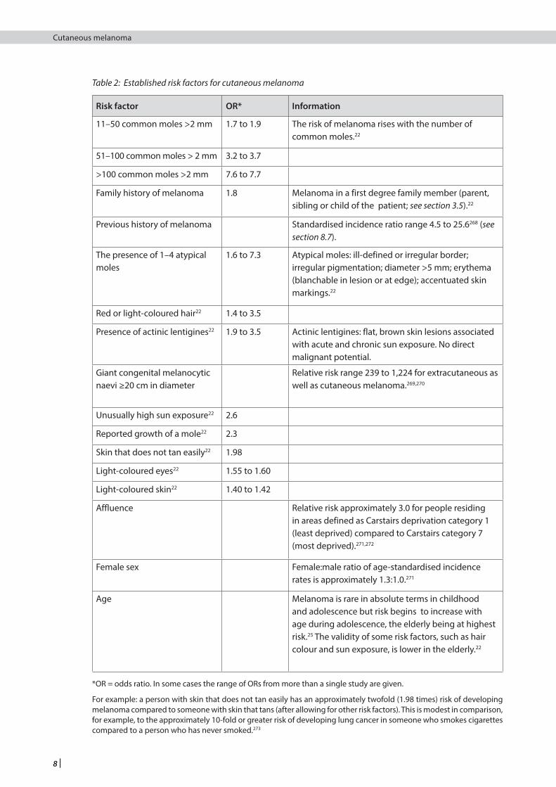
3.4.2 RISK FACTORS
Risk factors for melanoma have been identified mainly from case control studies (see Table 2). The strength of a risk factor is usually expressed in terms of an odds ratio (OR). In the context of this guideline, the OR is the ratio of the odds in favour of exposure to a risk factor in people with melanoma to the odds in favour of exposure to the same risk factor among people who have not developed melanoma. For relatively rare diseases such as melanoma, the OR can be thought of as being equivalent to the relative risk, that is, the ratio of the incidence rate of melanoma among exposed individuals to the incidence rate among unexposed individuals. The higher the OR (or relative risk), the stronger the association between the risk factor and melanoma. This is important from the perspective of an individual, but from a public health perspective a lower OR for a commonly occurring risk factor may be more important than a higher OR for a risk factor which occurs rarely in the population.
1+
2+
2++
3
1+
8 |
Cutaneous melanoma
Table 2: Established risk factors for cutaneous melanoma
Risk factor OR* Information
11–50 common moles >2 mm 1.7 to 1.9 The risk of melanoma rises with the number of common moles.22
51–100 common moles > 2 mm 3.2 to 3.7
>100 common moles >2 mm 7.6 to 7.7
Family history of melanoma
1.8 Melanoma in a first degree family member (parent, sibling or child of the patient; see section 3.5).22
Previous history of melanoma Standardised incidence ratio range 4.5 to 25.6268 (see section 8.7).
The presence of 1–4 atypical moles
1.6 to 7.3 Atypical moles: ill-defined or irregular border; irregular pigmentation; diameter >5 mm; erythema (blanchable in lesion or at edge); accentuated skin markings.22
Red or light-coloured hair22 1.4 to 3.5
Presence of actinic lentigines22 1.9 to 3.5 Actinic lentigines: flat, brown skin lesions associated with acute and chronic sun exposure. No direct malignant potential.
Giant congenital melanocytic naevi ≥20 cm in diameter
Relative risk range 239 to 1,224 for extracutaneous as well as cutaneous melanoma.269,270
Unusually high sun exposure22 2.6
Reported growth of a mole22 2.3
Skin that does not tan easily22 1.98
Light-coloured eyes22 1.55 to 1.60
Light-coloured skin22 1.40 to 1.42
Affluence Relative risk approximately 3.0 for people residing in areas defined as Carstairs deprivation category 1 (least deprived) compared to Carstairs category 7 (most deprived).271,272
Female sex Female:male ratio of age-standardised incidence rates is approximately 1.3:1.0.271
Age Melanoma is rare in absolute terms in childhood and adolescence but risk begins to increase with age during adolescence, the elderly being at highest risk.25 The validity of some risk factors, such as hair colour and sun exposure, is lower in the elderly.22
*OR = odds ratio. In some cases the range of ORs from more than a single study are given.
For example: a person with skin that does not tan easily has an approximately twofold (1.98 times) risk of developing melanoma compared to someone with skin that tans (after allowing for other risk factors). This is modest in comparison, for example, to the approximately 10-fold or greater risk of developing lung cancer in someone who smokes cigarettes compared to a person who has never smoked.273
| 9
Cutaneous melanoma 3 • Prevention, surveillance and genetics
RHealthcare professionals and members of the public should be aware of the risk factors for melanoma.
Individuals identified as being at higher risk should be advised about appropriate methods of sun protection, educated about the diagnostic features of cutaneous melanoma and encouraged to perform self examination of the skin.
3.5 GENETICS
It is estimated that 1–2% of melanomas are attributable to the inheritance of melanoma susceptibility genes.25 Mutations in cyclin-dependant kinase inhibitor 2A (CDKN2A) are associated with an increase risk of melanoma.25,26 Prevalence of CDKN2A mutations in affected families varies between countries.26-28 Cyclin-dependant kinase 4 (CDK4) mutations have also been implicated but have a low prevalence worldwide.26 In Scotland the prevalence of CDKN2A mutations in families with two or more first degree relatives affected by melanoma is approximately 22% (7 in 32 families).29 Mutations in CDKN2A are also associated with a risk of pancreatic cancer in some families and therefore a family history of pancreatic cancer and melanoma may increase the likelihood of identifying a CDKN2A mutation.25,27,28
A systematic review of clinical practice guidelines found that most guidelines do not cover genetic testing in their discussion, but where they do there is consensus that this should be offered in the context of genetic counselling.28
There may be additional benefits for patients to undergo genetic counselling for genetic testing as a higher compliance in self examination has been reported after genetic testing.30 People with mutations in CDKN2A may have a higher risk of smoking-related cancers and so should be advised to abstain from smoking tobacco.31
RGenetic testing for mutations in CDKN2A should be offered to an affected individual who has a first degree relative affected by melanoma or pancreatic cancer.
2++
34
1_
2+
10 |
Cutaneous melanoma
4 Diagnosis and prognostic indicators
The vast majority of melanomas are visible, if not to the patient, then at least to friends, family or health professionals. Members of the general public and health professionals should be aware of the signs suggestive of melanoma. In Scotland, melanomas occur more commonly in men than women. The most frequent site is the leg for women and the trunk in men. A small number of patients have occult primary lesions and present with metastatic disease. Up to ten percent of melanomas can be amelanotic (non-pigmented) or hypomelanotic, increasing diagnostic difficulty.
4 All patients with a diagnosis of melanoma should be discussed at a specialist multidisciplinary team (MDT) meeting.
4.1 TYPES OF MELANOMA
Melanomas are subdivided into types on the basis of clinical features and pathology.
4.1.1 SUPERFICIAL SPREADING MALIGNANT MELANOMA
Superficial spreading malignant melanoma (SSMM) is the most frequently encountered type of melanoma; characteristically an asymmetrical pigmented lesion with variable pigmentation and sometimes an irregular outline. Patients may have noted growth, a change in sensation and/or colour, crusting, bleeding or inflammation of the lesion. The duration of the symptoms varies from a few months to several years.
4.1.2 NODULAR MELANOMA
The second most common type is nodular melanoma (NM). This usually has a shorter presentation and a greater tendency to bleed and/or ulcerate.
4.1.3 LENTIGO MALIGNA MELANOMA
The next most frequent is the melanoma that occurs most often in sun-damaged skin on the head and neck of older patients. This is the only type that has a clearly recognised and often lengthy pre-invasive (in situ) lesion termed lentigo maligna (LM) before progressing, in some instances, to an invasive lentigo maligna melanoma (LMM).
4.1.4 ACRAL LENTIGINOUS MELANOMA
Acral lentiginous melanoma (ALM) occurs on sites including the palms, soles and beneath the nails.
4.1.5 DESMOPLASTIC TYPE MELANOMA
Desmoplastic type melanoma is uncommon.
It is important to distinguish between pure and mixed subtypes of desmoplastic melanoma (DM). Pure DM is thought to be associated with a more favourable outcome and lower incidence of positive sentinel lymph node biopsy (SLNB) (2.2% versus 15.8% in mixed DM, and 17.5% in conventional melanoma).32 Similar figures were reported in another study, with 1/92 patients with pure DM having a positive SNLB compared with 7/39 patients with mixed subtype.33 However, a small single centre study described higher local recurrence rates in pure DM (28/118) compared with mixed DM (18/124).34,35
4.1.6 PIGMENT SYNTHESISING (ANIMAL TYPE) MELANOMA
Pigment synthesising melanoma (also known as animal type melanoma) or low-grade hypermelanotic melanoma, is rare. It should be considered an indolent type of melanoma where there is little incidence of systemic metastases despite frequent positive SLNB.36-38
3
3
| 11
Cutaneous melanoma 4 • Diagnostics and prognostic indicators
4
3
1+
1+
3
3
2+
4.2 CLINICAL DIAGNOSIS
Suspicious pigmented lesions are best examined in a good light with or without magnification and should be assessed using the 7-point checklist (see Table 4) or ABCDE systems (see Table 3).39,40 The presence of any major feature in the 7-point checklist, or any of the features in the ABCDE system, is an indication for referral. The presence of minor features should increase suspicion. It is accepted that some melanomas will have no major features.
Table 3: The 7 -point checklist lesion system
Major features Minor features
• change in size of lesion • inflammation
• irregular pigmentation • itch/altered sensation
• irregular border • lesion larger than others
• oozing/crusting of lesion
Table 4: The ABCDE lesion system
A Geometrical Asymmetry in two axes
B Irregular Border
C At least two different Colours in lesion
D Maximum Diameter >6 mm
E Evolution/change in lesion
Clinical diagnosis of melanoma is difficult and the accuracy of diagnosis may vary according to a clinician’s level of experience, with reports of considerable variation in sensitivity from 50–86% and an inverse relationship between sensitivity and experience.41-43
High-magnification dermoscopy is more sensitive than non-dermatoscopic diagnosis when used by clinicians with experience of the technique.44 45
Training clinicians to be experts in hand-held dermoscopy improves diagnostic accuracy but it may diminish the sensitivity of the diagnosis of non-expert or untrained dermatologists.46-48 Observational studies have compared excision and pathological assessment to using other preoperative assessment methods of diagnosis including magnetic resonance imaging (MRI), high resolution ultrasound (US) and digital imaging of possible melanomas.49-52 These studies failed to show significant benefit.
R Clinicians should be familiar with the 7-point or the ABCDE checklist for assessing lesions.
4 Assess all pigmented skin lesions that are either referred for assessment or identified during follow up in secondary or tertiary care, using dermoscopy carried out by healthcare professionals trained in this technique.
4.3 DELAY IN DIAGNOSIS
Nine observational studies exploring delay were identified.41,53-60 Significant delays (greater than three months) in diagnosis of invasive melanoma are usually patient rather than physician related.41,53-60 Delay was defined differently in each study, with some including both patient and physician components.
All of the studies identified show inconsistency between Breslow thickness (see section 4.7.2) and delay, although melanomas diagnosed incidentally by health professionals were consistently thinner than those noted by patients themselves.56
12 |
Cutaneous melanoma
Several studies showed longer delays in older patients,42,58 in men, in rural versus urban dwellers and in those with plantar melanomas.42,59
There is inconsistency in findings regarding patients’ knowledge of melanoma and delay. Two observational studies found that delay in presentation was shorter if the patient was aware of possibility of malignancy.56,60 Conversely, another study found that delays were longer in those with greater knowledge, perhaps due to false reassurance caused by greater knowledge (see section 3.4.1).42
Physician delay accounts for a very small part of the total delay in diagnosis.41 Medical delays were shorter and the Breslow thickness was less when patients were seen by dermatologists as opposed to general practitioners.41
RHealth professionals should be encouraged to examine patients’ skin during other clinical examinations.
4 Emphasis should be given to the recognition of early melanoma by both patients and health professionals.
4.4 EDUCATING HEALTH PROFESSIONALS ABOUT DIAGNOSIS
An Australian RCT demonstrated a decrease in the number of benign lesions excised by general practitioners (GPs) after being given algorithms and cameras as aids to diagnosis.61 In an American RCT, the use of a booklet, magnifying and measuring tools and feedback sessions improved the ability of primary care residents to triage suspicious lesions.62
4 Targeted education can enhance health professionals’ ability to diagnose melanoma.
4.5 BIOPSY OF SUSPICIOUS LESIONS
The optimal specimen for full histological evaluation of a suspected melanoma is a complete excision with a 2 mm surround of normal skin and a cuff of fat.63 This enables assessment of the entire lesion (see section 5.1). Elliptical excisions should be performed along the long axis in the line of a natural skin crease or longitudinally in limbs. The exact surgical margins of excision should be recorded on the operation note.
Non-excisional biopsy may lead to inadequate histology.64-68 The least useful type of biopsy is the superficial shave variety. Two large studies demonstrate that non-excisional biopsy of the primary lesion has no effect on prognosis.65,69
Management of invasive lentigo maligna melanoma may have to be approached differently to superficial spreading melanoma. The frequently facial site and large diameter of such lesions may render full excision difficult or excessively destructive. In these instances incisional biopsy(s) of the most clinically suspicious areas are appropriate, but this may not detect all areas of invasion, and may underestimate depth.70
RA suspected melanoma should be excised with a 2 mm margin and a cuff of fat.
If complete excision cannot be performed as a primary procedure an incisional or punch biopsy of the most suspicious area is advised.
A superficial shave biopsy is inappropriate for suspicious pigmented lesions.
4 GPs should refer urgently all patients in whom melanoma is a strong possibility rather than carry out a biopsy in primary care.
Newly-diagnosed patients should receive both verbal and written information about melanoma including the treatment options and support services available to them.
2+
2+
2+
2+
1+
1_
3
| 13
Cutaneous melanoma 4 • Diagnostics and prognostic indicators
4.6 PATHOLOGICAL DIAGNOSIS
4.6.1 HANDLING A SUSPECTED MELANOMA
The volume of evidence addressing the handling of suspected melanomas is small. Recommendations on how to describe and select tissue blocks from a suspected melanoma are available from standard surgical pathology textbooks.71
Appropriate treatment, follow up and prognostication for patients with melanoma are entirely dependent on accurate pathological diagnosis and microscopic staging. The macroscopic description of the specimen, together with adequate and appropriate methods of block selection, is central to this process.
RThe macroscopic description of a suspected melanoma should:
• state the biopsy type, whether excision, incision, or punch
• describe and measure the biopsy (in mm)
• state the size of the lesion in mm and describe the lesion in detail (shape, pattern of pigment distribution, presence or absence of a nodular component and presence or absence of ulceration)
• state the clearance of the lesion (in mm) from the nearest lateral margin and the deep margin.
Selection of tissue blocks:
• the entire lesion should be submitted for histopathological examination
• the lesion should be sectioned transversely at 3 mm intervals and the blocks loaded into labelled cassettes
• cruciate blocks should not be routinely selected (they limit the assessment of low power architectural features such as symmetry)
• cruciate blocks may be used to assess margins in very large LM excisions.
A photograph of the macroscopic specimen may be of great value, especially if the precise origins of labelled blocks are drawn onto the photograph to permit exact orientation.
4.7 PROGNOSTIC INDICATORS/CORE MICROSCOPIC DATASET ITEMS
Histological reporting of primary cutaneous malignant melanoma and regional lymph nodes should follow the dataset produced by the Royal College of Pathologists (RCPath). The microscopic core items for the pathology report are summarised in this section. Further details are available from the RCPath dataset.72
4.7.1 HISTOGENETIC TYPE
The majority of studies do not demonstrate a significant association between histogenetic subtype and patient outcome in the common melanoma types when matched for Breslow thickness. However, in pigment synthesising melanoma and pure desmoplastic melanoma, histogenetic type does appear to play a role in determining the likelihood of recurrence.
4 The histogenetic type should be included in the pathology report.
14 |
Cutaneous melanoma
4.7.2 BRESLOW THICKNESS
A strong association between tumour thickness and prognosis was originally demonstrated by Breslow73 and has since been verified in many large scale studies of melanoma.74-78 Breslow thickness is the single most important prognostic variable in primary cutaneous melanoma.72 It is recommended that Breslow thickness is measured to a minimum of one decimal place but to allow for accurate staging, two decimal places should be used in cases sitting close to the boundary between pT1/2, pT2/3, and pT3/4 as defined by the American Joint Committee on Cancer (AJCC) (see Table 6).72,79
RAn accurate measurement of the Breslow thickness should be included in the pathology report for any melanoma that has an invasive component.
4.7.3 ULCERATION
A small study of 177 participants with melanomas of intermediate thickness (1.51 to 3.99 mm) identified epidermal ulceration as one of four variables that predicted visceral and bony metastases.80 Ulceration has been shown to act as a prognostic variable after adjustment for other variables.75,76 A study of 1,042 patients identified epidermal ulceration as a significant prognostic variable and this was incorporated into a mathematical model for predicting recurrence and survival at three, five and ten years.74 Some studies also show that increasing breadth of epidermal ulceration is associated with an increasingly unfavourable prognosis.74
RThe presence or absence of histological evidence of epidermal ulceration should be noted in the pathology report.
4.7.4 MITOTIC RATE
The most recent AJCC guideline specifically uses the presence of mitotic activity in the dermal component of a melanoma to distinguish pT1a from pT1b tumours (see Table 6). Both the AJCC and RCPath provide guidance on how to measure and report mitotic rates. This should be documented as mitoses per square millimetre and should be recorded as a 0 or a whole number. The presence of any mitotic activity (irrespective of how many high-power fields have been used during the assessment) should always be given a figure of at least 1.72,79
RMitotic rate is used as a defining criterion for pT1b melanomas and should be recorded in the pathology report.
4.7.5 LYMPHOVASCULAR INVASION
Lymphovascular invasion (LVI) is a core dataset item from the RCPath and should be stated in the report. It is important to exclude retraction artifact and it is not important to separate lymphatic or vascular invasion.72
4.7.6 MICROSCOPIC SATELLITES/IN-TRANSIT METASTASIS
Microsatellites are defined by AJCC as any discontinuous nest of intralymphatic metastatic cells greater than 0.05 mm in diameter that are clearly separated by normal dermis (not fibrosis or inflammation) from the main invasive component of melanoma by a distance of at least 0.3 mm.81 Macrosatellite metastases are defined as discrete separate nodules within 2 cm of the primary tumor and are considered intralymphatic extensions of the primary tumor, whereas in-transit metastases are defined as any dermal or subcutaneous disease 2 cm or more from the primary tumor but not beyond the draining regional nodal basin.79
The presence of microsatellites upstages a melanoma to pTN2c.79 The RCPath supports the view that microsatellites do not have to be present within the lymphatic system.72
A systematic review found that the prognosis for patients with microsatellites is essentially identical to that for patients with macrosatellites.82 There was no demonstrable difference in survival for patients with satellites compared to those with in-transit metastases.
4
2+
2++
4
2++
4
2+
4
1+
2+
| 15
Cutaneous melanoma 4 • Diagnostics and prognostic indicators
A prospective cohort study of 258 patients with clinical stage I melanoma found that 13 out of 14 patients with histological evidence of lymphatic invasion developed in-transit metastases after a median interval of 10 months and concluded that lymphatic invasion correlates strongly with early locoregional cutaneous relapse.83
A study of 140 patients with thick melanomas reported that the identification of lymphatic invasion was associated with an increased risk of metastasis but not with overall survival.84 However, in a series of 17,600 patients the presence of microsatellites had a profound negative impact on prognosis and in the current AJCC staging system the presence of satellites upstages the tumour from I or II to IIIb or IIIc.79
Identifying lymphovascular invasion and/or microscopic satellites confers considerable prognostic value. The presence of lymphatic invasion accurately predicts early cutaneous relapse and should be included as a stratification criterion for the selection of patients for adjuvant therapy. The histological identification of microsatellites also defines a subset of patients at much greater risk of relapse. The presence of microsatellites correlates strongly with occult metastatic disease in regional lymph nodes.
RIdentification of microscopic satellites upstages the pN status of melanoma according to the AJCC cancer staging manual (7th edition) and should be included in the pathology report. The defining criteria should be strictly adhered to and the presence or absence of microsatellites should be stated in the pathology report.
4.7.7 RADIAL VERSUS VERTICAL GROWTH PHASE
Tumour growth phase correlates strongly with clinical outcome.75,85 A study of 501 patients with primary melanomas identified a subgroup of 122 as being in radial growth phase only. No patients in this subgroup showed evidence of metastatic disease during a minimum follow-up period of 100 months. The OR for a patient with radial growth phase melanoma surviving for eight years was given as 1.0.75 A second study evaluated 624 patients, of whom 161 had melanoma displaying radial only growth phase characteristics. None of the patients developed metastatic disease at long-term follow up (median 13.7 years).85 The definitions of growth phase are discussed in more detail in the RCPath dataset.72
R The growth phase characteristics should be stated in the pathology report of all melanomas.
4.7.8 TUMOUR INFILTRATING LYMPHOCYTES
The association between survival advantage and the presence of tumour infiltrating lymphocytes (TIL) within the vertical growth phase component is unclear. Although one study demonstrated a strong correlation,75 the presence of an inflammatory response loses independent prognostic strength on multivariate modelling.74
TILs are an AJCC prognostic item and are included in the RCPath dataset.
Tumour infiltrating lymphocytes are a core dataset item and should be recorded in the pathology report.
4.7.9 REGRESSION
There is an adverse association between histological evidence of regression and outcome, but the strength of this relationship is disputed.74,75 86 One large study identified tumour regression in the radial growth phase as a variable that retained predictive strength after multivariate analysis.75 In a subsequent study of 1,042 patients the significance of tumour regression was subsumed by the other clinical and histological features studied.74 Extensive late regression might indicate that the melanoma has, at some time, been significantly thicker than it now appears. Tumours with this feature are liable to be understaged.86
If the zone of regression is deeper than the deepest melanoma cell then this should not alter the formal Breslow thickness; Breslow thickness should be measured to the deepest tumour cell as per the original definition. Regression is defined by the RCPath as variable destruction of melanoma cells, inflammatory response, fibrosis and melanin laden macrophages. The RCPath suggest that severely dysplastic nevi and in situ melanoma which show convincing features of established regression should be considered for MDT discussion.72
2++
2++
4
2+
4
2++
4
2++
2+
16 |
Cutaneous melanoma
R If the presence or absence of regression is apparent it should be included in the pathology report.
4.7.10 CLARK LEVEL
The Clark level has been replaced by mitotic index/count for defining pT1a and pT1b tumours in the 7th edition of the AJCC staging system (see section 4.7.4). In cases where there is no ulceration present and mitotic activity cannot be assessed, if the tumour has a Clark level of 4 or of 5 then the tumour is staged as pT1b according to the AJCC.72,79
RIf the pT1a/pT1b status cannot be determined through the presence of ulceration and/or mitotic activity then a Clark level of 4 or 5 can be used to upstage the tumour. Clark level only need be documented in these cases.
4.7.11 BRAF STATUS
4 Serine/threonine-protein kinase B-Raf (BRAF) status should be requested in all patients with advanced disease and recorded on the pathology report (see section 9.3.1).
4.8 SPECIALIST PATHOLOGY REPORTING
Significant discrepancy exists between general pathologists, dermatopathologists as well as between experts in pigmented lesion pathology, in the reporting of melanocytic tumours.87-89 Both under- and over-diagnosis of malignancy is recognised and, for melanoma, there is poor agreement on the assessment of prognostic parameters.
4 Pathologists responsible for reporting melanocytic lesions must be aware of the diagnostic pitfalls in this area. Participation in appropriate continuing professional development (CPD) activity is advisable.
Cases where significant diagnostic doubt exists should be referred for specialist dermatopathology opinion.
4.9 MELANOMA PATHOLOGY REPORT
Table 5: Core features of a pathology report for invasive melanoma
Clinical data/macroscopic description Histological data
Clinical site Histogenetic type
Specimen type Breslow thickness
Size of specimen in three dimensions Ulceration
Size of lesion in three dimensions Mitotic index
Atypical features Lymphovascular space invasion
Microsatellites/in-transit metastatic cells
Perineural invasion
Growth phase
Tumour infiltrating lymphocytes
Regression
Clark level (if pT1a/b staging not possible from mitotic index/ulceration)
Margins peripheral and deep
Tumour stage (pT)
BRAF status (if applicable)
2++
4
2++
| 17
Cutaneous melanoma 4 • Diagnostics and prognostic indicators
4.10 PATHOLOGICAL EXAMINATION AND REPORTING OF THERAPEUTIC AND SENTINEL LYMPH NODE DISSECTION SPECIMENS
Detailed protocols for dissection of therapeutic lymph node dissection specimens are available in standard textbooks of surgical pathology.90,91
The surgical report for completion and therapeutic lymph node dissections (see section 5.3) should identify both macroscopic and microscopic features.
Macroscopic features which should be recorded include:• the size of the specimen in three dimensions• the presence (and size) or absence of a macroscopic abnormality, and • the presence or absence of a localisation marker.
The microscopic features which should be recorded include:• the exact number of nodes identified within the specimen • the number of nodes containing metastatic disease and whether the atypical node is involved or not • the presence or absence of extracapsular spread, and • whether the margin of the specimen is involved by tumour.72
When macroscopic examination reveals tumour within a node, a single block of tissue is sufficient to confirm the observation. Nodes that appear tumour free should be serially sliced (if large) and all of the tissue processed. Small nodes may be processed intact and levelled to ensure thorough examination.72
Sentinel lymph nodes (SLN) are processed using either lymphoscintigraphy and/or blue dye to trace the afferent lymphatic channels and node. Protocols giving further details are available.91-93 Nodes identified by lymphoscintigraphy (usually technetium-99) should be fixed in formalin for 24 hours to allow for radioactive decay.72
When dye has been used, the sentinel node should be examined macroscopically to determine whether any staining has occurred. The node should then be processed according to the European Organisation for Research and Treatment of Cancer (EORTC) trial protocol.72
Additional information to be recorded in the pathology report for SLNs include dye observed in tissue (macroscopic) and the number of SLNs involved in the tumour and the location of deposit: subcapsular, parenchymal and/or extracapsular spread (microscopic).72
Although immunohistochemistry (IHC) facilitates the detection of melanoma in sentinel nodes, the possibility of false positive results, for example the misinterpretation of capsular naevus cells, remains. This can be minimised by careful evaluation of the immunochemical preparations in the context of the corresponding haematoxylin and eosin stained section. The AJCC (7th edition) considers it acceptable to diagnose nodal metastases solely on IHC staining for melanoma-associated markers in situations where corresponding atypical cells are not always seen on haematoxylin and eosin sections.79
Groups of sections at multiple levels throughout the sentinel node are sometimes examined, but there is no evidence that such rigorous sampling increases the diagnostic yield. Detecting melanoma cells in SLNs using polymerase chain reaction (PCR) techniques cannot be recommended at present due to concerns regarding both sensitivity and specificity.94
4
4
18 |
Cutaneous melanoma
5 Surgical management and staging
5.1 WIDE LOCAL EXCISION SURGERY FOR PRIMARY MELANOMA
Historically very wide margins of excision were advocated in the management of melanoma. Appreciation of Breslow thickness as a prognostic indicator (see section 4.7.2) supports the concept of a conservative approach to surgery, with narrowing of the margins of excision.101-104 The safety of these narrower margins has been demonstrated in a series of studies.105-108
A comparison of 1 cm and 3 cm margins for tumours up to 2 mm thick found no overall survival difference between the two groups.95 A small number of patients with lesions thicker than 1 mm developed local recurrence.96-98
A 1 cm margin should therefore be adequate for melanomas less than 1 mm thick. For lesions 1–2 mm thick a width excision of 1–2 cm should be considered, in the context of a full clinical assessment.
Lentigo maligna, (a variant of melanoma in situ), should also be surgically removed, given the risk of invasion. Currently 5 mm surgical margins are recommended, although a case series reported that 26% of lentigo maligna required greater margins to achieve clearance as atypical cells may extend beyond the visible edge.99 There is limited evidence from case series that Mohs micrographic surgery (MMS) may reduce the size of the defect in lentigo maligna.100 For patients for which surgery is not an option, there is some evidence for the use of radiotherapy and topical imiquimod for the treatment of lentigo maligna.99,101 Cryotherapy and topical-5-fluorouracil have also been used but there is no recently published evidence.100,102
Evidence-based recommendations on excision margins for melanoma can be found in the NICE guideline on assessment and management of melanoma.45
R• Consider a clinical margin of at least 0.5 cm when excising stage 0 melanoma.
• If excision for stage 0 melanoma does not achieve an adequate histological margin, discuss further management with the multidisciplinary team.
• Offer excision with a clinical margin of at least 1 cm to people with stage I melanoma.
• Offer excision with a clinical margin of at least 2 cm to people with stage II melanoma.
The suggested width of excision at sites of aesthetic and functional importance requires clinical consideration and discussion with the MDT. The deep excision margin should incorporate adipose tissue down to, but not including, the deep fascia.103,104 No evidence was identified on optimal timing of wide excision in patients with melanoma.
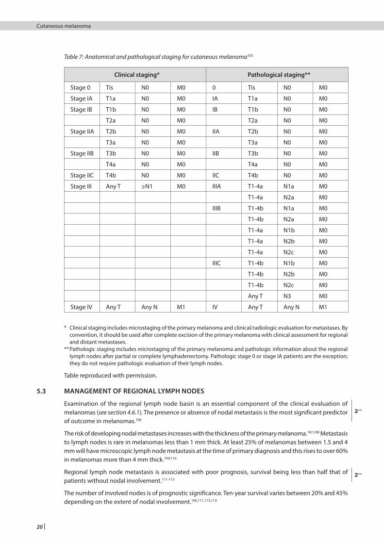
5.2 STAGING MELANOMA
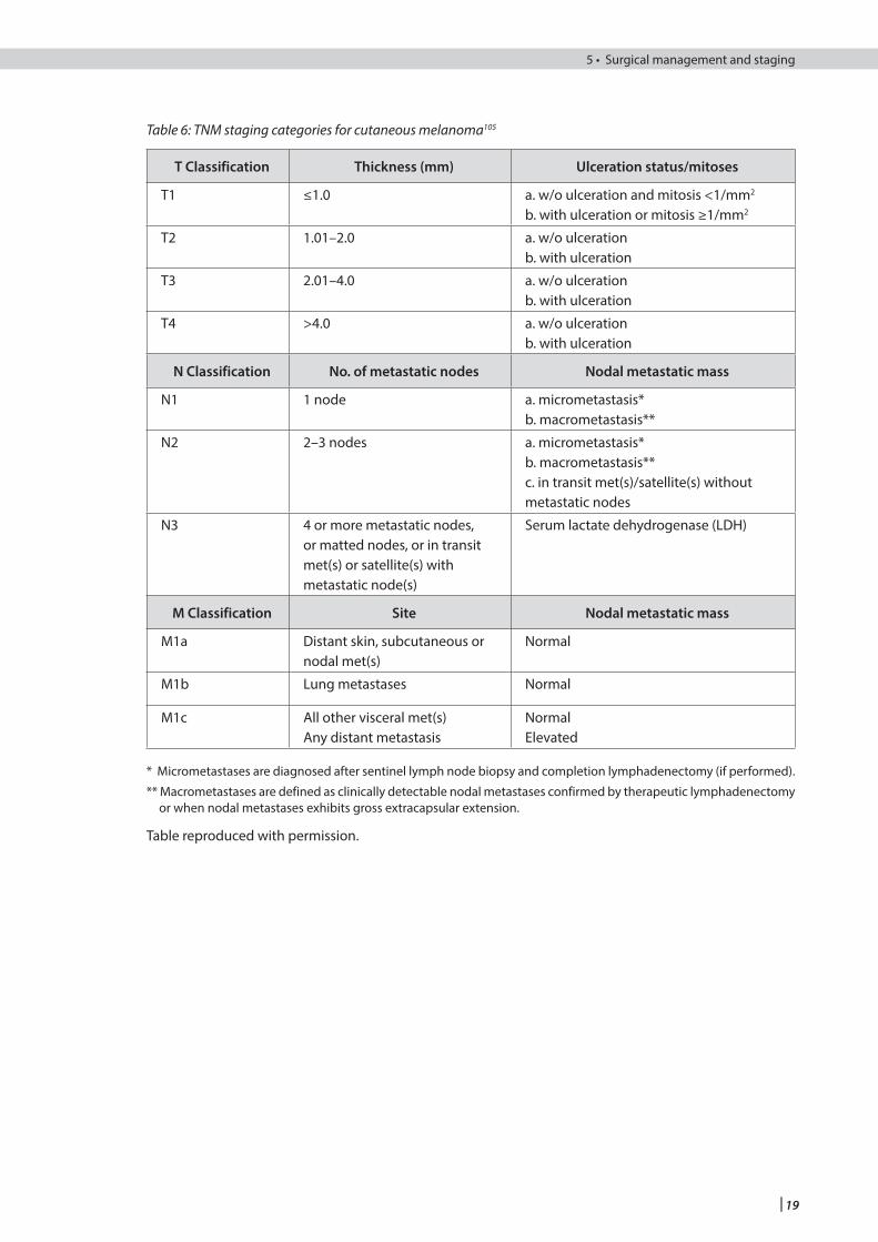
Melanoma should be staged using the tumour, node, metastasis (TNM) staging classification described by the American Joint Committee on Cancer (AJCC)105 (see Tables 6 and 7).
1+
34
1++
3
1+
3
4
| 19
Cutaneous melanoma 5 • Surgical management and staging
Table 6: TNM staging categories for cutaneous melanoma105
T Classification Thickness (mm) Ulceration status/mitoses
T1 ≤1.0 a. w/o ulceration and mitosis <1/mm2
b. with ulceration or mitosis ≥1/mm2
T2 1.01–2.0 a. w/o ulcerationb. with ulceration
T3 2.01–4.0 a. w/o ulcerationb. with ulceration
T4 >4.0 a. w/o ulcerationb. with ulceration
N Classification No. of metastatic nodes Nodal metastatic mass
N1 1 node a. micrometastasis*b. macrometastasis**
N2 2–3 nodes a. micrometastasis*b. macrometastasis**c. in transit met(s)/satellite(s) withoutmetastatic nodes
N3 4 or more metastatic nodes, or matted nodes, or in transit met(s) or satellite(s) with metastatic node(s)
Serum lactate dehydrogenase (LDH)
M Classification Site Nodal metastatic mass
M1a Distant skin, subcutaneous or nodal met(s)
Normal
M1b Lung metastases Normal
M1c All other visceral met(s)Any distant metastasis
NormalElevated
* Micrometastases are diagnosed after sentinel lymph node biopsy and completion lymphadenectomy (if performed).
** Macrometastases are defined as clinically detectable nodal metastases confirmed by therapeutic lymphadenectomy or when nodal metastases exhibits gross extracapsular extension.
Table reproduced with permission.
20 |
Cutaneous melanoma
Table 7: Anatomical and pathological staging for cutaneous melanoma105
Clinical staging* Pathological staging**
Stage 0 Tis N0 M0 0 Tis N0 M0
Stage IA T1a N0 M0 IA T1a N0 M0
Stage IB T1b N0 M0 IB T1b N0 M0
T2a N0 M0 T2a N0 M0
Stage IIA T2b N0 M0 IIA T2b N0 M0
T3a N0 M0 T3a N0 M0
Stage IIB T3b N0 M0 IIB T3b N0 M0
T4a N0 M0 T4a N0 M0
Stage IIC T4b N0 M0 IIC T4b N0 M0
Stage III Any T ≥N1 M0 IIIA T1-4a N1a M0
T1-4a N2a M0
IIIB T1-4b N1a M0
T1-4b N2a M0
T1-4a N1b M0
T1-4a N2b M0
T1-4a N2c M0
IIIC T1-4b N1b M0
T1-4b N2b M0
T1-4b N2c M0
Any T N3 M0
Stage IV Any T Any N M1 IV Any T Any N M1
* Clinical staging includes microstaging of the primary melanoma and clinical/radiologic evaluation for metastases. By convention, it should be used after complete excision of the primary melanoma with clinical assessment for regional and distant metastases.
** Pathologic staging includes microstaging of the primary melanoma and pathologic information about the regional lymph nodes after partial or complete lymphadenectomy. Pathologic stage 0 or stage IA patients are the exception; they do not require pathologic evaluation of their lymph nodes.
Table reproduced with permission.
5.3 MANAGEMENT OF REGIONAL LYMPH NODES
Examination of the regional lymph node basin is an essential component of the clinical evaluation of melanomas (see section 4.6.1). The presence or absence of nodal metastasis is the most significant predictor of outcome in melanomas.106
The risk of developing nodal metastases increases with the thickness of the primary melanoma.107,108 Metastasis to lymph nodes is rare in melanomas less than 1 mm thick. At least 25% of melanomas between 1.5 and 4 mm will have microscopic lymph node metastasis at the time of primary diagnosis and this rises to over 60% in melanomas more than 4 mm thick.109,110
Regional lymph node metastasis is associated with poor prognosis, survival being less than half that of patients without nodal involvement.111-113
The number of involved nodes is of prognostic significance. Ten-year survival varies between 20% and 45% depending on the extent of nodal involvement.106,111,113,114
2++
2++
| 21
Cutaneous melanoma 5 • Surgical management and staging
5.3.1 MANAGEMENT OF PALPABLE LYMPH NODES
Fine needle aspiration/open biopsy
Patients with melanoma who have palpable lymph node(s) either at their first presentation or at a follow- up visit should have fine needle aspiration cytology (FNAC). If the first sample is unsatisfactory or negative with persistent suspicion, it should be repeated with ultrasound guidance, if required. If doubt persists an open biopsy can be performed.114,115
4 If there is palpable lymphadenopathy FNAC should be used to obtain cytological confirmation of metastases.
If open biopsy is undertaken the incision must be placed in the same line as for a potential radical lymphadenectomy.
Therapeutic lymph node dissection
Confirmation of metastatic melanoma in a palpable lymph node is an indication for radical dissection of that lymph node basin.
Therapeutic lymph node dissection is beneficial in controlling locoregional disease. The risk of recurrence in the dissected node field remains, particularly with head and neck melanomas.116,117
Head and neck melanomas have the most variable pattern of lymph node metastasis and require a variety of types of neck dissection that may include the parotid or the posterior occipital chain nodes.117
RTherapeutic lymph node dissection requires complete and radical removal of all draining lymph nodes to allow full pathological examination.
4 Patients with a confirmed metastatic lymph node(s) should be radiologically staged prior to lymph node dissection.
Regional lymph node dissection carries a well defined and significant morbidity and should be undertaken only by surgeons with appropriate expertise.
Patients should be advised of the risk of lymphoedema following lymph node dissection. If lymphoedema occurs, patients should be referred to a lymphoedema specialist.
5.3.2 MANAGEMENT OF NON-PALPABLE LYMPH NODES
The high incidence of occult metastasis in clinically impalpable nodes has prompted surgeons to investigate regional lymph nodes.
Sentinel lymph node biopsy
The sentinel lymph node is defined as the first node in the lymphatic basin that drains the lesion and is the node at greatest risk for the development of metastasis.118 Biopsy of this node can assist in staging patients at risk of metastatic disease.
4
2++
22 |
Cutaneous melanoma
The standard for sentinel lymph node biopsy (SLNB) is a triple diagnostic approach of lymphoscintography, blue dye dermal infiltration and localisation using a hand-held gamma probe.118-123 Performing SLNB requires appropriate surgical expertise,118 specialist nuclear medicine services and the availability of serial sectioning and immunohistochemistry techniques (see section 4.10).
Sentinel lymph node biopsy can determine the presence or absence of metastasis within the regional lymph node basin124-126 and it is a useful staging tool in melanomas >1 mm thick.78 In thick melanomas (>4 mm) it can identify a subset of melanomas which are node negative and therefore offer a better prognosis.126
There are no randomised control studies addressing the most effective way of managing patients with a positive sentinel lymph node biopsy. All of the observational studies reviewed were retrospective, had very small numbers and had significant selection bias127,128
People who choose to have SLNB may benefit from more accurate staging, giving a better indication of outcome (survival and risk of relapse). Table 8 outlines possible advantages and disadvantages of SLNB and may help inform discussion with patients on whether or not to proceed with SLNB.45
Table 8: Possible advantages and disadvantages of SLNB 45
Possible advantages Possible disadvantages
The operation helps to find out whether the cancer has spread to the lymph nodes. It is better than ultrasound scans at finding very small cancers in the lymph nodes.
The purpose of the operation is not to cure the cancer. There is no good evidence that people who have the operation live longer than people who do not have it.
The operation can help predict what might happen in the future. For example, in people with a primary melanoma that is between 1 and 4 mm thick:• around 1 out of 10 die within 10 years if the
sentinel lymph node biopsy is negative• around 3 out of 10 die within 10 years if the
sentinel lymph node biopsy is positive.
The result needs to be interpreted with caution. Of every 100 people who have a negative sentinel lymph node biopsy, around 3 will subsequently develop a recurrence in the same group of lymph nodes.
People who have had the operation may be able to take part in clinical trials of new treatments for melanoma. These trials often cannot accept people who haven’t had this operation.
A general anaesthetic is needed for the operation
The operation results in complications in between 4 and 10 out of every 100 people who have it.
Table reproduced with permission.
RSLNB should be considered as a staging technique in patients with stage IB-IIC melanoma with a Breslow thickness of >1 mm. It should not be offered to patients with stage IB melanoma where Breslow thickness is ≤1 mm.
Patients should be given detailed verbal and written information regarding the possible advantages and disadvantages of the SLNB procedure to allow them to make an informed decision.
2+
2++
4
1+
4
4
| 23
Cutaneous melanoma 5 • Surgical management and staging
Completion lymphadenectomy
Current practice is to consider completion lymphadenectomy in patients with a positive sentinel lymph node. No good-quality evidence was identified to determine whether completion lymphadenectomy provides better survival than clinical observation with or without serial ultrasound. Additionally there is insufficient evidence to characterise pathological features of the positive sentinel lymph node to instruct the decision regarding further management.128
NICE compiled the following table to inform discussion with patients on whether to proceed with completion lymphadenectomy.45
Table 9: Possible advantages and disadvantages of completion lymphadenectomy
Possible advantages Possible disadvantages
Removing the rest of the lymph nodes before cancer develops in them reduces the chance of the cancer returning in the same part of the body.
Lymphoedema (long-term swelling) may develop, and is most likely if the operation is in the groin and least likely in the head and neck.
The operation is less complicated and safer than waiting until cancer develops in the remaining lymph nodes and then removing them.
In 4 out of 5 people, cancer will not develop in the remaining lymph nodes, so there is a chance that the operation will have been done unnecessarily.
People who have had the operation may be able to take part in clinical trials of new treatments to prevent future melanoma. These trials often cannot accept people who have not had this operation.
There is no evidence that people who have this operation live longer than people who do not have it.
Having any operation can cause complications.
Table reproduced with permission.
RPatients with a positive sentinel lymph node should be offered appropriate counselling regarding the advantages and disadvantages of completion lymphadenectomy.
4 Following lymphadenectomy all patients should have access to specialist lymphoedema services.
1+
4
24 |
Cutaneous melanoma
6 Further investigations and non-surgical staging
Further investigation to determine precisely the extent of the disease is important in terms of prognosis, treatment, entry into clinical trials, research and audit.
Following pathological microstaging of a patient’s melanoma (see section 4.7) the presence of metastatic spread can be determined using three techniques:
• Surgical: assessment of the impalpable node by sentinel node biopsy and of the palpable node (see section 5)
• Imaging: conventional radiography, ultrasound scanning (US), computed tomography (CT), magnetic resonance imaging (MRI) and positron emission tomography–computed tomography (PET-CT)
• Blood Tests: routine haematology, tumour markers, liver function tests and lactate dehydrogenase (LDH).
6.1 IMAGING TECHNIQUES
6.1.1 CROSS SECTIONAL IMAGING
No good quality evidence was identified on which patients should undergo imaging as part of staging. Patients with stage IIC tumours have a worse prognosis compared to patients with other stage II tumours, with prognoses more comparable to stage III disease.79 Stage IIC tumours are therefore treated like stage III tumours in two guidelines.45,129 A guideline from NICE recommends that patients with stage IIC (without SLNB) and stage III disease should be offered CT imaging, due to the high risk of occult disease.45 Guidelines from the British Association of Dermatology (BAD) advise against routine imaging for patients with stages I or II melanoma because of the low incidence of true-positive and high incidence of false-positive findings, with the exception of patients with high-risk primary melanoma.130
It is the consensus opinion of the guideline development group that patients with stage IIC disease and above should be offered initial staging imaging.
No RCTs comparing CT and PET-CT in the staging of melanoma were identified. A meta-analysis of retrospective and prospective studies on the diagnostic accuracy of PET-CT and CT reported that PET-CT is the most sensitive and specific initial staging modality for the detection of distant metastases in patients with melanoma (sensitivity 80%, 95% credible interval (CrI) 53% to 93%; specificity 87%, 95% CrI 54% to 97% versus CT sensitivity 51%, 95% CrI 54% to 76%; specificity 69%, 95% CrI 30% to 92%).131 Further systematic reviews found PET-CT to have a sensitivity of 68–87% and specificity of 92–98% in patients with stage III or stage IV disease132 and specificity of 89% in patients with stage III disease.133 Many of the included studies, however, were retrospective and of poor quality, with wide inclusion criteria and insufficient reporting of withdrawals. Several potential sources of bias were also identified including referral bias, verification bias and review bias. The studies reported on diagnostic accuracy but did not include patient relevant outcomes.
Whilst PET-CT would seem to have a higher sensitivity and specificity for the detection of metastases, the quality of the evidence does not support its routine use as a first-line imaging modality in the staging of melanoma. This, in addition to its relative cost and limited availability in Scotland,134 inform the consensus opinion of the guideline development group that its use should currently be restricted to patients with indeterminate findings on CT or for those being considered for a major surgical resection.
R Staging CT should be offered to patients with stage IIC or above melanoma.
4 Staging CT should include head, chest, abdomen and pelvis. The neck should be included in patients with head and neck melanoma.
4 PET-CT should only be considered for patients with indeterminate findings on CT or for patients who are being considered for major surgical resection, after discussion with the specialist multidisciplinary team.
4
2++
2+
| 25
Cutaneous melanoma 6 • Further investigations and non-surgical staging
6.1.2 IDENTIFYING BRAIN METASTASES
No high-quality evidence was found on the optimal imaging modality for identifying brain metastases specifically in patients with melanoma. Evidence from reviews of studies of imaging on a variety of primary tumours suggest that contrast MRI is more sensitive than contrast CT in detecting brain metastases.135,136
Given that patients are likely to have a CT of the chest, abdomen and pelvis during staging of melanoma, and taking tolerability, cost and availability into consideration (see section 6.1.1), it is the consensus opinion of the guideline development group that CT should be the first-line imaging modality for identifying brain metastases.
RCT of the head with contrast should be used as the first-line imaging modality for identifying brain metastases.
MRI of the head should be considered where CT findings are equivocal.
If patients are being considered for locoregional treatment of brain metastases, contrast MRI should be performed to identify further lesions which may alter management.
6.2 LABORATORY INVESTIGATIONS
Investigations such as full blood counts (FBC) and liver function tests (LFT) are not helpful in identifying asymptomatic patients with distant disease.137,138 Elevated LDH in the absence of clinical symptoms or signs is the first indicator of stage IV disease in 12.5% of patients. By the time other blood parameters are significantly deranged, the patient will have other manifestations of metastasis.137,138 For patients with advanced disease, LDH is now included in the AJCC classification system.78 The evidence and availability of tumour markers such as S100 protein, melanoma inhibitory activity (MIA) protein and tyrosinase mRNA are limited. Investigating these markers is not routinely indicated.139
RRoutine blood tests are not indicated in staging asymptomatic patients with melanoma, with the exception of LDH in patients with stage IV disease, which is part of routine classification.
4
3
26 |
Cutaneous melanoma
7 Adjuvant treatment of stage II and III melanoma
Pathological features of primary melanoma, particularly Breslow thickness and ulceration, make it possible to identify patients with stage II disease who are at high risk of local or systemic recurrence (see section 4.7). Once patients have had melanoma recurrence in the local regional lymph nodes (stage III disease), over 50% will subsequently develop further metastatic spread. These observations support attempts to identify adjuvant treatment such as chemotherapy, immunotherapy and radiotherapy, given after complete clinical surgical clearance of melanoma.
7.1 ADJUVANT RADIOTHERAPY FOR RESECTED STAGE III MELANOMA
A single randomised phase 3 trial comparing adjuvant radiotherapy and observation was carried out in 250 patients who had undergone complete lymphadenectomy and were thought to be at high risk of local recurrence. Risk of lymph node relapse was significantly reduced in the adjuvant radiotherapy group (hazard ratio (HR) 0.56, 95% confidence interval (CI) 0.32 to 0.98, p=0.041) but no differences were noted for relapse-free or overall survival.140 Adjuvant radiotherapy is known to be associated with a risk of both short-term (dermatitis) and long-term (lymphoedema) toxicity. Results from trials on long-term radiotherapy complications are awaited. A case series suggested a significant increase in morbidity including lymphoedema rate as a complication of adjuvant radiotherapy.141
RConsider adjuvant radiotherapy for patients with completely resected stage IIIB or IIIC melanoma after discussion of the risk of local recurrence and the benefits and risks of radiotherapy including risk of significant adverse effects.
7.2 IMMUNOTHERAPY
7.2.1 INTERFERON
The observation that a large number of primary melanomas undergo partial regression and a small number of patients experience total regression of the whole melanoma has led to the concept of using either specific or non-specific immune stimulation as therapy for melanoma.
Adjuvant interferon alpha has been used in at least 10 large RCTs involving over 5,000 patients.142-151 Interferon dosage, frequency and route of administration and total duration of therapy all varied, but no trial reported significant overall survival benefit for interferon-treated patients. Several of the larger studies do report longer disease-free intervals after surgery145-147 but there is no evidence of a dose or duration of treatment effect. Toxic effects of interferon include extreme lassitude, muscle aches, headache, rigors, nausea, vomiting, and marrow toxicity, the latter being the cause of death in two patients in the first reported high-dose study.
RAdjuvant interferon should not be used for patients with AJCC stage II and III melanoma other than in a trial setting.
A number of well-designed trials of adjuvant immunotherapy (including ipilmumab, nivolumab and pembrolizumab) are ongoing.
7.3 IMMUNOSUPPRESSION
Numerous studies have investigated the relationship between immunosuppression and melanoma incidence. A poor-quality systematic review of population studies found that compared to the general population, there is a 2.4-fold (95% CI, 2.0 to 2.9) increased incidence of melanoma after transplantation.152 A meta-analysis also found that inflammatory bowel disease was associated with a 37% increased risk of melanomas compared to the general population.153 In addition, cohort studies have shown that patients with HIV have an increased risk of melanoma (standardized rate ratio of 2.6 (95% CI, 1.9 to 3.6),154 patients with a history of non-Hodgkin lymphoma (NHL) have a risk of subsequent melanoma that is increased 1.8 to 2.4 times,155 and patients with chronic lymphocytic leukaemia (CLL) have an increased risk of 2.3 to 3.1 times that of controls.156
1++
3
1_
2++
2+
1++
| 27
Cutaneous melanoma 7 • Adjuvant treatment of stage II and III melanoma
Although iatrogenic immunosuppression has been associated with increased risk of malignancy there is little data that is specific to melanoma. A population-based cohort study found that patients with rheumatoid arthritis treated with tumour necrosis factor (TNF) inhibitors had an increased risk of melanoma compared with patients with rheumatoid arthritis not treated with TNF inhibitors (HR 1.5, 95% confidence interval 1.0 to 2.2).157 A case-control study found that the use of TNF-alpha antagonists was independently associated with an increased melanoma risk in patients with inflammatory bowel disease (OR 1.9, 95% CI, 1.1 to 3.3)158 however, in a second cohort, the adjusted odds ratio was non-significant (OR 1.3, 95% CI, 0.6 to 2.7).159
Several studies have investigated the relationship between immunosuppression and melanoma prognosis. A retrospective review of immunosuppressed transplant patients found that those with thick melanoma (>3 mm) had a significantly poorer melanoma cause-specific survival rate.160 A second retrospective review found that the outcome for post-transplant patients with melanoma was significantly worse for those with tumours of Breslow thickness >2 mm.161 A further retrospective review found that patients taking immunosuppressants at the time of diagnosis of melanoma had a higher mortality than controls (42% v 23%, p=0.01) suggesting that immunosuppressive therapy may be associated with a more aggressive disease course.162 There is limited data on the prognosis for patients who were diagnosed with melanoma before having a transplant.152
A case-series has described the spontaneous regression of advanced melanoma in patients on long-term azothioprine for autoimmune disease on withdrawal of the immunsuppression.163
4 All patients with melanoma and a history of immunosuppression should have an MDT approach to care and minimising the immunosuppressive therapy should be considered where possible.
3 2++
3
2_
28 |
Cutaneous melanoma
8 Follow up of patients with stage I, II and III melanoma
8.1 INTRODUCTION
The purpose of the follow up-clinic is to:• provide reassurance and psychological counselling• provide comprehensive information about all aspects of the patient’s melanoma • detect recurrent disease • teach patients how to self examine for local and nodal recurrence • detect new primary melanomas.
No RCTs on follow-up methods were identified. Eight retrospective studies164-171 and one prospective cohort study172 were identified highlighting the following issues:• who should be followed up?• how frequently should patients be followed up?• for how long should patients be followed up?• how are recurrences detected?
8.2 SITE OF INITIAL RECURRENCE
Large retrospective studies show that between 60% and 80% of first recurrences are local and/or nodal.165-174
8.3 TIMING AND RATE OF RECURRENCE
The timing and rate of recurrence of melanoma is well recognised (see Table 10).170,173
Table 10: Timing and rate of recurrence of melanoma
Tumour thickness Recurrence rate Median recurrence time
<1.5 mm 2–19% 25–32 months
>1.5 mm 47–66% 12–16 months
The annual risk of recurrence for tumours <1.5 mm thick remains <6% for the first five years dropping to under 1% for the next five years. Tumours >1.5 mm thick have a higher risk of recurrence in the first year, dropping to <2% after year five.164,169,170 Overall most studies indicate that about 80% of recurrences occur within the first three years164,173,174 but up to 16% of first recurrences have been reported to occur after five years170 and late recurrence (more than ten years) is well recognised.175-178
Increased mitotic rate has also been found to be associated with risk of recurrence and survival, (see Figure 1 and section 4.7.4). 179
4
3
3
1+
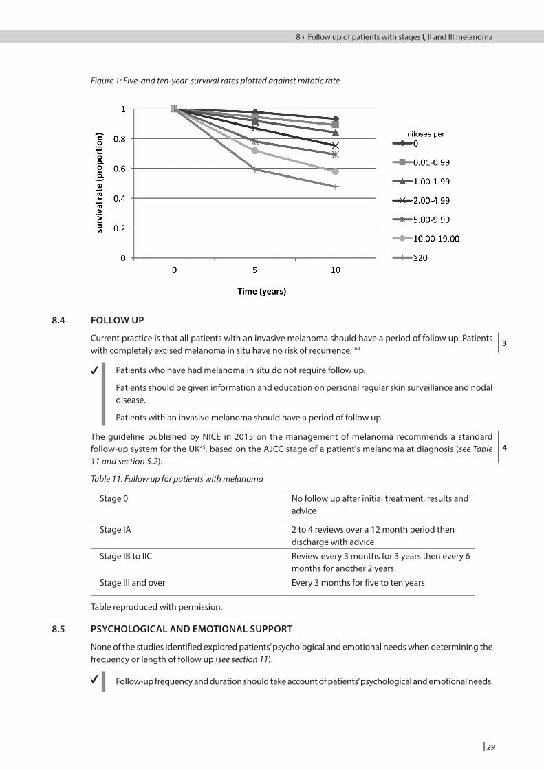
| 29
Cutaneous melanoma 8 • Follow up of patients with stages I, II and III melanoma
Figure 1: Five-and ten-year survival rates plotted against mitotic rate
8.4 FOLLOW UP
Current practice is that all patients with an invasive melanoma should have a period of follow up. Patients with completely excised melanoma in situ have no risk of recurrence.164
4 Patients who have had melanoma in situ do not require follow up.
Patients should be given information and education on personal regular skin surveillance and nodal disease.
Patients with an invasive melanoma should have a period of follow up.
The guideline published by NICE in 2015 on the management of melanoma recommends a standard follow-up system for the UK45, based on the AJCC stage of a patient's melanoma at diagnosis (see Table 11 and section 5.2).
Table 11: Follow up for patients with melanoma
Stage 0 No follow up after initial treatment, results and advice
Stage IA 2 to 4 reviews over a 12 month period then discharge with advice
Stage IB to IIC Review every 3 months for 3 years then every 6 months for another 2 years
Stage III and over Every 3 months for five to ten years
Table reproduced with permission.
8.5 PSYCHOLOGICAL AND EMOTIONAL SUPPORT
None of the studies identified explored patients’ psychological and emotional needs when determining the frequency or length of follow up (see section 11).
4 Follow-up frequency and duration should take account of patients’ psychological and emotional needs.
3
4
30 |
Cutaneous melanoma
8.6 SECOND PRIMARIES
Three retrospective studies found second primaries in 1.2%, 2% and 7% of their patients.164,170,180 The timing of discovery ranged from synchronous with the initial melanoma to more than 10 years later. The second primaries were usually thinner. One paper estimated that the Scottish patients in their study had a 200-fold increase in risk of developing a second melanoma compared to the general population180 (see Table 2, section 3.4.2).
8.7 DETECTING RECURRENCES
Large retrospective studies have shown that 90% of recurrent disease in patients with stage I and II melanoma is detected solely by signs or symptoms noted either by the patient or the physician, with imaging techniques detecting the remainder.165,167,168,181,182 Patients’ own detection rates in between clinic visits are generally in the range of 33–72%164,166,168,171,182,183 but in one study where patients with stage I-III melanoma were meticulously educated in self-examination techniques the rate of self detection rose to 100%.184 The rate of self detection in one prospective trial was much lower at 17%.183 Three retrospective studies indicate that the overall survival time is the same for patient-detected recurrences as for those detected in the clinic.168,171,182
8.7.1 ROUTINE LABORATORY TESTS
Routine laboratory tests (full blood count and liver function tests) do not detect asymptomatic recurrent disease in patients with stage I-III melanoma.137,165-167,184
Lactate dehydrogenase is a marker of liver metastases and tumour burden in patients with stage IV disease that indicates a poor prognosis.182,185,186 Two studies have looked at LDH as a first indicator of metastases. An elevated LDH was the first indicator in 12.5% of patients with stage III disease (with a sensitivity of 73%) when tested for every three months.138 In a prospective cohort study of stages I-IV disease an elevated LDH was the first indicator in 2% of recurrences when patients were tested every 12 months (stages I and II melanoma) or every six months (stages III and IV melanoma).172
8.7.2 SURVEILANCE IMAGING
Overall, the quality of evidence for the method (including imaging), frequency and duration of surveillance in patients with melanoma is very low in all clinical outcomes of interest.45 This is demonstrated by the lack of a consensus on an approach to surveillance from a wide variety of different dermatological and oncological organisations.187
A meta-analysis of prospective and retrospective studies addressing the diagnostic accuracy of imaging modalities for surveillance of patients with melanoma reported that PET-CT has the highest sensitivity and specificity for detecting distant metastases (sensitivity 86%, 95% CrI 76% to 93%; specificity 91%, 95% CrI 79% to 97%), compared to CT (sensitivity 63%, 95% CrI 46% to 77%; specificity 78%, 95% CrI 58% to 90%).131 However, many of the included studies were retrospective and of poor quality with wide inclusion criteria and insufficient reporting of withdrawals. Several potential sources of bias were also identified including referral bias, verification bias and review bias. A further review of the role of PET-CT in the surveillance of patients with malignant melanoma found a sensitivity of 96% and specificity of 92%.188 There was no direct comparison to CT. The authors criticised the lack of high quality prospective studies in this field and highlighted a number of possible biases in the studies, especially referral bias. Neither of these reviews included data on patient outcomes and the studies looked at diagnostic accuracy only.
Whilst surveillance imaging has potential benefits, there are also considerable potential disadvantages that need to be taken into account. These are summarised in the NICE guideline on melanoma (see Table 12). 45
4
3
3
2++
4
2+
4
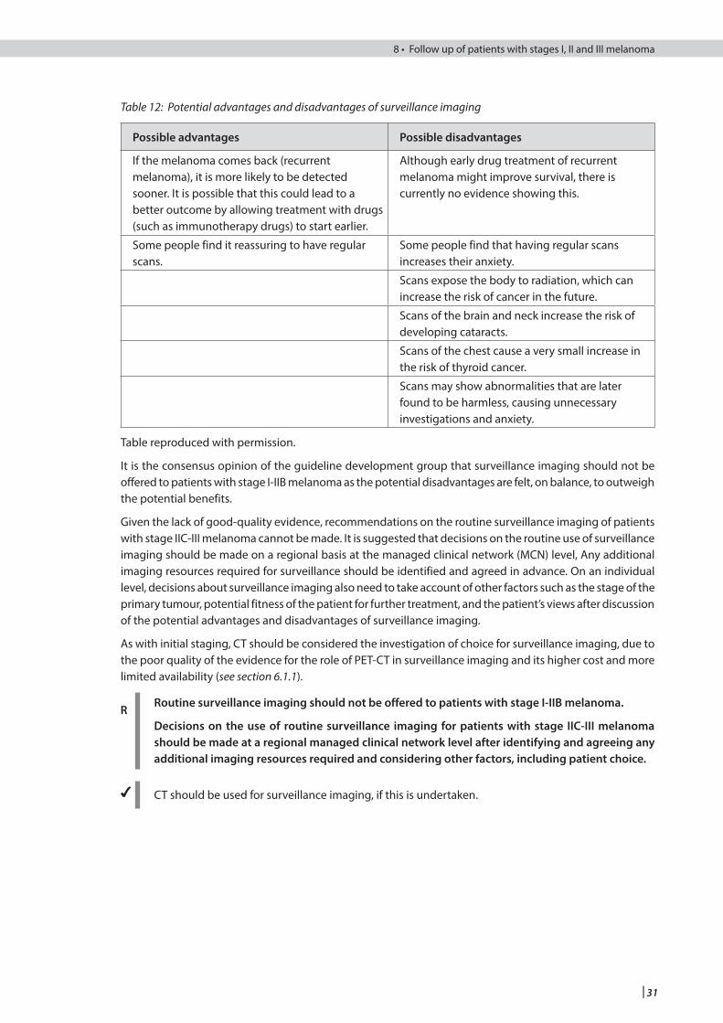
| 31
Cutaneous melanoma 8 • Follow up of patients with stages I, II and III melanoma
Table 12: Potential advantages and disadvantages of surveillance imaging
Possible advantages Possible disadvantages
If the melanoma comes back (recurrent melanoma), it is more likely to be detected sooner. It is possible that this could lead to a better outcome by allowing treatment with drugs (such as immunotherapy drugs) to start earlier.
Although early drug treatment of recurrent melanoma might improve survival, there is currently no evidence showing this.
Some people find it reassuring to have regular scans.
Some people find that having regular scans increases their anxiety.
Scans expose the body to radiation, which can increase the risk of cancer in the future.
Scans of the brain and neck increase the risk of developing cataracts.
Scans of the chest cause a very small increase in the risk of thyroid cancer.
Scans may show abnormalities that are later found to be harmless, causing unnecessary investigations and anxiety.
Table reproduced with permission.
It is the consensus opinion of the guideline development group that surveillance imaging should not be offered to patients with stage I-IIB melanoma as the potential disadvantages are felt, on balance, to outweigh the potential benefits.
Given the lack of good-quality evidence, recommendations on the routine surveillance imaging of patients with stage IIC-III melanoma cannot be made. It is suggested that decisions on the routine use of surveillance imaging should be made on a regional basis at the managed clinical network (MCN) level, Any additional imaging resources required for surveillance should be identified and agreed in advance. On an individual level, decisions about surveillance imaging also need to take account of other factors such as the stage of the primary tumour, potential fitness of the patient for further treatment, and the patient’s views after discussion of the potential advantages and disadvantages of surveillance imaging.
As with initial staging, CT should be considered the investigation of choice for surveillance imaging, due to the poor quality of the evidence for the role of PET-CT in surveillance imaging and its higher cost and more limited availability (see section 6.1.1).
RRoutine surveillance imaging should not be offered to patients with stage I-IIB melanoma.
Decisions on the use of routine surveillance imaging for patients with stage IIC-III melanoma should be made at a regional managed clinical network level after identifying and agreeing any additional imaging resources required and considering other factors, including patient choice.
4 CT should be used for surveillance imaging, if this is undertaken.
32 |
Cutaneous melanoma
9 Management of advanced (unresectable stage IIIC or IV) melanoma
9.1 INTRODUCTION
Recent years have seen the development of several new treatment options for patients with advanced melanoma including BRAF and mitogen-activated protein kinase (MEK) inhibitors and several novel immunotherapies. All of these treatments are associated with significantly improved outcomes although the optimal choice, sequence and combination of therapies are still to be determined. It is now also recognised that there are several different genomic subtypes of melanoma 189 although translating this increased understanding into the development of other new therapies for patients with melanoma remains under investigation.
4 All patients with advanced melanoma should be tested for mutations in BRAF and have their management discussed at a specialist MDT in order to determine the optimal management strategy taking into account patient fitness, co-morbidity, disease burden and overall aim of treatment.
All patients with advanced melanoma should be offered the opportunity to participate in clinical trials.
9.2 SURGERY
Metastasectomy may be an option for patients with distant skin, node and visceral metastases. In subcutaneous metastases prevention of ulceration of superficial lesions is best prevented by resection when the lesions are at a size where skin closure is possible. Surgery of single or localised metastases has been shown to be associated with improved survival.190 The proportion of patients suitable for metastasectomy ranges from 10% to 25%.191-193 Five-year survival of 14-33% was described in one retrospective review for those with distant subcutaneous and lung metastases respectively. This study showed prognostic significance for Breslow thickness, number of metastases and prior disease-free interval.191
4 Metastasectomy should be considered in patients with stage IV disease
9.3 SYSTEMIC THERAPY
9.3.1 BRAF AND MEK INHIBITORS
Development of BRAF inhibitors (vemurafenib and dabrafenib) as single agents or in combination with a MEK inhibitor (cobimetanib and trametinib) represents a major advance for patients with advanced melanoma.
Two open label RCTs demonstrated that BRAF inhibitors (vemurafenib and dabrafanib) improved response and progression-free survival (PFS) compared to chemotherapy alone in patients with unresectable stage IIIC or stage IV BRAF mutation-positive melanoma with a response rate of 48% and 50% versus 5% and 6%; PFS 5.3 and 5.1 months versus 1.6 and 2.7 months respectively.194,195 Response is further improved with the combination of a BRAF inhibitor (vemurafenib or dabrafenib) and a MEK inhibitor (cobimentinib or trametinib), with an improved response rate and PFS compared to a BRAF inhibitors alone (response rate 64–68% versus 45–51% for BRAF inhibitors alone and PFS 9.3 to 11.4 months versus 6.2 to 8.8 months).196-198
The toxicity profile for BRAF inhibitors compared to combination BRAF and MEK inhibitors is diverse: grade 3-4 toxicity rates range from 28–63% for BRAF inhibitor alone and 35–65% for combination therapy.194-198
Vemurafenib and dabrafenib are accepted for use by the SMC as monotherapy for the treatment of patients with BRAF V600 mutation-positive unresectable or metastatic melanoma as first-line therapy (see section 12.4). Trametinib in combination with dabrafenib is approved for use in the treatment of adult patients with unresectable or metastatic melanoma with a BRAF V600 mutation.
RTrametinib in combination with dabrafenib is recommended for patients with unresectable stage IIIC or stage IV melanoma with a BRAF V600 mutation.
1_
1+
| 33
Cutaneous melanoma 9 • Management of advanced (unresectable stage IIIC or IV) melanoma
9.3.2 IMMUNOTHERAPIES
Development of novel immunotherapies (ipilimumab, pembrolizumab and nivolumab) as single agents or in combination represents a major advance for patients with advanced melanoma.
Several RCTs have demonstrated that novel immunotherapies are effective in improving outcomes in patients with unresectable stage IIIC or stage IV melanoma.
A trial comparing ipilimumab to glycoprotein100 (gp100) for second-line therapy found that ipilimumab was associated with improved overall survival (OS) of 10.1 months versus 6.4 months (HR 0.66; p=0.003). 199
Compared to chemotherapy for first-line treatment, nivolumab had a PFS of 5.1 versus 2.2 months, HR 0.43, 95% CI 0.34 to 0.56; one-year OS was 72.9% versus 42.1%, HR 0.42, p<0.001.200
Ipilimumab has also been compared to nivolumab and pembrolizumab in RCTs.
Pembrolizumab (two-weekly or three-weekly) was associated with an improved six-month PFS of 47.3% (two-weekly) or 46.4% (three-weekly) compared to 26.5% for ipilimumab, HR 0.58; p,0.001; one-year OS was 74.1%, 68.4% or 58.2% respectively HR 0.63; p=0.0005 for two-weekly pembrolizumab, HR 0.69; p=0.0036 for three-weekly pembrolizumab; the response rate was 33.7% versus 32.9% versus 11.9% (p<0.001 for both comparisons).201
The combination of nivolumab and ipilimumab improved outcomes compared to ipilimumab or nivolumab alone (PFS 11.5 months (combination) versus 2.9 months (ipilimumab) versus 6.9 months (nivolumab), HR 0.42; p<0.001). This study also confirmed that the outcomes for nivolumab were significantly improved compared to ipilimumab; PFS 6.9 months versus 2.9 months, HR 0.57 (p<0.00001).191
All of the novel immunotherapy agents are associated with a significant risk of autoimmune toxicity including colitis. Grade 3–4 toxicity rates are generally lower with single agent nivolumab (11.7%) and pembrolizumab (10.1–13.3%), higher with ipilimumab (10–19.9%) and highest with the combination of nivolumab and ipilimumab (55%).199-203
While there is evidence of efficacy for novel immunotherapies, optimal choice, sequence and combination of therapies are still to be determined. Ipilimumab, pembrolizumab and nivolumab monotherapy and ipilimumab/nivolumab combination therapy have been considered and accepted for use by the SMC (with restrictions) (see section 12.4).
RIpilimumab, pembrolizumab and nivolumab monotherapy or ipilimumab/nivolumab combination therapy are recommended for patients with unresectable stage IIIC and IV melanoma.
9.4 ISOLATED LIMB PERFUSION
Isolated limb perfusion (ILP) is a surgical technique that allows localised delivery of a high dose of chemotherapy (usually melphalan). Minimal systemic leakage occurs, confining toxicity to the limb. ILP has been used in two clinical situations:204,205
• adjuvant treatment for high-risk primary melanoma• therapeutic treatment for major limb recurrence of melanoma.
Isolated limb perfusion is a significant surgical undertaking and should only be made available in centres where a high number of such operations are performed each year. One centre can provide this service for a population of approximately five million people.
4 ILP should be performed in a specialist centre.
9.4.1 ADJUVANT TREATMENT
A prospective multicentre RCT involving 832 patients showed that prophylactic ILP with melphalan cannot be recommended in patients with a high-risk primary limb melanoma.206
4 ILP should not be used as an adjuvant treatment.
1+
1+
1+
1+
34 |
Cutaneous melanoma
9.4.2 THERAPEUTIC TREATMENT
A multicentre RCT207 and reviews208,209 suggest that hyperthermic ILP with melphalan alone or melphalan plus TNF alpha can produce a complete though short lived response rate ranging from 50 to 90% in patients with limb recurrence (in-transit metastases).
4 ILP is a treatment option for patients with bulky disease confined to one limb.
9.5 ABLATIVE THERAPIES
9.5.1 CARBON DIOXIDE LASER ABLATION
A carbon dioxide laser delivers short wavelength energy in a focussed light beam to destroy tumour nodules. It can be applied under local anaesthetic, can be repeated and provides effective local disease control.210-212
4 Carbon dioxide laser ablation can be considered for multiple lesions of trunk or abdomen and for limb disease.
Other similar treatments are available and vary locally.
9.5.2 ELECTROCHEMOTHERAPY
Electrochemotherapy (ECT) uses short electric pulses to increase the absorption of either intralesional or intravenous chemotherapy.213 It can be used in patients who have had previous surgery, radiotherapy and isolated limb perfusion/infusion, and may provide further treatment options when others have been exhausted.214,215
Meta-analyses report a high response rate to ECT in patients with cutaneous metastases.213,214 ECT had a complete response of 56.8% and, for complete response and partial response combined, an objective response of 80.6% in patients with melanoma compared to 8% and 19.9% for chemotherapy alone for all tumour types (with no significant difference found between tumour type).214 ECT was well tolerated with 90% of patients reported to be amenable to further treatment if needed.213 Minor side effects from treatment were muscle spasms, skin changes, nausea and fatigue.215
Data on long-term survival or quality of life is limited.215 No studies were identified comparing the efficacy of ECT with recently developed immunotherapies.
No evidence on the cost effectiveness of ECT was identified. It is currently only available in one centre in Scotland: NHS Tayside. Delivery of ECT requires specialist equipment and training.
RElectrochemotherapy should be considered as a treatment option for patients with cutaneous melanoma metastases after multidisciplinary team discussion and careful consideration of alternative systemic therapy options, or when other options have been exhausted.
9.6 RADIOTHERAPY
9.6.1 RADIOSENSITIVITY
There is evidence that melanoma cells in vitro have a spectrum of radio sensitivity and that melanoma should not be considered a uniformly radio resistant disease.216 Experimental studies have suggested that atypical, large radiotherapy fraction sizes may be more efficacious than standard treatments but at present there are no randomised trials to support the routine use of large fraction sizes.216,217
1+
4
1+
1+
43
1+
4
| 35
Cutaneous melanoma 9 • Management of advanced (unresectable stage IIIC or IV) melanoma
9.6.2 BONE METASTASES
Studies looking at the treatment of bone metastases usually include only a small percentage of patients with melanoma. Recommendations have been extrapolated from the data available from studies of bone metastases from various tumour types. When using single fractions to palliate pain from bone metastases, an 8 Gy fraction is effective and provides superior pain relief to lower doses.218 There does not appear to be an advantage to using 20 Gy in four fractions over an 8 Gy single fraction.219 Some patients may benefit from higher dose, fractionated regimens, although this has not been fully established.220
R Single-dose radiotherapy of a least 8 Gy may be an effective treatment for bone metastases.
9.6.3 SPINAL CORD COMPRESSION
There is no clear evidence to support or refute the use of radiotherapy (in combination with other treatments) to alleviate the pain and neurological deficit associated with spinal cord compression caused by metastatic melanoma.221,222
The value of surgical intervention in such patients has been established.222,226 Patients with symptoms of spinal cord compression should be referred urgently to an appropriate surgeon.223
4 If a patient presents with spinal cord compression consideration should be given to available medical oncology options. BRAF testing should be considered if this has not already been done, and targeted BRAF therapy should be considered in new cases.
9.6.4 BRAIN METASTASES
Although central nervous system (CNS) involvement by melanoma is a common finding at autopsy, brain metastases are diagnosed in only approximately 10% of patients before death.224 For cerebral metastases from all tumour types, good performance status, favourable response to corticosteroid treatment, and the absence of systemic disease are statistically significant predictive factors for a better survival.225
Postoperative radiotherapy has been used as adjuvant treatment following the resection of CNS disease. However, no survival benefit from postoperative radiotherapy has been demonstrated.224,226 Radiotherapy without surgery, combined with corticosteroids appears to palliate the symptoms of some patients with inoperable cerebral metastases from melanoma but there is no evidence of a survival benefit.216,226,227 Radiosurgery (stereotactic radiotherapy) has been used to treat patients with inoperable disease who are fit enough to undergo this procedure, and the results may be equivalent to surgery alone.228
Two non-comparative studies were identified on BRAF inhibitors in patients with brain metastases,229,230 which do not alter the current recommendation on the use of BRAF inhibitors (see section 9.3.1).
Two Cochrane reviews addressing brain metastases were identified. The first concluded that adding whole-brain radiotherapy (WBRT) to surgery or stereotactic radiosurgery (SRS) did not show a survival benefit over surgery or SRS alone.231 The other concluded that there was low-quality evidence that adding upfront WBRT to surgery or SRS decreases any intracranial disease progression at one year but no clear evidence of an effect on overall and progression free survival.232
RPatients with good performance status, favourable response to corticosteroid treatment, absence of systemic disease and who harbour favourable CNS disease should be considered for surgical resection of their CNS disease.
If surgery is not possible, patients should be considered for systemic therapy.
4 All patients with brain-limited metastasis should tested for BRAF mutations and their management discussed at a neuro-oncology multidisciplinary team to determine the optimal choice of treatment including systemic or targeted therapy, surgery or stereotactic radiosurgery.
2+
2++
4
42+
1+
2+
3
34
36 |
Cutaneous melanoma
9.7 SPECIALIST PALLIATIVE CARE
The General Medical Council has stated that basic palliative care skills are required by every member of the medical profession.233 Clinical Standards for the provision of both basic and specialist palliative care are available.234 Specialist palliative care is an integral component of the care of patients with advanced malignancy, required at varying times during their illness. SIGN guideline number 106 (Control of pain in patients with cancer) covers pain control in patients with all cancers.235
Patients who develop metastatic melanoma require input from a number of agencies both within and outwith the health service. They may need rehabilitative, functional, social and/or financial support services, most of which are available in specialist palliative care settings, as well as in primary care and cancer centres. The evaluation of the effectiveness of specialist palliative care involves assessment of the different dimensions of care provided, such as pain and other symptom control, psychological care, care of the family and carers, rehabilitation and terminal care.
Three RCTs were identified that included all carcinomas, which, in the context of palliative care, are reasonable to relate to patients with melanoma.236-238 The first two studies looked at the effect of co-ordinating all services available within the NHS, local authorities and the voluntary sector through the addition of nurse co-ordinators. A total of 203 cancer patients expected to live for less than one year were randomly assigned to either the intervention or the routine services group. Patients assigned to the intervention group spent fewer days in hospital, required fewer home visits and their family were less likely to feel angry about their relative’s death.236,237 The third RCT used place of death as the outcome measure in a study of 434 patients with incurable malignant disease.238 The intervention group had inpatient and outpatient hospital services provided by the palliative medicine unit, the unit served as a link to community services, predefined guidelines maintained communication between services and community staff took part in an educational programme. Significantly more intervention group patients died at home and spent less time in nursing homes in their last months of life.
A systematic review of the effectiveness of specialist palliative care teams identified 18 studies, including five RCTs.239 Specialist palliative care teams were associated with more time spent at home by patients, satisfaction of patients and their carers, symptom control, a reduction in the number of inpatient hospital days, a reduction in overall cost, and with the patient dying where they wished.
RPatients with advanced melanoma require a co-ordinated multiprofessional approach with input from a specialist palliative care team.
Patients with poorly controlled symptoms should be referred to specialist palliative care at any point in the cancer journey.
1+
1+
| 37
Cutaneous melanoma 10 • Melanoma in women
10 Melanoma in women
10.1 PREGNANCY
Pregnancy is frequently associated with increased activity of benign melanocytes leading to pigmentary changes. This has led to concern that pregnancy is deleterious for women with melanoma.
The prognoses of women with thickness-matched melanomas who embarked on a pregnancy after apparently successful surgical treatment of AJCC stage I or II melanoma have been compared.240-243 No difference in disease-free or overall survival is found between women who have, and women who have not, become pregnant after melanoma treatment. Prognosis is mainly dependent on tumour thickness.240-243
There is no substantial evidence of an effect of pregnancy in women with stage III and IV melanoma, but as the prognosis for these groups is already poor, the possibility of a maternal death during pregnancy or when the child is an infant is high.
The placenta of an infant born to a mother with stage III or IV melanoma should be examined for the presence of melanoma metastases. If they are present there is a 20% risk of death of the baby from transplacental melanoma.244-246
Women who develop melanoma during a pregnancy show a greater mean thickness of the primary lesion at the time of excision than age-matched non-pregnant women.242,243 This suggests either delayed diagnosis or accelerated growth due to the hormonal and immunological environment of pregnancy. There is no evidence to support the suggestion that it is physiological for melanocytic naevi to change during pregnancy.247
There are no good data on prognosis for women who embark on a pregnancy having had a melanoma diagnosed and treated during a previous pregnancy. One paper reports that patients with stage I or II disease have no greater recurrence rate than non-pregnant age-matched controls but that those with nodal disease have significantly higher recurrence rates.248
4 Women with a significant risk of recurrence (localised disease of ≥1mm thickness) who wish to become pregnant after surgery for stage I or II melanoma should be advised to delay pregnancy for two years postsurgery, as the likelihood of recurrence is highest during this period.
Pregnant women who present with growing or changing pigmented lesions should be treated as non-pregnant women.
10.2 ORAL CONTRACEPTION AFTER MELANOMA TREATMENT
Meta-analysis provides no evidence that use of the oral contraceptive is a risk factor for melanoma.249 Five large studies indicate that oral contraceptive use by women after surgery for stage I or II melanoma does not adversely affect their prognosis.248,250-254
4 Women who have had a melanoma treated should select contraception in the same way as women who have not had a melanoma
10.3 HORMONE REPLACEMENT THERAPY AFTER MELANOMA TREATMENT
Five case controlled studies show no effect of hormone replacement therapy (HRT) as a risk factor for melanoma.251,252,255-257
4 Women who have had stage I and II melanoma and who wish to take HRT should be treated as women who have not had melanoma.
2++
2++
2+
2++
3
4
38 |
Cutaneous melanoma
11 Provision of information
This section reflects the issues likely to be of most concern to patients and their carers. These points are provided for use by health professionals when discussing melanoma with patients and carers and in guiding the production of locally produced information materials.
11.1 INFORMATION PROVISION
An RCT of patients with stage I melanoma suggests that a structured information programme to inform patients about melanoma progression and treatment options increased patients’ knowledge of the disease, the risk factors involved and possible preventative measures.258 The study reported no difference in psychological variables. A second RCT found that facilitated education programmes for patients with stage I and II melanoma, in which one element was an information programme about cancer recurrence, had a positive effect on coping behaviour and affective distress values.259 A prospective cohort study with patients with metastatic disease found that those who understood the expected outcomes of their disease had higher quality of life scores and longer survival periods.260
The provision of information to patients increases their knowledge of the disease, can enhance coping behaviour and reduce levels of affective distress.
R Patients should receive targeted information throughout their journey of care.
4 Healthcare professionals working with cancer patients should have training in communication skills.
11.2 COMMUNICATION
A Cochrane review, an RCT, three cohort studies and a survey were identified that cover a wide range of issues related to communication skills, all demonstrating strongly that communication skills training for healthcare professionals is of lasting benefit.261-266
The following have all been shown to be potentially effective communication tools or strategies:
• health related quality of life measurements • needs assessment tools • recorded consultation • audio of general information • summary letter as follow up • presence of support person • actively encouraging questions and a question prompt list • patient-held record.
R Communication skills training should be provided across the multidisciplinary team.
4 Information needs should be resourced and provided using a variety of media, to meet individual patient/carer needs.
11.3 PATIENT SUPPORT GROUPS
Patients benefit from psychoeducational interventions provided in a structured group, facilitated by qualified personnel.258-260,267 The studies suggest that facilitated groups can help patients cope better at all stages of their disease, increase knowledge levels and reduce affective distress.
4 Health service patient support groups should be structured, facilitated by trained professionals and incorporate health education.
Information on all patient support groups should be made easily available to patients.
1_
1+
2
1+
2+
3
2+
1_
| 39
Cutaneous melanoma 11 • Provision of information
11.4 CHECKLIST FOR PROVISION OF INFORMATION
This section gives examples of the information patients/carers may find helpful at the key stages of the patient journey. The checklist was designed by members of the guideline development group based on their experience and their understanding of the evidence base. The checklist is neither exhaustive nor exclusive.
In primary care
• Advise patients that removal or biopsy of the tumour may occur at the initial visit. • Advise patients that it is appropriate to refer them to a specialist and how long they should expect
to wait for an appointment.• Explain how photo triage (if available) can be of benefit.
At the specialist clinic
• Explain to patients how a diagnosis will be reached including: – clinical examination – types of biopsy and the need for local anaesthetic – how, when and by whom biopsy results will be given.
• With any surgical procedure, whether small biopsy or large excision, explain about surgical complications which include: pain, swelling, bleeding, bruising, loss of function and unpredictable scarring, including keloid scarring.
• Advise patients about how long they should expect to spend at the hospital. • Be clear about the time between biopsy results and treatment. • Describe what treatments will be offered. • Where possible, give patients written information about appointment waiting times and details of
who to contact if appointment is not received within 2 weeks.
At the specialist clinic once the diagnosis is known
• Explain the nature of the patient’s particular cutaneous melanoma in precise terms. • Explain what further treatment options there are and which are appropriate for the patient.• Explain whether any other tests are appropriate, such as scans. • Give as much information as possible about the likely prognosis. • Explain how the majority of melanomas arise. • Where appropriate, explain that the patient’s case will be referred to the MDT. • Explain whether other specialists will be involved in the treatment, such as, plastic surgeons, oral
and maxillofacial surgeons, oncologists, clinical nurse specialists, etc. • Explain what might be involved in any one particular treatment eg flaps, grafts, complex
reconstruction, sentinel lymph node biopsy. • Explain what might be involved in recuperation and rehabilitation and realistic timescales for
recovery (including scar potential and healing time, especially in visible areas).• Try to give the patient some idea of the time to their definitive treatment, acknowledging that this
might be difficult if other specialists are involved.
40 |
Cutaneous melanoma
At follow up
• Discuss how well the treatment went and whether any further treatment is needed: surgery, radiotherapy or input from oncologists.
• Discuss the prognosis in light of the definitive treatment. • Discuss the risk of recurrence and how the patient might detect this, and whether any tests are
indicated to detect recurrence. • Advise the patient about the likely length of follow up. • Ensure patients are aware of the support available from a clinical nurse specialist and other health
professionals eg Maggie’s centres, MacMillan Cancer Support or camouflage clinic and refer if appropriate.
• Allow sufficient time to discuss the following with patients: – psychological adjustment after a diagnosis and treatment for skin cancer – anxiety and low mood – coping strategies – being visibly different/stigma – use of camouflage and cosmetics– assessment and management of lymphoedema for those patients at risk.
• Advise patients to bring a written list of questions or concerns. A proforma that addresses these aspects can focus the discussion time.
• Offer patient education about self care for example: – self checking, skin examination, checking lymph nodes and getting to know their body– what to look for, eg features of abnormal skin lesions and what actions to take if they are
concerned (it is also useful to detect any other health issues that require medical assessment).– discuss prevention including:
- use of high-factor sunscreen and protective clothing- the damaging effects of sun beds- the need for precautions while working and taking holidays in the UK.
• Provide patients with written information leaflets and advise them how they can access self-help groups (see section 11.5).
11.5 SOURCES OF FURTHER INFORMATION
11.5.1 GENERAL INFORMATION
NHS inform A national health information service for Scotland.Website: www.nhsinform.scotwww.nhsinform.scot/illnesses-and-conditions/cancerThe NHS inform cancer page is full of practical and emotional support to help you or someone you know who is living with cancer.
Skin cancer (melanoma)www.nhsinform.scot/illnesses-and-conditions/cancer/cancer-types-in-adults/skin-cancer-melanoma NHS inform can help you find detailed information on different cancers, make your own customised cancer leaflet, find support groups and more.
Care, support and rightsInformation on accessing healthand care services can be found here.Tel: 0800 22 44 88 (8am–10pm)www.nhsinform.scot/care-support-and-rights/
| 41
Cutaneous melanoma 11 • Provision of information
11.5.2 ORGANISATIONS SPECIFIC TO SKIN CONDITIONS
British Association of Dermatologists (BAD) Willan House, 4 Fitzroy Square, London W1T 5HQTel: 0207 383 0266Email: [email protected] of the aims of the British Association of Dermatologists is to raise awareness of all aspects of skin disease. This charity provides a range of patient information leaflets.
British Skin Foundation4 Fitzroy Square, London W1T 5HQTel: 020 7391 6341www.britishskinfoundation.org.ukThe British Skin Foundation supports research into skin conditions. It provides information on the treatment of skin cancers.
Changing Faces ScotlandTel: 0845 4500 640 (Monday to Thursday, 8.30am-3pm) Email: [email protected] www.changingfaces.org.ukChanging Faces provide psychological support to people and families who are living with conditions, marks or scars that affect their appearance.
MASScot (Melanoma Action and Support Scotland)A skin cancer charity208 Clyde Street Glasgow G1 4JY, Tel: 0141 221 9878Email: [email protected] number: SCIO 040286www.masscot.org.ukMASScot is a Scottish charity run by skin cancer patient volunteers. They provide qualified and insured therapists, near to you, free of charge. MASScot campaigns for improvements in prevention, detection and care, and works with primary and secondary schools to promote sun awareness. They aim to make the public aware of the dangers of sunburn.
11.5.3 ORGANISATIONS SPECIFIC TO CANCER
Cancer Support Centre, Cancer Support ScotlandThe Calman Centre, 75 Shelley Road, Glasgow G12 0ZEFreephone: 0800 652 4531Tel: 0141 337 8199Email: [email protected] Calman Cancer Support Centre provides emotional and practical support on a one-to-one basis and through community-based groups. It provides complementary and talking therapies to anyone affected by cancer.
Cancer Research UKAngel Building, 407 St John Street, London EC1V 4AD Tel: 0808 800 4040 (Monday to Friday, 9am–5pm)www.cancerresearchuk.orgCancer research UK funds research into cancer, campaigns on cancer issues and produces patient information leaflets.
42 |
Cutaneous melanoma
Macmillan Cancer Support89 Albert Embankment, London, SE1 7UQTel: 0808 808 00 00 (Monday to Friday, 9am–8pm).www.macmillan.org.ukThird floor, 132 Rose Street, Edinburgh. EH2 3JDTel: 0131 260 3720Email: [email protected] Macmillan supports people with cancer and their families with specialist information, treatment and care.
Maggie’s Cancer Caring Centres ScotlandThe Gatehouse,10 Dumbarton Road, Glasgow G11 6PATel: 0300 123 1801Email: [email protected]’s provides practical, emotional and social support to people with cancer, their family and friends. Built alongside NHS cancer hospitals and staffed with professional experts, Maggie’s Centres are warm and welcoming, full of light and open space, with a big kitchen table at their heart. In Scotland there are Maggie’s Centres in Glasgow, Airdrie, Edinburgh, Kirkaldy, Dundee, Aberdeen and Inverness.
Marie Curie Cancer Care in Scotland133 Balornock Road, Stobhill Hospital Grounds, Glasgow G21 3USTel: 0131 561 3900Email: [email protected] www.mariecurie.org.ukMarie Curie Cancer Care is dedicated to the cure of people affected by cancer and improving their quality of life through its caring services, research and education.
Teenage Cancer TrustThird floor, 93 Newman Street, London. W1T 3EZTel: 020 7612 0370Email: [email protected]
11.5.4 CANCER NETWORKS IN SCOTLAND
Scotland’s cancer networks offer a range of support and advice to patients and families, including support groups and written information.
North of Scotland Cancer Network (NOSCAN)Rosehill Annexe, Aberdeen Royal Infirmary Site, Cornhill Road, Aberdeen AB25 2ZGTel: 01224 552 745www.noscan.scot.nhs.uk
South East Scotland Cancer Network (SCAN)Pentland House, 47 Robb’s Loan, Edinburgh EH14 1TYTel: 0131 465 7681www.scan.scot.nhs.uk
West of Scotland Cancer Network (WOSCAN)1st Floor, St Mungo Building, Glasgow Royal Infirmary, 84 Castle Street, Glasgow G4 0SFTel: 0141 211 1145www.woscan.scot.nhs.uk
| 43
Cutaneous melanoma 12 • Implementing the guideline
12 Implementing the guideline
This section provides advice on the resource implications associated with implementing the key clinical recommendations, and advice on audit as a tool to aid implementation.
12.1 IMPLEMENTATION STRATEGY
Implementation of national clinical guidelines is the responsibility of each NHS board and is an essential part of clinical governance. Mechanisms should be in place to review care provided against the guideline recommendations. The reasons for any differences should be assessed and addressed where appropriate. Local arrangements should then be made to implement the national guideline in individual hospitals, units and practices.
Implementation of this guideline will be encouraged and supported by SIGN. The implementation strategy for this guideline encompasses the following tools and activities.
12.2 RESOURCE IMPLICATIONS OF KEY RECOMMENDATIONS
No recommendations are considered likely to reach the £5 million threshold which warrants full cost impact analysis.
12.3 AUDITING CURRENT PRACTICE
The Cancer quality performance indicators (QPIs) have been developed by Healthcare Improvement Scotland in collaboration with the three Regional Cancer Networks (NOSCAN, SCAN and WOSCAN) and the NHS Information Services Division (ISD). QPIs will be kept under regular review and be responsive to changes in clinical practice and emerging evidence.
The overarching aim of the cancer quality work programme is to ensure that activity at NHS board level is focused on areas most important in terms of improving survival and patient experience whilst reducing variance and ensuring safe, effective and person-centred cancer care.
Further information on QPIs can be found on the Healthcare Improvement Scotland website www.healthcareimprovementscotland.org/our_work/cancer_care_improvement/cancer_qpis.aspx
12.4 HEALTH TECHNOLOGY ASSESSMENT ADVICE FOR NHSSCOTLAND
In 2013 the SMC advised that ipilimumab is accepted for use in NHSScotland for the treatment of advanced (unresectable or metastatic) melanoma in adults who have received prior therapy.
In 2013 the SMC advised that vemurafenib is accepted for restricted use as first-line treatment of BRAF V600 mutation-positive unresectable or metastatic melanoma.
In 2014 the SMC advised that ipilimumab is accepted for use in NHSScotland for the treatment of advanced (unresectable or metastatic) melanoma in adults (first-line use).
In 2015 SMC advised that dabrafenib is accepted for restricted use as monotherapy treatment for adult patients with unresectable or metastatic melanoma with a BRAF V600 mutation. It is restricted for use only in those patients who have received no prior therapy.
In 2015 SMC advised that it had approved pembrolizumab as monotherapy for the treatment of advanced (unresectable or metastatic) melanoma in adults previously untreated with ipilimumab
In 2016 SMC advised that it had approved nivolumab as monotherapy for the treatment of advanced (unresectable or metastatic) melanoma in adults previously untreated with nivolumab.
In 2016 SMC advised that it had approved trametinib in combination with dabrafenib for the first-line treatment of adult patients with unresectable or metastatic melanoma with a BRAF V600 mutation.
In 2016 SMC advised that nivolumab in combination with ipilimumab is accepted for the first-line treatment of adult patients with unresectable or metastatic melanoma.
44 |
Cutaneous melanoma
13 The evidence base
13.1 SYSTEMATIC LITERATURE REVIEW
The evidence base for this guideline was synthesised in accordance with SIGN methodology. A systematic review of the literature was carried out using an explicit search strategy devised by a SIGN Information Officer. Databases searched include Medline, Embase, Cinahl, PsycINFO and the Cochrane Library. The year range covered was 2004–2016. Internet searches were carried out on various websites including the US National Guidelines Clearinghouse. The main searches were supplemented by material identified by individual members of the development group. Each of the selected papers was evaluated by two Evidence and Information Scientists using standard SIGN methodological checklists before conclusions were considered as evidence by the guideline development group.
13.1.1 LITERATURE SEARCH FOR PATIENT ISSUES
At the start of the guideline development process, a SIGN Evidence and Information Scientist conducted a literature search for qualitative and quantitative studies that addressed patient issues of relevance to early management of patients with melanoma. Databases searched include Medline, Embase, Cinahl and PsycINFO, and the results were summarised by the SIGN Patient Involvement Officer and presented to the guideline development group.
13.1.2 LITERATURE SEARCH FOR COST-EFFECTIVENESS EVIDENCE
The guideline development group identified key questions with potential cost-effectiveness implications, based on the following criteria, where it was judged particularly important to gain an understanding of the additional costs and benefits of different treatment strategies:
• treatments which may have a significant resource impact• opportunities for significant disinvestment or resource release• the potential need for significant service redesign• cost-effectiveness evidence could aid implementation of a recommendation.
A systematic literature search for economic evidence for these questions was carried out by a SIGN Evidence and Information Scientist covering the years 2004–2016. Databases searched include Medline, Embase, NHS Economic Evaluation Database (NHS EED) and Health Economics Evaluation Database (HEED). Each of the selected papers was evaluated by a Health Economist, and considered for clinical relevance by guideline group members.
Interventions are considered to be cost effective if they fall below the commonly-accepted UK threshold of £20,000 per quality-adjusted life year (QALY).
13.2 RECOMMENDATIONS FOR RESEARCH
The guideline development group was not able to identify sufficient evidence to answer all of the key questions asked in this guideline (see Annex 1). The following areas for further research have been identified:
• Large studies on the behaviour of lentigo maligna and eventual risk of invasion• A comparison of behaviour of lentigo maligna melanoma with superficial spreading malignant melanoma• Large cohort studies on pure desmoplastic subtype biological potential compared to superficial spreading
or mixed desmoplastic subtype, and for animal type versus superficial spreading• Prospective study comparison of CT and MRI for identifying brain metastases in patients with melanoma• RCTs on the efficacy of electrochemotherapy with immunotherapy versus immunotherapy alone• An RCT of the role of regular surveillance imaging on the survival of patients with stage III melanoma
compared to routine follow up including different imaging modalities and the optimal interval for imaging• Trials on the sequencing of immune agents and targeted agents• RCTs comparing the efficacy of combination BRAF inhibitors with novel immunotherapies• RCTs on the efficacy of BRAF inhibitors after immunotherapy.

| 45
Cutaneous melanoma 13 • The evidence base
13.3 REVIEW AND UPDATING
This guideline was issued in 2017 and will be considered for review in three years. The review history, and any updates to the guideline in the interim period, will be noted in the review report, which is available in the supporting material section for this guideline on the SIGN website: www.sign.ac.uk
Comments on new evidence that would update this guideline are welcome and should be sent to the SIGN Executive, Gyle Square, 1 South Gyle Crescent, Edinburgh, EH12 9EB (email: [email protected]).
46 |
Cutaneous melanoma
14 Development of the guideline
14.1 INTRODUCTION
SIGN is a collaborative network of clinicians, other healthcare professionals and patient organisations and is part of Healthcare Improvement Scotland. SIGN guidelines are developed by multidisciplinary groups of practising healthcare professionals using a standard methodology based on a systematic review of the evidence. Further details about SIGN and the guideline development methodology are contained in ‘SIGN 50: A Guideline Developer’s Handbook’, available at www.sign.ac.uk
This guideline was developed according to the 2015 edition of SIGN 50.
14.2 THE GUIDELINE DEVELOPMENT GROUP
Dr Ewan Brown (Chair) Consultant in Medical Oncology, Edinburgh Cancer Centre, Western General Hopsital, Edinburgh
Dr Stuart Ballantyne Clinical Lead for Radiology, Gartnavel General Hospital, Glasgow
Ms Juliet Brown Evidence and Information Scientist, Healthcare Improvement Scotland
Dr Richard Casasola Consultant Clinical Oncologist, Tayside Cancer Centre, Dundee
Dr Mark Darling Consultant Dermatologist, New Victoria Hospital, Glasgow
Ms Amanda Degabriele MacMillan Skin Cancer Clinical Nurse Specialist, Ninewells Hospital, Dundee
Dr Robert Dickie General Practitioner, The Group Practice, Isle of Lewis
Ms Sheena Dryden Clinical Nurse Specialist – Dermatology, NHS Lothian, Lauriston Buildings, Edinburgh
Ms Elaine Fletcher Specialist Registrar in Clinical Genetics, Western General Hospital, Edinburgh
Dr Susannah Fraser Consultant Dermatologist, Queen Margaret Hospital, Dunfermline and Victoria Hospital, Fife
Mr Stephen Heller-Murphy Programme Manager, SIGN
Dr Alex Holme Consultant Dermatologist, Royal Infirmary of Edinburgh
Ms Frances Kelly Lay representative, Wishaw
Dr Lucy Melly Consultant Pathologist, Southern General Hospital, Glasgow
Mr Owen Moseley Senior Health Economist, Healthcare Improvement Scotland
Dr Colin Moyes Consultant Dermatopathologist, Southern General Hospital, Glasgow
Mr Omar Quaba Consultant Plastic Surgeon, Ninewells Hospital, Dundee
Dr Sanjay Rajpara Consultant Dermatologist, Aberdeen Royal Infirmary
Dr Alan Simms Consultant Radiologist, St John’s Hospital, Livingston
Ms Leigh Smith Lay representative, Chair of Melanoma Action and Support Scotland
Ms Ailsa Stein Programme Manager, SIGN
Dr Lorna Thompson Health Services Researcher, Healthcare Improvement Scotland
Dr Ashita Waterston Consultant Medical Oncologist, Beatson West of Scotland Cancer Centre, Glasgow
| 47
Cutaneous melanoma 14 • Development of the guideline
The membership of the guideline development group was confirmed following consultation with the member organisations of SIGN. All members of the guideline development group made declarations of interest. A register of interests is available in the supporting material section for this guideline at www.sign.ac.uk
Guideline development and literature review expertise, support and facilitation were provided by SIGN Executive and Healthcare Improvement Staff. All members of the SIGN Executive make yearly declarations of interest A register of interests is available on the contacts page of the SIGN website www.sign.ac.uk
Euan Bremner Project Officer
Karen Graham Patient Involvement Officer
Karen King Distribution and Office Co-ordinator
Stuart Neville Publications Designer
Gaynor Rattray Guideline Co-ordinator
Dr Carolyn Sleith Evidence and Information Scientist, Healthcare Improvement Scotland
14.2.1 ACKNOWLEDGEMENTS
SIGN would like to acknowledge the guideline development group responsible for the development of SIGN 72: Cutaneous melanoma, on which this guideline is based.
14.3 CONSULTATION AND PEER REVIEW
A report of the consultation and peer review comments and responses is available in the supporting material section for this guideline on the SIGN website. All expert referees and other contributors made declarations of interest and further details of these are available on request from the SIGN Executive.
14.3.1 SPECIALIST REVIEWERS INVITED TO COMMENT ON THIS DRAFT
This guideline was also reviewed in draft form by the following independent expert referees, who were asked to comment primarily on the comprehensiveness and accuracy of interpretation of the evidence base supporting the recommendations in the guideline. The guideline group addresses every comment made by an external reviewer, and must justify any disagreement with the reviewers’ comments.
SIGN is very grateful to all of these experts for their contribution to the guideline.
Ms Marissa Collins Researcher in Health Economics, Glasgow Caledonian University, Glasgow
Professor Alan Denison Consultant Radiologist, Summerfield House, Aberdeen
Dr Val Doherty Dermatologist, Royal Infirmary of Edinburgh, Edinburgh
Mrs Wilma Ford MacMillan Skin Cancer Clinical Nurse Specialist, Southern General Hospital, Glasgow
Dr Matthew Hough Consultant Plastic Surgeon, Ninewells Hospital, Dundee.
Mrs Kirsty MacFarlane Principal Pharmacist, Scottish Medicines Consortium, Healthcare Improvement Scotland
Dr Colin Malone Locum Consultant Dermatologist, Dumfries and Galloway Royal Infirmary
Dr Megan Mowbray Consultant Dermatologist, Queen Margaret Hospital, Dunfermline
Dr Lisa Naysmith Consultant Dermatological Surgeon, Royal Infirmary of Edinburgh
48 |
Cutaneous melanoma
Dr Marianne Nicolson Consultant Medical Oncologist, Aberdeen Royal Infirmary, Aberdeen
Professor Mary Porteous Consultant Clinical Geneticist, Western General Hospital, Edinburgh
Dr Charlotte Proby Professor of Dermatology, University of Dundee
Dr James Vestey Consultant Dermatologist, Raigmore Hospital, Inverness
14.3.2 PUBLIC CONSULTATION
The draft guideline was also available on the SIGN website for a month to allow all interested parties to comment.
14.3.3 SIGN EDITORIAL GROUP
As a final quality control check, the guideline is reviewed by an editorial group comprising the relevant specialty representatives on SIGN Council to ensure that the specialist reviewers’ comments have been addressed adequately and that any risk of bias in the guideline development process as a whole has been minimised. The editorial group for this guideline was as follows. All members of SIGN Council make yearly declarations of interest. A register of interests is available on the SIGN Council Membership page of the SIGN website www.sign.ac.uk.
Dr Roberta James SIGN Programme Lead; Co-Editor
Professor John Kinsella Chair of SIGN; Co-Editor
Mr Alan Timmins Royal Pharmaceutical Society
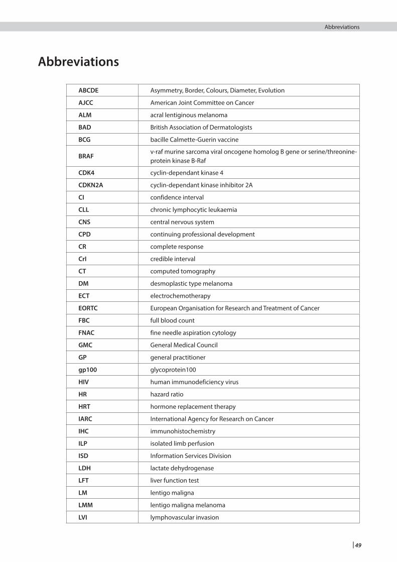
| 49
Cutaneous melanoma Abbreviations
Abbreviations
ABCDE Asymmetry, Border, Colours, Diameter, Evolution
AJCC American Joint Committee on Cancer
ALM acral lentiginous melanoma
BAD British Association of Dermatologists
BCG bacille Calmette-Guerin vaccine
BRAFv-raf murine sarcoma viral oncogene homolog B gene or serine/threonine-protein kinase B-Raf
CDK4 cyclin-dependant kinase 4
CDKN2A cyclin-dependant kinase inhibitor 2A
CI confidence interval
CLL chronic lymphocytic leukaemia
CNS central nervous system
CPD continuing professional development
CR complete response
CrI credible interval
CT computed tomography
DM desmoplastic type melanoma
ECT electrochemotherapy
EORTC European Organisation for Research and Treatment of Cancer
FBC full blood count
FNAC fine needle aspiration cytology
GMC General Medical Council
GP general practitioner
gp100 glycoprotein100
HIV human immunodeficiency virus
HR hazard ratio
HRT hormone replacement therapy
IARC International Agency for Research on Cancer
IHC immunohistochemistry
ILP isolated limb perfusion
ISD Information Services Division
LDH lactate dehydrogenase
LFT liver function test
LM lentigo maligna
LMM lentigo maligna melanoma
LVI lymphovascular invasion
50 |
Cutaneous melanoma
MA marketing authorisation
MCN managed clinical network
MDT multidisciplinary team meeting
MEK mitogen-activated protein kinase
MIA melanoma inhibitory activity
MMS mohs micrographic surgery
MRI magnetic resonance imaging
MTA multiple technology appraisal
NHL non-Hodgkin lymphoma
NICE National Institute for Health and Care Excellence
NM nodular melanoma
NOSCAN North of Scotland Cancer Network
OR odds ratio
OS overall survival
PCR polymerase chain reaction
PET positron emission tomography
PET-CT positron emission tomography–computed tomography
PFS progression free survival
QALY quality-adjusted life year
QPI quality performance indicator
RCPath Royal College of Pathologists
RCT randomised control trial
SCAN South East Scotland Cancer Network
SIGN Scottish Intercollegiate Guidelines Network
SLN sentinel lymph nodes
SLNB sentinel lymph node biopsy
SMC Scottish Medicines Consorium
SPC summary of product characteristics
SPF sun protection factor
SRS stereotactic radiosurgery
SSMM superficial spreading malignant melanoma
TIL tumour infiltrating lymphocytes
TNF tumour necrosis factor
TNM tumour, node, metastases
US ultrasound
UVA ultraviolet A
UVB ultraviolet B
WBRT whole-brain radiotherapy
WOSCAN West of Scotland Cancer Network
| 51
Cutaneous melanoma Annexes
This guideline is based on a series of structured key questions that define the target population, the intervention, diagnostic test, or exposure under investigation, the comparison(s) used and the outcomes used to measure efficacy, effectiveness, or risk. These questions form the basis of the systematic literature search.
Guideline section Key question
4.1 1. What is the rate/risk of local recurrence and/or metastasis and survival in patients with desmoplastic melanoma or animal type melanoma compared to patients with superficial spreading?Comparison: superficial spreadingOutcomes: local recurrence, metastasis, survival
5.3 2. In which patients with melanoma is sentinel lymph node biopsy an effective tool for staging, predicting outcome or guiding treatment options?
Patients with melanoma:a) <1 mm Breslow thickness with mitotic rate 1 or moreb) intermediate thickness 1 mm to 3.5 mmc) thick melanoma >3.5 mm
Interventions: SLNB (to exclude stage 3 disease)
Comparisons: observation, clinical follow up, high definition ultrasound
Outcomes: sensitivity/specificity for identification of microscopic metastases, overall survival (5 years, 10 years), disease free survival (5 years, 10 years),adverse events
5.3 3. In patients with malignant melanoma who are SLNB+ what is the most effective management strategy?Interventions: completion lymphadenectomy (clearance/complete lymph node dissection)Comparisons: serial ultrasound, observationOutcomes: recurrence, overall survival (5 years, 10 years), disease free survival (5 years, 10 years), adverse events
4.1 4. What evidence is there for treatment of lentigo maligna and lentigo maligna melanoma?
Patients with: lentigo maligna, lentigo maligna melanoma
Interventions: conventional surgery, Mohs micrographic surgery, radiotherapy, cryotherapy, imiquimod, 5-FU, observation
Comparisons: conventional surgery, Mohs micrographic surgery, radiotherapy, cryotherapy, imiquimod, 5-FU, observation
Outcomes: disease clearance/recurrence, development of invasive disease, adverse events, cosmesis, patient satisfaction.
6.1 5. In patients with malignant melanoma, who should undergo radiology imaging as part of their initial staging investigations?Patients with: melanoma stage I-II, stage IIIInterventions: radiological stagingComparisons: between stage I-II and stage IIIOutcomes: sensitivity, specificity, positive predictive value, false positive predictive value, patient anxiety, patient satisfaction
Annex 1 Key questions addressed in this update
52 |
Cutaneous melanoma
6.1 6. What is the best radiology modality for systemic staging of patients with malignant melanoma?
Interventions and comparisons: CT v PET/CT, PET/CT v no radiology, CT v no radiology
Outcomes: sensitivity, specificity, positive predictive value, false positive predictive value
6.19.6
7. Is CT or MRI better for diagnosing brain metastases in patients with malignant melanoma?
Outcomes: sensitivity, specificity, positive predictive value, false positive predictive value
8.4 8. What is the clinical and cost effectiveness of routine radiological follow up of patients with treated malignant melanoma?
Patients with: treated malignant melanoma – stage IIb or greater
Interventions: radiology follow up investigations at various intervals
Comparison: clinical follow up only
Outcomes: sensitivity, specificity, positive predictive value, false positive predictive value, patient anxiety, patient satisfaction
8.7 9. What is the best imaging modality for use in routine follow up for detection of recurrence in patients with malignant melanoma?
Patients with: treated malignant melanoma – stage IIb or greater
Interventions and comparisons: chest x-ray, CT, PET/CT, ultrasound
Outcomes: sensitivity, specificity, positive predictive value, false positive predictive value
9.5 10. What is the clinical and cost effectiveness of electrochemotherapy (ECT) in the management of patients with melanoma skin metastases?
Patients with: stage III/IV disease, cutaneous metastases
Interventions: ECT
Comparisons: isolated limb perfusion, laser, radiotherapy, surgery
Outcomes: response rates, overall survival (5 years, 10 years), progression free survival (5 years, 10 years), adverse effects
9.3 11. In patients with advanced melanoma (unresectable stage IIIC or stage IV) which is the most clinically and cost effective systemic therapy?
Interventions: • BRAF inhibitors (vemurafenib, dabrafenib)• MEK inhibitors (trametinib, cobimetinib) • immunotherapy (ipilimumab, pembrolizumab, nivolumab, interferon
interleukin-2)• chemotherapy (dacarbazine, paclitaxel, carboplatin)• cKIT inhibitors (imatinib)• combination treatment (eg vemurafenib/cobimetinib and dabrafenib/
trametinib)
Comparisons: chemotherapy, observation
Outcomes: overall survival (5 years, 10 years), progression free survival (5 years, 10 years), response rate, toxicity
| 53
Cutaneous melanoma Annexes
7.1 12. In patients with completely resected stage III melanoma what is the benefit from adjuvant radiotherapy?
Interventions: radiotherapy
Comparison: observation
Outcomes: overall survival (5 years, 10 years), progression free survival (5 years, 10 years), local recurrence, toxicity
7.3 13. What is the relationship between the immune system and melanoma?
54 |
Cutaneous melanoma
1 ISD. Cancer Incidence in Scotland (2013). Edinburgh: Information Services Division; 2015. [cited 22 Jul 2015]. Available from url: https://isdscotland.scot.nhs.uk/Health-Topics/Cancer/Publications/2015-04-28/2015-04-28-Cancer-Incidence-Report.pdf?20609682799
2 Joint Formulary Committee. British National Formulary: Guidance on Prescribing (online). [cited 08 January 2016]. Available from url: http:www.medicinescomplete.com
3 IARC monographs on the evaluation of carcinogenic risks to humans. Volume 55: solar and ultraviolet radiation. Lyon: International Agency for Research on Cancer 1992. [cited 05/01/2017].
4 Elwood JM, Jopson J. Melanoma and sun exposure: an overview of published studies. Int J Cancer 1997;73(2):198-203.
5 Whiteman DC, Whiteman CA, Green AC. Childhood sun exposure as a risk factor for melanoma: a systematic review of epidemiologic studies. Cancer Causes Control 2001;12(1):69-82.
6 Wang SQ, Setlow R, Berwick M, Polsky D, Marghoob AA, Kopf AW, et al. Ultraviolet A and melanoma: a review. J Am Acad Dermatol 2001;44(5):837-46.
7 Hill L, Ferrini RL. Skin cancer prevention and screening: summary of the American College of Preventive Medicine's practice policy statements. CA Cancer J Clin 1998;48(4):232-5.
8 Ferrini RL, Perlman M, Hill L. American College of Preventive Medicine policy statement: Screening for skin cancer. Am J Prev Med 1998;14(1):80-2.
9 Ness AR, Frankel SJ, Gunnell DJ, Smith GD. Are we really dying for a tan? BMJ 1999;319(7202):114-6.
10 NHMRC. Clinical practice guidelines for the management of cutaneous melanoma. Canberra: National Health and Medical Research Council; 1999. [cited 16 May 2003]. Available from url: http://www.health.gov.au/nhmrc/publications/pdf/cp68.pdf
11 The Cancer Council Australia, Australian Cancer Network, Ministry of Health NZ. Clinical Practice Guidelines for the Management of Melanoma in Australia and New Zealand. Wellington: The Cancer Council Australia and Australian Cancer Network, Sydney and New Zealand Guidelines Group 2008.
12 BAD. Stage One Melanoma Patient Information Leaflet. 2013;
13 Crane LA, Schneider LS, Yohn JJ, Morelli JG, Plomer KD. "Block the sun, not the fun": evaluation of a skin cancer prevention program for child care centers. Am J Prev Med 1999;17(1):31-7.
14 Richard MA, Martin S, Gouvernet J, Folchetti G, Bonerandi JJ, Grob JJ. Humour and alarmism in melanoma prevention: a randomized controlled study of three types of information leaflet. Br J Dermatol 1999;140(5):909-14.
15 Dey P, Collins S, Will S, Woodman CBJ. Randomised controlled trial assessing effectiveness of health education leaflets in reducing incidence of sunburn. BMJ 1995;311(7012):1062-3.
16 Hanrahan PF, Hersey P, Watson AB, Callaghan TM. The effect of an educational brochure on knowledge and early detection of melanoma. Aust J Public Health 1995;19:270-4.
17 Segan CJ, Borland R, Hill DJ. Development and evaluation of a brochure on sun protection and sun exposure for tourists. Health Educ J 1999;58(2):177-91.
18 Hanrahan PF, Hersey P, Menzies SW, Watson AB, D'Este CA. Examination of the ability of people to identify early changes of melanoma in computer-altered pigmented skin lesions. Arch Dermatol 1997;133(3):301-11.
19 Kiekbusch S, Hannich HJ, Isacsson A, Johannisson A, Lindholm LH, Sager E, et al. Impact of a cancer education multimedia device on public knowledge, attitudes, and behaviors: a controlled intervention study in Southern Sweden. J Cancer Educ 2000;15(4):232-6.
20 Sefton E, Glazebrook C, Garrud P, Zaki I. Educating patients about malignant melanoma: Computer-assisted learning in a pigmented lesion clinic. Br J Dermatol 2000;142(1):66-71.
21 Bastuji-Garin S, Grob JJ, Grognard C, Grosjean F, Guillaume JC. Melanoma prevention: evaluation of a health education campaign for primary schools. Arch Dermatol 1999;135(8):936-40.
22 Helfand M, Mahon S, Eden K. Screening for skin cancer. Rockville (MD): Agency for Healthcare Research and Quality; 2001. (AHRQ publication No. 01-S002). [cited 16 May 2003]. Available from url: http://hstat.nlm.nih.gov/hq/Hquest/db/3601/screen/DocTitle/odas/1/s/36462
23 Melia J, Harland C, Moss S, Eiser JR, Pendry L. Feasibility of targeted early detection for melanoma: a population-based screening study. Br J Cancer 2000;82(9):1605-9.
24 Katris P, Donovan RJ, Gray BN. The use of targeted and non-targeted advertising to enrich skin cancer screening samples. Br J Dermatol 1996;135(2):268-74.
25 Goldstein AM, Chidambaram A, Halpern A, Holly EA, Guerry ID, Sagebiel R, et al. Rarity of CDK4 germline mutations in familial melanoma. Melanoma Res 2002;12(1):51-5.
26 Bruno W, Ghiorzo P, Battistuzzi L, Ascierto PA, Barile M, Gargiulo S, et al. Clinical genetic testing for familial melanoma in Italy: a cooperative study. J Am Acad Dermatol 2009;61(5):775-82.
27 Wadt KA, Aoude LG, Krogh L, Sunde L, Bojesen A, Gronskov K, et al. Molecular characterization of melanoma cases in Denmark suspected of genetic predisposition. PLoS One 2015;10(3):e0122662.
28 Watts CG, Dieng M, Morton RL, Mann GJ, Menzies SW, Cust AE. Clinical practice guidelines for identification, screening and follow-up of individuals at high risk of primary cutaneous melanoma: a systematic review. Br J Dermatol 2015;172(1):33-47.
29 Lang J, Boxer M, MacKie RM. CDKN2A mutations in Scottish families with cutaneous melanoma: results from 32 newly identified families. Br J Dermatol 2005;153(6):1121-5.
30 Glanz K, Volpicelli K, Kanetsky PA, Ming ME, Schuchter LM, Jepson C, et al. Melanoma genetic testing, counseling, and adherence to skin cancer prevention and detection behaviors. Cancer Epidemiol Biomarkers Prev 2013;22(4):607-14.
31 Helgadottir H, Hoiom V, Jonsson G, Tuominen R, Ingvar C, Borg A, et al. High risk of tobacco-related cancers in CDKN2A mutation-positive melanoma families. J Med Genet 2014;51(8):545-52.
32 Pawlik TM, Ross MI, Prieto VG, Ballo MT, Johnson MM, Mansfield PF, et al. Assessment of the role of sentinel lymph node biopsy for primary cutaneous desmoplastic melanoma. Cancer 2006;106(4):900-6.
33 Hawkins WG, Busam KJ, Ben-Porat L, Panageas KS, Coit DG, Gyorki DE, et al. Desmoplastic melanoma: a pathologically and clinically distinct form of cutaneous melanoma. Ann Surg Oncol 2005;12(3):207-13.
References
| 55
Cutaneous melanoma References
34 Maurichi A, Miceli R, Camerini T, Contiero P, Patuzzo R, Tragni G, et al. Pure desmoplastic melanoma: a melanoma with distinctive clinical behavior. Ann Surg 2010;252(6):1052-7.
35 Arora A, Lowe L, Su L, Rees R, Bradford C, Cimmino VC, et al. Wide excision without radiation for desmoplastic melanoma. Cancer 2005;104(7):1462-7.
36 Ludgate MW, Fullen DR, Lee J, Rees R, Sabel MS, Wong SL, et al. Animal-type melanoma: a clinical and histopathological study of 22 cases from a single institution. Br J Dermatol 2010;162(1):129-36.
37 Mandal RV, Murali R, Lundquist KF, Ragsdale BD, Heenan P, McCarthy SW, et al. Pigmented epithelioid melanocytoma: favorable outcome after 5-year follow-up. Am J Surg Pathol 2009;33(12):1778-82.
38 Zembowicz A, Carney JA, Mihm MC. Pigmented epithelioid melanocytoma: a low-grade melanocytic tumor with metastatic potential indistinguishable from animal-type melanoma and epithelioid blue nevus. Am J Surg Pathol 2004;28(1):31-40.
39 Healsmith MF, Bourke JF, Osborne JE, Graham-Brown RA. An evaluation of the revised seven-point checklist for the early diagnosis of cutaneous malignant melanoma. Br J Dermatol 1994;130(1):48-50.
40 Fitzpatrick TB, Rhodes AR, Sober AJ, Mihm MC. Primary malignant melanoma of the skin: the call for action to identify persons at risk; to discover precursor lesions; to detect early melanomas. Pigment Cell 1988;9(110):7.
41 Duff CG, Melsom D, Rigby HS, Kenealy JM, Townsend PL. A 6 year prospective analysis of the diagnosis of malignant melanoma in a pigmented-lesion clinic: even the experts miss malignant melanomas, but not often. Br J Plast Surg 2001;54(4):317-21.
42 Richard MA, Grob JJ, Avril MF, Delaunay M, Gouvernet J, Wolkenstein P, et al. Delays in diagnosis and melanoma prognosis (II): the role of doctors. Int J Cancer 2000;89(3):280-5.
43 Gerbert B, Maurer T, Berger T, Pantilat S, McPhee SJ, Wolff M, et al. Primary care physicians as gatekeepers in managed care: Primary care physicians' and dermatologists' skills at secondary prevention of skin cancer. Arch Dermatol 1996;132(9):1030-8.
44 Bafounta ML, Beauchet A, Aegerter P, Saiag P. Is dermoscopy (epiluminescence microscopy) useful for the diagnosis of melanoma? Results of a meta-analysis using techniques adapted to the evaluation of diagnostic tests. Arch Dermatol 2001;137(10):1343-50.
45 National Institute for Health and Clincial Excellence. Melanoma: assessment and management. London: NICE; 2015. [cited 05/01/2017]. Available from url: https://www.nice.org.uk/guidance/ng14
46 Westerhoff K, McCarthy WH, Menzies SW. Increase in the sensitivity for melanoma diagnosis by primary care physicians using skin surface microscopy. Br J Dermatol 2000;143(5):1016-20.
47 Binder M, Schwarz M, Winkler A, Steiner A, Kaider A, Wolff K, et al. Epiluminescence microscopy. A useful tool for the diagnosis of pigmented skin lesions for formally trained dermatologists. Arch Dermatol 1995;131(3):286-91.
48 Benelli C, Roscetti E, Pozzo VD, Gasparini G, Cavicchini S. The dermoscopic versus the clinical diagnosis of melanoma. Eur J Dermatol 1999;9(6):470-6.
49 Maurer J, Knollmann FD, Schlums D, Garbe C, Vogl TJ, Bier J, et al. Role of high-resolution magnetic resonance imaging for differentiating melanin-containing skin tumors. Invest Radiol 1995;30(11):638-43.
50 Lassau N, Spatz A, Avril MF, Tardivon A, Margulis A, Mamelle G, et al. Value of high-frequency US for preoperative assessment of skin tumors. Radiographics 1997;17(6):1559-65.
51 Chwirot BW, Chwirot S, Redzinski J, Michniewicz Z. Detection of melanomas by digital imaging of spectrally resolved ultraviolet light-induced autofluorescence of human skin. Eur J Cancer 1998;34(11):1730-4.
52 Aitken JF, Pfitzner J, Battistutta D, O'Rourke PK, Green AC, Martin NG. Reliability of computer image analysis of pigmented skin lesions of Australian adolescents. Cancer 1996;78(2):252-7.
53 O'Donnell B, Dervan P, Codd M, Powell F, Lawlor D, O'Loughlin S. A clinicopathological correlation of 134 stage 1 and 79 non-invasive cutaneous melanomas presenting over a decade (1984-1993) at the Mater Misericordiae Hospital, Dublin. Ir J Med Sci 1998;167(3):132-5.
54 Lennon GM, Griffin M, O'Briain DS, Cassidy M, Caldwell M, Young M, et al. Malignant melanoma lately diagnosed. Ir Med J 1989;82(3):109-11.
55 Baccard M, Havard S, Souques M. Prospective study of the incidence of melanoma in the Paris region in 1994. Melanoma Res 1997;7(4):335-8.
56 Oliveria SA, Christos PJ, Halpern AC, Fine JA, Barnhill RL, Berwick M. Patient knowledge, awareness, and delay in seeking medical attention for malignant melanoma. J Clin Epidemiol 1999;52(11):1111-6.
57 Richard MA, Grob JJ, Avril MF, Delaunay M, Gouvernet J, Wolkenstein P, et al. Delays in diagnosis and melanoma prognosis (I): the role of patients. Int J Cancer 2000;89(3):271-9.
58 Franke W, Neumann NJ, Ruzicka T, Schulte KW. Plantar malignant melanoma - A challenge for early recognition. Melanoma Res 2000;10(6):571-6.
59 Bennett DR, Wasson D, MacArthur JD, McMillen MA. The effect of misdiagnosis and delay in diagnosis on clinical outcome in melanomas of the foot. J Am Coll Surg 1994;179(3):279-84.
60 Rampen FH, Rumke P, Hart AA. Patients' and doctors' delay in the diagnosis and treatment of cutaneous melanoma. Eur J Surg Oncol 1989;15(2):143-8.
61 Del Mar CB, Green AC, Battistutta D. Do public media campaigns designed to increase skin cancer awareness result in increased skin excision rates? Aust N Z J Public Health 1997;21(7):751-4.
62 Gerbert B, Bronstone A, Wolff M, Maurer T, Berger T, Pantilat S, et al. Improving primary care residents' proficiency in the diagnosis of skin cancer. J Gen Intern Med 1998;13(2):91-7.
63 Calonje E. ACP best practice no 162. The histological reporting of melanoma. J Clin Pathol 2000;53(8):587-90.
64 Griffiths RW, Briggs JC. Biopsy procedures, primary wide excisional surgery and long term prognosis in primary clinical stage I invasive cutaneous malignant melanoma. Ann R Coll Surg Engl 1985;67(2):75-8.
65 Lees VC, Briggs JC. Effect of initial biopsy procedure on prognosis in Stage 1 invasive cutaneous malignant melanoma: review of 1086 patients. Br J Surg 1991;78(9):1108-10.
66 Pariser RJ, Divers A, Nassar A. The relationship between biopsy technique and uncertainty in the histopathologic diagnosis of melanoma. Dermatol Online J 1999;5(2):4.
67 Lederman JS, Sober AJ. Does biopsy type influence survival in clinical stage I cutaneous melanoma? J Am Acad Dermatol 1985;13(6):983-7.
68 Witheiler DD, Cockerell CJ. Sensitivity of diagnosis of malignant melanoma: a clinicopathologic study with a critical assessment of biopsy techniques. Exp Dermatol 1992;1(4):170-5.
56 |
Cutaneous melanoma
69 Bong JL, Herd RM, Hunter JA. Incisional biopsy and melanoma prognosis. J Am Acad Dermatol 2002;46(5):690-4.
70 Coleman WP, Davis RS, Reed RJ, Krementz ET. Treatment of lentigo maligna and lentigo maligna melanoma. J Dermatol Surg 1980;6(6):476-9.
71 Rosai J. Rosai and Ackerman’s Surgical Pathology. St Louis (MO): Mosby; 2011.
72 Slater D, Walsh M. Dataset for the histological reporting of primary cutaneous malignant melanoma and regional lymph nodes. London: The Royal College of Pathologists; 2014.
73 Breslow A. Thickness, cross-sectional areas and depth of invasion in the prognosis of cutaneous melanoma. Ann Surg 1970;172(5):902-8.
74 Cochran AJ, Elashoff D, Morton DL, Elashoff R. Individualized prognosis for melanoma patients. Hum Pathol 2000;31(3):327-31.
75 Clark WH, Jr., Elder DE, Guerry Dt, Braitman LE, Trock BJ, Schultz D, et al. Model predicting survival in stage I melanoma based on tumor progression. J Natl Cancer Inst 1989;81(24):1893-904.
76 Barnhill RL, Fine JA, Roush GC, Berwick M. Predicting five-year outcome for patients with cutaneous melanoma in a population-based study. Cancer 1996;78(3):427-32.
77 Balch CM, Buzaid AC, Atkins MB, Cascinelli N, Coit DG, Fleming ID, et al. A new American Joint Committee on Cancer staging system for cutaneous melanoma. Cancer 2000;88(6):1484-91.
78 Balch CM, Buzaid AC, Soong SJ, Atkins MB, Cascinelli N, Coit DG, et al. Final version of the American Joint Committee on Cancer staging system for cutaneous melanoma. J Clin Oncol 2001;19(16):3635-48.
79 Edge S, Compton C. The American Joint Committee on Cancer: the 7th edition of the AJCC cancer staging manual and the future of TNM. Ann Surg Oncol 2010; 17: 1471-4.
80 Day CL, Jr., Mihm MC, Jr., Lew RA, Harris MN, Kopf AW, Fitzpatrick TB, et al. Prognostic factors for patients with clinical stage I melanoma of intermediate thickness (1.51 - 3.39 mm). A conceptual model for tumor growth and metastasis. Ann Surg 1982;195(1):35-43.
81 Balch CM. Microscopic satellites around a primary melanoma: another piece of the puzzle in melanoma staging. Ann Surg Oncol 2009;16(5):1092-4.
82 Gershenwald JE, Buzaid AC, Ross MI. Classification and staging of melanoma. Clin Lab Med 2000;20(4):785-815.
83 Borgstein PJ, Meijer S, van Diest PJ. Are locoregional cutaneous metastases in melanoma predictable? Ann Surg Oncol 1999;6(3):315-21.
84 Massi D, Borgognoni L, Franchi A, Martini L, Reali UM, Santucci M. Thick cutaneous malignant melanoma: a reappraisal of prognostic factors. Melanoma Res 2000;10(2):153-64.
85 Guerry IDP, Synnestvedt M, Elder DE, Schultz D. Lessons from tumor progression: The invasive radial growth phase of melanoma is common, incapable of metastasis, and indolent. J Invest Dermatol 1993;100(3):342S-5S.
86 Blessing K, McLaren KM, McLean A, Davidson P. Thin malignant melanomas (less than 1.5 mm) with metastasis: a histological study and survival analysis. Histopathology 1990;17(5):389-95.
87 Cook MG. Diagnostic discord with melanoma. J Pathol 1997;182(3):247-9.
88 Corona R, Mele A, Amini M, De Rosa G, Coppola G, Piccardi P, et al. Interobserver variability on the histopathologic diagnosis of cutaneous melanoma and other pigmented skin lesions. J Clin Oncol 1996;14(4):1218-23.
89 Weinstock MA, Barnhill RL, Rhodes AR, Brodsky GL, Abell E, Hurley J, et al. Reliability of the histophalogic diagnosis of melanocytic dysplasia. Arch Dermatol 1997;133(8):953-8.
90 Rosai J. Ackerman's Surgical Pathology. 8th. St Louis (MO): Mosby; 1996.
91 Cochran AJ. Sentinel lymph node pathology. In: Kirkham N, Lemoine NR, editors. Progress in pathology. London: Greenwich Medical Media; 2001.
92 ADASP. Recommendations for processing and reporting lymph node specimens submitted for evaluation of metastatic disease. Am J Surg Pathol 2001;25(7):961-3.
93 Cochran AJ. Surgical pathology remains pivotal in the evaluation of 'sentinel' lymph nodes. Am J Surg Pathol 1999;23(10):1169-72.
94 Gutzmer R, Kaspari M, Brodersen JP, Mommert S, Volker B, Kapp A, et al. Specificity of tyrosinase and HMB45 PCR in the detection of melanoma metastases in sentinel lymph node biopsies. Histopathology 2002;41(6):510-8.
95 Veronesi U, Cascinelli N, Adamus J, Balch C, Bandiera D, Barchuk A, et al. Thin stage I primary cutaneous malignant melanoma. Comparison of excision with margins of 1 or 3 cm. N Engl J Med 1988;318(18):1159-62.
96 Cohn-Cedermark G, Rutqvist LE, Andersson R, Breivald M, Ingvar C, Johansson H, et al. Long term results of a randomized study by the Swedish Melanoma Study Group on 2-cm versus 5-cm resection margins for patients with cutaneous melanoma with a tumor thickness of 0.8-2.0 mm. Cancer 2000;89:1495-501.
97 Ringborg U, Andersson R, Eldh J, Glaumann B, Hafstrom L, Jacobsson S, et al. Resection margins of 2 versus 5 cm for cutaneous malignant melanoma with a tumor thickness of 0.8 to 2.0 mm: randomized study by the Swedish Melanoma Study Group. Cancer 1996;77:1809-14.
98 Heaton KM, Sussman JJ, Gershenwald JE, Lee JE, Reintgen DS, Mansfield PF, et al. Surgical margins and prognostic factors in patients with thick (>4 mm) primary melanoma. Ann Surg Oncol 1998;5(4):322-8.
99 Sharma TR, Bordeaux JS. Management of Lentigo Maligna: Update on Surgical and Medical Treatments. Curr Dermatol Rep 2014;3:86-90.
100 McLeod M, Choudhary S, Giannakakis G, Nouri K. Surgical treatments for lentigo maligna: a review. Dermatol Surg 2011;37(9):1210-28.
101 Tzellos T, Kyrgidis A, Mocellin S, Chan AW, Pilati P, Apalla Z. Interventions for melanoma in situ, including lentigo maligna. Cochrane Database of Systematic Reviews 2014, Issue 12.
102 Mahendran R, Newton-Bishop J. Survey of UK current practice in the treatment of lentigo melanoma. Br J Dermatol 2001;144:71-6.
103 Olsen G. Removal of fascia: cause of more frequent metastases of malignant melanomas of the skin to regional lymph nodes? Cancer 1964;17:1159-64.
104 Kenady DE, Brown BW, McBride CM. Excision of underlying fascia with a primary malignant melanoma: effect on recurrence and survival rates. Surgery 1982;92(4):615-8.
105 Compton CC, Byrd DR, Garcia-Aguilar J, Kurtzman SH, Olawaiye A, Washington MK. AJCC Cancer Staging Atlas: A companion to the Seventh Edition of the AJCC cancer staging manual and handbook. 2nd. Chicago: Springer; 2012.
106 Balch CM, Soong SJ, Gershenwald JE, Thompson JF, Reintgen DS, Cascinelli N, et al. Prognostic factors analysis of 17,600 melanoma patients: validation of the American Joint Committee on Cancer melanoma staging system. J Clin Oncol 2001;19(16):3622-34.
| 57
Cutaneous melanoma References
107 Stankard C, Cruse CW, Cox C, Wells KE, King J, Reintgen DS. The concept of lymph node dissections in patients with malignant melanoma. Ann Plast Surg 1992;28(1):33-8.
108 Balch CM. Surgical management of melanoma: Results of prospective randomized trials. Ann Surg Oncol 1998;5(4):301-9.
109 Veronesi U, Adamus J, Bandiera DCb, Brennhovd IO, Caceres E, Cascinelli N, et al. Stage I melanoma of the limbs. Immediate versus delayed node dissection. Tumori 1980;66(3):373-96.
110 McCarthy WH, Shaw HM, Cascinelli N, Santinami M, Belli F. Elective lymph node dissection for melanoma: two perspectives. World J Surg 1992;16(2):203-13.
111 Woods JE. Management of malignant melanoma of the head and neck. Mayo Clin Proc 1989;64(7):861-3.
112 Balch CM, Soong SJ, Murad TM, Ingalls AL, Maddox WA. A multifactorial analysis of melanoma: III. Prognostic factors in melanoma patients with lymph node metastases (stage II). Ann Surg 1981;193(3):377-88.
113 Coit DG, Rogatko A, Brennan MF. Prognostic factors in patients with melanoma metastatic to axillary or inguinal lymph nodes. A multivariate analysis. Ann Surg 1991;214(5):627-36.
114 National Health and Medical Research Centre. Clinical practice guidelines for the management of cutaneous melanoma. Canberra; 1999. [cited 16 May 2003].
115 Roberts D, Anstey A, Barlow R, Cox N, Newton Bishop J, Corrie P. UK guidelines for the management of cutaneous melanoma. Br J Dermatol 2002(146):7-17.
116 Jonk A, Kroon B, Mooi W, al. E. Value of therapeutic neck dissection in patients with melanoma. Diag Oncol 1993(3):268-70.
117 O'Brien CJ, Gianoutsos MP, Morgan MJ. Neck dissection for cutaneous malignant melanoma. World J Surg 1992;16(2):222-6.
118 Morton DL, Wen DR, Wong JH, Economou JS, Cagle LA, Storm FK, et al. Technical details of intraoperative lymphatic mapping for early stage melanoma. Arch Surg 1992;127(4):392-9.
119 Karakousis CP, Grigoropoulos P. Sentinel node biopsy before and after wide excision of the primary melanoma. Ann Surg Oncol 1999;6(8):785-9.
120 Bostick P, Essner R, Glass E, Kelley M, Sarantou T, Foshag LJ, et al. Comparison of blue dye and probe-assisted intraoperative lymphatic mapping in melanoma to identify sentinel nodes in 100 lymphatic basins. Arch Surg 1999;134(1):43-9.
121 Gershenwald JE, Tseng CH, Thompson W, Mansfield PF, Lee JE, Bouvet M, et al. Improved sentinel lymph node localization in patients with primary melanoma with the use of radiolabeled colloid. Surgery 1998;124(2):203-10.
122 Morton DL, Thompson JF, Essner R, Elashoff R, Stern SL, Nieweg OE, et al. Validation of the accuracy of intraoperative lymphatic mapping and sentinel lymphadenectomy for early-stage melanoma: a multicenter trial. Multicenter Selective Lymphadenectomy Trial Group. Ann Surg 1999;230(4):453-63; discussion 63-5.
123 Morton DL. Sentinel lymphadenectomy for patients with clinical stage I melanoma. J Surg Oncol 1997;66(4):267-9.
124 Reintgen D, Cruse CW, Wells K, Berman C, Fenske N, Glass F, et al. The orderly progression of melanoma nodal metastases. Ann Surg 1994;220(6):759-67.
125 Clary BM, Mann B, Brady MS, Lewis JJ, Coit DG. Early recurrence after lymphatic mapping and sentinel node biopsy in patients with primary extremity melanoma: A comparison with elective lymph node dissection. Ann Surg Oncol 2001;8(4):328-37.
126 Gershenwald JE, Mansfield PF, Lee JE, Ross MI. Role for lymphatic mapping and sentinel lymph node biopsy in patients with thick (> or = 4 mm) primary melanoma. Ann Surg Oncol 2000;7(2):160-5.
127 Chang SB, Askew RL, Xing Y, Weaver S, Gershenwald JE, Lee JE, et al. Prospective assessment of postoperative complications and associated costs following inguinal lymph node dissection (ILND) in melanoma patients. Ann Surg Oncol 2010;17(10):2764-72.
128 Kyrgidis A, Tzellos T, Mocellin S, Apalla Z, Lallas A, Pilati P, et al. Sentinel lymph node biopsy followed by lymph node dissection for localised primary cutaneous melanoma. Cochrane Database of Systematic Reviews 2015;5:CD010307.
129 Pflugfelder A, Kochs C, Blum A, Schadendorf D. Malignant melanoma S3 - guideline "diagnosis, therapy and follow-up of melanoma". JDDG 2013;11 (Suppl 6):116.
130 Marsden JR, Newton-Bishop JA, Burrows L, Cook M, Corrie PG, Cox NH, et al. Revised U.K. guidelines for the management of cutaneous melanoma 2010. Br J Dermatol 2010;163(2):238-56.
131 Xing Y, Bronstein Y, Ross MI, Askew RL, Lee JE, Gershenwald JE, et al. Contemporary diagnostic imaging modalities for the staging and surveillance of melanoma patients: a meta-analysis J Natl Cancer Inst 2011(2):129-42.
132 Schroer-Gunther MA, Wolff RF, Westwood ME, Scheibler FJ, Schurmann C, Baumert BG, et al. F-18-fluoro-2-deoxyglucose positron emission tomography (PET) and PET/computed tomography imaging in primary staging of patients with malignant melanoma: a systematic review. Systematic reviews 2012;1:62.
133 Rodriguez Rivera A, Alabbas H, Ramjaun A, Meguerditchian A. Value of positron emission tomography scan in stage III cutaneous melanoma: a systematic review and meta-analysis. Surg Oncol 2014;23(1):11-6.
134 ISD. Radiology services costs. [cited 15 October 2015]. Available from url: http://www.isdscotland.org/Health-Topics/Finance/Costbook/Speciality-Costs/Radiology.asp
135 Fink KR, Fink JR. Imaging of brain metastases. Surg Neurol Int 2013;4(Suppl 4):S209-19.
136 Soffietti R, Cornu P, Delattre J, Grant R, Graus F, Grisold W, et al. Brain metastases. Handb Clin Neurol 2006:437-45.
137 Huang CL, Provost N, Marghoob AA, Kopf AW, Levin L, Bart RS. Laboratory tests and imaging studies in patients with cutaneous malignant melanoma. J Am Acad Dermatol 1998;39(3):451-63.
138 Finck SJ, Giuliano AE, Morton DL. LDH and melanoma. Cancer 1983;51(5):840-3.
139 Brochez L, Naeyaert JM. Serological markers for melanoma. Br J Dermatol 2000;143(2):256-68.
140 Burmeister BH, Henderson MA, Ainslie J, Fisher R, Di Iulio J, Smithers BM, et al. Adjuvant radiotherapy versus observation alone for patients at risk of lymph-node field relapse after therapeutic lymphadenectomy for melanoma: a randomised trial. Lancet Oncol 2012;13(6):589-97.
141 Agrawal S, Kane JM, 3rd, Guadagnolo BA, Kraybill WG, Ballo MT. The benefits of adjuvant radiation therapy after therapeutic lymphadenectomy for clinically advanced, high-risk, lymph node-metastatic melanoma. Cancer 2009;115(24):5836-44.
142 Creagan ET, Dalton RJ, Ahmann DL, Jung SH, Morton RF, Langdon Jr RM, et al. Randomized, surgical adjuvant clinical trial of recombinant interferon alfa-2a in selected patients with malignant melanoma. J Clin Oncol 1995;13(11):2776-83.
58 |
Cutaneous melanoma
143 Meyskens FL, Jr., Kopecky KJ, Taylor CW, Noyes RD, Tuthill RJ, Hersh EM, et al. Randomized trial of adjuvant human interferon gamma versus observation in high-risk cutaneous melanoma: a Southwest Oncology Group study. J Natl Cancer Inst 1995;87(22):1710-3.
144 Kirkwood JM, Strawderman MH, Ernstoff MS, Smith TJ, Borden EC, Blum RH. Interferon alfa-2b adjuvant therapy of high-risk resected cutaneous melanoma: The Eastern Cooperative Oncology Group trial EST 1684. J Clin Oncol 1996;14(1):7-17.
145 Grob JJ, Dreno B, de la Salmoniere P, Delaunay M, Cupissol D, Guillot B, et al. Randomised trial of interferon alpha-2a as adjuvant therapy in resected primary melanoma thicker than 1.5 mm without clinically detectable node metastases. French Cooperative Group on Melanoma. Lancet 1998;351(9120):1905-10.
146 Pehamberger H, Soyer HP, Steiner A, Kofler R, Binder M, Mischer P, et al. Adjuvant interferon alfa-2a treatment in reseeted primary stage II cutaneous melanoma. J Clin Oncol 1998;16(4):1425-9.
147 Kirkwood JM, Ibrahim JG, Sondak VK, Richards J, Flaherty LE, Ernstoff MS, et al. High- and low-dose interferon alfa-2b in high-risk melanoma: first analysis of intergroup trial E1690/S9111/C9190. J Clin Oncol 2000;18(12):2444-58.
148 Cameron DA, Cornbleet MC, Mackie RM, Hunter JA, Gore M, Hancock B, et al. Adjuvant interferon alpha 2b in high risk melanoma - the Scottish study. Br J Cancer 2001;84(9):1146-9.
149 Eggermont AM, Keilholz U, Testori A, Cook M, Lienard D, Ruiter DJ. The EORTC melanoma group translational research program on prognostic factors and ultrastaging in association with the adjuvant therapy trials in stage II and stage III melanoma. European Organization for Research and Treatment of Cancer. Ann Surg Oncol 2001;8(9 Suppl):38S-40S.
150 Cascinelli N, Belli F, MacKie RM, Santinami M, Bufalino R, Morabito A. Effect of long-term adjuvant therapy with interferon alpha-2a in patients with regional node metastases from cutaneous melanoma: a randomised trial. Lancet 2001;358(9285):866-9.
151 Hancock BW, Harris S, Wheatley K, Gore M. Adjuvant interferon-alpha in malignant melanoma: Current status. Cancer Treat Rev 2000;26(2):81-9.
152 Dahlke E, Murray CA, Kitchen J, Chan A-W. Systematic review of melanoma incidence and prognosis in solid organ transplant recipients. Transplant Res 2014;3:10.
153 Singh S, Nagpal SJ, Murad MH, Yadav S, Kane SV, Pardi DS, et al. Inflammatory bowel disease is associated with an increased risk of melanoma: a systematic review and meta-analysis. Clin Gastroenterol Hepatol 2014;12(2):210-8.
154 Patel P, Hanson DL, Sullivan PS, Novak RM, Moorman AC, Tong TC, et al. Incidence of types of cancer among HIV-infected persons compared with the general population in the United States, 1992-2003. Ann Intern Med 2008;148(10):728-36.
155 Kubica AW, Brewer JD. Melanoma in immunosuppressed patients. Mayo Clin Proc 2012;87(10):991-1003.
156 Brin L, Zubair AS, Brewer JD. Optimal management of skin cancer in immunosuppressed patients. Am J Clin Dermatol 2014;15(4):339-56.
157 Raaschou P, Simard JF, Asker Hagelberg C, Askling J. Rheumatoid arthritis, anti-tumour necrosis factor treatment, and risk of squamous cell and basal cell skin cancer: cohort study based on nationwide prospectively recorded data from Sweden. BMJ 2016;352:i262.
158 Long MD, Martin CF, Pipkin CA, Herfarth HH, Sandler RS, Kappelman MD. Risk of melanoma and nonmelanoma skin cancer among patients with inflammatory bowel disease. Gastroenterology 2012;143(2):390-9 e1.
159 Nyboe Andersen N, Pasternak B, Basit S, Andersson M, Svanstrom H, Caspersen S, et al. Association between tumor necrosis factor-alpha antagonists and risk of cancer in patients with inflammatory bowel disease. JAMA 2014;311(23):2406-13.
160 Brewer JD, Christenson LJ, Weaver AL, Dapprich DC, Weenig RH, Lim KK, et al. Malignant melanoma in solid transplant recipients: collection of database cases and comparison with surveillance, epidemiology, and end results data for outcome analysis. Arch Dermatol 2011;147(7):790-6.
161 Matin RN, Mesher D, Proby CM, McGregor JM, Bouwes Bavinck JN, del Marmol V, et al. Melanoma in organ transplant recipients: clinicopathological features and outcome in 100 cases. Am J Transplant 2008;8(9):1891-900.
162 Frankenthaler A, Sullivan RJ, Wang W, Renzi S, Seery V, Lee MY, et al. Impact of concomitant immunosuppression on the presentation and prognosis of patients with melanoma. Melanoma Res 2010;20(6):496-500.
163 Dillon P, Thomas N, Sharpless N, Collichio F. Regression of advanced melanoma upon withdrawal of immunosuppression: case series and literature review. Med Oncol 2010;27(4):1127-32.
164 Dicker TJ, Kavanagh GM, Herd RM, Ahmad T, McLaren KM, Chetty U, et al. A rational approach to melanoma follow-up in patients with primary cutaneous melanoma. Scottish Melanoma Group. Br J Dermatol 1999;140(2):249-54.
165 Weiss M, Loprinzi CL, Creagan ET, Dalton RJ, Novotny P, O'Fallon JR. Utility of follow-up tests for detecting recurrent disease in patients with malignant melanomas. JAMA 1995;274(21):1703-5.
166 Basseres N, Grob JJ, Richard MA, Thirion X, Zarour H, Noe C, et al. Cost-effectiveness of surveillance of stage I melanoma. A retrospective appraisal based on a 10-year experience in a dermatology department in France. Dermatology 1995;191(3):199-203.
167 Bastien M, Tessier MH, Legoux B, Litoux P, Bureau B, Dreno B. Usefulness of paraclinical follow-up in stage I melanoma. Arch Dermatol 1997;133(11):1462-3.
168 Mooney MM, Kulas M, McKinley B, Michalek AM, Kraybill WG. Impact on survival by method of recurrence detection in stage I and II cutaneous melanoma. Ann Surg Oncol 1998;5(1):54-63.
169 Kelly JW, Blois MS, Sagebiel RW. Frequency and duration of patient follow-up after treatment of a primary malignant melanoma. J Am Acad Dermatol 1985;13(5 Pt 1):756-60.
170 McCarthy WH, Shaw HM, Thompson JF, Milton GW. Time and frequency of recurrence of cutaneous stage I malignant melanoma with guidelines for follow-up study. Surg Gynecol Obstet 1988;166(6):497-502.
171 Baughan CA, Hall VL, Leppard BJ, Perkins PJ. Follow-up in stage I cutaneous malignant melanoma: an audit. Clin Oncol (R Coll Radiol) 1993;5(3):174-80.
172 Garbe C, Paul A, Kohler-Spath H, Ellwanger U, Stroebel W, Schwarz M, et al. Prospective evaluation of a follow-up schedule in cutaneous melanoma patients: recommendations for an effective follow-up strategy. J Clin Oncol 2003;21(3):520-9.
173 Martini L, Brandani P, Chiarugi C, Reali UM. First recurrence analysis of 840 cutaneous melanomas: a proposal for a follow-up schedule. Tumori 1994;80(3):188-97.
174 Fusi S, Ariyan S, Sternlicht A. Data on first recurrence after treatment for malignant melanoma in a large patient population. Plast Reconstr Surg 1993;91(1):94-8.
175 Tahery DP, Moy RL. Lack of predictive factors in late recurrence of stage I melanoma. Int J Dermatol 1992;31(9):629-31.

| 59
Cutaneous melanoma References
176 McEwan L, Smith JG, Matthews JP. Late recurrence of localized cutaneous melanoma: its influence on follow-up policy. Plast Reconstr Surg 1990;86(3):527-34.
177 Pearlman NW, Takach TJ, Robinson WA, Ferguson J, Cohen AL. A case-control study of late recurrence of malignant melanoma. Am J Surg 1992;164(5):458-60; discussion 60-1.
178 McCarthy WH, Shaw HM, McCarthy SW, Rivers JK, Thompson JF. Cutaneous melanomas that defy conventional prognostic indicators. Semin Oncol 1996;23(6):709-13.
179 Thompson JF, Soong SJ, Balch CM, Gershenwald JE, Ding S, Coit DG, et al. Prognostic significance of mitotic rate in localized primary cutaneous melanoma: an analysis of patients in the multi-institutional American Joint Committee on Cancer melanoma staging database. J Clin Oncol 2011;29(16):2199-205.
180 Burden AD, Vestey JP, Sirel JM, Aitchison TC, Hunter JA, MacKie RM. Multiple primary melanoma: risk factors and prognostic implications. BMJ 1994;309(6951):375.
181 Jillella A, Mani S, Nair B, Poo WJ, Bolognia J, Ariyan S, et al. The role for close follow-up of melanoma patients with AJCC stages I-III: A preliminary analysis (Meeting abstract). Proc Annu Meet Cent Soc Clin Res U S 1995;14:A1311.
182 Franzke A, Probst-Kepper M, Buer J, Duensing S, Hoffmann R, Wittke F, et al. Elevated pretreatment serum levels of soluble vascular cell adhesion molecule 1 and lactate dehydrogenase as predictors of survival in cutaneous metastatic malignant melanoma. Br J Cancer 1998;78(1):40-5.
183 Ruark DS, Shaw HM, Ingvar C, McCarthy WH, Thompson JF. Who detects the first recurrence in stage I cutaneous malignant melanoma: patient or doctor?: 156. Melanoma Res 1993;3(1):44.
184 Shumate CR, Urist MM, Maddox WA. Melanoma recurrence surveillance. Patient or physician based? Ann Surg 1995;221(5):566-9; discussion 9-71.
185 Sirott MN, Bajorin DF, Wong GY, Tao Y, Chapman PB, Templeton MA, et al. Prognostic factors in patients with metastatic malignant melanoma. A multivariate analysis. Cancer 1993;72(10):3091-8.
186 Heimdal K, Hannisdal E, Gundersen S. Regression analyses of prognostic factors in metastatic malignant melanoma. Eur J Cancer Clin Oncol 1989;25(8):1219-23.
187 Trotter SC, Sroa N, Winkelmann RR, Olencki T, Bechtel M. A Global Review of Melanoma Follow-up Guidelines. J Clin Aesthet Dermatol 2013;6(9):18-26.
188 Danielsen M, Hojgaard L, Kjaer A, Fischer BM. Positron emission tomography in the follow-up of cutaneous malignant melanoma patients: a systematic review. Am J Nucl Med Mol Imaging 2013(1):17-28.
189 Network. CGA. Genomic Classification of Cutaneous Melanoma. Cell 2015;161(7):1681-96.
190 Brand CU, Ellwanger U, Stroebel W, Meier F, Schlagenhauff B, Rassner G, et al. Prolonged survival of 2 years or longer for patients with disseminated melanoma. An analysis of related prognostic factors. Cancer 1997;79(12):2345-53.
191 Manola J, Atkins M, Ibrahim J, Kirkwood J. Prognostic factors in metastatic melanoma: A pooled analysis of Eastern Cooperative Oncology Group trials. J Clin Oncol 2000;18(22):3782-93.
192 Karakousis CP, Velez A, Driscoll DL, Takita H. Metastasectomy in malignant melanoma. Surgery 1994;115(3):295-302.
193 Wong JH, Skinner KA, Kim KA, Foshag LJ, Morton DL. The role of surgery in the treatment of nonregionally recurrent melanoma. Surgery 1993;113(4):389-94.
194 Chapman PB, Hauschild A, Robert C, Haanen JB, Ascierto P, Larkin J, et al. Improved survival with vemurafenib in melanoma with BRAF V600E mutation. N Engl J Med 2011;364(26):2507-16.
195 Hauschild A, Grob JJ, Demidov LV, Jouary T, Gutzmer R, Millward M, et al. Dabrafenib in BRAF-mutated metastatic melanoma: a multicentre, open-label, phase 3 randomised controlled trial. Lancet 2012;380(9839):358-65.
196 Larkin J, Ascierto PA, Dreno B, Atkinson V, Liszkay G, Maio M, et al. Combined vemurafenib and cobimetinib in BRAF-mutated melanoma. N Engl J Med 2014;371(20):1867-76.
197 Long GV, Stroyakovskiy D, Gogas H, Levchenko E, de Braud F, Larkin J, et al. Combined BRAF and MEK inhibition versus BRAF inhibition alone in melanoma. N Engl J Med 2014;371(20):1877-88.
198 Robert C, Karaszewska B, Schachter J, Rutkowski P, Mackiewicz A, Stroiakovski D, et al. Improved overall survival in melanoma with combined dabrafenib and trametinib. N Engl J Med 2015;372(1):30-9.
199 Hodi FS, O'Day SJ, McDermott DF, Weber RW, Sosman JA, Haanen JB, et al. Improved survival with ipilimumab in patients with metastatic melanoma. N Engl J Med 2010;363(8):711-23.
200 Robert C, Long GV, Brady B, Dutriaux C, Maio M, Mortier L, et al. Nivolumab in previously untreated melanoma without BRAF mutation. N Engl J Med 2015;372(4):320-30.
201 Robert C, Schachter J, Long GV, Arance A, Grob JJ, Mortier L, et al. Pembrolizumab versus Ipilimumab in Advanced Melanoma. N Engl J Med 2015;25(372):2521-32.
202 Larkin J, Chiarion-Sileni V, Gonzalez R, Grob JJ, Cowey CL, Lao CD, et al. Combined Nivolumab and Ipilimumab or Monotherapy in Untreated Melanoma. N Engl J Med 2015:1270-1.
203 Weber JS, D'Angelo SP, Minor D, Hodi FS, Gutzmer R, Neyns B, et al. Nivolumab versus chemotherapy in patients with advanced melanoma who progressed after anti-CTLA-4 treatment (CheckMate 037): a randomised, controlled, open-label, phase 3 trial. Lancet Oncol 2015;16(4):375-84.
204 Krementz ET, Carter RD, Sutherland CM, Campbell M. The use of regional chemotherapy in the management of malignant melanoma. World J Surg 1979;3(3):289-304.
205 Lejeune FJ. Isolation perfusion of the limbs for malignant melanoma. 38th Clinical Conference, Advances in the Biology and Clinical Management of Melanoma 1995; 13-4.
206 Koops HS, Vaglini M, Suciu S, Kroon BBR, Thompson JF, Gohl J, et al. Prophylactic isolated limb perfusion for localized, high-risk limb melanoma: Results of a multicenter randomized phase III trial. J Clin Oncol 1998;16(9):2906-12.
207 Lienard D, Eggermont AM, Koops HS, Kroon B, Towse G, Hiemstra S, et al. Isolated limb perfusion with tumour necrosis factor-alpha and melphalan with or without interferon-gamma for the treatment of in-transit melanoma metastases: a multicentre randomized phase II study. Melanoma Res 1999;9(5):491-502.
208 Vrouenraets BC, Nieweg OE, Kroon BBR. Thirty-five years of isolated limb perfusion for melanoma: Indications and results. Br J Surg 1996;83(10):1319-28.
209 Lienard D, Eggermont AM, Kroon BBR, Koops HS, Lejeune FJ. Isolated limb perfusion in primary and recurrent melanoma: Indications and results. Semin Surg Oncol 1998;14(3):202-9.
210 Hill S, Thomas JM. Treatment of cutaneous metastases from malignant melanoma using the carbon-dioxide laser. Eur J Surg Oncol 1993;19(2):173-7.
211 Lingam MK, McKay AJ. Carbon dioxide laser ablation as an alternative treatment for cutaneous metastases from malignant melanoma. Br J Surg 1995;82(10):1346-8.
60 |
Cutaneous melanoma
212 Hill S, Thomas JM. Use of the carbon dioxide laser to manage cutaneous metastases from malignant melanoma. Br J Surg 1996;83(4):509-12.
213 Spratt DE, Gordon Spratt EA, Wu S, DeRosa A, Lee NY, Lacouture ME, et al. Efficacy of skin-directed therapy for cutaneous metastases from advanced cancer: a meta-analysis. J Clin Oncol 2014;32(28):3144-55.
214 Mali B, Jarm T, Snoj M, Sersa G, Miklavcic D. Antitumor effectiveness of electrochemotherapy: a systematic review and meta-analysis. Eur J Surg Oncol 2013;39(1):4-16.
215 National Institute for Health and Clinical Excellence. Electrochemotherapy for metastases in the skin from tumours of non-skin origin and melanoma. London: NICE; 2013. (IPG 446. ). [cited 05/01/2017]. Available from url: https://www.nice.org.uk/Guidance/IPG446
216 Geara FB, Ang KK. Radiation therapy for malignant melanoma. Surg Clin North Am 1996;76:1383-98.
217 Overgaard J. Radiation treatment of malignant melanoma. Int J Radiat Oncol Biol Phys 1980;6(1):41-4.
218 Jeremic B, Shibamoto Y, Acimovic L, Milicic B, Milisavljevic S, Nikolic N, et al. A randomized trial of three single-dose radiation therapy regimens in the treatment of metastatic bone pain. Int J Radiat Oncol Biol Phys 1998;42(1):161-7.
219 Nielsen OS, Bentzen SM, Sandberg E, Gadeberg CC, Timothy AR. Randomized trial of single dose versus fractionated palliative radiotherapy of bone metastases. Radiother Oncol 1998;47(3):233-40.
220 Ratanatharathorn V, Powers WE, Moss WT, Perez CA. Bone metastasis: review and critical analysis of random allocation trials of local field treatment. Int J Radiat Oncol Biol Phys 1999;44(1):1-18.
221 Kirova YM, Chen J, Rabarijaona LI, Piedbois Y, Le Bourgeois JP. Radiotherapy as palliative treatment for metastatic melanoma. Melanoma Res 1999;9(6):611-3.
222 Levack P, Collie D, Gibson A, Graham J, Grant R, hurman D, et al. A prospective audit of the diagnosis, management and outcome of malignant cord compression. Edinburgh: Scottish Executive Department of Health, Clinical Resources and Audit Group; 2001. [cited 16 May 2003]. Available from url: http://www.show.scot.nhs.uk/crag/committees/CEPS/reports/F%20Report%20copy%206-2-02.PDF
223 Scottish Executive Department of Health. Scottish referral guidelines for suspected cancer. Edinburgh: Scottish Executive Department of Health; 2002. [cited 16 May 2003]. Available from url: http://www.show.scot.nhs.uk/sehd/cancerinscotland/DocumentsScottish%20Referral%20Guidelines%20for%20Suspected%20Cancer.pdf
224 Wronski M, Arbit E. Surgical treatment of brain metastases from melanoma: a retrospective study of 91 patients. J Neurosurg 2000;93(1):9-18.
225 Lagerwaard FJ, Levendag PC, Nowak PJ, Eijkenboom WM, Hanssens PE, Schmitz PI. Identification of prognostic factors in patients with brain metastases: a review of 1292 patients. Int J Radiat Oncol Biol Phys 1999;43(4):795-803.
226 Gupta G, Robertson AG, MacKie RM. Cerebral metastases of cutaneous melanoma. Br J Cancer 1997;76(2):256-9.
227 Ewend MG, Carey LA, Brem H. Treatment of melanoma metastases in the brain. Semin Surg Oncol 1996;12(6):429-35.
228 Grob JJ, Regis J, Laurans R, Delaunay M, Wolkenstein P, Paul K, et al. Radiosurgery without whole brain radiotherapy in melanoma brain metastases. Eur J Cancer 1998;34(8):1187-92.
229 Fennira F, Pages C, Schneider P, Sidina I, Viguier M, Basset-Seguin N, et al. Vemurafenib in the French temporary authorization for use metastatic melanoma cohort: a single-centre trial. Melanoma Res 2014;24(1):75-82.
230 Flaherty L, Hamid O, Linette G, Schuchter L, Hallmeyer S, Gonzalez R, et al. A single-arm, open-label, expanded access study of vemurafenib in patients with metastatic melanoma in the United States. Cancer J 2014;20(1):18-24.
231 Patil CG, Pricola K, Sarmiento JM, Garg SK, Bryant A, Black KL. Whole brain radiation therapy (WBRT) alone versus WBRT and radiosurgery for the treatment of brain metastases. Cochrane Database of Systematic Reviews 2012;9:CD006121.
232 Soon YY, Tham IW, Lim KH, Koh WY, Lu JJ. Surgery or radiosurgery plus whole brain radiotherapy versus surgery or radiosurgery alone for brain metastases. Cochrane Database of Systematic Reviews 2014;3:CD009454.
233 General Medical Council. Tomorrow’s doctors. Recommendations on undergraduate medical education. London; 2002. [cited 16 may 2003]. Available from url: http://www.gmc-uk.org/med_ed/tomdoc.htm
234 CSBS. Specialist palliative care. Edinburgh: Clinical Standards Board for Scotland; 2002. [cited 16 May 2003]. Available from url: http://www.healthcareimprovementscotland.org/previous-resources/standards/specialist-palliative_care.aspx
235 Scottish Intercollegiate Guideline Network (SIGN). Control of pain in adults with cancer. Edinburgh: SIGN; 2008. (SIGN publication number 106). [cited 4 May 2015]. Available from url: http://www.sign.ac.uk/pdf/SIGN106.pdf
236 Addington-Hall JM, MacDonald LD, Anderson HR, Chamberlain J, Freeling P, Bland JM, et al. Randomised controlled trial of effects of coordinating care for terminally ill cancer patients. BMJ 1992;305(6865):1317-22.
237 Raftery JP, Addington-Hall JM, MacDonald LD, Anderson HR, Bland JM, Chamberlain J, et al. A randomized controlled trial of the cost-effectiveness of a district co-ordinating service for terminally ill cancer patients. Palliat Med 1996;10(2):151-61.
238 Jordhoy MS, Fayers P, Saltnes T, Ahlner-Elmqvist M, Jannert M, Kaasa S. A palliative-care intervention and death at home: a cluster randomised trial. Lancet 2000;356(9233):888-93.
239 Hearn J, Higginson IJ. Do specialist palliative care teams improve outcomes for cancer patients: a systematic literature review. Palliat Med 1998;12(5):317-32.
240 Wong JH, Sterns EE, Kopald KH, Nizze JA, Morton DL. Prognostic significance of pregnancy in stage I melanoma. Arch Surg 1989;124(10):1227-30; discussion 30-1.
241 Slingluff CL, Jr., Reintgen DS, Vollmer RT, Seigler HF. Malignant melanoma arising during pregnancy. A study of 100 patients. Ann Surg 1990;211(5):552-7; discussion 8-9.
242 MacKie RM, Bufalino R, Morabito A, Sutherland C, Cascinelli N. Lack of effect of pregnancy on outcome of melanoma. For The World Health Organisation Melanoma Programme. Lancet 1991;337(8742):653-5.
243 Travers RL, Sober AJ, Berwick M, Mihm MC, Jr., Barnhill RL, Duncan LM. Increased thickness of pregnancy-associated melanoma. Br J Dermatol 1995;132(6):876-83.
244 Potter JF, Schoeneman M. Metastasis of maternal cancer to the placenta and fetus. Cancer 1970;25(2):380-8.
245 Baergen RN, Johnson D, Moore T, Benirschke K. Maternal melanoma metastatic to the placenta: a case report and review of the literature. Arch Pathol Lab Med 1997;121(5):508-11.
| 61
Cutaneous melanoma References
246 Ferreira CM, Maceira JM, Coelho JM. Melanoma and pregnancy with placental metastases. Report of a case. Am J Dermatopathol 1998;20(4):403-7.
247 Grin CM, Driscoll MS, Grant-Kels JM. The relationship of pregnancy, hormones, and melanoma. Semin Cutan Med Surg 1998;17(3):167-71.
248 Shiu MH, Schottenfeld D, Maclean B, Fortner JG. Adverse effect of pregnancy on melanoma: a reappraisal. Cancer 1976;37(1):181-7.
249 Gefeller O, Hassan K, Wille L. Cutaneous malignant melanoma in women and the role of oral contraceptives. Br J Dermatol 1998;138(1):122-4.
250 Danforth DN, Jr., Russell N, McBride CM. Hormonal status of patients with primary malignant melanoma: a review of 313 cases. South Med J 1982;75(6):661-4.
251 Lederman JS, Lew RA, Koh HK, Sober AJ. Influence of estrogen administration on tumor characteristics and survival in women with cutaneous melanoma. J Natl Cancer Inst 1985;74(5):981-5.
252 Osterlind A, Tucker MA, Stone BJ, Jensen OM. The Danish case-control study of cutaneous malignant melanoma. III. Hormonal and reproductive factors in women. Int J Cancer 1988;42(6):821-4.
253 Stevens RG, Lee JA, Moolgavkar SH. No association between oral contraceptives and malignant melanomas. N Engl J Med 1980;302(17):966.
254 Karagas MR, Stukel TA, Dykes J, Miglionico J, Greene MA, Carey M, et al. A pooled analysis of 10 case-control studies of melanoma and oral contraceptive use. Br J Cancer 2002;86(7):1085-92.
255 Zanetti R, Franceschi S, Rosso S, Bidoli E, Colonna S. Cutaneous malignant melanoma in females: the role of hormonal and reproductive factors. Int J Epidemiol 1990;19(3):522-6.
256 Le MG, Cabanes PA, Desvignes V, Chanteau MF, Mlika N, Avril MF. Oral contraceptive use and risk of cutaneous malignant melanoma in a case-control study of French women. Cancer Causes Control 1992;3(3):199-205.
257 Smith MA, Fine JA, Barnhill RL, Berwick M. Hormonal and reproductive influences and risk of melanoma in women. Int J Epidemiol 1998;27(5):751-7.
258 Brandberg Y BM, Bolund C, Michelson H, Mansson, Brahme E. Ringborg U. Sjoden P. O. Information to patients with malignant melanoma: A randomized group study. Patient Educ Couns 1994;23:97-105.
259 Fawzy NW. A psychoeducational nursing intervention to enhance coping and affective state in newly diagnosed malignant melanoma patients. Cancer Nurs 1995;18(6):427-38.
260 Butow PN, Coates AS, Dunn SM. Psychosocial predictors of survival in metastatic melanoma. J Clin Oncol 1999;17(7):2256-63.
261 Lecouturier J, Jacoby A, Bradshaw C, Lovel T, Eccles M. Lay carers' satisfaction with community palliative care: results of a postal survey. South Tyneside MAAG Palliative Care Study Group. Palliat Med 1999;13(4):275-83.
262 Wright EP, Kiely MA, Lynch P, Cull A, Selby PJ. Social problems in oncology. Br J Cancer 2002;87(10):1099-104.
263 Fellowes D, Wilkinson S, Moore P. Communication skills training for health care professionals working with cancer patients, their families and/or carers. Cochrane Database of Systematic Reviews 2004(2):CD003751.
264 Bruera E, Pituskin E, Calder K, Neumann CM, Hanson J. The addition of an audiocassette recording of a consultation to written recommendations for patients with advanced cancer: A randomized, controlled trial. Cancer 1999;86(11):2420-5.
265 Hinton J. An assessment of open communication between people with terminal cancer, caring relatives, and others during home care. J Palliat Care 1998;14(3):15-23.
266 Smeenk FW, de Witte LP, van Haastregt JC, Schipper RM, Biezemans HP, Crebolder HF. Transmural care. A new approach in the care for terminal cancer patients: its effects on re-hospitalization and quality of life. Patient Educ Couns 1998;35(3):189-99.
267 Brandberg Y, Bergenmar M, Michelson H, Mansson-Brahme E, Sjoden PO. Six-month follow-up of effects of an information programme for patients with malignant melanoma. Patient Educ Couns 1996;28(2):201-8.
268 Goggins WB, Tsao H. A population-based analysis of risk factors for a second primary cutaneous melanoma among melanoma survivors. Cancer 2003;97:639-43.
269 Marghoob AA, Schoenbach SP, Kopf AW, Orlow SJ, Nossa R, Bart RS. Large congenital melanocytic nevi and the risk for the development of malignant melanoma. Arch Dermatol 1996;132:170-5.
270 Swerdlow AJ, English JS, Qiao Z. The risk of melanoma in patients with congenital nevi: a cohort study. J Am Acad Dermatol 1995;32:595-9.
271 Harris V, Sandridge A, Black RJ, Brewster DH, Gould A. Cancer Registration Statistics Scotland, 1986 - 1995. Edinburgh: National Health Service in Scotland, Information and Statistics Division; 1998.
272 Carstairs V, Morris R. Deprivation and health in Scotland. Health Bull (Edinb) 1990;48:162-75.
273 International Agency for Research on Cancer. IARC monographs on the evaluation of the carcinogenic risk of chemical to humans. Volume 38: tobacco smoking. Lyon: The Agency; 1986

ISBN 978 1 909103 49 8 www.sign.ac.uk
www.healthcareimprovementscotland.org
Edinburgh Office | Gyle Square |1 South Gyle Crescent | Edinburgh | EH12 9EB Telephone 0131 623 4300 Fax 0131 623 4299
Glasgow Office | Delta House | 50 West Nile Street | Glasgow | G1 2NPTelephone 0141 225 6999 Fax 0141 248 3776
The Healthcare Environment Inspectorate, the Scottish Health Council, the Scottish Health Technologies Group, the Scottish Intercollegiate Guidelines Network (SIGN) and the Scottish Medicines Consortium are key components of our organisation.