shoulder injuries stuart lisle, md primary care sports medicine fellow university of new mexico...
TRANSCRIPT

SHOULDER INJURIESStuart Lisle, MD
Primary Care Sports Medicine Fellow
University of New Mexico
10/15/14

Disclosures
I wish!
Overview
Anatomy Epidemiology Instability Biceps Rotator Cuff/Impingement Acromioclavicular Joint Adhesive Capsulitis

Anatomy

Epidemiology
Shoulder pain- 3rd most common MSK complaint behind low back pain and cervical pain
Shoulder Instability
Translation of the humeral head against the glenoid
Instability, Subluxation, Dislocation Anterior, Posterior, Multidirectional Traumatic, Atraumatic
Anterior Instability
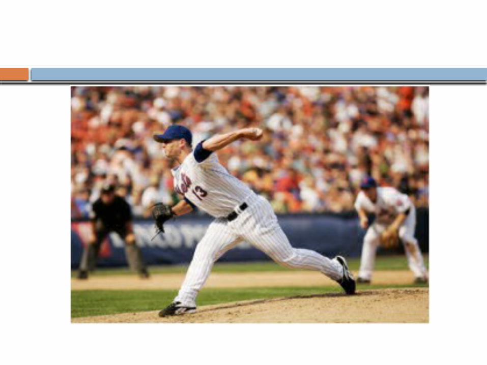
By far most common Typically trauma to arm in position of
abduction, extension, external rotation (person throwing) or by a blow to the posterior shoulder
Present with abnormal contour and fullness at anterior shoulder; arm abducted, internally rotated


Anterior Instability
Exams--Apprehension -Relocation-Load and Shift
Diagnostics--X-ray Views: AP, axillary and scapular-Y-can be performed before for diagnosis or after reduction for confirmation of relocation depending on clinical setting

Apprehension/Relocation
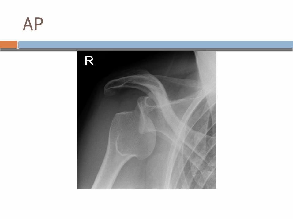
AP

Axillary

Scapular-Y

Anterior Instability
Treatment (several methods)--Stimson technique-Traction on arm at the wrist and forward flexion with counter traction at the chest-Westing, Milch, Kocher…
Surgery? -often depends on age and activity level
Associated Injuries--Hill-Sachs- compression of ant glenoid on post humerous-Bankhart- lesion on ant glenoid


Posterior Dislocation
Much less common Flexion, adduction, internal rotation-
offensive lineman “Lightning strikes and seizures” Easy to miss, especially on AP film Reduction is more difficult- apply traction
in line and try to manipulate humeral head back into place


Biceps Tendonitis
Primary occurs as inflammatory condition at bicipital groove
Secondary (more common) results from changes to surrounding structures like rotator cuff impingement or tears
Overuse injury Tender to palpation along anterior aspect of
shoulder, that may radiate down biceps Exam- Yergason’s, Speeds and possibly Neer’s
and Hawkin’s due to impingement association

Speed’s
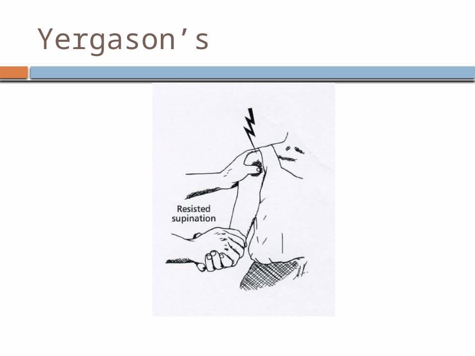
Yergason’s

Neer’s

Hawkins’
Bicep’s Rupture
Forceful elbow flexion against resistance or abrupt eccentric contraction
Pain, swelling over anterior arm “Popeye” deformity Elderly may be asymptomatic Treat with pain control and therapy for
mobility in elderly Surgery may be performed for young/active
or those concerned with cosmesis (who would?!)


SLAP Lesion
Superior Labrum Anterior and Posterior Can be insidious and acute trauma Traction from overhead throwing
athletes, fall on outstretched arm Pain with overhead activities; popping,
clicking, catching (difficult to differentiate from rotator cuff pathology)
Exams debatable- O’Brien’s, biceps load, anterior slide


O’Brien’s
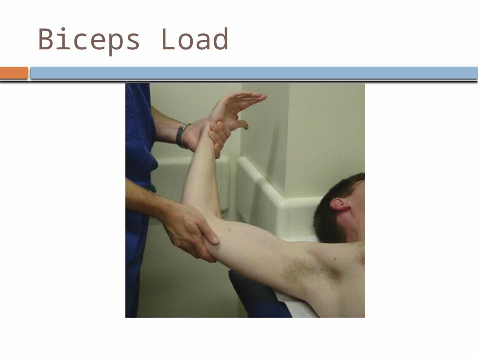
Biceps Load

Anterior Slide

SLAP Treatment
Rest, ice, NSAID’s Physical Therapy focusing on rotator cuff
strength and scapular stability Surgical referral if fails conservative
treatment

Impingement/Rotator Cuff Syndrome
Spectrum including subacromial bursitis, rotator cuff tendinopathy, rotator cuff partial tears
Subacromial impingement occurs on rotator cuff from undersurface of acromion and coracoclavicular ligament (cuff fatigue, tendinopathy, AC spurring)
Internal impingement occurs from rotator cuff on superior glenoid
Coracoid impingement occurs between cuff and a prominent coracoid
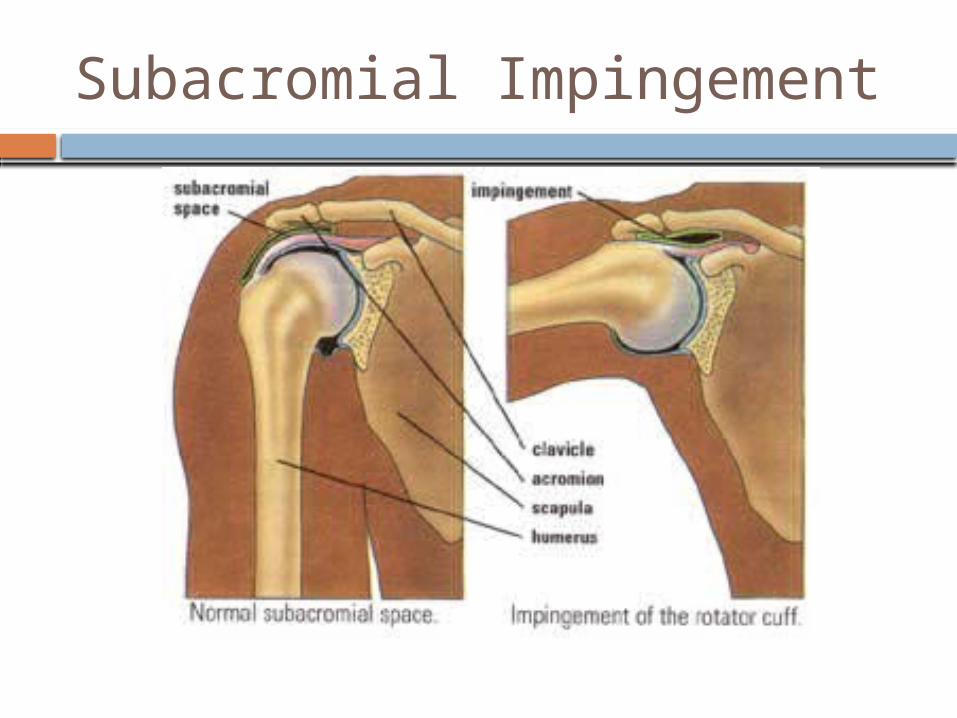
Subacromial Impingement
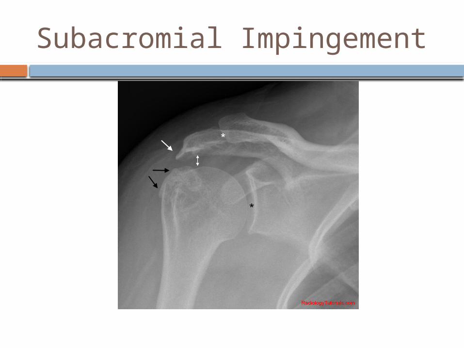
Subacromial Impingement
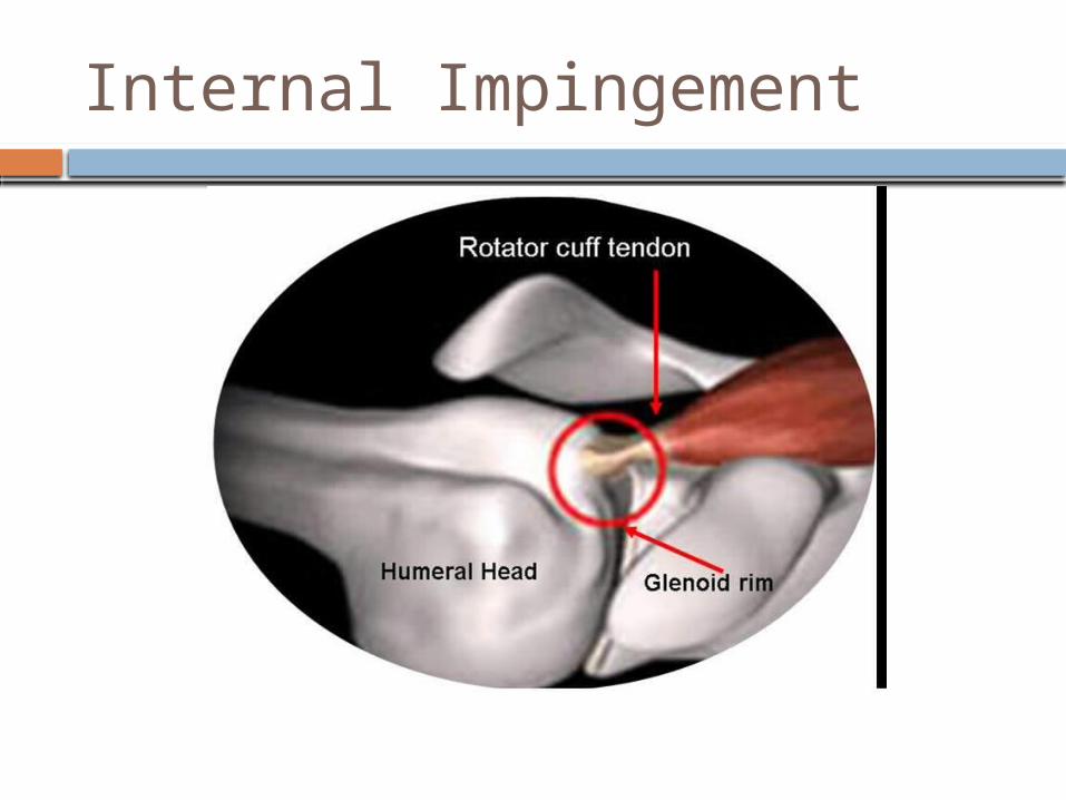
Internal Impingement

Internal Impingement

Coracoid Impingement

Impingement/Rotator Cuff Syndrome
History- -SI- anterior shoulder pain, radiates to lateral shoulder; pain with overhead activities; pain at night, when lying on affected side-II- posterior or deep pain; pain in throwing motion-CI- anterior pain, exacerbated by forward flexion and internal rotation
Exam- Neer’s, Hawkins’, Painful arc X-rays- AP, Outlet, Axillary- to look for GH arthritis,
at AC and coracoid MRI will show tendinopathy, tears (full or partial),
subacromial bursitis
Impingement/Rotator Cuff Syndrome
Treatment- NSAIDs and PT to strengthen cuff and scapular stabilizers; corticosteroid injection for subacromial impingement or bursitis
Surgery can be option if failure to improve, but majority improve with conservative therapy

Rotator Cuff Tears
MRI studies show 34% of asymptomatic individuals have rotator cuff tears (>60 yrs- 26% have partial thickness tears and 28% have full thickness)
Acute from traumatic event or chronic tendinopathy that progresses to tear
Presentation similar to subacromial impingement -anterolateral shoulder pain-overhead activites-night pain-weakness
Supraspinatus most common

RC Tears
Exam-palpate for atrophy (chronic)-external/internal rotation, flexion, abduction-belly off test (subscapularis)-external rotation lag sign (supraspinatus and infraspinatus)-shrug sign (better negative predictive value)-drop-arm sign

Belly Off

External Rotation Lag Sign

Shrug Sign
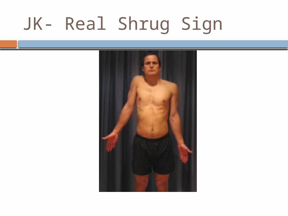
JK- Real Shrug Sign

Rotator Cuff Tears
Imaging-X-rays: AP may show humeral head proximal migration (chronic tears); look for signs of arthritis or calcific tendonitis-MRI: can distinguish full vs partial thickness; level of fat infiltration and atrophy (not good for surgery)-U/S: cheaper, but tech dependent (not common here)
Rotator Cuff Tears
Treatment-Individualized based on age/activity level-Conservative Non-Surgical: similar as for impingement (PT, NSAIDs, injection); less successful for patient’s with symptoms >1yr or significant weakness-Surgical referral recommended for younger/active and those with acute traumatic tears

Acromioclavicular Joint
AC Sprain/Separation- trauma (acute or repetitive) causing damage/tearing of acromioclavicular and coracoclavicular ligaments
Tenderness over AC joint; possibly elevation of clavicle on palpation
Classification: -Type I: sprain of AC ligament (CC intact)-Type II: tear of AC (CC intact); slight elevation of clavicle on xray-Type III: complete tear of AC and CC ligs and elevation of clavicle-Types IV-VI: keeps getting worse and damage to surrounding structures

AC Separation

Grade 3

AC Sprain
History- fall on shoulder or on outstretched arm (hockey player checked into boards or FB player landing on shoulder; cyclist falling off bike)
Exam- cross arm test and O’Brien’s if localizes to AC joint
Treatment- sling, ice, analgesics for Type I, II and usually III (sometimes III needs surgery); IV-VI need surgery
Recovery- 1 to 6 weeks (or keep playing…)
Adhesive Capsulitis
“Frozen Shoulder” Pain and gradual loss of active AND
passive ROM caused by soft tissue contracture
Idiopathic; more common in women and diabetics
Clinical diagnosis, but imaging can help rule out other causes; loss of flexion and external rotation >50% compared to unaffected side

Adhesive Capsulitis
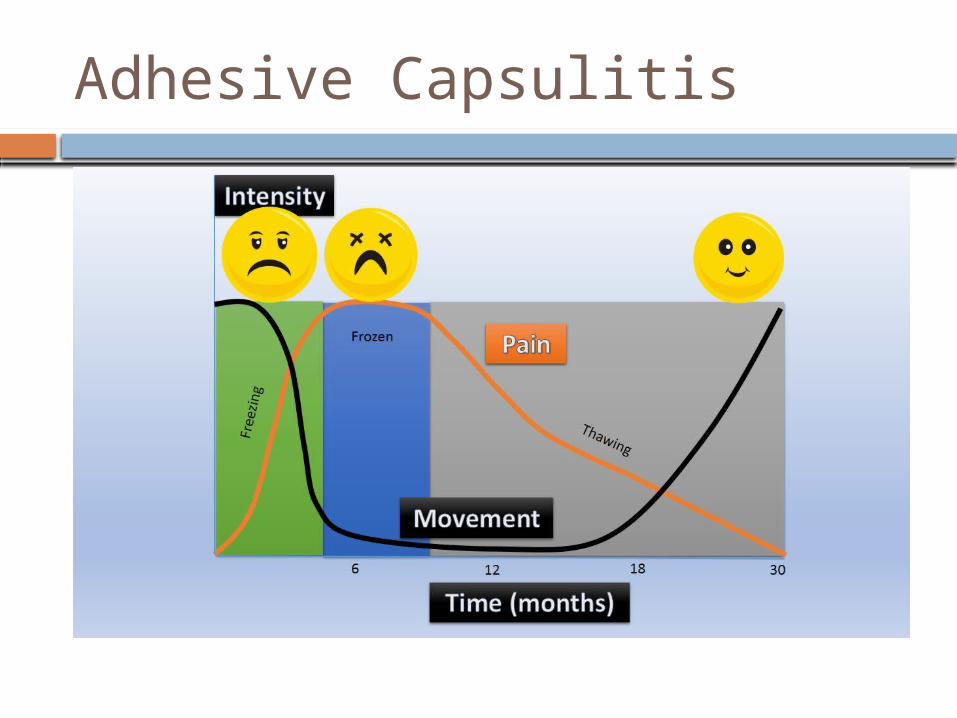
Stages-1: Pain with active and passive ROM (<3 mo)-2: “Freezing Stage” pain and progressive loss of ROM (3-9 mo)-3: “Frozen Stage” significant stiffness, minimal pain (9-15 mo)-4: “Thawing Stage” progressive improved ROM and minimal pain
Adhesive Capsulitis
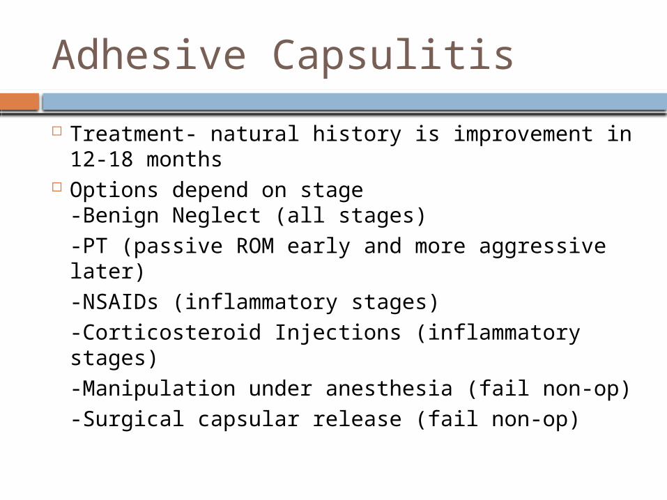
Treatment- natural history is improvement in 12-18 months
Options depend on stage-Benign Neglect (all stages)-PT (passive ROM early and more aggressive later)-NSAIDs (inflammatory stages)-Corticosteroid Injections (inflammatory stages)-Manipulation under anesthesia (fail non-op)-Surgical capsular release (fail non-op)
Adhesive Capsulitis

The End…Whew!
Questions??
References
Google Images, a lot. Madden, Christopher C. et al. Netter’s Sports
Medicine. 2010. Medscape. “Rotator Cuff Pathology.” O’Connor, Francis G. et al. ACSM’s Sports
Medicine, A Comprehensive Review. 2013. O’Kane, John W. et al. “The Evidence-Based
Shoulder Evaluation.” Extremity and Joint Conditions. Current Sports Medicine Reports. 2014 American College of Sports Medicine.