short-term clinical and angiographic outcome in child with ... · sdh showed with severe and...
TRANSCRIPT
130 Copyright © 2014 Korean Neurotraumatology Society
Introduction
Traumatic pseudoaneurysms are rare cases, and they oc-cupy less than 1% of all intracranial aneurysms. In spite of their high mortality of 30%, it is difficult to diagnose for their rarity.1)
The development of traumatic pseudoaneurysm depends on the mechanism of injury. It can occur after a penetrating or closed head injury, and also after an intracranial surgery such as transsphenoidal surgery, sinus surgery, ventricular taps, stereotactic brain biopsy, and endoscopic third ventric-ulostomy.4,5,12,14)
Here, we recently experienced the case of traumatic pseu-doaneurysm treated with endovascular embolization in a child. We always should keep in mind that the case of falx subdural hematoma (SDH) after head trauma has the possi-bility of traumatic pseudoaneurysm, and it requires vascu-lar work-up.
Case Report
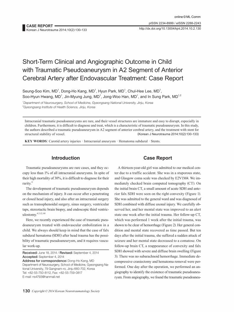
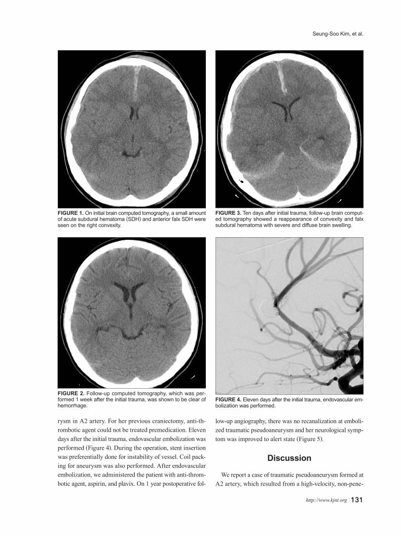
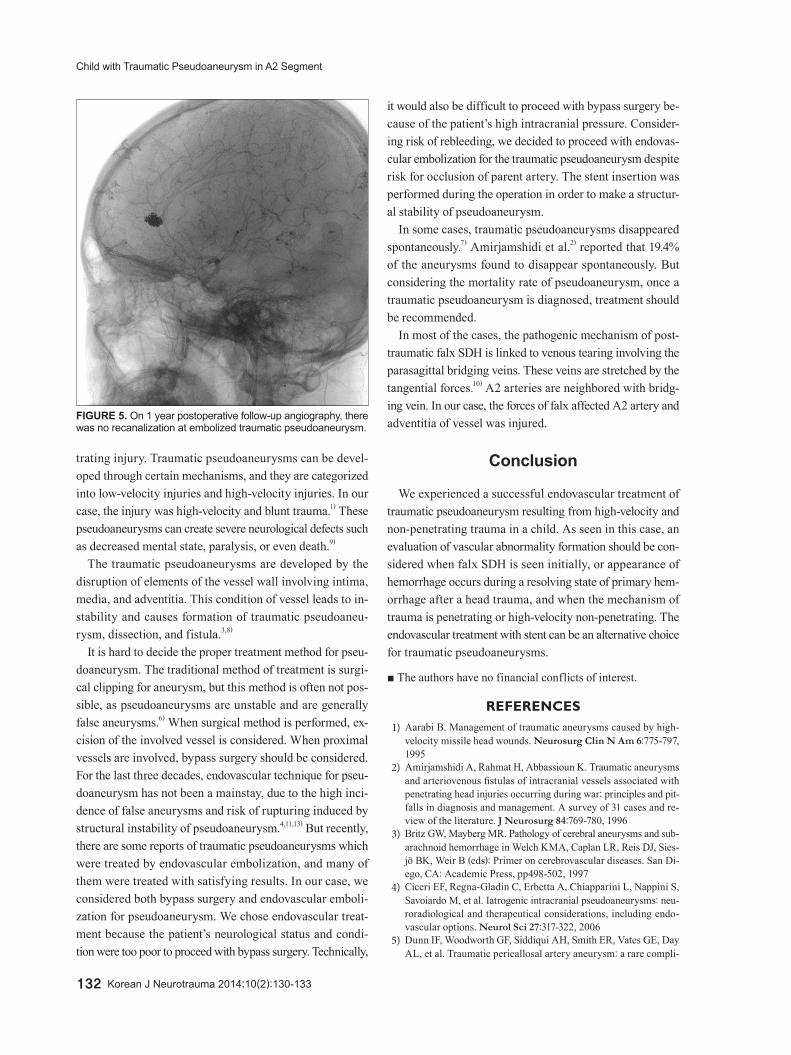
A thirteen-year-old girl was admitted to our medical cen-ter due to a traffic accident. She was in a stuporous state, and Glasgow coma scale was checked by E2V1M4. We im-mediately checked brain computed tomography (CT). On the initial brain CT, a small amount of acute SDH and ante-rior falx SDH were seen on the right convexity (Figure 1). She was admitted to the general ward and was diagnosed of SDH combined with diffuse axonal injury. We carefully ob-served her, and her mental state was improved to an alert state one week after the initial trauma. Her follow-up CT, which was performed 1 week after the initial trauma, was shown to be clear of hemorrhage (Figure 2). Her general con-dition and mental state recovered as time passed. But ten days after the initial trauma, she suffered a sudden attack of seizure and her mental state decreased to a comatose. On follow-up brain CT, a reappearance of convexity and falx SDH showed with severe and diffuse brain swelling (Figure 3). There was no subarachnoid hemorrhage. Immediate de-compressive craniectomy and hematoma removal were per-formed. One day after the operation, we performed an an-giography to identify the existence of traumatic pseudoaneu-rysm. From angiography, we found the traumatic pseudoaneu-
Short-Term Clinical and Angiographic Outcome in Child with Traumatic Pseudoaneurysm in A2 Segment of Anterior Cerebral Artery after Endovascular Treatment: Case Report
Seung-Soo Kim, MD1, Dong-Ho Kang, MD1, Hyun Park, MD1, Chul-Hee Lee, MD1, Soo-Hyun Hwang, MD1, Jin-Myung Jung, MD1, Jong-Woo Han, MD1, and In Sung Park, MD1,2
1Department of Neurosurgery, School of Medicine, Gyeongsang National University, Jinju, Korea 2Gyeongsang Institute of Health Science, Jinju, Korea
Intracranial traumatic pseudoaneurysms are rare, and their vessel structures are immature and easy to disrupt, especially in children. Furthermore, it is difficult to diagnose and treat, which is a characteristic of traumatic pseudoaneurysm. In this study, the authors described a traumatic pseudoaneurysm in A2 segment of anterior cerebral artery, and the treatment with stent for structural stability of vessel. (Korean J Neurotrauma 2014;10(2):130-133)
KEY WORDS: Carotid artery injuries ㆍIntracranial aneurysm ㆍHematoma subdural ㆍStents.
Received: June 18, 2014 / Revised: September 4, 2014Accepted: September 4, 2014Address for correspondence: Dong-Ho Kang, MDDepartment of Neurosurgery, School of Medicine, Gyeongsang Na-tional University, 79 Gangnam-ro, Jinju 660-702, KoreaTel: +82-55-750-8112, Fax: +82-55-759-0817E-mail: [email protected]
CASE REPORTKorean J Neurotrauma 2014;10(2):130-133
pISSN 2234-8999 / eISSN 2288-2243
http://dx.doi.org/10.13004/kjnt.2014.10.2.130
online © ML Comm
Seung-Soo Kim, et al.
http://www.kjnt.org 131
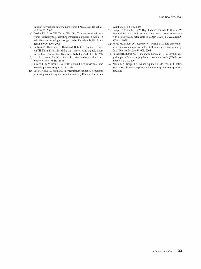
rysm in A2 artery. For her previous craniectomy, anti-th-rombotic agent could not be treated premedication. Eleven days after the initial trauma, endovascular embolization was performed (Figure 4). During the operation, stent insertion was preferentially done for instability of vessel. Coil pack-ing for aneurysm was also performed. After endovascular embolization, we administered the patient with anti-throm-botic agent, aspirin, and plavix. On 1 year postoperative fol-
low-up angiography, there was no recanalization at emboli-zed traumatic pseudoaneurysm and her neurological symp-tom was improved to alert state (Figure 5).
Discussion
We report a case of traumatic pseudoaneurysm formed at A2 artery, which resulted from a high-velocity, non-pene-
FIGURE 1. On initial brain computed tomography, a small amount of acute subdural hematoma (SDH) and anterior falx SDH were seen on the right convexity.
FIGURE 2. Follow-up computed tomography, which was per-formed 1 week after the initial trauma, was shown to be clear of hemorrhage.
FIGURE 3. Ten days after initial trauma, follow-up brain comput-ed tomography showed a reappearance of convexity and falx subdural hematoma with severe and diffuse brain swelling.
FIGURE 4. Eleven days after the initial trauma, endovascular em-bolization was performed.
132 Korean J Neurotrauma 2014;10(2):130-133
Child with Traumatic Pseudoaneurysm in A2 Segment
trating injury. Traumatic pseudoaneurysms can be devel-oped through certain mechanisms, and they are categorized into low-velocity injuries and high-velocity injuries. In our case, the injury was high-velocity and blunt trauma.1) These pseudoaneurysms can create severe neurological defects such as decreased mental state, paralysis, or even death.9)
The traumatic pseudoaneurysms are developed by the disruption of elements of the vessel wall involving intima, media, and adventitia. This condition of vessel leads to in-stability and causes formation of traumatic pseudoaneu-rysm, dissection, and fistula.3,8)
It is hard to decide the proper treatment method for pseu-doaneurysm. The traditional method of treatment is surgi-cal clipping for aneurysm, but this method is often not pos-sible, as pseudoaneurysms are unstable and are generally false aneurysms.6) When surgical method is performed, ex-cision of the involved vessel is considered. When proximal vessels are involved, bypass surgery should be considered. For the last three decades, endovascular technique for pseu-doaneurysm has not been a mainstay, due to the high inci-dence of false aneurysms and risk of rupturing induced by structural instability of pseudoaneurysm.4,11,13) But recently, there are some reports of traumatic pseudoaneurysms which were treated by endovascular embolization, and many of them were treated with satisfying results. In our case, we considered both bypass surgery and endovascular emboli-zation for pseudoaneurysm. We chose endovascular treat-ment because the patient’s neurological status and condi-tion were too poor to proceed with bypass surgery. Technically,
it would also be difficult to proceed with bypass surgery be-cause of the patient’s high intracranial pressure. Consider-ing risk of rebleeding, we decided to proceed with endovas-cular embolization for the traumatic pseudoaneurysm despite risk for occlusion of parent artery. The stent insertion was performed during the operation in order to make a structur-al stability of pseudoaneurysm.
In some cases, traumatic pseudoaneurysms disappeared spontaneously.7) Amirjamshidi et al.2) reported that 19.4% of the aneurysms found to disappear spontaneously. But considering the mortality rate of pseudoaneurysm, once a traumatic pseudoaneurysm is diagnosed, treatment should be recommended.
In most of the cases, the pathogenic mechanism of post-traumatic falx SDH is linked to venous tearing involving the parasagittal bridging veins. These veins are stretched by the tangential forces.10) A2 arteries are neighbored with bridg-ing vein. In our case, the forces of falx affected A2 artery and adventitia of vessel was injured.
Conclusion
We experienced a successful endovascular treatment of traumatic pseudoaneurysm resulting from high-velocity and non-penetrating trauma in a child. As seen in this case, an evaluation of vascular abnormality formation should be con-sidered when falx SDH is seen initially, or appearance of hemorrhage occurs during a resolving state of primary hem-orrhage after a head trauma, and when the mechanism of trauma is penetrating or high-velocity non-penetrating. The endovascular treatment with stent can be an alternative choice for traumatic pseudoaneurysms.
■ The authors have no financial conflicts of interest.
REFERENCES1) Aarabi B. Management of traumatic aneurysms caused by high-
velocity missile head wounds. Neurosurg Clin N Am 6:775-797, 1995
2) Amirjamshidi A, Rahmat H, Abbassioun K. Traumatic aneurysms and arteriovenous fistulas of intracranial vessels associated with penetrating head injuries occurring during war: principles and pit-falls in diagnosis and management. A survey of 31 cases and re-view of the literature. J Neurosurg 84:769-780, 1996
3) Britz GW, Mayberg MR. Pathology of cerebral aneurysms and sub-arachnoid hemorrhage in Welch KMA, Caplan LR, Reis DJ, Sies-jö BK, Weir B (eds): Primer on cerebrovascular diseases. San Di-ego, CA: Academic Press, pp498-502, 1997
4) Ciceri EF, Regna-Gladin C, Erbetta A, Chiapparini L, Nappini S, Savoiardo M, et al. Iatrogenic intracranial pseudoaneurysms: neu-roradiological and therapeutical considerations, including endo-vascular options. Neurol Sci 27:317-322, 2006
5) Dunn IF, Woodworth GF, Siddiqui AH, Smith ER, Vates GE, Day AL, et al. Traumatic pericallosal artery aneurysm: a rare compli-
FIGURE 5. On 1 year postoperative follow-up angiography, there was no recanalization at embolized traumatic pseudoaneurysm.
Seung-Soo Kim, et al.
http://www.kjnt.org 133
cation of transcallosal surgery. Case report. J Neurosurg 106(2 Sup-pl):153-157, 2007
6) Golshani K, Britz GW, Yoo A, West GA. Traumatic cerebral aneu-rysms secondary to penetrating intracranial injuries in Winn HR (ed): Youmans neurological surgery, ed 6. Philadelphia, PA: Saun-ders, pp4000-4003, 2011
7) Halbach VV, Higashida RT, Hieshima GB, Goto K, Norman D, New-ton TH. Dural fistulas involving the transverse and sigmoid sinus-es: results of treatment in 28 patients. Radiology 163:443-447, 1987
8) Hart RG, Easton JD. Dissections of cervical and cerebral arteries. Neurol Clin 1:155-182, 1983
9) Kieck CF, de Villiers JC. Vascular lesions due to transcranial stab wounds. J Neurosurg 60:42-46, 1984
10) Lee M, Kim MS, Yoon SW. Interhemispheric subdural hematoma presenting with falx syndrome after trauma. J Korean Neurotrau-
matol Soc 6:158-161, 201011) Lempert TE, Halbach VV, Higashida RT, Dowd CF, Urwin RW,
Balousek PA, et al. Endovascular treatment of pseudoaneurysms with electrolytically detachable coils. AJNR Am J Neuroradiol 19: 907-911, 1998
12) Rayes M, Bahgat DA, Kupsky WJ, Mittal S. Middle cerebral ar-tery pseudoaneurysm formation following stereotactic biopsy. Can J Neurol Sci 35:664-668, 2008
13) Rückert RI, Rutsch W, Filimonow S, Lehmann R. Successful stent-graft repair of a vertebrojugular arteriovenous fistula. J Endovasc Ther 8:495-500, 2001
14) Zanini MA, Borges RA, Nunes Aquino GH, de Freitas CC. Iatro-genic cortical aneurysm post-craniotomy. Br J Neurosurg 24:214-215, 2010