short communication - university of pittsburgh
TRANSCRIPT

Short CommunicationDifferential Viral Protein Expression in Kaposi’sSarcoma-Associated Herpesvirus-Infected Diseases
Kaposi’s Sarcoma, Primary Effusion Lymphoma, andMulticentric Castleman’s Disease
Carlo Parravicini,*† Bala Chandran,‡
Mario Corbellino,§ Emilio Berti,§ Marco Paulli,¶
Patrick S. Moore,*i and Yuan Chang*From the Department of Pathology,* College of Physicians and
Surgeons, Columbia University, New York, New York; the
Department of Pathology,† Lugio Sacco Hospital, Milan, Italy; the
Department of Microbiology, Molecular Genetics and
Immunology,‡ The University of Kansas Medical Center, Kansas
City, Kansas; the Institute of Infectious Disease and Tropical
Medicine,§ University of Milan, Luigi Sacco Hospital, Milan, Italy;
the Department of Pathology,¶ IRCCS Ospedale San Matteo,
University of Pavia, Pravia, Italy; and the Division of
Epidemiology,i Columbia University, New York, New York
Kaposi’s sarcoma (KS)-associated herpesvirus (KSHV)is linked to KS, primary effusion lymphomas (PEL),and a subset of multicentric Castleman’s disease(MCD). Transcript mapping studies using PEL celllines have allowed preliminary classification of viralgene expression into constitutive (class I) and induc-ible (class II/III) categories. To determine whetherviral gene expression differs in vivo , we examinedtissue sections of KSHV-infected disorders, using spe-cific antibodies against proteins that are representa-tive of the different expression classes of KSHV genes.ORF73/LANA appears to be a surrogate marker forKSHV infection because it is constitutively expressedin vitro and in vivo in all KSHV-infected cells. Expres-sion of vIRF1, vIL6, and PF-8 proteins in the infectedB cells of MCD lymph nodes reproduces the expres-sion pattern observed in TPA-stimulated KSHV-in-fected B-cell lines. In contrast, the protein expressionof the inducible viral genes that we tested in KS andPEL biopsies is restricted to PF-8 and vIL6, respec-tively. The tightly restricted expression of KSHV pro-teins in vivo differs from the dysregulated expressionof inducible KSHV genes in vitro and suggests thatviral gene expression in KSHV-infected cell lines does
not accurately reflect what occurs in diseased tissues.These differences may be related to either cell-specificor immune restriction of viral replication. (Am JPathol 2000, 156:743–749)
Kaposi’s sarcoma (KS)-associated herpesvirus (KSHV) isa human g-herpesvirus involved in the pathogenesis ofKS, primary effusion lymphoma (PEL), and a subset ofmulticentric Castleman’s disease (MCD).1–3 KSHV canbe stably propagated in vitro in B-cell lines derived fromPELs, but currently it is not readily transmissible at hightiter.4–8 Similar to other herpesviruses, KSHV infectioncan be characterized as lytic or latent. During lytic repli-cation, virions are packaged and released from the cell.This process requires DNA synthesis together with ex-pression of virion structural protein genes and is believedto result in cell death. Latent infection, however, is char-acterized by the persistence of the viral genome as acovalently closed circular episome with limited viral geneexpression.9,10 Latent KSHV in cells derived from PELcan be induced into lytic replication by chemical treat-ment with tetradecanoyl phorbol acetate (TPA) or bu-tyrate.10,11
PEL-derived cell lines stimulated with phorbol esters orbutyrate reveal three distinct classes of KSHV messengerRNAs (mRNAs) corresponding to constitutive (type I),constitutive/inducible (type II), and lytic/inducible (typeIII) transcripts.12 The expression of selected genes canbe further modulated by treatment with cycloheximide,phosphonoacetic acid, or both.13 Most of the viral cyto-
Supported by National Institutes of Health grants CA67391, CA75911,and CA82056; II Programma Nazionale di Ricerca sull’AIDS1998-ISSProject Number 30.A.0.50; and the Lucy Pang Yoa Chang Foundation.
Accepted for publication November 14, 1999.
Address reprint requests to Yuan Chang, P&S 14–442, Dept. of Pathol-ogy, Columbia University, College of Physicians and Surgeons, 630 West168th Street, New York, NY 10032. E-mail: [email protected].
American Journal of Pathology, Vol. 156, No. 3, March 2000
Copyright © American Society for Investigative Pathology
743

kines and signal transduction genes that are unique toKSHV are type II or III, inducible genes.12 These genesencode proteins that have been postulated to play acentral role in the pathogenesis of KSHV-related dis-eases.
A major drawback to studies on viral gene expressionin tissue culture cell lines is that expression patterns insuch systems may not reflect viral gene expression ininfected tissues in situ. For example, Epstein-Barr virus(EBV) demonstrates different programs of virus latency indifferent cell types.14 It is currently unknown whether thepattern of gene transcription observed in vitro in PEL celllines is present in different types of KSHV-infected cells invivo. Recent immunohistochemistry studies have shownthat KS spindle cell, mantle zone B cells in MCD lymphnodes and PEL cells express KSHV LANA,15–17 whereasvIL-6 is detectable in only a subpopulation of KSHV-infected hematopoietic cells.18,19 In this study we used apanel of antibodies directed against KSHV LANA(ORF73),20 vIL6 (K2),18 vIRF1 (ORFK9),21 and processiv-ity factor-8 (PF-8) (ORF59).22 These proteins representtype I-constitutive (ORF73/LANA), type II-constitutive/in-ducible (ORFK9/vIRF1 and ORF K2/vIL6), and type III-lytic/inducible (ORF59/PF-8) genes of KSHV.12 Our re-sults demonstrate that some KSHV genes can becomedysregulated in tissue culture and that tissue-specificpatterns of expression are not represented by tissueculture studies. Furthermore, different KSHV-associateddiseases show distinct patterns of viral gene expression.Therefore KSHV, like EBV, exhibits multiple programs forviral gene expression.
Materials and Methods
Cell Lines
BC-1,4 BCP-1,5 and BCBL-18 KSHV-infected cells weremaintained at 37°C and 5% CO2 in 1640 RPMI (GIBCO/BRL, Grand Island, NY) supplemented with 10% to 20%fetal calf serum (GIBCO/BRL). Induction of viral geneexpression was performed by treatment of cells with 20ng/ml of TPA (Sigma Chemical Co., St. Louis, MO).Ramos (EBV2/KSHV2) and P3HRI (EBV1/KSHV2) cells(ATCC, Gaithersburg, MD) were used as KSHV-negativecontrol cell lines. Cytospins, cell pellets, or both wereharvested after 48 hours. Cytospins were air dried over-
night, fixed in acetone for 4 minutes at room temperature,air dried for 30 minutes, and then either processed forimmunohistochemistry or stored at 280°C until use. Cellpellets were fixed overnight in 4% paraformaldehyde in0.1 mol/L phosphate-buffered saline (PBS; pH7.4) andembedded in paraffin.
Tissue Cases and Controls
Fifteen KS skin lesions, 10 lymph nodes from patientswith MCD, and biopsies from four cases of PEL wereinvestigated. KS biopsies included both classical (n 5 7)and human immunodeficiency virus (HIV)-associated(n 5 8) cases and were representative of patch (n 5 5),plaque (n 5 7), and nodular (n 5 3) stages of the dis-ease. All 10 MCD cases were of the plasma cell variant,six from HIV-seronegative and four from HIV-seropositiveindividuals. In contrast, all four PEL cases were acquiredimmune deficiency syndrome (AIDS)-related. Frozen ma-terial (required for PF-8 immunostaining) was availablefor 8 of 15 KS lesions and 7 of 10 MCD cases. Frozenmaterial was available from one of the four PEL cases.Frozen and paraffin-embedded sections from normal ton-sils (n 5 5) and skin biopsies (n 5 5) were used ascontrols.
Antibodies
The panel of antibodies against KSHV used in this studyspecifically recognizes viral proteins representative ofdifferent classes of transcripts involved in virus replica-tion (see Table 1). When applied to KSHV-infected cells,monoclonal antibodies and sera directed against differ-ent portions of the same KSHV protein and/or raised indifferent species showed the same staining pattern andlabeled the same percentage of cells, with a range ofvariability of less than 5% (data not shown). Immunohis-tochemical procedures to reveal KSHV expression onparaffin-embedded biopsies were optimized by usingsections from formalin-fixed/paraffin-embedded pelletsof KSHV-infected PEL-derived (BC-1, BCP-1, BCBL-1)cell lines and uninfected control cells (RAMOS). The re-sults on paraffin-embedded sections were comparedwith the results obtained on acetone-fixed cytospins ofthe same cell lines (data not shown). For a given anti-
Table 1. Antibodies against KSHV Proteins
Antibody Antigen/Source (reference)KSHV
gene/protein Transcription classImmuno-
histochemistry*
R UK163 (rabbit) BCBL-1 cells (20) ORF73/LANA Class I—constitutive F/P(MW)R 535 (rabbit) GST-conj rec. protein (21) ORFK9/v-IRF1 Class II—constitutive/inducible F/P(MW)R 537 (rabbit) GST-conj rec. proteinS 544 (sheep) GST-conj rec. proteinR 394 (rabbit) synthetic peptide, AA 218-432 (18) ORFK2/v-IL6 Class II—constitutive/inducible F/PS 546 (sheep) GST-conj rec. proteinM 11D1 (mouse
monoclonal)TPA-induced BCBL-1 (22) ORF59/PF-8 Class III—lytic/inducible F
*F, reactive on cytospins and frozen sections; P, reactive on paraffin sections; MW, microwave treatment required; GST-conj rec., GST-conjugatedrecombinant.
744 Parravicini et alAJP March 2000, Vol. 156, No. 3

body, the conditions were considered satisfactory when,in repeated experiments, the number of immunostainedcells was the same on paraffin-embedded sections andcytospins, with a range of variability of less than 5% (datanot shown). To obtain the same percentage of stainedcells on the paraffin-embedded sections and on the cy-tospins, antibodies against LANA and vIRF1 required amicrowave-ethylenediaminetetraacetic acid (EDTA) pre-treatment of paraffin sections,23 whereas the reactivity ofantibodies against vIL6 did not require this treatment. Themonoclonal antibody M11D1 against KSHV PF-8 was theonly reagent that was nonreactive on paraffin sectionsand required frozen materials (see Table 1).
Monoclonal antibodies or rabbit antisera against CD3,CD4, CD8, CD45, CD20, CD23, CD5, CD79, CD30, EMA,Ki67, CD34, vWF:Fact.VIII (DAKO, Glostrupp, Denmark),and CD138 (VIth Workshop on Human leukocyte Anti-gens, Osaka, Japan) were used to detect specific lym-phocyte subpopulations as well as spindle cells in KSlesions.
Immunohistochemistry
Antibody binding was revealed using peroxidase-labeledgoat anti-mouse or goat anti-rabbit antisera (DAKO) fol-lowed by tyramide amplification (DuPont/NEN, Boston,MA). Reactions were developed using diaminobenzidine(DAB; Sigma) or amino ethyl carbazole (AEC; DAKO) aschromogenic substrates, and sections were counter-stained with hematoxylin. For double immunostaining, thesecond primary antibody was revealed using alkalinephosphatase-conjugated rabbit anti-mouse or swine anti-rabbit antisera and Fast Blue (Sigma) as chromogen. Forimmunofluorescence, fluorescein-isothiocyanate (FITC)-conjugated goat anti-mouse or goat anti-rabbit antisera(Southern Biotechnology, Birmingham, AL) were usedalone or, for double staining, in combination with biotin-ylated horse anti-mouse (Vector Laboratories, Burlin-game, CA) or goat anti-rabbit antisera (Southern Biotech-nology) followed by Avidin Texas Red (Vector).
Results
Protein Expression in PEL-Derived BC-1,BCP-1, and BCBL-1 Cell Lines
Protein expression patterns were examined in three celllines (BC-1, BCP-1, and BCBL-1) before and after TPAtreatment (summarized in Table 2). Although LANA wasexpressed in all cells (Figure 1A) regardless of TPA treat-ment and appears to be a marker for KSHV infection,examination of the immunohistochemical profile for theremaining proteins reveals a tightly regulated expressionpattern. The BC-1 cell line had constitutive low-level ex-pression of vIRF1 and vIL6 (class II genes) but lackedexpression of PF-8 (class III). In contrast, low-level PF-8protein expression was present in BCBL-1 and BCP-1cells without TPA treatment, which is consistent withthese cell lines being producer cell lines with a low per-centage of cells spontaneously entering into full lytic
replication. The percentages of cells expressing vIRF1and PF-8 increased approximately 20-fold after TPAtreatment for all three cell lines, whereas vIL6, which wasexpressed at higher constitutive levels, increased only 3-to 5-fold (Table 3). We found that the vIL6 staining patternin KSHV-infected cell lines was less than initially report-ed,18 probably due to technical artifacts in the earlierstudy that were related to antibody dilution and develop-ment. In TPA-stimulated BCBL-1 cells, double-stainingstudies demonstrated that more than 80% of cells ex-pressing vIRF1 coexpressed PF-8, as compared with10% of PF-8-positive cells expressing vIL6 (Table 4). Allantibodies against KSHV proteins were nonreactive byimmunohistochemistry to RAMOS and P3HRI cells, aswell as KSHV-negative tissue controls.
Expression of Viral Proteins in KSHV-RelatedDiseases
Kaposi’s Sarcoma
KSHV protein expression is highly restricted in KSlesions. LANA is the only protein examined that wasexpressed in most KS tumor cells. In KS lesions, regard-less of histological stage, the nuclei of spindle and en-dothelial KS cells were positive for LANA in a typicalspeckled immunoreactivity pattern (Figure 2B). Intratu-moral arterioles and extralesional normal tissues, includ-ing epidermis and skin adnexa, were LANA negative(Figure 2C). All clinical stages of KS, including patch,plaque, and nodular lesions, were positive for LANA ex-pression. In all KS lesions, most CD451 infiltrating leu-kocytes were CD681 monocytic cells, with only a fewand irregularly distributed CD31 T or CD201 B lympho-cytes. Of these CD451/CD681 monocytes, less than 1%were LANA positive (Figure 2E), and none expressedPF-8 or other KSHV proteins. No expression of vIRF1 orvIL6 was seen in any of the KS lesions. In four of fivenodular lesions for which frozen material was available,the nuclei of rare (less than 1%) tumor spindle and en-dothelial cells were positive for PF-8 (Figure 2F).
PEL
Comparison of tumor biopsies to unstimulated celllines shows several important differences between PELcells growing in vivo and in vitro. As in KS lesions and PELcell lines, LANA has a speckled nuclear pattern in virtu-ally all PEL biopsy cells (Figure 1E). In contrast, less than
Table 2. Expression of KSHV Proteins in InfectedPEL-Derived Cell Lines versus KS, PEL, andmCD Tissue Samples
KSHVprotein
PEL-derivedcell lines* KS MCD PEL
LANA 1 1 1 1v-IRF1 1 2 1 2v-IL6 1 2 1 1PF-8 1 1 1 2
*BCBL-1, BCP-1, BC-1.
KSHV Protein Expression in Vivo and in Vitro 745AJP March 2000, Vol. 156, No. 3

Figure 1. A: LANA is constitutively expressed in nearly all BCBL-1 cells as a speckled nuclear pattern (inset). Similar expression is seen also in BC-1 and BCP-1cells (DAB/hematoxylin counterstain). B: vIRF1 in BCBL-1 cells is localized to the cytoplasm in one cell (right) and in the nucleus of another (left), consistentwith nuclear translocation. C: Only 1% of BCBL-1 cells express vIRF1 without TPA (upper panel) but up to .20% (lower panel) after 48 hours of TPA stimulation(DAB/hematoxylin counterstain). D: With TPA stimulation (upper panel) BCBL-1 cells may express nuclear PF-8 alone (brown), cytoplasmic vIRF1 alone (blue),or may coexpress both proteins. In some cells double stained for PF-8 and vIRF1, the latter shows a pseudolinear intracytoplasmic pattern reminiscent of roughendoplasmic reticulum positivity (lower panel) (AEC/FastBlue; no counterstain). E: Section of myocardium with infiltrating PEL. All neoplastic cells expressnuclear LANA (AEC/hematoxylin counterstain). F: Peritoneal biopsy with infiltrating PEL. Only a few tumor cells are immunopositive for vIL6 (DAB/hematoxylincounterstain). G: Hyalinization of germinal centers, targetoid mantle zone, and increased vascularity in a lymph node biopsy from plasma cell variant,KSHV-related multicentric Castleman’s disease (H&E stain). H: KSHV LANA protein expression is restricted to a subpopulation of mantle zone lymphocytes thatshow a speckled nuclear staining pattern (inset) (DAB/hematoxylin counterstain). I: Double-stained section of an MCD lymph node showing that nearly all ofthe KSHV-infected mantle zone lymphocytes expressing LANA (light brown) are negative for CD79 (blue), with only a few double-positive cells (inset). Similarfindings are obtained by double staining with other B-cell antigens expressed by normal mantle zone lymphocytes. (DAB/FastBlue; no counterstain). J: MCDlymph node section with expression of vIL6 (brown) restricted to a subpopulation of mantle zone cells having immunoblastic morphology (inset) (DAB/hematoxylin counterstain). K: In double-stained sections, vIRF1 (blue) in mantle zone cells is also positive for LANA (inset). Double-stained lymphocytes,however, account for only 10% to 30% of the LANA-positive cells (DAB/FastBlue; no counterstain). L: MCD frozen section, same case as in C, shows that onlyrare cells at the border between mantle zone and germinal center express PF-8 (DAB/hematoxylin counterstain).
746 Parravicini et alAJP March 2000, Vol. 156, No. 3

5% of the cells were positive for vIL6 (Figure 1F), and noimmunostaining for vIRF1 protein was detected. BecausevIRF1 can be detected at low levels in resting (ie, un-stimulated) PEL cell lines, these data suggest that genedysregulation has occurred in adaptation of the cells toculture.
MCD
In contrast to KS and PEL lesions, a broad pattern ofKSHV protein expression is present in MCD tissues. Ex-pression of KSHV proteins is confined to the follicularmantle zone, in which 10% to 30% of cells are positive forLANA (Figure 1H). Double staining demonstrates that asubpopulation of these cells expresses vIL6 and vIRF1.The number of cells expressing these latter viral proteins,however, was highly variable among different follicles ofthe same lymph node, ranging from 5% to 25% of theLANA-positive cells (Figure 1, J and K). In four cases forwhich frozen material was available, rare LANA-positivecells of the mantle zone were also positive for PF-8 (Fig-ure 1L). Outside the follicles, cells expressing KSHV pro-teins were an inconspicuous and occasional finding.When present, they were within poorly defined aggre-gates of B-lymphocytes in the paracortical areas or assingle isolated cells in the marginal sinus. In all MCDcases, cells expressing KSHV proteins had large, irreg-ular or round nuclei with marginated chromatin and me-dium-sized nucleoli (Figure 1J, inset). Cells expressingLANA were only occasionally positive for CD20 or CD79(Figure 1I) but always negative for CD23 or CD5 or for theT-cell antigen CD3. In all but one MCD case, in which asingle double-stained cell was observed, CD681 mono-cytes were also negative.
Immunohistochemical results on KS, PEL, and CD bi-opsies are summarized in Table 2. Extralesional normaltissues in the biopsies used in this study were consistentlynegative with all antibodies against KSHV proteins tested.
Discussion
The pattern of viral gene expression observed in vitro inPEL-derived cells is reproduced in vivo in MCD but not inKS nor in PEL. In MCD lymph nodes, cells expressingLANA are confined to the mantle zone of the lymphoidfollicles.24 These KSHV-infected cells are negative forT-cell or monocytic markers and are indistinguishable bymorphology from normal mantle zone lymphocytes, al-though only a minority express CD20 or CD79 B-cellmarkers. A subset of the LANA-positive mantle zonecells, most of which have an immunoblastic morphology,expresses vIL6 as well as vIRF1 and, to lesser extent,PF-8. KSHV-infected mantle zone cells therefore repro-duce the pattern of viral gene expression observed inTPA-stimulated PEL-derived cell lines, in which a subsetof cells expresses class II and III genes.6,8 As we re-ported previously19 and as was subsequently confirmedby Staskus et al,25 expression of vIL6 appears to be re-stricted to hematopoietic cells and, together with other in-ducible KSHV proteins, may be responsible for the hyper-plastic changes typically observed in MCD lymph nodes.
Of the proteins assayed in this study, we found thatexpression is restricted to vIL6 alone in PEL tissue biop-sies. In all 4 cases studied, vIL6 was detectable in lessthan 5% of the LANA-positive neoplastic cells, whereasvIRF1 and PF-8 proteins were not expressed. A differ-ence in viral gene expression patterns between tissueculture cells and the parental tumor has been docu-mented in EBV infection, in which cell lines derived fromBurkitt’s lymphomas expressing type 1 latency in vivoacquire type 2 or type 3 latency in vitro (see14 for review).Our findings suggest that KSHV protein expression maybe different in vitro and in vivo. Therefore caution isneeded in extending cell culture gene expression studiesto patient tissues. Our findings also suggest that in vivoand in vitro expression of vIL6 can occur independentlyfrom the activation of the lytic replicative cascade.
Table 3. KSHV Protein Expression in Infected Cell Lines with and without TPA Stimulation
KSHV protein Staining pattern
% BCBL-1 % BC-1 % BCP-1
TPA2 TPA1 TPA2 TPA1 TPA2 TPA1
ORF73 Nuclear, speckled 100 100 100 100 100 100v-IRF1 Nuclear and cytoplasmic, diffuse 1 24 ,1 20 7 25v-IL6 Cytoplasmic, diffuse ,1 2 ,1 6 4 15PF-8 Nuclear, diffuse or membrane 1 20 ;0 25 ;0 26
Table 4. Coexpression of Inducible/Lytic KSHV Proteins in BCBL1 Cells with and without TPA Treatment
KSHV proteins TPA2 (%) TPA1 (%) Summary
PF-8/v-IL6 0.1 1.8 80% vIL61 are PF-81PF-8 alone 2.3 18 10% PF-81 are vIL61v-IL6 alone 0.1 0.5PF-8/v-IRF1 0.7 18 90% vIRF1 are PF-81PF-8 alone 0.2 3.8 80% PF-81 are vIRF1v-IRF1 alone 0.1 2.5
KSHV Protein Expression in Vivo and in Vitro 747AJP March 2000, Vol. 156, No. 3
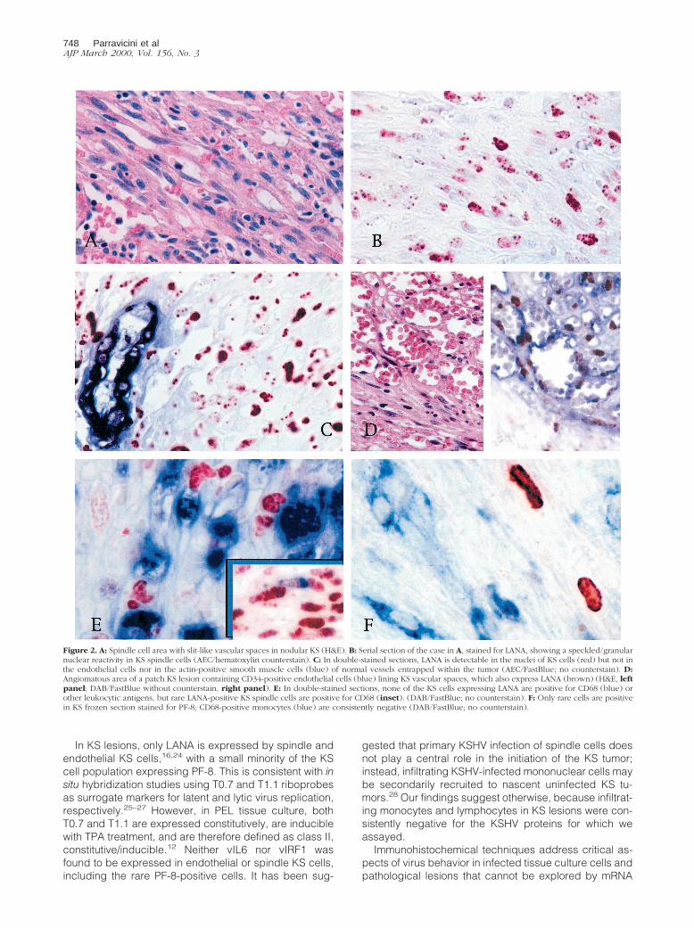
In KS lesions, only LANA is expressed by spindle andendothelial KS cells,16,24 with a small minority of the KScell population expressing PF-8. This is consistent with insitu hybridization studies using T0.7 and T1.1 riboprobesas surrogate markers for latent and lytic virus replication,respectively.25–27 However, in PEL tissue culture, bothT0.7 and T1.1 are expressed constitutively, are induciblewith TPA treatment, and are therefore defined as class II,constitutive/inducible.12 Neither vIL6 nor vIRF1 wasfound to be expressed in endothelial or spindle KS cells,including the rare PF-8-positive cells. It has been sug-
gested that primary KSHV infection of spindle cells doesnot play a central role in the initiation of the KS tumor;instead, infiltrating KSHV-infected mononuclear cells maybe secondarily recruited to nascent uninfected KS tu-mors.28 Our findings suggest otherwise, because infiltrat-ing monocytes and lymphocytes in KS lesions were con-sistently negative for the KSHV proteins for which weassayed.
Immunohistochemical techniques address critical as-pects of virus behavior in infected tissue culture cells andpathological lesions that cannot be explored by mRNA
Figure 2. A: Spindle cell area with slit-like vascular spaces in nodular KS (H&E). B: Serial section of the case in A, stained for LANA, showing a speckled/granularnuclear reactivity in KS spindle cells (AEC/hematoxylin counterstain). C: In double-stained sections, LANA is detectable in the nuclei of KS cells (red) but not inthe endothelial cells nor in the actin-positive smooth muscle cells (blue) of normal vessels entrapped within the tumor (AEC/FastBlue; no counterstain). D:Angiomatous area of a patch KS lesion containing CD34-positive endothelial cells (blue) lining KS vascular spaces, which also express LANA (brown) (H&E, leftpanel; DAB/FastBlue without counterstain, right panel). E: In double-stained sections, none of the KS cells expressing LANA are positive for CD68 (blue) orother leukocytic antigens, but rare LANA-positive KS spindle cells are positive for CD68 (inset). (DAB/FastBlue; no counterstain). F: Only rare cells are positivein KS frozen section stained for PF-8; CD68-positive monocytes (blue) are consistently negative (DAB/FastBlue; no counterstain).
748 Parravicini et alAJP March 2000, Vol. 156, No. 3

expression studies. Although extensive mRNA mappingof viral gene expression has been performed in KSHV-infected cell lines, this is the first report to compare theprotein expression of a panel of constitutive and induc-ible genes in KSHV-associated disorders. Our studyshows that KS, PEL, and MCD are characterized bydiffering patterns of KSHV protein expression. Currently,it is unclear whether these differences reflect the in vivoestablishment of alternative types of latency, which en-able KSHV to escape immunological surveillance, orwhether these differences in patterns of expression aredue to a cell/tissue-specific control of KSHV gene expres-sion. These differences are disease specific and arelikely to be relevant to understanding the pathogenesis ofKSHV-infected diseases.
Acknowledgments
We thank Antoine Gessain (Institut Pasteur, Paris) forproviding one of the PEL cases used in this study.
References
1. Chang Y, Cesarman E, Pessin MS, Lee F, Culpepper J, Knowles DM,Moore PS. Identification of herpesvirus-like DNA sequences in AIDS-associated Kaposi’s sarcoma. Science 1994, 266:1865–1869
2. Cesarman E, Chang Y, Moore PS, Said JW, Knowles DM: Kaposi’ssarcoma-associated herpesvirus-like DNA sequences in AIDS- re-lated body-cavity-based lymphomas. N Engl J Med 1995, 332:1186–1191
3. Soulier J, Grollet L, Oksenhendler E, Cacoub P, Cazals HD, BabinetP, d’Agay MF, Clauvel JP, Raphael M, Degos L, et al: Kaposi’ssarcoma-associated herpesvirus-like DNA sequences in multicentricCastleman’s disease. Blood 1995, 86:1276–1280
4. Cesarman E, Moore PS, Rao RH, Inghirami G, Knowles DM, Chang Y.In vitro establishment and characterization of two acquired immuno-deficiency syndrome-related lymphoma cell lines (BC-1 and BC-2)containing Kaposi’s sarcoma-associated herpesvirus-like (KSHV)DNA sequences. Blood 1995, 86:2708–2714
5. Boshoff C, Gao SJ, Healy LE, Matthews S, Thomas AJ, Coignet L,Warnke RA, Strauchen JA, Matutes E, Kamel OW, Moore PS, WeissRA, Chang Y: Establishing a KSHV1 cell line (BCP-1) from peripheralblood and characterizing its growth in Nod/SCID mice. Blood 1998,91:1671–1679
6. Miller G, Heston L, Grogan E, Gradoville L, Rigsby M, Sun R, SheddD, Kushnaryov VM, Grossberg S, Chang Y: Selective switch betweenlatency and lytic replication of Kaposi’s sarcoma herpesvirus andEpstein-Barr virus in dually infected body cavity lymphoma cells.J Virol 1997, 71:314–324
7. Renne R, Blackbourn D, Whitby D, Levy J, Ganem D: Limited trans-mission of Kaposi’s sarcoma-associated herpesvirus in cultured cells.J Virol 1998, 72:5182–5188
8. Renne R, Zhong W, Herndier B, McGrath M, Abbey N, Kedes D,Ganem D: Lytic growth of Kaposi’s sarcoma-associated herpesvirus(human herpesvirus 8) in culture. Nat. Med. 1996, 2:342–346
9. Roizman B: The human herpesviruses. The family Herpesviridae . TheFamily Herpesviridae. Edited by B Roizman, RJ Whitley, C Lopez.New York, Raven Press, 1993, pp 1–9
10. Miller G, Rigsby MO, Heston L, Grogan E, Sun R, Metroka C, Levy JA,Gao SJ, Chang Y, Moore P: Antibodies to butyrate-inducible antigensof Kaposi’s sarcoma- associated herpesvirus in patients with HIV-1infection. N Engl J Med 1996, 334:1292–1297
11. Offermann MK, Lin JC, Mar EC, Shaw R, Yang J, Medford RM.Antioxidant-sensitive regulation of inflammatory-response genes inKaposi’s sarcoma cells. J Acquir Immune Defic Syndr Hum Retrovirol1996, 13:1–11
12. Sarid R, Flore O, Bohenzky RA, Chang Y, Moore PS: Transcriptionmapping of the Kaposi’s sarcoma-associated herpesvirus (humanherpesvirus 8) genome in a body cavity-based lymphoma cell line(BC-1). J Virol 1998, 72:1005–1012
13. Sun R, Lin SF, Staskus K, Gradoville L, Grogan E, Haase A, Miller G:Kinetics of Kaposi’s sarcoma-associated herpesvirus gene expres-sion. J Virol 1999, 73:2232–2242
14. Rickinson AB, Kieff E: Epstein-Barr virus. Fields Virology, Edited byBN Fields, DM Knipe, PM Howley. Philadelphia: Lippincott-Raven,1956, pp. 2397–2446
15. Kedes DH, Lagunoff M, Renne R, Ganem D: Identification of the geneencoding the major latency-associated nuclear antigen of the Kapo-si’s sarcoma-associated herpesvirus. J Clin Invest 1997, 100:2606–2610
16. Kellam P, Bourboulia D, Dupin N, Shotton C, Fisher C, Talbot S,Boshoff C, Weiss RA: Characterization of monoclonal antibodiesraised against the latent nuclear antigen of human herpesvirus 8.J Virol 1999, 73:5149–5155
17. Rainbow L, Platt GM, Simpson GR, Sarid R, Gao SJ, Stoiber H,Herrington CS, Moore PS, Schulz TF: The 222- to 234-kilodalton latentnuclear protein (LNA) of Kaposi’s sarcoma-associated herpesvirus(human herpesvirus 8) is encoded by orf73 and is a component of thelatency-associated nuclear antigen. J Virol 1997, 71:5915–5921
18. Moore PS, Boshoff C, Weiss RA, Chang Y: Molecular mimicry ofhuman cytokine and cytokine response pathway genes by KSHV.Science 1996, 274:1739–1744
19. Parravicini C, Corbellino M, Paulli M, Magrini U, Lazzarino M, MoorePS, Chang Y: Expression of a virus-derived cytokine, KSHV vIL-6, inHIV-seronegative Castleman’s disease. Am J Pathol 1997, 151:1517–1522
20. Moses AV, Fish KN, Ruhl R, Smith PP, Strussenberg JG, Zhu L,Chandran B, N JA: Long-term infection and transformation of dermalmicrovascular endothelial cells by human herpesvirus 8. J Virol 1999,73:6892–6902
21. Jayachandra S, Low KG, Thlick A, Yu J, Ling PD, Chang Y, Moore PS:Three unrelated viral transforming proteins (vIRF, EBNA2 and E1A)induce the MYC oncogene through the interferon-responsive PRFelement using different transcription coadaptors. Proc Natl Acad SciUSA 1999, 96:11566–11571
22. Chan SR, Bloomer C, Chandran B: Identification and characterizationof human herpesvirus-8 lytic cycle-associated ORF 59 protein andthe encoding cDNA by monoclonal antibody. Virology 1998, 240:118–126
23. Cattoretti G, Pileri S, Parravicini C, Becker M, Poggi S, Bifulco C, KeyG, D’Amato L, Sabattini E, Feudale E, Rilke F: Antigen unmasking onformalin-fixed, paraffin-embedded tissue sections. J Pathol 1993,171:83–98
24. Dupin N, Fisher C, Kellam P, Ariad S, Tulliez M, Franck N, van ME,Salmon D, Gorin I, Escande JP, Weiss RA, Alitalo K, Boshoff C:Distribution of human herpesvirus-8 latently infected cells in Kaposi’ssarcoma, multicentric Castleman’s disease, and primary effusion lym-phoma. Proc Natl Acad Sci USA 1999, 96:4546–4551
25. Staskus KA, Sun R, Miller G, Racz P, Jaslowski A, Metroka C, BrettSH, Haase AT: Cellular tropism and viral interleukin-6 expressiondistinguish human herpesvirus 8 involvement in Kaposi’s sarcoma,primary effusion lymphoma, and multicentric Castleman’s disease.J Virol 1999, 73:4181–4187
26. Sturzl M, Blasig C, Schreier A, Neipel F, Hohenadl C, Cornali E,Ascherl G, Esser S, Brockmeyer NH, Ekman M, Kaaya EE, TschachlerE, Biberfeld P: Expression of HHV-8 latency-associated T0.7 RNA inspindle cells and endothelial cells of AIDS-associated, classical andAfrican Kaposi’s sarcoma. Int J Cancer 1997, 72:68–71
27. Staskus KA, Zhong W, Gebhard K, Herndier B, Wang H, Renne R,Beneke J, Pudney J, Anderson DJ, Ganem D, Haase AT: Kaposi’ssarcoma-associated herpesvirus gene expression in endothelial(spindle) tumor cells. J Virol 1997, 71:715–719
28. Blasig C, Zietz C, Haar B, Neipel F, Esser S, Brockmeyer NH,Tschachler E, Colombini S, Ensoli B, Sturzl M: Monocytes in Kaposi’ssarcoma lesions are productively infected by human herpesvirus 8.J Virol 1997, 71:7963–7968
KSHV Protein Expression in Vivo and in Vitro 749AJP March 2000, Vol. 156, No. 3