short bouts of mechanical loading are as … · mineralization, bioreactors, stem cells, ... in...
TRANSCRIPT

45 www.ecmjournal.org
A Sittichokechaiwut et al. Load vs. Dex induced matrix production by hMSCsEuropean Cells and Materials Vol. 20 2010 (pages 45-57) DOI: 10.22203/eCM.v020a05 ISSN 1473-2262
Abstract
Dexamethasone (Dex) is used widely to inducedifferentiation in human mesenchymal stem cells (hMSCs);however, using a pharmaceutical agent to stimulate hMSCdifferentiation is not the best choice for engineered tissuetransplantation due to potential side-effects. The goal ofthe present study was to investigate the effects of dynamiccompressive loading on differentiation and mineralizedmatrix production of hMSCs in 3D polyurethane scaffolds,using a loading regimen previously shown to stimulatemineralised matrix production of mature bone cells (MLO-A5). hMSCs were seeded in polyurethane scaffolds andcultured in standard culture media with or without Dex.Cell-seeded scaffolds were compressed at 5% global strainfor 2 h on day 9 and then every 5 days in a media-filledsterile chamber. Samples were tested for mRNA expressionof alkaline phosphatase (ALP), osteopontin (OPN),collagen type 1 (col 1) and runt-related transcription factor-2 (RUNX-212 h) after the first loading, cell viability byMTS assay and alkaline phosphatase activity at day 12 ofculture and cell viability, collagen content by Sirius redand calcium content by alizarin red at day 24 of culture.Neither Dex nor loading had significant effects on cellviability. Collagen content was significantly higher (p<0.01)in the loaded group compared with the non-loaded groupin all conditions. There was no difference in ALP activityor the amount of collagen and calcium produced betweenthe non-loaded group supplemented with Dex and theloaded group without Dex. We conclude that dynamicloading has the ability to stimulate osteogenic differentiationof hMSC in the absence of glucocorticoids.
Keywords: Bone, repair/regeneration, bone -mineralization, bioreactors, stem cells, osteogenesis,collagen, biomaterials – scaffolds.
*Address for correspondence:Gwendolen ReillyDepartment of Materials Science and EngineeringKroto Research Institute, Broad LaneUniversity of Sheffield, Sheffield, S3 7HQ, U.K.
E-mail: [email protected]
Introduction
Tissue engineering, in which a patient’s undifferentiatedcells or stem cells are seeded into a biocompatible scaffoldin controllable environments in vitro is the subject of muchrecent research focus. It is hoped that such techniqueswill allow the regeneration or replacement of ageingtissues without the need for human organs. To engineer abone tissue replacement, cells need to be grown in a 3-Dscaffold and provided with sufficient nutrients and stimuli.Bioreactors for cell culture that provide mechanical andchemical stimuli can be used to induce growth anddifferentiation of cells, and may enhance extracellularmatrix deposition and mineralization of the tissueconstructs (Cowie et al., 2006; Huang et al., 2004; Krekeet al., 2008; Mauney et al., 2004b; Meinel et al., 2004;Simmons et al., 2003; Sumanasinghe et al., 2006).
Stem cells are an attractive source of cells for use intissue engineering and regenerative medicine. They differfrom progenitor cells in their theoretically limitlesscapacity for self-renewal and multilineage differentiation,whereas progenitor cells’ self renewal capability iscomparatively reduced (Weissman, 2000). In theory, thiscapacity for self renewal could provide an unlimitedsource of donor materials for transplantation (Heng et al.,2004). Stem cells can be derived from the inner cell massof an embryo blastocyst (embryonic stem cells (ESCs))or from adult tissues (adult stem cells (ASCs)), such asbone marrow-derived mesenchymal stem cells (MSCs)(Heath, 2000). However, there is some controversy as towhether ‘MSCs’ are true stem cells or skeletal stem cells,able to regenerate only skeletal tissues such as bone,cartilage and marrow fat cells and having only limitedself-renewal capacity (Bianco et al., 2006; Robey andBianco, 2006). Because of the doubts regarding the stemcell status of MSC the term ‘multipotent mesenchymalstromal cells’ has been suggested however for the purposeof this manuscript the term MSC will be retained(Dominici et al., 2006). MSCs are easier to obtain andproliferate more rapidly than fully differentiatedosteoblasts. Bone tissue engineering may requireimplantation of pre-differentiated osteogenic progenitorcells or osteoblasts, rather than undifferentiated stem cells,in order to prevent non-specific tissue differentiation ofstem cells and accelerate tissue integration (Yoshikawaet al., 1996).
To direct MSCs along the osteogenic lineage, non-protein-based chemical compounds such asDexamethasone (Dex), a synthetic glucocorticoid, arewidely used because they are cost-effective to produce
SHORT BOUTS OF MECHANICAL LOADING ARE AS EFFECTIVE ASDEXAMETHASONE AT INDUCING MATRIX PRODUCTION BY HUMAN BONE
MARROW MESENCHYMAL STEM CELLS
A. Sittichokechaiwut1, J.H. Edwards2, A.M. Scutt3, and G.C. Reilly2*
1 Department of Preventive Dentistry, Naresuan University, Thailand2 Kroto Research Institute, Department of Materials Science and Engineering, University of Sheffield, U.K.
3 School of Medicine and Biomedical Sciences, University of Sheffield, U.K.

46 www.ecmjournal.org
A Sittichokechaiwut et al. Load vs. Dex induced matrix production by hMSCs
(Lecoeur and Ouhayoun, 1997; Porter et al., 2003;Yoshikawa et al., 1996). Dex has been shown to act atboth early and late stages of osteogenic differentiation toaccelerate osteoblastic maturation by mechanisms whichare still unclear (Porter et al., 2003), as well to stimulateadipogenesis and chondrogenesis in vitro depending onwhich co-factors are added to the media (Osyczka et al.,2002). In continuous treatment with Dex, MSCs have beenshown to upregulate expression of osteocalcin and bonesialoprotein (Aubin, 1999), alkaline phosphatase activity(Peter et al., 1998) and matrix mineralization (Dieudonneet al., 1999; Maniatopoulos et al., 1988). However, Dexhas also been shown to downregulate expression ofcollagen type I and enhance maturation of adipocytes inculture (Beresford et al., 1992) and glucocorticoids induceapoptosis of osteoblasts (Weinstein et al., 1998)
Mechanical forces have been shown previously in vivoto play an important role in bone formation by inducingosteoprogenitor cells of the marrow stroma to differentiateinto osteoblasts (Holtorf et al., 2005; Turner et al., 1998).They are also well demonstrated to regulate bone growthin vivo (Klein-Nulend et al., 2005). However, the effectsof mechanical loading on differentiation of human MSCare not well understood due to the variety of mechanicalstimuli and loading systems used. Previously, wedemonstrated that mature osteoblasts from a mouse cellline respond to mechanical loading in an in vitro 3-Denvironment by increasing bone matrix production andupregulating matrix protein gene expression(Sittichockechaiwut et al., 2009). We predicted that theloading system and protocol used in that study would alsohave the potential to induce osteogenic differentiation andbone matrix production by hMSCs. Other studies usingfluid flow as a stimulus have shown that mechanical stimulican have a more potent effect than Dex on early osteogenicdifferentiation (Yourek et al., 2010). The hypothesis in thepresent study is that short bouts of dynamic compressiveloading, will have the ability to stimulate osteogenicdifferentiation of hMSC as a potential alternative totreatment with Dex, thereby avoiding the potential negativeeffects of Dex on long-term osteoblast differentiation.
Materials and Methods
Human mesenchymal stem cell preparationFrozen human mononuclear cells from bone marrowaspirates were obtained from 5 donors (Table 1) fromregistered companies (StemCell Technologies Inc., Basel,Switzerland, denoted STEMCELL or Lonza Biologics,Cambridge, UK, denoted LONZA). Mononuclear cellswere plated in T25 flasks with a minimum of 105 per flask.α-MEM media (Invitrogen, Paisley, UK) supplementedwith 10% foetal calf serum (FCS), Penicillin/Streptomycin(100 μg/ml, respectively, Fungizone (25 μg/ml) and L-glutamine 100 μg/ml (all supplements from Sigma Aldrich,Dorset, UK). After the cells had adhered (7-10 days) thenon-adherent hematopoietic cells were washed awayleaving fibroblast-like cells with a spindle-likemorphology. When the cells had reached 80% confluence,
they were subcultured at a density of 5x10⁵ cells perscaffold. Cells were used at passage 2 for all experimentsin this study.
Scaffold preparationThe polymer scaffolds used in this study were based onpolyether polyurethane (PU) provided commercially byCaligen Foam Ltd, Lancashire, UK, foam reference:XE1700V (kindly donated by Professor Anthony J. Ryan,Department of Chemistry University of Sheffield, UK).The pore size of the foam varies between 150-1000 μm(mean 400 μm). The strut width varies from 43-96 μm(mean 65 μm). The Young’s modulus of elasticity testedby a single cycle of loading to 50% strain at 0.2 mm/secusing mechanical testing machine ELF3200 (BOSE, USA)was 2.87±0.02 KPa (Sittichokechaiwut and Reilly, 2009).All samples were cut into cylinders with diameters of 10mm and heights of 5 mm and were subsequently sterilizedusing 70% ethanol. Prior to cell seeding, the scaffolds werewashed with phosphate buffered saline (PBS) and werethen immersed in culture media for 10 minutes. Thescaffolds were removed from the media, gently squeezedto remove excess media and placed in 1 cm internaldiameter medical grade stainless steel rings (produced atthe School of Medicine, University of Sheffield) whichsupport the scaffolds while initial cell attachment occurs.
Cell seeding and culture in 3-D scaffolds5 × 105 cells in 50 μl of media were seeded onto the top ofeach sterile scaffold in the steel ring. The scaffolds werethen incubated for 1 h to allow the cells to attach afterwhich sufficient basal culture media was added to thecultures to cover the scaffolds. After incubation overnightthe cell-seeded scaffolds were removed from the ring andmedical grade stainless steel wire holders made fromLEOWIRE® (0.8 mm in diameter, Leone Orthodontic andImplantology, Florence, Italy) were placed over them (Fig.1) to ensure the scaffolds were kept fully immersed in themedia, fresh media was added to cover the scaffolds. 50μg/ml Ascorbic acid-2-phosphate supplement was addedon day 1 and 5mM βGP was added on day 4 to all samples,10nM Dex was added on day 4 to selected samples tomodulate MSCs differentiation. The cell-seeded scaffoldswere cultured in the incubator for the experimental periodand were supplied with fresh media and additionalsupplements every 3 days.
Dynamic cyclic compressive stimulation
Table 1. Information on the human bone marrowmononuclear cells obtained from five different donors.
Donor Company Age Gender A STEMCELL 20 Female B LONZA 44 Male C STEMCELL 23 Male D STEMCELL 28 Male E STEMCELL 23 Male

47 www.ecmjournal.org
A Sittichokechaiwut et al. Load vs. Dex induced matrix production by hMSCs
Cyclic compression was performed in a BioDynamic™chamber mounted on a ELF3200 mechanical testingmachine (BOSE, Eden Prairie, MN, USA). Thebiodynamic chamber and all circuit components weresterilized by autoclave. Under the laminar flow hood, thesample to be loaded was removed from the well plate andplaced into the chamber, between two compressive platens.The chamber was filled with 200 ml of loading media andthen was mounted onto the mechanical testing machine.The cell-seeded scaffolds were dynamically loaded incompression using a sine wave at 1Hz, 5% strain on day 9and then every 5 days up to and including day 19. Theregimen was based on that which previously upregulatedmineralised matrix in mature osteoblasts(Sittichockechaiwut et al., 2009), but with a longer pre-loading culture period. We previously showed that neither0.5 or 1 h of loading upregulated collagenous matrix inmature osteoblasts and that 2.5% strain caused less matrixupregulation while 10% reduced cell number(Sittichokechaiwut and Reilly, 2009). The experiment wasrepeated 3 times (N=2 per individual experiment). 6samples from 3 donors (donor A, B and D; detail of donorsshown in Table1) were tested for cell viability by MTSassay, collagen by Sirius red and calcium by alizarin red atday 24 of culture. In separate experiments 6 samples from3 donors were used to assay the gene expression of matrixprotein type I collagen (Col1), osteopontin (OPN), alkalinephosphatase (ALP) and runt-related transcription factor 2
(Runx-2), 12 h after a single bout of 2 h of loading on day9 (donor C, D and E). ALP activity was measured on day12, 3 days after the first bout of loading (donor C, D andE). The experimental timeline and conditions aresummarised in Fig. 1. The loading force and displacementdata were automatically recorded by WinTest software(BOSE). During loading, a paired-non-loaded sample waskept in a sterile media-filled T75 flask in the sameconditions as the loaded sample, with the exception ofmechanical stimulation, at room temperature. Betweenloading cycles, both loaded and non-loaded controlsamples were cultured in an incubator under standardconditions.
MTS assay for cell viabilityMTS assay is a colourimetric method to determine thenumber of viable cells in culture (Ng et al., 2005). Theyellow MTS tetrazolium compound is reduced by live cellsinto a pink formazan product that is soluble in culturemedium. The cell-seeded scaffolds were washed with PBSuntil there was no colour in the solution and were thenplaced in the 10 mm diameter stainless steel rings. Assayswere performed by adding 0.5 ml of 1:10 MTS (Promega,Southampton, UK) in PBS, directly to the scaffolds andto an empty scaffold for the blank control, incubating for3 h at 37ºC. The solution was removed and its absorbanceread at 490nm with a 96-well plate reader. The quantity offormazan product as measured by the plate reader is
Fig. 1. (A) Summary of experimental timeline and conditions. Cells were seeded in a small volume of media intoPU scaffolds contained in steel rings in 12 well plates (B) and topped up with media after 1 h. On day 1 of culturethe rings were removed and scaffolds moved to 6 well plates where they were held immersed in media by steel wire(C). (D) Fluorescent micrograph of human MSCs (the cells are hES-MP™ 002.5 from Cellartis (Gothenburg,Sweden) not from the experiments described) attached to the struts of a polyurethane scaffold 24 h after seeding,stained with TRITC phalloidin (actin cytoskeleton) and DAPI (nucleus) the scaffold displays red autofluorescence.

48 www.ecmjournal.org
A Sittichokechaiwut et al. Load vs. Dex induced matrix production by hMSCs
directly proportional to the number of metabolically activecells that were present in the culture.
Calcium and collagen stainingAfter the MTS assay, scaffolds were washed with PBS,then fixed with 10% formalin for 10 min at roomtemperature. The solution was removed and scaffolds werewashed with PBS, cut into 5-6 pieces and all pieces froma single scaffold placed in a well of a six-well plate. AlizarinRed (Sigma Aldrich, Dorset, UK), a dye that combineswith calcium to form a bright red colour (Gregory et al.,2004), was dissolved in distilled water 1 mg/ml, adjustedto pH 5.5 with ammonium hydroxide and added to eachwell, samples were placed under mild shaking for 30 minat room temperature. The dye was then removed andsamples were washed with distilled water. The cultureswere observed qualitatively under light microscopy. Forquantitative analysis, the samples in each well weredestained with 5% perchloric acid, under mild shaking for15 minutes. Optical density was then measured at 490 nmusing a 96-well plate reader. After alizarin Red destaining,all samples were washed with distilled water and air-dried.1 mg/ml of Sirius red dye (Sigma Aldrich), a strong anionicdye used for measuring collagen (Tullberg-Reinert andJundt, 1999), in saturated picric acid solution was addedto each well and placed under mild shaking for 18 h. Thedye solution was removed and washed with distilled waterto remove unbound dye. The bound dye was observedqualitatively under light microscopy. For quantitativeanalysis, the scaffolds in each well were destained with0.2 M NaOH/methanol, in a 1:1 ratio, under mild shakingfor 15 minutes. Optical density was then measured at 490nm.
Messenger ribonucleic acid (mRNA) isolation andreverse transcriptase polymerase chain reaction (RT-PCR)12 h after loading, cellular mRNA was extracted using aDynabeads® mRNA DIRECTTM kit (Invitrogen) accordingto the manufacturer’s instructions. The isolated mRNA wasreversely transcribed and amplified using the JumpStartTM
RED HT RT-PCR kit (Sigma Aldrich) using primersequences (MWG Biotech, London, UK) as in Table 2.The conditions were RT – 50°C for 30 min, RT inactivation– 94°C for 3 min, denaturation – 94°C for 15 s, annealing– 58.5°C for 30 s, extension – 72°C for 1 min, finalextension – 72°C for 10 min for 28 amplification cycles,for all primer sets. RT-PCR products were visualized on aFlashGelTM system (Lonza, Berkshire, UK) and the relativeband density of the RT-PCR products was quantified usingImageJ software (National Institutes of Health, Bethesda,MD, USA).
Alkaline phosphatase (ALP) activityTotal ALP was measured spectrophotometrically with p-nitrophenol phosphate as a substrate and normalized torelative cell number by MTS assay. Cell assay buffer wasmade up by mixing 1.5 M Tris (pH 9.0), 1 mM ZnCl2 and1mM Mg Cl2 in ddH2O.. Samples were washed with PBSand then digestion buffer (1:10 of cell assay buffer inddH2O and 1% Triton X-100) was added to the samples.The scaffold samples were squeezed for 1 min, then theextracted solution was transferred to a 1.5 mlmicrocentrifuge tube and incubated for 30 min at 37°Cand then overnight at 4°C. 10 μl of extracted solution wasmixed with 190 μl of ALP solution containing 37.1 mg p-nitrophenol phosphate (Sigma Aldrich) in 20 ml of cell
Fig. 2. mRNA expression of RUNX2, OPN, ALP and COL1 12 h after a single bout of 2 h of loading. Top: an examplegel of 3 experiments, bottom: mean ± standard deviation (S.D.) for band density for the mRNA of interest normalisedto GAPDH within the same experiment. Loading had a statistically significant effect on COL1 expression but Dex hadno significant effect whereas RUNX2, OPN and ALP were upregulated significantly by Dex treatment compared tono-Dex treatment. (p<0.05, Two-way ANOVA, N=3)

49 www.ecmjournal.org
A Sittichokechaiwut et al. Load vs. Dex induced matrix production by hMSCs
assay buffer. The mixture was transferred into a well of a96 well plate and incubated for 10 min at room temperature.The plate was read at a wavelength of 410 nm at 1 minand 5 min, ALP levels were expressed as nmol of p-nitrophenol.
Statistical analysesStatistical analyses were performed using Minitab™software. Two-way ANOVA was used to test differencesbetween multiple treatments followed by a Tukey’s post-hoc pair-wise comparison or a Student’s paired t-test fornon paired groups (Dex vs. no-Dex treatment) and pairedgroups (loaded vs. non-loaded) respectively. Differenceswere considered statistically significant if the p-value wasless than 0.05.
Results
mRNA expressionDex treatment significantly upregulated ALP, Runx-2 andOPN mRNA expression (Fig 2). In contrast, loading alonehad no significant effect despite small apparent effects ofloading in the no-Dex groups (Fig. 2). Interestingly, Col1mRNA showed a different pattern of expression withloading having a significant effect on mRNA productionbut Dex having no significant effect either by two-wayANOVA or pair-wise comparisons within the treatmentgroups. Col1 mRNA in the loaded group was significantlyhigher than in the non-loaded group with no-Dex treatment,by paired t-test, although this was not the case within theDex treated groups.
Alkaline phosphatase activityThe relative number of metabolically active cells in eachscaffold was analysed by MTS assay prior to alkalinephosphatase assay and there were no differences betweentreatment groups (Fig. 3a). There was little difference inALP activity between the 3 donors assayed (Fig. 3b) andall followed the same pattern with respect to the treatmentconditions so were pooled for statistical analysis (Fig. 3c).As would be expected the enzyme activity of ALPmeasured on day 12 (3 days after the first bout of loading)
Fig. 3. Cell viability (A), ALP activity per donor (B)and ALP activity per viable cell (C) at day 12 of culture(3 days after first bout of loading), mean ± S.D.Samples subjected to mechanical loading, treated withDex or both exhibited significantly higher ALP activitycompared with static non-loaded controls in basalmedium with no-Dex (p<0.05, Two-way ANOVA,N=6, 2 samples per donor). The effect of loading wasonly apparent in the no-Dex treated group (*p<0.05,Tukey’s post-hoc pair-wise comparison).
mRNA Base pairs Primer sequences
GAPDH 702 Forward 5'-GGG CTG CTT TTA ACT CTG GT-3' Reverse 5'-TGG CAG GTT TTT CTA GAC GG -3'
Osteopontin 416 Forward 5'-AGC CAG GAC TCC ATT GAC TCG AAC-3'
Reverse 5'-GTT TCA GCA CTC TGG TCA TCC AGC-3'
RUNX2 125 Forward 5'-AGA TGA TGA CAC TGC CAC CTC TG-3'
Reverse 5'-GGG ATG AAA TGC TTG GGA ACT GC-3'
Alkaline phosphatase 162 Forward 5'-ACC ATT CCC ACG TCT TCA CAT TTG-3'
Reverse 5'-AGA CAT TCT CTC GTT CAC CGC C-3'
Type I collagen 461 Forward 5'-GGA CAC AAT GGA TTG CAA GG-3'
Reverse 5'-TAA CCA CTG CTC CAC TCT GG-3'
Table 2. Human primer sequences for RT-PCR.

50 www.ecmjournal.org
A Sittichokechaiwut et al. Load vs. Dex induced matrix production by hMSCs
Fig. 4. Sirius red (collagen) and alizarin red (calcium) staining of samples. Images show representative stainedscaffolds (one scaffold per well, cut into cross sections) and light micrographs of random areas of scaffolds. Theamount of collagen in scaffolds appeared higher in loaded samples supplemented with Dex compared to other groups.
followed similar trends to the mRNA expressed on day 9.Samples subjected to mechanical loading, treated with Dexor Dex and loading exhibited significantly higher ALPactivity compared with static non-loaded controls in basalmedium with no-Dex. The effect of loading was significantonly in the no-Dex treated group.
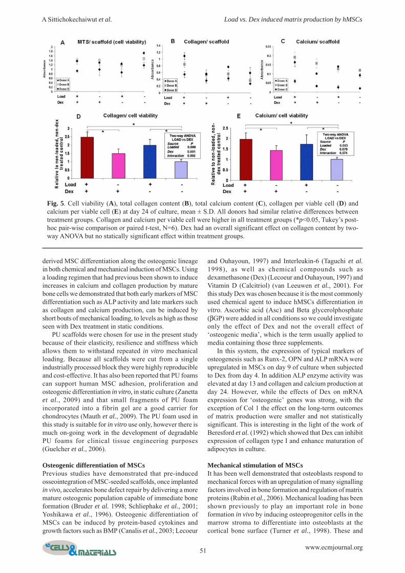
Cell viability, collagen and calcium productionStaining of scaffolds after 24 days of culture showed thatalthough there was some extra deposition of matrix on theoutside of the scaffold, as is commonly seen in 3D cultureof MSCs, collagen and calcium were present throughoutthe scaffold in each section (Fig. 4). Examination of thescaffolds by light microscopy showed qualitativedifferences in both Sirius red staining for collagen andalizarin red staining for calcium. Prior to staining therelative number of metabolically active cells was analysedby MTS (Fig. 5a) and as for the day 12 assay on a differentgroup of cells (Fig. 3a) there were no significant effects ofthe treatments on relative viable cell number.
Samples were analysed by individual donor and therewere differences between different donors (Figs. 5a-c) ashas been previously shown in the alkaline phosphataseresponses of human MSCs to BMP-2 (Diefenderfer et al.,2003) and bioactive glass (Reilly et al., 2007). In particularcells from donor A had fewer metabolically active cells atday 24 and a very low calcium content, below the limit atwhich the plate reader measures absorbance linearly.However, all donors showed similar relative differences
between treatment groups. Data were normalised to aninternal control for statistical analyses and the results forthe donor cells with low calcium values were not includedin the calcium analyses.
Collagen per viable cell was higher in all treatmentgroups with loading (2 fold), Dex (1.5 fold) or bothtreatments (2.5 fold) compared to non-loaded without Dex(Fig. 5d). A two-way ANOVA confirmed that both theeffect of Dex and the effect of loading were statisticallysignificant and there was no interaction between them,suggesting that the two effects are independent. Paired t-tests indicated that collagen content was significantlyhigher in the loaded group compared with the non-loadedgroup regardless of whether Dex was added to the media.Similarly to Col 1 mRNA there was no significant effectof Dex treatment alone within the non-loaded group.Calcium content per cell followed the same trend ascollagen content with the effect of loading beingstatistically significant although in this case the overalleffect of Dex was not statistically significant. Within theno-Dex treated groups, loading caused a 2 fold increase incalcium deposition, having an effect as large as that seenwith Dex and loading combined.
Discussion
Scaffold materialsIn the present study we used an inert polyurethane foamas a scaffold and demonstrated that it supports bone marrow
51 www.ecmjournal.org
A Sittichokechaiwut et al. Load vs. Dex induced matrix production by hMSCs
derived MSC differentiation along the osteogenic lineagein both chemical and mechanical induction of MSCs. Usinga loading regimen that had previous been shown to induceincreases in calcium and collagen production by maturebone cells we demonstrated that both early markers of MSCdifferentiation such as ALP activity and late markers suchas collagen and calcium production, can be induced byshort bouts of mechanical loading, to levels as high as thoseseen with Dex treatment in static conditions.
PU scaffolds were chosen for use in the present studybecause of their elasticity, resilience and stiffness whichallows them to withstand repeated in vitro mechanicalloading. Because all scaffolds were cut from a singleindustrially processed block they were highly reproducibleand cost-effective. It has also been reported that PU foamscan support human MSC adhesion, proliferation andosteogenic differentiation in vitro, in static culture (Zanettaet al., 2009) and that small fragments of PU foamincorporated into a fibrin gel are a good carrier forchondrocytes (Mauth et al., 2009). The PU foam used inthis study is suitable for in vitro use only, however there ismuch on-going work in the development of degradablePU foams for clinical tissue engineering purposes(Guelcher et al., 2006).
Osteogenic differentiation of MSCsPrevious studies have demonstrated that pre-inducedosseointegration of MSC-seeded scaffolds, once implantedin vivo, accelerates bone defect repair by delivering a moremature osteogenic population capable of immediate boneformation (Bruder et al. 1998; Schliephake et al., 2001;Yoshikawa et al., 1996). Osteogenic differentiation ofMSCs can be induced by protein-based cytokines andgrowth factors such as BMP (Canalis et al., 2003; Lecoeur
and Ouhayoun, 1997) and Interleukin-6 (Taguchi et al.1998), as well as chemical compounds such asdexamethasone (Dex) (Lecoeur and Ouhayoun, 1997) andVitamin D (Calcitriol) (van Leeuwen et al., 2001). Forthis study Dex was chosen because it is the most commonlyused chemical agent to induce hMSCs differentiation invitro. Ascorbic acid (Asc) and Beta glycerolphosphate(βGP) were added in all conditions so we could investigateonly the effect of Dex and not the overall effect of‘osteogenic media’, which is the term usually applied tomedia containing those three supplements.
In this system, the expression of typical markers ofosteogenesis such as Runx-2, OPN and ALP mRNA wereupregulated in MSCs on day 9 of culture when subjectedto Dex from day 4. In addition ALP enzyme activity waselevated at day 13 and collagen and calcium production atday 24. However, while the effects of Dex on mRNAexpression for ‘osteogenic’ genes was strong, with theexception of Col 1 the effect on the long-term outcomesof matrix production were smaller and not statisticallysignificant. This is interesting in the light of the work ofBeresford et al. (1992) which showed that Dex can inhibitexpression of collagen type I and enhance maturation ofadipocytes in culture.
Mechanical stimulation of MSCsIt has been well demonstrated that osteoblasts respond tomechanical forces with an upregulation of many signallingfactors involved in bone formation and regulation of matrixproteins (Rubin et al., 2006). Mechanical loading has beenshown previously to play an important role in boneformation in vivo by inducing osteoprogenitor cells in themarrow stroma to differentiate into osteoblasts at thecortical bone surface (Turner et al., 1998). These and
Fig. 5. Cell viability (A), total collagen content (B), total calcium content (C), collagen per viable cell (D) andcalcium per viable cell (E) at day 24 of culture, mean ± S.D. All donors had similar relative differences betweentreatment groups. Collagen and calcium per viable cell were higher in all treatment groups (*p<0.05, Tukey’s post-hoc pair-wise comparison or paired t-test, N=6). Dex had an overall significant effect on collagen content by two-way ANOVA but no statically significant effect within treatment groups.

52 www.ecmjournal.org
A Sittichokechaiwut et al. Load vs. Dex induced matrix production by hMSCs
related findings led researchers to hypothesise thatosteogenic precursors respond to mechanical loading andmuch recent research supports this. For example in cellmonolayers, oscillatory fluid flow (Arnsdorf et al., 2009)has been shown to induce the upregulation of transcriptionfactors involved in the osteogenic differentiation pathwaysof MSCs, and laminar flow to upregulate BMP-2 and OPNmRNA (Yourek et al., 2010). Cyclic stretching of hMSCson a flexible membrane resulted in upregulation of bothbone transcription factors, matrix proteins and ALP
(Diederichs et al., 2009; Jagodzinski et al., 2004) inaddition to markers of cartilage (Friedl et al., 2007).
However, it is becoming increasingly clear thatmechanotransduction pathways operate differently in 2Dcompared to 3D culture (Reilly and Engler, 2009) andunderstanding 3D culture is not only important to bettermimic the physiological environment but also necessaryto produce tissue in bulk for tissue engineering. Recentefforts to study the mechanical effects of osteogenesis in3D have led to the development of multiple models which
Table 3. Example of previous studies of osteogenic responses of MSCs to mechanical stimulation in 3-Denvironments.
Reference Type of stimulation
Loading procedure Culture period
Outcome (loaded samples compared with non loaded
Cartmell et al. (2003)
Continuous
flow
Flow induced shear stress on cell-seeded human trabecular bone scaffold.
7 days Upregulation of RUNX2, OCN, ALP mRNA expression
Grayson et al. (2008)
Flow induced shear stress on cell-seeded decellularized bone matrix.
35 days Greater bone volume by µCT. Increasing of total protein, ALP, BSP and OPN
Datta et al. (2005)
Flow induced shear stress on cell-seeded titanium fiber mesh scaffold.
16 days Higher calcium content
Jagodzinski et al. (2008)
Continuous flow and
intermittent strain
Continuous flow through cell-seeded demineralised bone matrix with some constructs subjected to additional cyclic scaffold compression
21 days Higher cell number (both loading conditions) maintenance of osteocalcin production (cyclic compression only).
Sumanasinghe et al. (2006)
Intermittent strain
Uniaxial cyclic tensile strain on cell-seeded collagen gels.
14 days Upregulation of BMP-2 gene expression.
Byrne et al. (2008)
Uniaxial cyclic tensile strain on cell-seeded collagen-glycosaminoglycan scaffolds
7 days Upregulation of OPN gene expression.
Mauney et al. (2004)
4-point bending on cell-seeded demineralised bone matrix
16 days Higher ALP activity.
Sittichokechaiwut et al.
(Current results)
Cyclic dynamic compressive strain on cell-seeded polyurethane scaffolds
24 days Higher ALP activity, COL1 gene expression, collagen and calcium content.

53 www.ecmjournal.org
A Sittichokechaiwut et al. Load vs. Dex induced matrix production by hMSCs
can be broadly divided into continuous loading systemssuch as perfusion flow bioreactors, or short termstimulation systems such as stretch, compression andbending of cell seeded constructs, or short periods of fluidflow through constructs. Examples of such studies aresummarised in Table 3, though this is not intended to bean extensive review of all previous research in this area.Here we examined the short term effects of a single loadingbout and the long term effects of repeated loading withinthe same system.
For continuous perfusion systems, it is difficult toseparate the effects of improved nutrient perfusion fromthe effects of the shear stress induced by fluid flow, in factboth effects appear to be involved in osteoblast responsesto fluid flow in monolayer (Donahue et al., 2003). In thissystem we chose to use an open cell highly porous foamso that our cells could survive well enough in long termstatic culture to see the effects of a specific loading regimen.While our loading regimen would have temporarilyimproved nutrient perfusion in the loaded scaffold, thisoccurred for only 6 h within a 576 h (24 day) culture periodtherefore, we believe nutrient perfusion played only aminor role in this system.
mRNA expression and ALP activityRUNX2 (Cbfa1) gene expression was performed in thisstudy as it is specific to the early differentiation of MSCsinto the osteogenic lineage, and is a modulator of boneformation by fully differentiated osteoblasts (Karsenty,2001). Osteopontin is a bone matrix protein that isupregulated during human MSC differentiation in vitro(Lian et al., 1998) and responsive to mechanical loadingin mature bone cells (You et al., 2001) and human MSCs(Sharp et al., 2009; Yourek et al., 2010). ALP has beenwidely used as a marker of MSCs differentiation towardthe osteogenic lineage, as increases in enzymatic activityand expression of both the gene and protein correspond toan osteoblastic phenotype (Grayson et al., 2008; Mauneyet al., 2004a; Wozniak et al., 2000). It peaks during thematrix formation phase and declines thereafter, while OPNand OCN peak in the late maturation or early mineralizationphases (Lian et al., 1998). For each of the three genes thereappeared to be more mRNA in loaded than non-loadedsamples within the no-Dex treatment group however theband density measurement did not show a statisticallysignificant difference. Previous studies in which thesegenes have been shown to be upregulated by mechanicalstimulation have usually used higher strains or longerloading periods then used here (Table 3). For exampleRunx2 has been shown to be upregulated by mechanicalloading of hMSCs after cyclic loading at 8% strain intension or 10% in compression (Friedl et al., 2007;Jagodzinski et al., 2008; Jagodzinski et al., 2004).
Two hours of dynamic compressive loading at 5%strain doubled the expression of Col1 mRNA in both Dexand non-Dex treated groups compared to non-loadedconstructs suggesting that mechanical loading could beused to stimulate collagen synthesis in situations where itis preferable not to use Dex. This suggests that Col1 mRNAexpression is more sensitive to loading and less sensitiveto Dex than the other ‘ostoegenic’ genes investigated.
However, these responses are likely to be specific to cellsource, scaffold and loading regimen for instance Friedlet al. (2007), Jagodzinski et al. (2008) and Jagodzinski etal. (2004) also saw a strong effect of cyclic stretching ofhMSCs (monolayer) on Col1 expression however in theircase the effect was much stronger in Dex treated cultures,while Byrne et al. (2008) saw no effect of cyclic stretchingon Col1 expression by rat MSCs in collagen-glycosaminoglycan porous scaffolds when all cells weretreated with Dex, but Grellier et al. (2009) saw a decreasein Col1 expression together with an increase in ALPexpression after a fluid flow stimulus.
ALP activity was measured 3 days after the loadingbout and in this case the loading-induced difference waslarger than that for ALP mRNA (40% higher) andstatistically significant. ALP activity induced by just 2 hof compression loading was as high as that induced by 8days of Dex treatment (culture days 4 to 12). The effectsof Dex in our 3D scaffold (50% higher ALP) arecomparable with its effects on the same source of humanMSCs in standard 2D tissue culture plastic (40% higher inDex treated cultures by day 14, data not shown). Mauneyet al. (2004a) have shown that ALP activity in vitro waselevated by cyclic mechanical loading for cells culturedin 10nM Dex, but this effect was abolished with 100nMconcentration, suggesting that Dex can inhibit thismechanism at high concentrations.
Matrix production and mineralizationCollagen and calcium were distributed over the scaffoldstruts, although cell bodies were not localised in theseexperiments, it is likely there were distributed throughoutthis deposited ECM as in our previous study on matureosteoblasts (Sittichockechaiwut et al., 2009). In agreementwith the ColI mRNA data, collagen and calcium depositiondoubled in mechanically stimulated MSCs when comparedto non-loaded samples with no Dex (Fig. 2). Although therewas some effect of Dex, constructs treated with Dex onlyhad no higher collagen and calcium content than constructssubjected to loading only (no-Dex). This indicates thatmatrix synthesis and mineralization can be elevated bymechanical stimulation independent of Dex. There is someindication of an additive effect of the two stimuli with thehighest production being seen in constructs subjected toboth loading and Dex though this is not significantly higherthan loading treatment alone. Others have shown thatperfusion culture improves matrix production (Table 3)and implied that some of the reason for this is theproduction of flow induced shear stresses, but here weshow that continuous matrix perfusion is not necessary toinduce a robust matrix forming response. Interestingly, ina study in which continuous perfusion and dynamiccompression were applied to 3D hMSC constructs in thesame bioreactor conditions, perfusion upregulated cellproliferation only and bouts of dynamic compressioncombined with perfusion were required to maintainproduction of the bone matrix gene osteocalcin(Jagodzinski et al., 2008). There is also evidence that whenMSC-containing constructs are implanted in vivo, shortbouts of compression loading enhance mineralisation(Duty et al., 2007), including in a scaffold implanted

54 www.ecmjournal.org
A Sittichokechaiwut et al. Load vs. Dex induced matrix production by hMSCs
without cells and infiltrated by the animal’s precursor cells(Roshan-Ghias et al., 2010). This implies that bonedifferentiation will be stimulated if hMSC seededconstructs are implanted into load bearing sites in vivo.
A limitation of our scaffold is that although we apply aglobal strain of 5%, individual cells will experience verydifferent substrate strain (tensile, compressive shear) andfluid flow regimens depending on how well they areattached to the scaffold and the scaffold strut orientationin relation to the loading direction, while other cells arestress-shielded. This may not be of high importance whenoptimising conditions for growth of tissue engineered bone,where as long as the construct as a whole produces matrixthis is a positive result. However, it does make difficult toelucidate the mechanotransduction mechanisms that triggerthe response. Models such as those being developed tobetter understand the strains and shear stresses experiencedwithin porous scaffolds (Lacroix et al., 2006; O’Brien etal., 2007) would help to elucidate what mechanical forcescells are experiencing within this complex system.
In conclusion, this study has shown that a combinationof short bouts of cyclic loading and rest periods canimprove matrix production by human bone marrow derivedMSCs in engineered bone constructs in vitro. The datasuggest that a short mechanical stimulus is an additionalor alternative tool for establishing precultivation conditionsprior to clinical implantation of tissue engineered bone,which has implications in the design of mechanical loadingregimens in bioreactor culture. However, given the varietyof scaffolds and loading regimens under currentinvestigation and the contrasting stimulus-specific resultspresented in the literature further research is needed tounderstand how cell-lineage specific effects ondifferentiation can be consistently obtained usingmechanical stimuli.
Acknowledgements
We gratefully acknowledge Professor Sheila Mac Neil whoprovided access to cell culture facilities and reagents. BoseElectroForce systems group supported this research byproviding the ELF 3200 for an evaluation period. AS wassupported by a Royal Thai Government Scholarship andthe research was partially funded by a Royal Society ofLondon Research Grant and the University of SheffieldDevolved Funds scheme.
References
Arnsdorf EJ, Tummala P, Kwon RY, Jacobs CR (2009)Mechanically induced osteogenic differentiation – the roleof RhoA, ROCKII and cytoskeletal dynamics. J Cell Sci122: 546-553.
Aubin JE (1999) Osteoprogenitor cell frequency in ratbone marrow stromal populations: role for heterotypic cell-cell interactions in osteoblast differentiation. J CellBiochem 72: 396-410.
Beresford JN, Bennett JH, Devlin C, Leboy PS, OwenME (1992) Evidence for an inverse relationship betweenthe differentiation of adipocytic and osteogenic cells inrat marrow stromal cell cultures. J Cell Sci 102: 341-351.
Bianco P, Kuznetsov SA, Riminucci M, Gehron RobeyP (2006) Postnatal skeletal stem cells. Methods Enzymol419: 117-148.
Bruder SP, Kraus KH, Goldberg VM, Kadiyala S(1998) The effect of implants loaded with autologousmesenchymal stem cells on the healing of canine segmentalbone defects. J Bone Joint Surg Am, 80:985-996.
Byrne EM, Farrell E, McMahon LA, Haugh MG,O’Brien FJ, Campbell VA, Prendergast PJ, O’Connell BC(2008) Gene expression by marrow stromal cells in aporous collagen-glycosaminoglycan scaffold is affectedby pore size and mechanical stimulation. J Mater Sci MaterMed 19: 3455-3463.
Cartmell SH, Porter BD, Garcia AJ, Guldberg RE(2003) Effects of medium perfusion rate on cell-seededthree-dimensional bone constructs in vitro. Tissue Eng 9:1197-1203.
Canalis E, Economides AN, Gazzerro E (2003) Bonemorphogenetic proteins, their antagonists, and the skeleton.Endocr Rev 24: 218-235.
Cowie R, Walker RD, Scutt A (2006) The use of high-frequency, low-intensity vibration to stimulate theproliferation and differentiation of primary rat bonemarrow cells. Cytotherapy 8: 63-63.
Datta N, Holtorf HL, Sikavitsas VI, Jansen JA, MikosAG (2005) Effect of bone extracellular matrix synthesizedin vitro on the osteoblastic differentiation of marrowstromal cells. Biomaterials 26: 971-977.
Diederichs S, Freiberger F, van Griensven M (2009)Effects of repetitive and short time strain in human bonemarrow stromal cells. J Biomed Mater Res A 88: 907-915.
Diefenderfer DL, Osyczka AM, Garino JP, Leboy PS(2003) Regulation of BMP-induced transcription incultured human bone marrow stromal cells. J Bone JointSurg Am, 85-A Suppl 3: 19-28.
Dieudonne SC, Kerr JM, Xu T, Sommer B, DeRubeisAR, Kuznetsov SA, Kim IS, Gehron Robey P, Young MF(1999) Differential display of human marrow stromal cellsreveals unique mRNA expression patterns in response todexamethasone. J Cell Biochem, 76:231-243.
Dominici M, Le Blanc K, Mueller I, Slaper-CortenbachI, Marini F, Krause D, Deans R, Keating A, Prockop D,Horwitz E (2006) Minimal criteria for defining multipotentmesenchymal stromal cells. The International Society forCellular Therapy position statement. Cytotherapy 8: 315-317.
Donahue TLH, Haut TR, Yellowley CE, Donahue HJ,Jacobs CR (2003) Mechanosensitivity of bone cells tooscillating fluid flow induced shear stress may bemodulated by chemotransport. J Biomech 36: 1363-1371.
Duty AO, Oest ME, Guldberg RE (2007) Cyclicmechanical compression increases mineralization of cell-seeded polymer scaffolds in vivo. J Biomech Eng 129: 531-539.
Friedl G, Schmidt H, Rehak I, Kostner G, SchauensteinK, Windhager R (2007) Undifferentiated humanmesenchymal stem cells (hMSCs) are highly sensitive tomechanical strain: transcriptionally controlled early osteo-chondrogenic response in vitro. Osteoarthritis Cartilage,15:1293-1300.

55 www.ecmjournal.org
A Sittichokechaiwut et al. Load vs. Dex induced matrix production by hMSCs
Grayson WL, Bhumiratana S, Cannizzaro C, Chao PH,Lennon DP, Caplan AI, Vunjak-Novakovic G (2008)Effects of initial seeding density and fluid perfusion rateon formation of tissue-engineered bone. Tissue Eng PartA 14: 1809-1820.
Gregory CA, Gunn WG, Peister A, Prockop DJ (2004)An alizarin red-based assay of mineralization by adherentcells in culture: comparison with cetylpyridinium chlorideextraction. Anal Biochem 329: 77-84.
Grellier M, Bareille R, Bourget C, Amedee J (2009)Responsiveness of human bone marrow stromal cells toshear stress. J Tissue Eng Regen Med 3: 302-309.
Guelcher SA, Patel V, Gallagher KM, Connolly S,Didier JE, Doctor JS, Hollinger JO (2006) Synthesis andin vitro biocompatibility of injectable polyurethane foamscaffolds. Tissue Eng 12: 1247-1259.
Heath CA (2000) Cells for tissue engineering. TrendsBiotechnol 18: 17-19.
Heng BC, Cao T, Stanton LW, Robson P, Olsen B(2004) Strategies for directing the differentiation of stemcells into the osteogenic lineage in vitro. J Bone MinerRes 19: 1379-1394.
Holtorf HL, Jansen JA, Mikos AG (2005) Flowperfusion culture induces the osteoblastic differentiationof marrow stroma cell-scaffold constructs in the absenceof dexamethasone. J Biomed Mater Res A 72: 326-334.
Huang CY, Hagar KL, Frost LE, Sun Y, Cheung HS(2004) Effects of cyclic compressive loading onchondrogenesis of rabbit bone-marrow derivedmesenchymal stem cells. Stem Cells 22: 313-323.
Jagodzinski M, Drescher M, Zeichen J, HankemeierS, Krettek C, Bosch U, van Griensven M (2004) Effectsof cyclic longitudinal mechanical strain anddexamethasone on osteogenic differentiation of humanbone marrow stromal cells. Eur Cell Mater 7: 35-41.
Jagodzinski M, Breitbart A, Wehmeier M, Hesse E,Haasper C, Krettek C, Zeichen J, Hankemeier S (2008)Influence of perfusion and cyclic compression onproliferation and differentiation of bone marrow stromalcells in 3-dimensional culture. J Biomech 41: 1885-1891.
Karsenty G (2001) Minireview: transcriptional controlof osteoblast differentiation. Endocrinology 142: 2731-2733.
Klein-Nulend J, Bacabac RG, Mullender MG (2005)Mechanobiology of bone tissue. Pathol Biol (Paris) 53:576-580.
Kreke MR, Sharp LA, Lee YW, Goldstein AS (2008)Effect of intermittent shear stress on mechanotransductivesignaling and osteoblastic differentiation of bone marrowstromal cells. Tissue Eng Part A 14: 529-537.
Lacroix D, Chateau A, Ginebra MP, Planell JA (2006)Micro-finite element models of bone tissue-engineeringscaffolds. Biomaterials 27: 5326-5334.
Lecoeur L, Ouhayoun JP (1997) In vitro induction ofosteogenic differentiation from non-osteogenicmesenchymal cells. Biomaterials 18: 989-993.
Lian JB, Stein GS, Stein JL, van Wijnen AJ (1998)Transcriptional control of osteoblast differentiation.Biochem Soc Trans 26: 14-21.
Maniatopoulos C, Sodek J, Melcher AH (1988) Boneformation in vitro by stromal cells obtained from bonemarrow of young adult rats. Cell Tissue Res 254: 317-330.
Mauney JR, Blumberg J, Pirun M, Volloch V, Vunjak-Novakovic G, Kaplan DL (2004a) Osteogenicdifferentiation of human bone marrow stromal cells onpartially demineralized bone scaffolds in vitro. Tissue Eng10: 81-92.
Mauney JR, Sjostorm S, Blumberg J, Horan R, O’LearyJP, Vunjak-Novakovic G, Volloch V, Kaplan DL (2004b)Mechanical stimulation promotes osteogenicdifferentiation of human bone marrow stromal cells on 3-D partially demineralized bone scaffolds in vitro. CalcifTissue Int 74: 458-468.
Mauth C, Bono E, Haas S, Paesold G, Wiese H, MaierG, Boos N, Graf-Hausner U (2009) Cell-seededpolyurethane-fibrin structures – a possible system forintervertebral disc regeneration. Eur Cell Mater 18: 27-38.
Meinel L, Karageorgiou V, Fajardo R, Snyder B,Shinde-Patil V, Zichner L, Kaplan D, Langer R, Vunjak-Novakovic G (2004) Bone tissue engineering using humanmesenchymal stem cells: effects of scaffold material andmedium flow. Ann Biomed Eng 32: 112-122.
Ng KW, Leong DT, Hutmacher DW (2005) Thechallenge to measure cell proliferation in two and threedimensions. Tissue Eng 11: 182-191.
O’Brien FJ, Harley BA, Waller MA, Yannas IV, GibsonLJ, Prendergast PJ (2007) The effect of pore size onpermeability and cell attachment in collagen scaffolds fortissue engineering. Technol Health Care 15: 3-17.
Osyczka AM, Noth U, O’Connor J, Caterson EJ, YoonK, Danielson KG, Tuan RS (2002) Multilineagedifferentiation of adult human bone marrow progenitorcells transduced with human papilloma virus type 16 E6/E7 genes. Calcif Tissue Int 71: 447-458.
Peter SJ, Liang CR, Kim DJ, Widmer MS, Mikos AG(1998) Osteoblastic phenotype of rat marrow stromal cellscultured in the presence of dexamethasone, beta-glycerolphosphate, and L-ascorbic acid. J Cell Biochem71: 55-62.
Porter RM, Huckle WR, Goldstein AS (2003) Effectof dexamethasone withdrawal on osteoblasticdifferentiation of bone marrow stromal cells. J CellBiochem 90: 13-22.
Reilly GC, Engler AJ (2009) Intrinsic extracellularmatrix properties regulate stem cell differentiation. JBiomech 1: 1.
Reilly GC, Radin S, Chen AT, Ducheyne P (2007)Differential alkaline phosphatase responses of rat andhuman bone marrow derived mesenchymal stem cells to45S5 bioactive glass. Biomaterials 28: 4091-4097.
Robey PG, Bianco P (2006) The use of adult stem cellsin rebuilding the human face. J Am Dent Assoc, 137:961-972.
Roshan-Ghias A, Terrier A, Bourban PE, Pioletti DP(2010) In vivo cyclic loading as a potent stimulatory signalfor bone formation inside tissue engineering scaffold. EurCell Mater 19: 41-49.

56 www.ecmjournal.org
A Sittichokechaiwut et al. Load vs. Dex induced matrix production by hMSCs
Rubin J, Rubin C, Jacobs CR (2006) Molecularpathways mediating mechanical signaling in bone. Gene367: 1-16.
Schliephake H, Knebel JW, Aufderheide M, TauscherM (2001) Use of cultivated osteoprogenitor cells toincrease bone formation in segmental mandibular defects:an experimental pilot study in sheep. Int J Oral MaxillofacSurg 30: 531-537.
Sharp LA, Lee YW, Goldstein AS (2009) Effect of low-frequency pulsatile flow on expression of osteoblasticgenes by bone marrow stromal cells. Ann Biomed Eng37: 445-453.
Simmons CA, Matlis S, Thornton AJ, Chen S, WangCY, Mooney DJ (2003) Cyclic strain enhances matrixmineralization by adult human mesenchymal stem cellsvia the extracellular signal-regulated kinase (ERK1/2)signaling pathway. J Biomech 36: 1087-1096.
Sittichokechaiwut A, Reilly GC (2009). Developmentof a culture system to modulate tissue engineered boneformation by varying loading conditions. Proc 7th IntConfManufact Res (ICMR 09), pp 443-448.
Sittichockechaiwut A, Scutt AM, Ryan AJ, BonewaldL, Reilly GC (2009) Use of rapidly mineralizing osteoblastsand short periods of mechanical loading to acceleratematrix maturation in 3D scaffolds. Bone 44: 822-829.
Sumanasinghe RD, Bernacki SH, Loboa EG (2006)Osteogenic differentiation of human mesenchymal stemcells in collagen matrices: effect of uniaxial cyclic tensilestrain on bone morphogenetic protein (BMP-2) mRNAexpression. Tissue Eng 12: 3459-3465.
Taguchi Y, Yamamoto M, Yamate T, Lin SC, MocharlaH, DeTogni P, Nakayama N, Boyce BF, Abe E, ManolagasSC (1998) Interleukin-6-type cytokines stimulatemesenchymal progenitor differentiation toward theosteoblastic lineage. Proc Assoc Am Physicians 110: 559-574.
Tullberg-Reinert H, Jundt G (1999) In situmeasurement of collagen synthesis by human bone cellswith a Sirius red-based colorimetric microassay: effectsof transforming growth factor beta2 and ascorbic acid 2-phosphate. Histochem Cell Biol 112: 271-276.
Turner CH, Owan I, Alvey T, Hulman J, Hock JM(1998) Recruitment and proliferative responses ofosteoblasts after mechanical loading in vivo determinedusing sustained-release bromodeoxyuridine. Bone 22: 463-469.
Vaccaro AR, Chiba K, Heller JG, Patel T, Thalgott JS,Truumees E, Fischgrund JS, Craig MR, Berta SC, WangJC (2002) Bone grafting alternatives in spinal surgery.Spine J 2: 206-215.
Van Leeuwen JP, Van Driel M, Van den Bemd GJ, PolsHA (2001) Vitamin D control of osteoblast function andbone extracellular matrix mineralization. Crit Rev EukaryotGene Expr 11: 199-226.
Weinstein RS, Jilka RL, Parfitt AM, Manolagas SC(1998) Inhibition of osteoblastogenesis and promotion ofapoptosis of osteoblasts and osteocytes by glucocorticoids– Potential mechanisms of their deleterious effects on bone.J Clin Invest 102: 274-282.
Weissman IL (2000) Translating stem and progenitorcell biology to the clinic: barriers and opportunities.Science 287: 1442-1446.
Wozniak M, Fausto A, Carron CP, Meyer DM, HruskaKA (2000) Mechanically strained cells of the osteoblastlineage organize their extracellular matrix through uniquesites of alphavbeta3-integrin expression. J Bone Miner Res15: 1731-1745.
Yoshikawa T, Ohgushi H, Tamai S (1996) Immediatebone forming capability of prefabricated osteogenichydroxyapatite. J Biomed Mater Res 32: 481-492.
You J, Reilly GC, Zhen X, Yellowley CE, Chen Q,Donahue HJ, Jacobs CR (2001) Osteopontin generegulation by oscillatory fluid flow via intracellular calciummobilization and activation of mitogen-activated proteinkinase in MC3T3-E1 osteoblasts. J Biol Chem 276: 13365-13371.
Yourek G, McCormick SM, Mao JJ, Reilly GC (2010)Shear stress induces osteogenic differentiation of humanmesenchymal stem cells. Regenerative Medicine, in press.
Zanetta M, Quirici N, Demarosi F, Tanzi MC,Rimondini L, Fare S (2009) Ability of polyurethane foamsto support cell proliferation and the differentiation of MSCsinto osteoblasts. Acta Biomater 5: 1126-1136.
Discussion with Reviewer
Reviewer I: Describe the hypothesised biologicalmechanism of how loading is able to induce osteogenicdifferentiation in a similar manner to, but in the absenceof glucocorticoids.Authors: At this stage it is not possible to tell if loadingand Dex initiate differentiation via the same pathways ordifferent pathways as multiple pathways are involved inboth stimuli. There is some evidence of an additive effectof Dex and loading (Fig. 5) indicating that there is probablymore than one pathway involved. If Dex and loading dopartially operate via the same mechanism a strongcandidate for the signalling pathway mediating this is theextracellular signal regulated pathway 1 and 2 (ERK1/2).ERK1/2 has been consistently shown to be upregulatedby a range of mechanical loading stimuli in both osteoblasts(You et al., 2001; text reference) and MSCs (Simmons etal., 2003, text reference; Glossop and Cartmell, 2009)including by cyclic hydrostatic pressure in a 3D PLGAconstructs (Kim et al., 2007). In preliminary experimentswe detected increased ERK1/2 phosphorylation in cellssubjected to the loading conditions described here (datanot shown). However, the contribution of ERK1/2signalling to osteoblast differentiation is very complex.While ERK1/2 has been shown to stimulate thedexamethasone-induced differentiation of MSCs intoosteoblasts, via phosphorylation of the osteogenictranscription factor RUNX2/Cbfa1 in monolayer culture(Jaiswal et al., 2000), and 3D culture (Farrell et al., 2006)it has also been shown to inhibit BMP-2 induceddifferentiation in monolayer (Osyczka and Leboy, 2005)and collagen induced osteogenic differentiation in 3D gels

57 www.ecmjournal.org
A Sittichokechaiwut et al. Load vs. Dex induced matrix production by hMSCs
(Lund et al., 2009). Therefore, there is likely to be acomplex relationship between the temporalphosphorylation of ERK1/2, the co-stimulation of otherpathways and the ultimate fate of human MSCs whichwarrants further and detailed investigation.
Additional References
Farrell E, O’Brien FJ, Doyle P, Fischer J, Yannas I,Harley BA, O’Connell B, Prendergast PJ, Campbell VA(2006) A collagen-glycosaminoglycan scaffold supportsadult rat mesenchymal stem cell differentiation alongosteogenic and chondrogenic routes. Tissue Eng 12: 459-468.
Glossop JR, Cartmell SH (2009) Effect of fluid flow-induced shear stress on human mesenchymal stem cells:Differential gene expression of IL1B and MAP3K8 inMAPK signaling. Gene Expr Patt 9: 381-388.
Jaiswal RK, Jaiswal N, Bruder SP, Mbalaviele G,Marshak DR, Pittenger MF (2000) Adult humanmesenchymal stem cell differentiation to the osteogenicor adipogenic lineage is regulated by mitogen-activatedprotein kinase. J Biol Chem 275: 9645-9652.
Kim SH, Choi YR, Park MS, Shin JW, Park KD, KimSJ, Lee JW (2007) Erk 1/2 activation in enhancedosteogenesis of human mesenchymal stem cells inpoly(lactic-glycolic acid) by cyclic hydrostatic pressure. JBiomed Mater Res 80A: 826-836.
Lund AW, Stegemann JP, Plopper GE (2009) Inhibitionof ERK promotes collagen gel compaction andfibrillogenesis to amplify the osteogenesis of humanmesenchymal stem cells in three-dimensional collagen Iculture. Stem Cells Dev 18: 331-41.
Osyczka AM, Leboy PS (2005) Bone morphogeneticprotein regulation of early osteoblast genes in humanmarrow stromal cells is mediated by extracellular signal-regulated kinase and phosphatidylinositol 3-kinasesignaling. Endocrinology 146: 3428-3437.