shining new light on the brain s bilingual signature : a
TRANSCRIPT

www.elsevier.com/locate/ynimg
NeuroImage 39 (2008) 1457–1471Shining new light on the brain’s “bilingual signature”: A functionalNear Infrared Spectroscopy investigation of semantic processing
Ioulia Kovelman,b Mark H. Shalinsky,c Melody S. Berens,a and Laura-Ann Petittoa,⁎
aDepartment of Psychology, University of Toronto Scarborough, Toronto, 1265 Military Trail, Toronto, ON Canada MIC 1A4bMassachusetts Institute of Technology, MA, USAcAnthinoula A. Martinos Center for Biomedical Imaging, Massachusetts General Hospital, Harvard Medical School, MA, USA
Received 25 May 2007; revised 9 October 2007; accepted 16 October 2007Available online 25 October 2007
Decades of research have shown that, from an early age, proficientbilinguals can speak each of their two languages separately (similar tomonolinguals) or rapidly switch between them (dissimilar to mono-linguals). Thus we ask, do monolingual and bilingual brains processlanguage similarly or dissimilarly, and is this affected by the languagecontext? Using an innovative brain imaging technology, functionalNear Infrared Spectroscopy (fNIRS), we investigated how adultbilinguals process semantic information, both in speech and in print,in a monolingual language context (one language at a time) or in abilingual language context (two languages in rapid alternation). Whileundergoing fNIRS recording, ten early exposed, highly proficientSpanish–English bilinguals completed a Semantic Judgment task inmonolingual and bilingual contexts and were compared to ten Englishmonolingual controls. Two hypotheses were tested: the SignatureHypothesis predicts that early, highly proficient bilinguals will recruitneural tissue to process language differently from monolinguals acrossall language contexts. The Switching Hypothesis predicts that bilingualswill recruit neural tissue to process language similarly to monolinguals,when using one language at a time. Supporting the SignatureHypothesis, in the monolingual context, bilinguals and monolingualsshowed differences in both hemispheres in the recruitment of DLPFC(BA 46/9) and IFC (BA 47/11), but similar recruitment of Broca’sarea (BA 44/45). In particular, in the monolingual context, bilingualsshowed greater signal intensity in channels maximally overlayingDLPFC and IFC regions as compared to monolinguals. In the bilingualcontext, bilinguals demonstrated a more robust recruitment of rightDLPFC and right IFC. These findings reveal how extensive earlybilingual exposure modifies language organization in the brain—thusimparting a possible “bilingual signature.” They further shedfascinating new light on how the bilingual brain may reveal thebiological extent of the neural architecture underlying all humanlanguage and the language processing potential not fully recruited inthe monolingual brain.© 2007 Elsevier Inc. All rights reserved.
⁎ Corresponding author.E-mail address: [email protected] (L.-A. Petitto).Available online on ScienceDirect (www.sciencedirect.com).
1053-8119/$ - see front matter © 2007 Elsevier Inc. All rights reserved.doi:10.1016/j.neuroimage.2007.10.017
Introduction
Can early dual language exposure modify neural tissue? Earlychildhood is a key period in human development whenneurological organization and reorganization take place (Dawsonand Fischer, 1994) and many early childhood experiences yield alifelong impact on brain organization (Fine et al., 2005; Johnsonand Newport, 1989; Neville and Bavelier, 2001; Mayberry andEichen, 1991; Newman et al., 2002; Newport, 1990; Ohnishi et al.,2001; Petersson et al., 2001; Petitto et al., 2000; Roder et al.,2002). Is early exposure to two languages one of these childhoodexperiences? Does extensive and maintained exposure to twolanguages from early life leave a “bilingual signature” on thehuman brain?
Early language experiences have been shown to result inpermanent behavioral and neurological changes. Individuals notexposed to any language before puberty (or even before age 7)commonly fail to achieve monolingual-like language proficiency,experience enhanced difficulty learning language later in life(Lenneberg, 1967; Mayberry and Eichen, 1991; Mayberry andFischer, 1989; Mayberry et al., 2002; Neville et al., 1997), anddisplay non-native language organization in the brain (Newmanet al., 2002). On the opposite end of the spectrum, early,extensive, and maintained bilingual exposure appears to yieldthe greatest dual language proficiency and the most similarneural organization between the two languages (e.g., Johnsonand Newport, 1989; Kovelman and Petitto, 2002; McDonald,2000; Petitto and Kovelman, 2003; Weber-Fox and Neville,1999).
Only a handful of researchers have conducted neuroimagingstudies that directly compare bilingual and monolingual brains(Mechelli et al., 2004; Rodriguez-Fornells et al., 2002). In the onlystructural-anatomical MRI study comparing bilinguals and mono-linguals (Mechelli et al., 2004), results showed that bilinguals hadan increase in gray matter volume in the left inferior parietal cortex,which was greatest in early exposed, highly proficient bilinguals.In one of the two functional studies, Rodriguez-Fornells et al.(2002) found that when bilinguals were required to ignore words in

1458 I. Kovelman et al. / NeuroImage 39 (2008) 1457–1471
one of their languages, they showed greater recruitment of the leftinferior frontal cortex (IFC; BA 44) and the left IFC area adjacentto the middle frontal gyrus (MFG; BA 46/9) than monolinguals.Given that the bilinguals (unlike the monolinguals) were requiredto ignore one of their languages in this study, the observed LIFC/MFG activation could be related to the switching and inhibitionnature of the task. Finally, in an fMRI neuroimaging studyconducted in our own laboratory, when performing a complexsyntactic task in one language at a time, bilinguals showed greateractivation within LIFC (BA 45) than monolinguals (Kovelmanet al., 2005).
Although it is commonly observed that early, extensive, andmaintained bilingual exposure typically results behaviorally inoptimal dual language competence (Johnson and Newport, 1989;Kovelman and Petitto, 2002; Weber-Fox and Neville, 1999), thereis controversy about the nature of the neural recruitment for thetwo languages, especially when exposure to two languages doesnot occur close in time (e.g., one language is acquired first, L1,followed by another language, L2). On one hand, some studieshave shown both subcortical activation differences and greaterfrontal and bilateral recruitment for L2 using a variety of languagetasks (text comprehension: Dehaene et al., 1997; Kim et al., 1997;semantic and grammatical processing of sentences: Hahne andFriederici, 2001; Wartenburger et al., 2003; Weber-Fox andNeville, 1999; phonological and lexical processing of words andsounds: Klein et al., 2006, 1995; Marian et al., 2003; Pillai et al.,2003). On the other hand, some studies have shown no neuralactivation differences between bilinguals’ L1 and L2 (textcomprehension: Perani et al., 1998; semantic and grammaticalprocessing of sentences: Friederici et al., 2002; phonological andlexical processing of words and sounds: Chee et al., 1999; Kleinet al., 1999, 1995). One reason for this controversy is that theextent and maintenance of dual language exposure can impact anindividual’s proficiency in each language. In turn, proficiency caninfluence dual language representation in the brains of bilinguals;age of acquisition also has influence on the representation oflanguage in bilingual brains (semantic and grammatical processingof sentences: Wartenburger et al., 2003; phonological and lexicalprocessing of words and sounds: Chee et al., 2004, 2001; Golestaniet al., 2006; Meschyan and Hernandez, 2006; also, see review byAbutalebi et al., 2001).
Another factor to consider when comparing bilinguals andmonolinguals is the types of language contexts in which aproficient bilingual adult routinely functions. Bilinguals caninteract in different contexts or “modes” (Grosjean, 1997). Forexample, when a bilingual is with a monolingual, he or shespeaks in only one language and is thus in Monolingual mode.When with other bilinguals, they often speak both of theirlanguages or function in a Bilingual mode. Thus, bilinguals mightfind themselves using either one language at a time or bothlanguages in rapid succession. High facility with this kind ofswitching across Bilingual and Monolingual language modes isobserved even in very young bilingual children (Genesee, 1989;Genesee et al., 1996; Holowka et al., 2002; Paradis et al., 2000;Petitto and Holowka, 2002; Petitto et al., 2003, 2001; Petitto andKovelman, 2003; Poulin-Dubois and Goodz, 2001). In particular,research in our own laboratory has observed how even veryyoung bilinguals can easily and systematically modulate theirlanguage choice when interacting with monolinguals of their twolanguages within the same context/environment (e.g., Petittoet al., 2001).
An additional intriguing factor to consider when comparingbilingual and monolingual language processing is the types oflexico-semantic usage that are possible for bilinguals but notmonolinguals. One of the most common forms of “code switching”observed in bilinguals (often called “language mixing” whenobserved in children) is the swapping of lexico-semantic itemsbelonging to one language when building a phrase in anotherlanguage. For example, one might say “Yesterday we ate crèmeglassée” (“ice-cream” in French spoken in Quebec; e.g., Grosjean,2001; Paradis et al., 2000; Petitto et al., 2001; Petitto and Kovelman,2003; Poplack, 1980). Current theories of bilingual lexico-semanticrepresentation have assumed the existence of a combined lexicalstore, in which each lexical item is connected to a number ofsemantic features in a common semantic store (Ameel et al., 2005;Dijkstra and Van Heuven, 2002; Green, 1998; Kroll and Sunderman,2003; Monsell et al., 1992). Words in two languages that shareoverlapping semantic representations within the common semanticstore are called “translation equivalents” (e.g., “mother” in Englishand “maman” in French). This idea is supported by the fact thatbilinguals can be semantically primed in one language to produce aword in the other language (Dijkstra and Van Heuven, 2002;Kerkhofs et al., 2006; Kroll and Sunderman, 2003) and can translateconcrete words, whose semantic representations overlap, faster thanabstract words, which are less likely to share semantic featuresacross languages (Van Hell and De Groot, 1998).
How do bilinguals avoid confusing their two languages as theyrapidly process their languages and/or move from one languagecontext to the next? Language switching has been thought to bedue to a mechanism, and possibly even an area in the brain, whichcan selectively activate only one language (Paradis, 1997). Thisidea was challenged both by the failure to locate a specific brainarea that controls the choice of language (Paradis, 1997) and alsobecause of the observation that language choice is not an “on”/“off” process. Rather, bilingual language usage appears to form acontinuum in which bilinguals selectively activate or inhibit theirtwo languages to a greater or lesser extent (Grosjean, 2001;Paradis, 1997). Currently, language switching is considered to be adynamic process in which the degree that each language isactivated and inhibited is modulated and dependent on thelanguage context (Green, 1998; Grosjean, 1997; Paradis, 1997).
Behavioral studies alone cannot answer the question of whetherbilingual language exposure yields neural differences as comparedto monolingual language exposure. Behavioral studies havepresented conflicting results, with some suggesting that even earlyexposed, highly proficient bilinguals perform significantly worsethan monolinguals on semantic tasks across different languagecontexts (Thomas and Allport, 2000; Von Studnitz and Green,2002), and others suggesting no such bilingual deficit (Caramazzaand Brones, 1980; Grosjean and Miller, 1994; Van Heuven et al.,1998). Additionally, as noted above, there are a limited number ofneuroimaging studies on bilingual switching and semanticlanguage processing across dual language contexts.
With regard to language switching, bilinguals in Chee et al.’s(2003) study showed different amounts of neural activation withinclassic semantic processing brain areas (in particular, middletemporal gyrus, MTG) across different language contexts, whileKlein et al. (1995) showed similar recruitment of both classiclanguage-dedicated brain areas (left IFC) as well as dorsolateralprefrontal cortex (DLPFC) irrespective of whether bilinguals wereusing one or two languages. Still other studies have shown thatbilinguals require increased recruitment of neural tissue classically

1459I. Kovelman et al. / NeuroImage 39 (2008) 1457–1471
associated with task switching and mediation of executiveprocesses, such as DLPFC and anterior cingulate cortex (AC) inbilingual versus monolingual contexts (Fugelsang and Dunbar,2005; Hernandez et al., 2000, 2001; Holtzheimer et al., 2005; Priceet al., 1999). As DLPFC might be involved in task monitoring,particularly during novel and/or attention demanding tasks, regard-less of whether task switching is involved, the lack or presence ofDLPFC activation during language switching tasks should beinterpreted with caution (Duncan and Owen, 2000; Fugelsanget al., 2006; Wager et al., 2004).
In the present study we examine and compare semanticprocessing in bilinguals and monolinguals. Using behavioral andneuroimaging techniques, we examine how bilinguals processprinted and auditory semantic information presented to them acrosstwo types of typically encountered contexts: (i) Monolingual mode—one language in isolation, and (ii) Bilingual mode—twolanguages in rapid succession. We studied a group of earlyexposed, highly proficient bilinguals who were carefully screenedfor early dual language exposure, dual language proficiency, anddual language maintenance, as well as a group of monolingualcontrols matched for gender and age. Our analyses includedcomparisons of behavioral performance (accuracy and reactiontime) and changes in hemodynamic response in participants asmeasured with an innovative brain imaging technology, functionalNear Infrared Spectroscopy (fNIRS).
Two hypotheses about how the bilingual brain processeslanguage were tested: the Neural Signature Hypothesis predictsthat early exposed, proficient bilinguals should process languagedifferently from monolinguals and recruit different neural tissueacross all contexts, including one language at a time (Monolingualmode) and two languages in rapid alternation (Bilingual mode).The difference would be expressed by bilinguals showing greateror lesser neural recruitment (greater or lesser intensity of thehemodynamic signal or presence versus absence of activation in aparticular area) of the classic language, cognitive attention/inhibition, and verbal working memory brain areas as comparedto monolinguals, regardless of language mode. The FunctionalSwitching Hypothesis predicts that early exposed, proficientbilinguals should process language similarly to monolinguals andrecruit similar neural tissue, but not across all contexts.Specifically, bilinguals and monolinguals should show similarneural profiles when processing one language at a time (Mono-lingual mode). Neural differences should emerge only in thecontext specific and unique to bilinguals: that is, when bilingualsare processing two languages in rapid alternation (Bilingual mode);in this mode bilinguals should show greater neural recruitment ofthe classic language areas and/or brain areas involved in taskswitching. Classic language brain areas would include left IFC (BA44/45), while classic verbal working memory/attention brain areaswould include left DLPFC (BA 46/9) and anterior IFC (BA 47/11).
Previous research has shown that there is no simple one-to-onecorrespondence between behavioral performance and neuralactivity. Such findings are particularly abundant in the bilingualliterature, where different language groups can show the samebehavioral performance, yet different neural profiles (Chee et al.,2004; Wartenburger et al., 2003). Therefore, our behavioralpredictions are not specific to either of the two neurologicalhypotheses. For both “Neural Signature” and “Functional Switch-ing” hypotheses, we predicted that bilinguals would make semanticdecisions with the same or slower reaction time and the same orlower accuracy as monolinguals.
fNIRS is among the world’s most innovative imaging technol-ogies. Like fMRI, fNIRS measures changes in the brain’s bloodoxygen level density (BOLD) while a person is performing specificcognitive tasks. An advantage over fMRI is that, in addition toBOLD, fNIRS also computes the deoxygenated and oxygenatedhemoglobin from the absorption measured at different wavelengthsusing the modified Beer–Lambert equation. While fNIRS cannotrecord deep into the human brain (∼4 cm depth), it has good spatialresolution that is excellent for studies of human higher cognition andlanguage, and it has better temporal resolution than fMRI (∼b5 sHR, sampling rate=10× per second). Unlike the large size of fMRI,fNIRS is very small, highly portable (the size of a desktopcomputer), and particularly child friendly (children and adults sitnormally in a comfortable chair, and babies can be studied whileseated on mom’s lap). Furthermore, fNIRS is virtually silent, unlikethe loud whirring of the fMRI that can make language processingstudies challenging. Most importantly, fNIRS tolerates movement.By contrast, fMRI does not tolerate movement and, thus, is difficultto use for studying language production. The fMRI’s restriction onmovement, its production of loud noises, and its restrictive testingchamber make it very challenging to use for studying infants andchildren as well as special populations of adults. With the advent offNIRS, new insights into the human child’s developing brainfunction with respect to higher cognition and language, as well asnew insights into the aging brain, can now be laid bare.
To the best of our knowledge, the present study is unique in itsdirect comparison of bilingual and monolingual semantic proces-sing while using modern neuroimaging technology and behavioraltechniques. It is also unique in its focus on bilinguals’ semanticperformance across multiple bilingual contexts and in its use ofboth auditory and visually presented stimuli. By doing so we hopeto provide insight into the nature of bilingual language processing,the impact that early and extensive bilingual exposure has on thebilingual brain, and the effectiveness of fNIRS in cognitiveneuroscience research.
Materials and methods
Participants
Bilingual participantsTen right-handed Spanish–English bilinguals participated in
this experiment (4 men, 6 women, see Table 1). All bilingualparticipants started receiving extensive and systematic exposure toboth English and Spanish before the age of 5. All bilingualparticipants had high, monolingual-like, language proficiency ineach of their two languages (as established with participantscreening methods described below, on which all participantsachieved the required accuracy of at least 80%). Half of theparticipants were exposed to both Spanish and English at homefrom birth, and the other half of the participants were exposed toSpanish at home from birth and extensive and maintained exposureto English in daycare or kindergarten beginning by ages 3–5. Allbilingual participants used English and Spanish consistently fromthe first onset of bilingual exposure to the present, had at least oneSpanish-speaking parent (most of the parents were native speakersof Spanish and late learners of English), and learned to read inEnglish within ages 5–7 and in Spanish within ages 5–12.Bilingual participants had no other exposure to a language outsideof English and Spanish until after age 10 and only in the format ofa “foreign” language class.
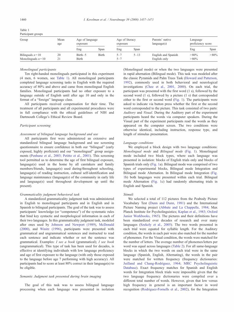
Table 1Participant groups
Group Meanage
Age of languageexposure
Age of literacyexposure
Parents' nativelanguage(s)
Languageproficiency score
Eng Span Eng Span Eng Span
Bilinguals n=10 20 Birth–5 Birth 5–7 5–13 English and Spanish N80% N80%Monolinguals n=10 21 Birth 5–7 English only N80%
1460 I. Kovelman et al. / NeuroImage 39 (2008) 1457–1471
Monolingual participantsTen right-handed monolinguals participated in this experiment
(4 men, 6 women, see Table 1). All monolingual participantscompleted language screening tasks in English with the requiredaccuracy of 80% and above and came from monolingual Englishfamilies. Monolingual participants had no other exposure to alanguage outside of English until after age 10 and only in theformat of a “foreign” language class.
All participants received compensation for their time. Thetreatment of all participants and all experimental procedures werein full compliance with the ethical guidelines of NIH andDartmouth College’s Ethical Review Board.
Participant screening
Assessment of bilingual language background and useAll participants first were administered an extensive and
standardized bilingual language background and use screeningquestionnaire to ensure confidence in both our “bilingual” (earlyexposed, highly proficient) and our “monolingual” group assign-ments (Penhune et al., 2003; Petitto et al., 2001). This screeningtool permitted us to determine the age of first bilingual exposure,language(s) used in the home by all caretakers and familymembers/friends, language(s) used during/throughout schooling,language(s) of reading instruction, cultural self-identification andlanguage maintenance (language(s) of the community in early lifeand language(s) used throughout development up until thepresent).
Grammaticality judgment behavioral taskA standardized grammaticality judgment task was administered
in English to monolingual participants and in English and inSpanish to bilingual participants. The goal of the task was to assessparticipants’ knowledge (or “competence”) of the systematic rulesthat bind key syntactic and morphological information in each oftheir two languages. In this grammaticality judgment task, modeledafter ones used by Johnson and Newport (1989), McDonald(2000), and Winitz (1996), participants were presented withgrammatical and ungrammatical sentences and instructed to readeach sentence and indicate whether or not the sentence wasgrammatical. Examples: I see a book (grammatical); I see book(ungrammatical). This type of task has been used for decades, iseffective at identifying individuals with low language proficiencyand age of first exposure to the language (with only those exposedto the language before age 7 performing with high accuracy). Allparticipants had to score at least 80% correct in their language(s) tobe eligible.
Semantic Judgment task presented during brain imaging
The goal of this task was to assess bilingual languageprocessing when each language was presented in isolation
(Monolingual mode) or when the two languages were presentedin rapid alternation (Bilingual mode). This task was modeled afterthe classic Pyramids and Palm Trees Task (Howard and Patterson,1992), commonly used in both behavioral and neurologicalinvestigations (Chee et al., 2001, 2000). On each trial, theparticipant was presented with the first word (1 s), followed by thesecond word (1 s), followed by a picture (1 s) that correspondedeither to the first or second word (Fig. 1). The participants wereasked to indicate via button press whether the first or the secondword corresponded to the picture. This task consisted of two parts:Auditory and Visual. During the Auditory part of the experimentparticipants heard the words via computer speakers. During theVisual part of the experiment participants read the words as theyappeared on the computer screen. The two conditions wereotherwise identical, including instruction, response type, andlength of stimulus presentation.
Language conditionsWe employed a block design with two language conditions:
Monolingual mode and Bilingual mode (Fig. 1). Monolingualmode included two block types, where each language waspresented in isolation: blocks of English trials only and blocks ofSpanish trials only (Fig. 1a). Bilingual mode was comprised of twotypes of experimental blocks, Bilingual mode Integration andBilingual mode Alternation. In Bilingual mode Integration (Fig.1b) both languages were presented within each trial. Bilingualmode Alternation (Fig. 1c) had randomly alternating trials inEnglish and Spanish.
StimuliWe selected a total of 112 pictures from the Peabody Picture
Vocabulary Test (Dunn and Dunn, 1981) and the InternationalPicture Naming project (Abbate and La Chappelle, 1984; MaxPlanck Institute for Psycholinguistics; Kaplan et al., 1983; OxfordJunior Workbooks, 1965). The pictures and their definitions havebeen standardized over decades of research and over manylanguages (Szekely et al., 2005). The two words presented ineach trial were equated for syllable length. For the Auditorycondition, the words in each pair were also matched for the numberof phonemes. For the Visual condition, the words were matched forthe number of letters. The average number of phonemes/letters perword was equal across languages (Table 2). For all same-languageblocks in which the two words on each trial were in the samelanguage (Spanish, English, Alternating), the words in the pairwere matched for written frequency (frequency dictionaries:Juilland and Chang-Rodríguez, 1964; MRC PsycholinguisticDatabase). Exact frequency matches for Spanish and Englishwords for Integration block trials were impossible given that thetwo language frequency dictionaries were compiled over adifferent total number of words. However, given that low versushigh frequency in general is an important factor in wordrecognition (Rodriguez-Fornells et al., 2002), for the Integration
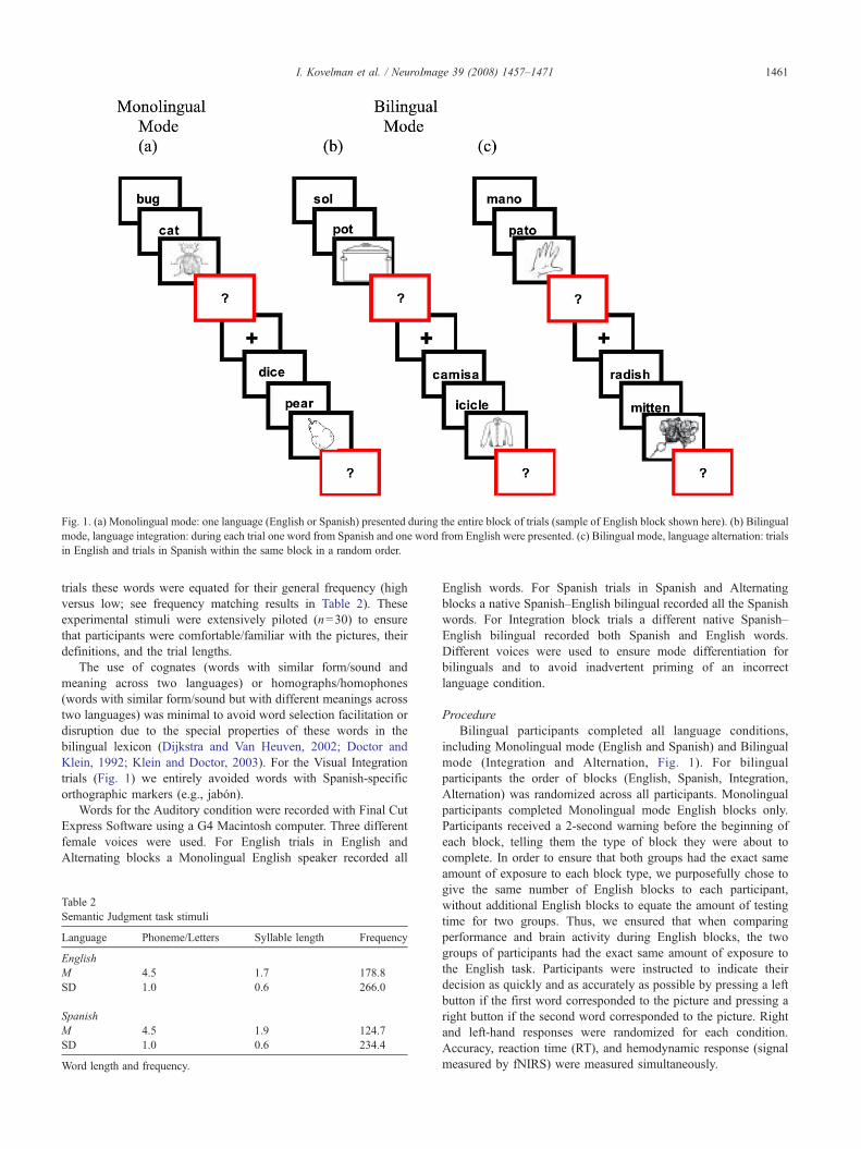
Fig. 1. (a) Monolingual mode: one language (English or Spanish) presented during the entire block of trials (sample of English block shown here). (b) Bilingualmode, language integration: during each trial one word from Spanish and one word from English were presented. (c) Bilingual mode, language alternation: trialsin English and trials in Spanish within the same block in a random order.
1461I. Kovelman et al. / NeuroImage 39 (2008) 1457–1471
trials these words were equated for their general frequency (highversus low; see frequency matching results in Table 2). Theseexperimental stimuli were extensively piloted (n=30) to ensurethat participants were comfortable/familiar with the pictures, theirdefinitions, and the trial lengths.
The use of cognates (words with similar form/sound andmeaning across two languages) or homographs/homophones(words with similar form/sound but with different meanings acrosstwo languages) was minimal to avoid word selection facilitation ordisruption due to the special properties of these words in thebilingual lexicon (Dijkstra and Van Heuven, 2002; Doctor andKlein, 1992; Klein and Doctor, 2003). For the Visual Integrationtrials (Fig. 1) we entirely avoided words with Spanish-specificorthographic markers (e.g., jabón).
Words for the Auditory condition were recorded with Final CutExpress Software using a G4 Macintosh computer. Three differentfemale voices were used. For English trials in English andAlternating blocks a Monolingual English speaker recorded all
Table 2Semantic Judgment task stimuli
Language Phoneme/Letters Syllable length Frequency
EnglishM 4.5 1.7 178.8SD 1.0 0.6 266.0
SpanishM 4.5 1.9 124.7SD 1.0 0.6 234.4
Word length and frequency.
English words. For Spanish trials in Spanish and Alternatingblocks a native Spanish–English bilingual recorded all the Spanishwords. For Integration block trials a different native Spanish–English bilingual recorded both Spanish and English words.Different voices were used to ensure mode differentiation forbilinguals and to avoid inadvertent priming of an incorrectlanguage condition.
ProcedureBilingual participants completed all language conditions,
including Monolingual mode (English and Spanish) and Bilingualmode (Integration and Alternation, Fig. 1). For bilingualparticipants the order of blocks (English, Spanish, Integration,Alternation) was randomized across all participants. Monolingualparticipants completed Monolingual mode English blocks only.Participants received a 2-second warning before the beginning ofeach block, telling them the type of block they were about tocomplete. In order to ensure that both groups had the exact sameamount of exposure to each block type, we purposefully chose togive the same number of English blocks to each participant,without additional English blocks to equate the amount of testingtime for two groups. Thus, we ensured that when comparingperformance and brain activity during English blocks, the twogroups of participants had the exact same amount of exposure tothe English task. Participants were instructed to indicate theirdecision as quickly and as accurately as possible by pressing a leftbutton if the first word corresponded to the picture and pressing aright button if the second word corresponded to the picture. Rightand left-hand responses were randomized for each condition.Accuracy, reaction time (RT), and hemodynamic response (signalmeasured by fNIRS) were measured simultaneously.
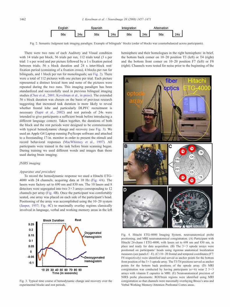
Fig. 2. Semantic Judgment task imaging paradigm. Example of bilinguals' blocks (order of blocks was counterbalanced across participants).
1462 I. Kovelman et al. / NeuroImage 39 (2008) 1457–1471
There were two runs of each Auditory and Visual conditionwith 14 trials per block, 56 trials per run, 112 trials total (3 s pertrial: 1 s per word and per picture followed by a 1 s fixation periodbetween trials; 56 s block duration and 24 s inter-block rest/fixation period (consisting of a fixation cross), 4 blocks per run forbilinguals, and 1 block per run for monolinguals; see Fig. 2). Therewere a total of 112 pictures with one picture per trial. Each picturerepresented a distinct lexical item and none of the pictures wererepeated during the two runs. This imaging paradigm has beenstandardized and successfully used in previous bilingual imagingstudies (Chee et al., 2001; Kovelman et al., in press). The extended56 s block duration was chosen on the basis of previous researchsuggesting that increased task duration is more likely to revealwhether frontal lobe and particularly DLPFC recruitment isnecessary (Sapir et al., 2002) and rest periods of 24s wereintended to give participants a sufficient break before introducing adifferent language context. Taken together, the durations of boththe block and the rest periods were designed to be commensuratewith typical hemodynamic change and recovery (see Fig. 3). Weused an Apple G4 Laptop running PsyScope software and attachedto a freestanding 17-in. monitor in order to present the stimuli andrecord behavioral responses (MacWhinney et al., 1997). Allparticipants were trained in the task before brain scanning began.During training we used different words and images than thoseused during brain imaging.
fNIRS imaging
Apparatus and procedureTo record the hemodynamic response we used a Hitachi ETG-
4000 with 24 channels, acquiring data at 10 Hz (Fig. 4A). Thelasers were factory set to 690 nm and 830 nm. The 10 lasers and 8detectors were segregated into two 3×3 arrays corresponding to 12channels per array (Fig. 4B). Once the participant was comfortablyseated, one array was placed on each side of the participant’s head.Positioning of the array was accomplished using the 10–20 system(Jasper, 1957; Fig. 4C) to maximally overlay regions classicallyinvolved in language, verbal and working memory areas in the left
Fig. 3. Typical time course of hemodynamic change and recovery over theexperimental blocks and rest periods.
hemisphere and their homologues in the right hemisphere: in brief,the bottom back corner on 10–20 position T3 (left) or T4 (right)and the bottom front corner on 10–20 position F7 (left) or F8(right). Channels were tested for noise prior to the beginning of the
Fig. 4. Hitachi ETG-4000 Imaging System, neuroanatomical probepositioning, and MRI neuroanatomical coregistration. (A) Participant withHitachi 24-chann l ETG-4000, with lasers set to 698 nm and 830 nm, inplace and ready for data acquisition. (B) The 3×3 optode arrays werepositioned on participants' heads using rigorous anatomical localizationmeasures (see panels C–E). (C) 10–20 frontal and temporal coordinates (F7/F8 respectively) were identified and served as anchor points for the bottomfront position of the 3×3 optode array. The T3/T4 positions served as anchorpoints for the bottom back positions of the optode array. (D) MRIcoregistration was conducted by having participants (n=6) wear 2 3×3arrays with vitamin E capsules in MRI. (E) Neuroanatomical precision ofNIRS probe placements: ROI/brain regions were identified using MRIcoregistration so that channels were maximally overlaying Broca's area andVerbal Working Memory/Attention Prefrontal Cortex areas.

1463I. Kovelman et al. / NeuroImage 39 (2008) 1457–1471
recording session. Digital photographs were taken of the arraypositioning prior to and after the recording session to identify if thearrays had moved during testing. An MPEG video recording wassynchronized with the testing session, so any apparent movementartifacts could be confirmed during off-line analysis.
ROI identification
In the 3×3 recording array, channels were considered as thearea between adjacent lasers and detectors, as is hardwired into theETG-4000 system. Each channel had two components, attenuationvalues from the 690 nm and 830 nm lasers. The attenuation valueswere converted to deoxy- and oxy-Hb values using the ModifiedBeer–Lambert equation. Once converted from laser attenuation,channels referred to the deoxy- and oxy-Hb changes in the regionsbetween the laser and detectors.
Our ROI included classic brain regions of interest for languageprocessing: Language PFC (channels maximally overlayingBroca’s area BA 44/45) and Verbal Working Memory/AttentionPFC (channels maximally overlaying DLPFC BA 46/9 and IFCBA 47/11).
There regions were identified with the help of PCA whichgrouped the channels into the Language PFC and Verbal WorkingMemory/Attention PFC principal components. The same channelswere grouped for every subject. Each channel was overlaying thesame brain area for every subject, as established by 10–20 probeplacement and MRI coregistration.
After the recording session, data were exported and analyzedusing Matlab (The Mathworks Inc.). Conversion of the raw data tohemoglobin values was accomplished in two steps. Under theassumption that scattering is constant over the path length, we firstcalculated the attenuation for each wavelength by comparing theoptical density of light intensity during the task to the calculatedbaseline of the signal. We then used the attenuation values for eachwavelength and sampled time points to solve the modified Beer–Lambert equation to convert the wavelength data to a meaningfuloxygenated and deoxygenated hemoglobin response (HbO and Hbrespectively) (Kohl et al., 1998).
MRI coregistration
For MRI (anatomical) coregistration, at another session, two3×3 arrays of Vitamin E tablets were constructed with the tabletsplaced precisely at each of the optode locations as described above.These Vitamin E arrays were then placed on to the participant’shead at precisely the same location as the optode array using the
Table 3Behavioral scores for Semantic Judgment task
Condition Monolinguals
# Correct (SD)
English Audio 26.3 (0.9)Video 26.8 (1.5)
Spanish AudioVideo
Integration AudioVideo
Alternation AudioVideo
10–20 coordinate system and secured in place with MRI safe tapeand straps. Using a Philips 3 T MRI, an anatomical scan was takenfrom 6 participants. The Vitamin E locations from these scans wereused as landmarks for coregistration and hence the recordedchannels, indicating that indeed the channels covered theanatomical locations anticipated by the 10–20 coordinate system(see Figs. 4D and E).
Foam padding was placed in the head coil to limit subject headmovement during image acquisition. T1-weighted three-dimen-sional magnetization-prepared rapid acquisition gradient echo (3D-MPRAGE) sagittal images were obtained with a Phillips 3 Tscanner. Scanning parameters were as follows: echo time (TE)=4.6 ms, repetition time (TR)=9.8 ms, flip angle=8°, acquisitionmatrix=256×256, 160 sagittal slices, and voxel size=1×1×1 mmwith no gap.
Results
Semantic Judgment task
Behavioral results
Bilinguals versus monolinguals. Bilinguals’ and monolinguals’accuracy and reaction time in English were compared using two2×2 mixed ANOVAs, one ANOVA for accuracy and one ANOVAfor reaction time (groups (between factor)×Audio and Videoconditions (within factor)). Bilinguals performed equally accu-rately (F(1,18)=0.8, pN0.01) and equally fast as monolinguals(F(1,18)=0.2, pN0.01). Both groups performed with the sameaccuracy, but faster (F(1,18)=58.2, pb0.01) when they read thewords rather than when they heard the words. Behavioral scoresfor this task are presented in Table 3. We used a two-standarddeviation cut-off method for analyzing our reaction time data. Inparticular, for each condition (Audio English, Audio Spanish,Audio Integration, Audio Alternation, Video English, VideoSpanish, Video Integration, Video Alternation) we established amean and standard deviation, and for each participant for eachcondition we eliminated reaction time data points, which werebelow or above two standard deviations for that condition.
Bilingual language modes. Bilinguals performed equally fast(F(1,18)=0.5, pN0.01), but with unequal accuracy across trials(F(3,27)=8.7, pb0.01) as was revealed by two 4×2 ANOVAs, oneANOVA for accuracy, and one ANOVA for reaction time (Languageconditions (within factor)×Audio and Video conditions (withinfactor)). Post hoc investigation showed that bilinguals performed
Bilinguals Monolinguals Bilinguals
# Correct (SD) RT ms (SD) RT ms (SD)
26.1 (1.1) 1000 (86) 919 (282)27.1 (0.9) 628 (124) 654 (132)26.4 (0.7) 955 (223)27 (0.8) 639 (98)
25.4 (0.5) 945 (220)25.7 (1.1) 673 (121)24.6 (1) 912 (236)26.9 (0.7) 652 (76)
1464 I. Kovelman et al. / NeuroImage 39 (2008) 1457–1471
better during English as compared to Integration trials (TukeyHonestly Significant Difference (HSD) pb0.05). However, as canbe seen in Table 3, these scores are within one point of each other(English mean=26 correct and Integration mean=25 correct out of atotal of 28 items, Audio andVideo conditions taken together for eachlanguage context and numbers are rounded for clarity). Therefore,we suggest that the readers interpret this statistical difference with adegree of caution as the statistical difference may not be entirelymeaningful and the participants may have essentially performedwith the same accuracy across all language trials and modes. Theparticipants performed more accurately (F(1,9)=51.4, pb0.01) andfaster (F(1,9)=54.4, pb0.01) when they read the words rather thanwhen they heard them.
Imaging resultsStatistical analyses were performed on the concentration of
HbO within a cortical region as this chromophore provided themost robust contrast-to-noise ratio across participants. HbO valuesfor each channel were plotted and inspected. The maximumpositive peak values were determined for each channel from 5 sbefore the onset of the trial until 10 s after the end of the trial.These values and baseline values (mean of 50 s preceding the firsttrial for each channel) were used for statistical analysis.
Task versus baseline. “Task” activation was defined as peakactivation during Monolingual mode blocks (English formonolinguals, and English and Spanish averaged for bilinguals;Audio and Video conditions averaged for both groups) and“Baseline” was defined as the mean of the 50-second period priorto the first task for each channel. A 2×24 MANOVA (task versusbaseline×24 channels) analysis revealed that all participants hadoverall greater brain activity during the task as compared tobaseline (F(15,40)=24, pb0.0001). The test of individual task–baseline contrasts for each channel confirmed that thisTaskNBaseline difference was significant for each channel withsignificance levels ranging from pb0.01 to pb0.0001.
ROI identification. We further used the Task activations (Englishfor monolinguals, English and Spanish for bilinguals, Audio andVideo averaged) in a Principal Component Analysis (PCA) for lefthemisphere channels as the first exploratory procedure to identifyregions of interest (ROI), the channels that overlay brain regionsparticularly involved with the current language task. The firstprincipal component explained 50% of the variance in the data andindicated two clusters: channels 1–8 had high coefficient loadings(0.680–0.880) and channels 9–12 had low coefficient loadings(0.280–0.580). Anatomical locations of channels 1–8 were examinedusing MRI anatomical coregistration scans. The anatomical coregis-tration analysis was conducted with MRIcro and Talairach Deamonanatomical localization software. Anatomical localization analysissuggested that most of the channels with high PCA coefficientloadings primarily overlay our originally hypothesized ROI areas.Channels 1 and 3 maximally overlay IFC (BA 44/45, includingclassic Broca’s area), channel 2 maximally overlays IFC (BA 47/11)and channels 4 and 7maximally overlayDLPFC (BA 46/9). Thus, wefurther narrowed down our selection of ROI channels to only these 5channels (1–4, 7). Henceforth in the analysis, we treat channels 1 and3 asmaximally overlaying Language PFC areas (BA 44/45), channels2, 4, and 7 as maximally overlaying Verbal Working Memory/Attention PFC areas (BA 47/11 and 46/9), and the remaining channels5–6 and 8–12 as maximally overlaying control areas.
Audio versus video. The first step in data analysis was to seewhether there was a significant difference between the Audio andVideo conditions that might preclude us from averaging these twoconditions for subsequent analysis presented in the paragraphsbelow. We explored potential differences between Audio andVideo conditions separately for each ROI and using the data frommonolinguals (English condition) and bilinguals (all languageconditions averaged). For each ROI we used a 2×2 repeated-measures ANOVA (Audio vs Video in Monolingual modeconditions (English for monolinguals; English and Spanishaveraged for bilinguals)×hemispheres). For the purposes of thisanalysis and all the subsequent comparisons for each individualparticipant we averaged brain activation for the groups of channelsbelonging to the same ROI (channels 1 and 3 for Language PFC,channels 2, 4, and 7 for Verbal Working Memory/Attention PFC,and channels 5, 6, 8, 9, 10, 11, and 12 for Control regions).
Language PFC. The results showed a significantly greater lefthemisphere Language PFC (BA 44/45) recruitment (F(1,19)=13.9,pb0.01), with no significant differences between Audio and Videoconditions (F(1,19)=0.9, pN0.05), or interaction between the twofactors (F(1,19)=1.8, pN0.05). Verbal Working Memory/AttentionPFC. The results showed no significant main effects of Audioversus Video comparison (F(1,19)=0.01, pN0.05), no significanteffect of hemisphere (F(1,19)=0.04, pN0.05) or interactionbetween these two factors (F(1,19)=1.9, pN0.05). Control areas.The results showed no significant main effects of Audio versusVideo comparison (F(1,19)=0.01, pN0.05), no significant effectof hemisphere (F(1,19)=0.02, pN0.05), or interaction between thetwo factors (F(1,19)=0.03, pN0.05). In summary, we observed nosignificant differences between the Audio and Video conditions,even though participants showed an overall greater recruitment ofleft hemisphere during Video conditions and a more even bilateralduring the Audio condition (Video mean activation LH=0.069,RH=0.057; Audio mean activation LH=0.063, RH=0.063),which is a classic finding given that visual language processingshould be more left-lateralized than auditory language processingthat should be more bilateral due to the general auditory processingof sound in both hemispheres. Therefore, for all subsequentanalyses, data were averaged across Audio and Visual conditions.
“Bilingual signature” and modes
Bilinguals versus monolingualsDo monolinguals differ from bilinguals in Monolingual mode,
when both groups are using only one language at a time? Similarity:As can be seen in Fig. 5A, when using one language at a time(Monolingual mode), both bilinguals and monolinguals had greaterleft than right hemisphere recruitment in the Language PFC (BA 44/45) brain area. This similarity between the groups is supported byabsence of any main effects found in a 3×2×2 mixed-measuresANOVA (group: monolinguals, bilinguals in Monolingual mode(between factor)×hemispheres (within factor)×ROI (LanguagePFC and Working Memory/Attention PFC, Control areas, withinfactor)), averaged across Audio and Video conditions. Non-significant main effects are as follows: group F(1,18)=0.2,pN0.05; hemispheres F(2,18)=2.6, pN0.05; and ROI F(2,17)=0.2, pN0.05. Differences: the significant 3-way interaction of groupby ROI by hemisphere can be seen in Fig. 5B (Wilks’ LambdaF(2,18)=5.0, pb0.05), suggesting that there was a difference inboth the left and right hemispheres in how the two groups recruited

Fig. 5. (A) Percent signal change in the left versus right hemispheres for Broca's area/Language PFC. There was no significant difference between bilinguals andmonolinguals in the recruitment of Broca's area (pN0.05). (B) Percent signal change in the left versus right hemispheres for Verbal Working Memory/AttentionPFC. For this ROI, there was a significant interaction between bilinguals and monolinguals in Monolingual mode, and Bilingual and Monolingual modes inbilinguals (pb0.05). Group and language mode similarity: Bilinguals (Monolingual mode) and monolinguals showed similar patterns of activation in let Broca/Language area and right Broca homologue. Group and language mode differences: Bilinguals (Monolingual mode) showed a different pattern of recruitment forleft Verbal Working Memory/Attention PFC and its right homologue as compared to monolinguals. Bilinguals (Bilingual mode) showed greater recruitment ofthe right hemisphere homologue of Broca's area and right hemisphere Working Memory/Attention PFC versus when in Monolingual mode.
1465I. Kovelman et al. / NeuroImage 39 (2008) 1457–1471
Verbal Working Memory/Attention PFC. Tukey Honestly Signifi-cant Differences (HSD) post hoc comparisons showed a signifi-cantly greater right homologue of Verbal Working Memory/Attention PFC recruitment in bilinguals as compared to mono-linguals (pb0.05). The means for each language mode for eachgroup can be found in Table 4.
Bilinguals in monolingual versus bilingual modesDo bilinguals differ in their use of neural resources towards
processing one language at a time (Monolingual mode) versusprocessing two languages in rapid alternation (Bilingual mode)?We used a 2×2×3 repeated-measures ANOVA (Bilingual versusMonolingual modes×hemispheres×ROI (Language PFC, VerbalWorking Memory/Attention PFC, Control areas)), averaged acrossAudio and Video conditions. We found a significant main effect ofhemisphere (F(1,9)=6.8, pb0.05), all other main effects were non-significant (mode F(1,9) = 0.3, pN0.05; ROI F(2,18) =0.2,pN0.05). Importantly, however, and as can be seen in Figs. 5Aand B, there were significant mode×hemisphere (F(1,9)=7.3,pb0.05) and hemisphere×ROI (F(2,18)=14.5, pb0.05) interac-tions, suggesting increased right hemisphere involvement during
Table 4Mean peak activation values (and SD) for monolinguals in Monolingual mode an
Group Mode Language PFC
L R
Monolinguals Monolingual 0.047 (0.030) 0.034 (0.026)Bilinguals Monolingual 0.065 (0.056) 0.051 (0.047)Bilinguals Bilingual 0.058 (0.082) 0.063 (0.093)
In Monolingual mode bilinguals and monolinguals had similar recruitment of Languintensity in right Verbal Working Memory/Attention PFC areas as compared to mobilinguals also showed greater signal intensity in right Verbal Working Memory/A
Bilingual mode as compared to Monolingual mode. Follow-up2×2 repeated-measures ANOVAs (mode×hemisphere) for eachROI showed that hemisphere by mode interaction was significantonly for Verbal Working Memory/Attention PFC (F(1,9)=5.1,pb0.05) and Control regions (F(1,9)=5.3, pb0.05). Mode×hemi-sphere interaction was not significant for Language PFC (F(1,9)=3.0, pN0.05), even though the means (Table 4) suggested that forthis brain region there was also an increased right hemisphereinvolvement during Bilingual mode. In summary and as can beseen in Table 4 and Fig. 5, left hemisphere involvement remainedrelatively constant across language modes for each brain region ofinterest, while right hemisphere involvement significantly in-creased in the Bilingual mode.
Monolingual mode in bilinguals: English versus SpanishIn order to explore any differences between the two Mono-
lingual mode conditions in bilinguals, Spanish and English, weused a 2×2×3 repeated-measures ANOVA (English and Spanishlanguages×hemispheres×brain area (Language PFC, VerbalWorking Memory/Attention PFC, Control areas)), averaged acrossAudio and Video conditions. There were no main effects of
d bilinguals in Monolingual and Bilingual modes
Verbal Working Memory/Attention PFC
Control regions
L R L R
0.059 (0.038) 0.042 (0.015) 0.051 (0.024) 0.049 (0.020)0.046 (0.035) 0.057 (0.043) 0.047 (0.020) 0.050 (0.020)0.040 (0.026) 0.07 (0.045) 0.047 (0.012) 0.061 (0.020)
age PFC regions. In Monolingual mode bilinguals also showed greater signalnolinguals (pb0.05). In Bilingual mode as compared to Monolingual modettention PFC areas (pb0.05).
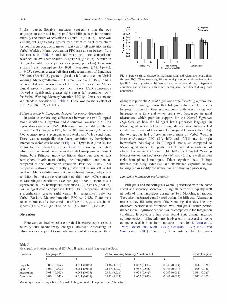
Fig. 6. Percent signal change during Integration and Alternation conditionsfor each ROI. There was a significant hemisphere by condition interaction(pb0.05), with greater right hemisphere recruitment during integrationcondition and relatively similar left hemisphere recruitment during bothconditions.
1466 I. Kovelman et al. / NeuroImage 39 (2008) 1457–1471
English versus Spanish languages suggesting that the twolanguages of early and highly proficient bilinguals yield the sameintensity and extent of activation (F(1,9)=0.7, pN0.05). There wasa slight, yet significantly greater recruitment of right hemispherefor both languages, due to greater right versus left activation in theVerbal Working Memory/Attention PFC area as can be seen fromthe means in Table 5 and follow-up post hoc comparisonsdescribed below (hemispheres F(1,9)=5.4, pb0.05). Similar toBilingual conditions comparison (see paragraph below), there wasa significant hemisphere by ROI interaction (F(2,18)=4.2,pb0.05), showing greater left than right recruitment of LanguagePFC area (BA 44/45), greater right than left recruitment of VerbalWorking Memory/Attention PFC area (BA 47/11; 46/9), and abalanced bilateral recruitment of the Control areas. For Mono-lingual mode comparison post hoc Tukey HSD comparisonshowed a significantly greater right versus left recruitment onlyfor Verbal Working Memory/Attention PFC (pb0.05), see meansand standard deviations in Table 5. There was no main effect ofROI (F(2,18)=0.2, pN0.05).
Bilingual mode in bilinguals: Integration versus AlternationIn order to explore any differences between the two Bilingual
mode conditions, Integration and Alternation, we used a 2×2×3repeated-measures ANOVA (Bilingual mode conditions×hemi-spheres×ROI (Language PFC, Verbal Working Memory/AttentionPFC, Control areas)), averaged across Audio and Video conditions.There was a marginally significant condition by hemisphereinteraction which can be seen in Fig. 6 (F(1,9)=10.0, p=0.06, themeans for the interaction are in Table 5), showing that whilebilinguals maintained the same level of left hemisphere recruitmentduring both Bilingual mode conditions, there was greater righthemisphere involvement during the Integration condition ascompared to the Alternation condition. Post hoc Tukey HSDcomparisons showed significantly greater right versus left VerbalWorking Memory/Attention PFC recruitment during Integrationcondition, but not during Alternation condition (pb0.05). Same asin Monolingual conditions (see paragraph above), there was asignificant ROI by hemisphere interaction (F(2,18)=4.5, pb0.05).For Bilingual mode comparison Tukey HSD comparison showeda significantly greater right versus left recruitment only forVerbal Working Memory/Attention PFC (pb0.05). There wereno main effects of either condition (F(1,9)=0.2, pN0.05), hemi-spheres (F(1,9)=3.2, pN0.05), or ROI (F(2,18)=0.1, pN0.05).
Discussion
Here we examined whether early dual language exposure bothneurally and behaviorally changes language processing inbilinguals as compared to monolinguals, and if so whether these
Table 5Mean peak activation values (and SD) for bilinguals in each language condition
Condition Language PFC Verbal Work
L R L
English 0.065 (0.056) 0.051 (0.047) 0.046 (0.035Spanish 0.045 (0.062) 0.051 (0.042) 0.039 (0.022Integration 0.058 (0.082) 0.063 (0.093) 0.041 (0.026Alternation 0.054 (0.043) 0.051 (0.047) 0.042 (0.021
Monolingual mode: English and Spanish; Bilingual mode: Integration and Alterna
changes support the Neural Signature or the Switching Hypothesis.The present findings show that bilinguals do neurally processlanguage differently than monolinguals both when using onelanguage at a time and when using two languages in rapidalternation, which provides support for the Neural SignatureHypothesis of how the bilingual brain processes language. InMonolingual mode, whereas bilinguals and monolinguals hadsimilar recruitment of the classic Language PFC areas (BA 44/45),the two groups had differential recruitment of Verbal WorkingMemory/Attention PFC (BA 46/9 and 47/11) and its righthemisphere homologue. In Bilingual mode, as compared toMonolingual mode, bilinguals had differential recruitment ofclassic Language PFC areas (BA 44/45) and Verbal WorkingMemory/Attention PFC areas (BA 46/9 and 47/11), as well as theirright hemisphere homologues. Taken together, these findingsindicate that early, extensive, and maintained exposure to twolanguages can modify the neural basis of language processing.
Language behavioral performance
Bilinguals and monolinguals overall performed with the samespeed and accuracy. Moreover, bilinguals performed equally wellin both of their languages during the two Monolingual modes.They also performed equally well during the Bilingual Alternationmode as they did during each of the Monolingual modes. The onlyobserved performance difference was bilinguals’ better perfor-mance in the English-only condition as compared to the Integrationcondition. It previously has been found that, during languagecomprehension, bilinguals are inadvertently processing somecomponents of both of their languages in parallel (Dijkstra et al.,1998; Doctor and Klein, 1992; Grosjean, 1997; Kroll andSunderman, 2003). Therefore, it is notable that bilinguals
ing Memory/Attention PFC Control regions
L R L
) 0.057 (0.043) 0.048 (0.019) 0.050 (0.020)) 0.059 (0.036) 0.043 (0.011) 0.050 (0.020)) 0.070 (0.045) 0.047 (0.012) 0.061 (0.020)) 0.057 (0.033) 0.047 (0.017) 0.052 (0.027)
tion.

1467I. Kovelman et al. / NeuroImage 39 (2008) 1457–1471
maintained the same efficacy of language processing as mono-linguals, and to some extent maintained the same successfulperformance as their task load became more complex duringBilingual mode.
Dijkstra and Van Heuven’s bilingual language model suggeststhat bilinguals’ should be able to equally effectively process onelanguage at a time versus two languages in rapid alternation. It isthe cognitive load incurred by the experimental task that mightresult in specific patterns of speed and accuracy during Bilingualversus Monolingual modes. Indeed, under some experimentalconditions, bilinguals perform with the same speed and accuracyduring both Bilingual and Monolingual modes (Caramazza andBrones, 1980; Grosjean and Miller, 1994; Van Heuven et al.,1998), whereas during other experimental conditions theirperformance declines during Bilingual mode tasks (Thomas andAllport, 2000; Von Studnitz and Green, 2002). We captured thesephenomena in our study: bilinguals performed with the same speedand accuracy during the Monolingual mode and the Bilingualmode Alternating condition (Table 3). These results suggest thatearly exposed and highly proficient bilinguals can perform withequal success in single and dual language contexts. However,experimental tasks such as the Bilingual mode Integrationcondition (Table 3) can take a toll on bilinguals’ accuracy ofperformance. Why might this be so? It is possibly due to anincreased attentional and working memory load that is required tokeep semantic items in two different languages activated inmemory at the same time.
Imaging findings
To the best of our knowledge, the results of the few functionalimaging studies outside of our laboratory that have directlycompared language processing in healthy, early exposed, andhighly proficient bilinguals versus monolinguals are in agreementwith our findings here. For example, Proverbio et al. (2002)compared Italian–Slovenian bilinguals and Italian monolingualsusing event-related potential (ERP) technology and a sentenceprocessing task. Bilinguals were shown to have a different neuralresponse as compared to monolinguals. Also consistent with ourfindings, Rodriguez-Fornells et al.’s (2002) fMRI work found thatthere was differential frontal lobe activation between bilinguals inBilingual mode and monolinguals in Monolingual mode. Together,these ERP and fMRI studies have converged with our fNIRS studyto suggest that the human neural organization and languageprocessing capacity can be molded by extensive dual languageexposure early in life.
When using only one language at a time, we found nodifference in how bilinguals and monolinguals recruited classicLanguage area PFC (BA 44/45). However, there was a hemisphericdifference in how bilinguals and monolinguals recruited WorkingMemory/Attention PFC (BA 47/11 and 46/9). In particular, therewas an overall greater bilateral recruitment of Working Memory/Attention PFC area in bilinguals as compared to monolinguals,when both groups were presented with only one language at a time(Monolingual mode). This greater recruitment, defined as theobserved significantly greater signal intensity of Working Memory/Attention PFC in bilinguals, was particularly significant in the righthemisphere. This greater signal intensity (as measured by thechange in oxygenated hemoglobin) was observed in fNIRSchannels maximally overlaying Working Memory/Attention PFCregions (DLPFC (BA 46/9) and IFC (BA 47/11)) in bilingual group
as compared to monolingual group. These findings support the ideathat dual language exposure can result in monolingual-likerecruitment of the classic language brain areas, such as left IFC,which incorporates Broca’s area (BA 44/45). The observed groupdifferences suggest that dual language processing may incur neuralchanges within brain regions that support working memory andattention associated with language processing. Prior memory andlanguage research has established that there are strong empiricallinks between DLPFC (BA 46/9) and working memory (Baddeley,2000; D’Esposito et al., 2000; Gabrieli et al., 1998; Smith andJonides, 1999). In particular, left DLPFC seems to be moreassociated with verbal working memory (e.g., Gabrieli et al.,1998), while right DLPFC seems to be more associated with visuo-spatial working memory, as well as cross-modal informationintegration (e.g., Bushara et al., 2001). Increased activation in leftIFC (BA 47/11) has been found in association with verbal tasks,such as semantic decision making (Roskies et al., 2001), whileincreased activation in right anterior IFC has been found inassociation with attention and selective inhibition for both verbaland non-verbal tasks (Aron et al., 2004). The differentialrecruitment of Verbal Working Memory/Attention PFC forbilinguals as compared to monolinguals that we found here islikely due to differences in the verbal working memory andattention resources required for dual language processing, such asselective attention allocation between competing linguistic infor-mation (Norman and Shallice, 1986). Alternatively, this activationdifference between the groups could simply be due to an overallgreater effort exerted by bilinguals. Although we cannot rule outthis possibility, bilinguals and monolinguals showed equally goodperformance on the task, which suggests the same level ofdifficulty for the two groups. Previous research has demonstratedequal level of performance and yet greater BOLD signal in agingand pathological populations, suggesting evidence of compensa-tion strategies. However, pathological and aging populations alsotypically undergo changes in brain vasculature, which makes itdifficult to interpret the exact nature of differences in BOLD signal,while here we studied young and healthy individuals with,hopefully, healthy and unaffected vascular system (see discussionby D’Esposito et al., 2003).
In bilingual language modes a bilingual needs to integrate andnegotiate cross-linguistic information. In our study, processing twolanguages in rapid succession was accompanied by an increase inboth Verbal Working Memory/Attention PFC (ROI analysis, Fig. 5;BA 46/9 and 47/11) as well as Language PFC (ROI analysis, BA44/45). This result is commensurate with our pilot fMRI findings,in which a comparable group of bilingual participants werescanned using an fMRI (Phillips 3 T) with the very same SemanticJudgment task (using identical screening and task administrationmethods as described in the Materials and methods section). UsingfMRI, we observed greater right DLPFC (BA 46) and right IFCrecruitment for both BA 47 (Attention PFC) and BA 45 (LanguagePFC) in bilinguals during Bilingual versus Monolingual mode (seeFig. 7, the data were analyzed with SPM 99 random effects model,Friston et al., 1995). As previously mentioned, increased activationin right hemisphere brain areas responsible for executive proces-sing has been observed in cross-domain information integration(Bushara et al., 2001). It also appears that semantic tasks whichrequire atypical “outside-the-box” information search and integra-tion also make selective reliance on right hemisphere frontal areas(e.g., generating unusual verbs in response to nouns, as in“dish”–“throw”; Seger et al., 2000). If information integration is a
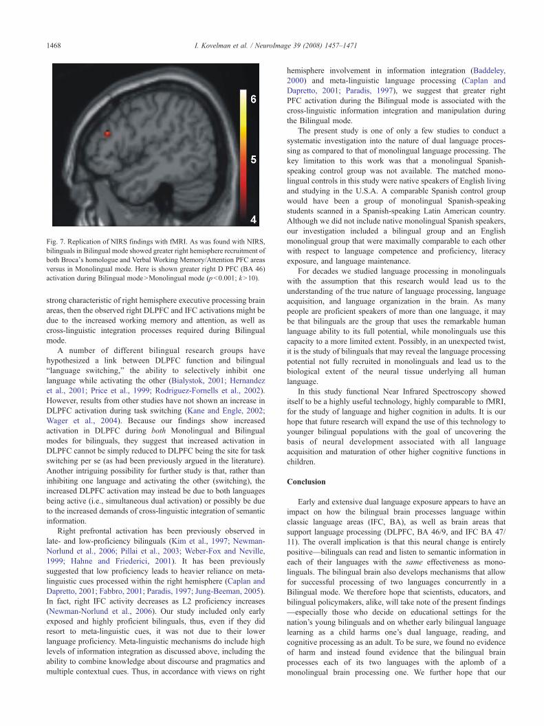
Fig. 7. Replication of NIRS findings with fMRI. As was found with NIRS,bilinguals in Bilingual mode showed greater right hemisphere recruitment ofboth Broca's homologue and Verbal Working Memory/Attention PFC areasversus in Monolingual mode. Here is shown greater right D PFC (BA 46)activation during Bilingual modeNMonolingual mode (pb0.001; kN10).
1468 I. Kovelman et al. / NeuroImage 39 (2008) 1457–1471
strong characteristic of right hemisphere executive processing brainareas, then the observed right DLPFC and IFC activations might bedue to the increased working memory and attention, as well ascross-linguistic integration processes required during Bilingualmode.
A number of different bilingual research groups havehypothesized a link between DLPFC function and bilingual“language switching,” the ability to selectively inhibit onelanguage while activating the other (Bialystok, 2001; Hernandezet al., 2001; Price et al., 1999; Rodriguez-Fornells et al., 2002).However, results from other studies have not shown an increase inDLPFC activation during task switching (Kane and Engle, 2002;Wager et al., 2004). Because our findings show increasedactivation in DLPFC during both Monolingual and Bilingualmodes for bilinguals, they suggest that increased activation inDLPFC cannot be simply reduced to DLPFC being the site for taskswitching per se (as had been previously argued in the literature).Another intriguing possibility for further study is that, rather thaninhibiting one language and activating the other (switching), theincreased DLPFC activation may instead be due to both languagesbeing active (i.e., simultaneous dual activation) or possibly be dueto the increased demands of cross-linguistic integration of semanticinformation.
Right prefrontal activation has been previously observed inlate- and low-proficiency bilinguals (Kim et al., 1997; Newman-Norlund et al., 2006; Pillai et al., 2003; Weber-Fox and Neville,1999; Hahne and Friederici, 2001). It has been previouslysuggested that low proficiency leads to heavier reliance on meta-linguistic cues processed within the right hemisphere (Caplan andDapretto, 2001; Fabbro, 2001; Paradis, 1997; Jung-Beeman, 2005).In fact, right IFC activity decreases as L2 proficiency increases(Newman-Norlund et al., 2006). Our study included only earlyexposed and highly proficient bilinguals, thus, even if they didresort to meta-linguistic cues, it was not due to their lowerlanguage proficiency. Meta-linguistic mechanisms do include highlevels of information integration as discussed above, including theability to combine knowledge about discourse and pragmatics andmultiple contextual cues. Thus, in accordance with views on right
hemisphere involvement in information integration (Baddeley,2000) and meta-linguistic language processing (Caplan andDapretto, 2001; Paradis, 1997), we suggest that greater rightPFC activation during the Bilingual mode is associated with thecross-linguistic information integration and manipulation duringthe Bilingual mode.
The present study is one of only a few studies to conduct asystematic investigation into the nature of dual language proces-sing as compared to that of monolingual language processing. Thekey limitation to this work was that a monolingual Spanish-speaking control group was not available. The matched mono-lingual controls in this study were native speakers of English livingand studying in the U.S.A. A comparable Spanish control groupwould have been a group of monolingual Spanish-speakingstudents scanned in a Spanish-speaking Latin American country.Although we did not include native monolingual Spanish speakers,our investigation included a bilingual group and an Englishmonolingual group that were maximally comparable to each otherwith respect to language competence and proficiency, literacyexposure, and language maintenance.
For decades we studied language processing in monolingualswith the assumption that this research would lead us to theunderstanding of the true nature of language processing, languageacquisition, and language organization in the brain. As manypeople are proficient speakers of more than one language, it maybe that bilinguals are the group that uses the remarkable humanlanguage ability to its full potential, while monolinguals use thiscapacity to a more limited extent. Possibly, in an unexpected twist,it is the study of bilinguals that may reveal the language processingpotential not fully recruited in monolinguals and lead us to thebiological extent of the neural tissue underlying all humanlanguage.
In this study functional Near Infrared Spectroscopy showeditself to be a highly useful technology, highly comparable to fMRI,for the study of language and higher cognition in adults. It is ourhope that future research will expand the use of this technology toyounger bilingual populations with the goal of uncovering thebasis of neural development associated with all languageacquisition and maturation of other higher cognitive functions inchildren.
Conclusion
Early and extensive dual language exposure appears to have animpact on how the bilingual brain processes language withinclassic language areas (IFC, BA), as well as brain areas thatsupport language processing (DLPFC, BA 46/9, and IFC BA 47/11). The overall implication is that this neural change is entirelypositive—bilinguals can read and listen to semantic information ineach of their languages with the same effectiveness as mono-linguals. The bilingual brain also develops mechanisms that allowfor successful processing of two languages concurrently in aBilingual mode. We therefore hope that scientists, educators, andbilingual policymakers, alike, will take note of the present findings—especially those who decide on educational settings for thenation’s young bilinguals and on whether early bilingual languagelearning as a child harms one’s dual language, reading, andcognitive processing as an adult. To be sure, we found no evidenceof harm and instead found evidence that the bilingual brainprocesses each of its two languages with the aplomb of amonolingual brain processing one. We further hope that our
1469I. Kovelman et al. / NeuroImage 39 (2008) 1457–1471
findings may excite cognitive neuroscientists to view bilinguallanguage processing as shedding new light on the full extent andvariability of the brain’s neural architecture underlying theremarkable human language capacity.
Acknowledgments
We thank Kevin Dunbar and the Pettito laboratory members,particularly Rachael Degenshein, Katherine White, Douglas McKen-ney, and Melissa Melendez. We also extend our thanks to all themembers of the Dartmouth Brain Imaging Center. L. A. Petitto (P.I.)thanks the National Institutes of Health (R01HD045822-01 andR21HD050558-01) and the Dana Foundation for funding thisresearch. We especially thank the Hitachi Medical Systems Japanfor their continuing technical support for our Hitachi functional NearInfrared Spectroscopy system. E-mail: [email protected].
References
Abbate, M.S., La Chappelle, N.B., 1984. Pictures, Please! An ArticulationSupplement. Communication Skill Builders, Tucson, AZ.
Abutalebi, J., Cappa, F.S., Perani, D., 2001. The bilingual brain asrevealed by functional neuroimaging. Biling. Lang. Cogn. 4 (2),179–190.
Ameel, E., Storms, G., Malt, B.C., Sloman, S.A., 2005. How bilingualssolve the naming problem. J. Mem. Lang. 53, 60–80.
Aron, A.R., Robbins, T.W., Poldrack, R.A., 2004. Inhibition and the rightinferior frontal cortex. Trends Cogn. Sci. 8 (4), 170–177.
Baddeley, A., 2000. The episodic buffer: a new component of workingmemory? Trends Cogn. Sci. 4 (11), 417–423.
Bialystok, E., 2001. Bilingualism in Development: Language, Literacy, andCognition. Cambridge University, New York.
Bushara, K.O., Grafman, J., Hallett, M., 2001. Neural correlates ofauditory–visual stimulus onset asynchrony detection. J. Neurosci. 21(1), 300–304.
Caplan, R., Dapretto, M., 2001. Making sense during conversation: an fMRIstudy. NeuroReport 12 (16), 3625–3632.
Caramazza, A., Brones, I., 1980. Semantic classification by bilinguals. Can.J. Psychol. 34 (1), 77–81.
Chee, M.W.L., Tan, E.W.L., Thiel, T., 1999. Mandarin and English singleword processing studied with functional magnetic resonance imaging. J.Neurosci. 19 (8), 3050–3056.
Chee, M.W.L., Weekes, B., Lee, K.M., Soon, C.S., Schreiber, A., Hoon, J.J.,Chee, M., 2000. Overlap and dissociation of semantic processing ofChinese characters, English words, and pictures: evidence from fMRI.NeuroImage 12 (4), 392–403.
Chee, M.W.L., Soon, C.S., Lee, H.L., 2001. Relative language proficiencymodulates BOLD signal change when bilinguals perform semanticjudgments. NeuroImage 13 (6), 1155–1163.
Chee, M.W.L., Soon, C.S., Lee, H.L., 2003. Common and segregatedneuronal networks for different languages revealed using functionalmagnetic resonance adaptation. J. Cogn. Neurosci. 15 (1), 85–97.
Chee, M.W.L., Soon, C.S., Lee, H.L, Pallier, C., 2004. Left insula activation:a marker for language attainment in bilinguals. PNAS 101 (42),15265–15270.
Dawson, G., Fischer, K.W., 1994. Human Behavior and the DevelopingBrain. Guilford Press, New York.
Dehaene, S., Dupoux, E., Mehler, J., Cohen, L., Paulesu, E., Perani, D., vande Moortele, P.F., Lehericy, S., Le Bihan, D., 1997. Anatomicalvariability in the cortical representation of first and second language.NeuroReport 8 (17), 3809–3815.
D’Esposito, M., Postle, B.R., Rypma, B., 2000. Prefrontal corticalcontributions to working memory: evidence from event-related fMRIstudies. Exp. Brain Res. 133 (1), 3–11.
D’Esposito, M., Deouell, L.Y., Gazzaley, A., 2003. Alterations in the BOLDfMRI signal with ageing and disease: a challenge for neuroimaging. Nat.Rev., Neurosci. 4 (11), 863–871.
Dijkstra, T., Van Heuven, W.J.B., 2002. The architecture of the bilingualword recognition system: from identification to decision. Biling. Lang.Cogn. 5 (3), 175–197.
Dijkstra, T., Van Heuven, W.J.B., Grainger, J., 1998. Simulating cross-language competition with the bilingual interactive activation model.Psychol. Belg. 38 (3–4), 177–196.
Doctor, E.A., Klein, D., 1992. Phonological processing in bilingual wordrecognition. In: Harris, R.J. (Ed.), Cognitive Processing in Bilinguals.Elsevier, Amsterdam, pp. 237–252.
Duncan, J., Owen, A.M., 2000. Common regions of the human frontal loberecruited by diverse cognitive demands. TrendsNeurosci. 23 (10), 475–483.
Dunn, L.M., Dunn, L.M., 1981. Peabody Picture Vocabulary Test-Revised.American Guidance Service, Circle Pines, MN.
Fabbro, F., 2001. The bilingual brain: cerebral representation of languages.Brain Lang. 79 (2), 211–222.
Fine, I., Finney, E.M., Boynton, G.M., Dobkins, K.R., 2005. Comparing theeffects of auditory deprivation and sign language within the auditory andvisual cortex. J. Cogn. Neurosci. 17, 1621–1637.
Friederici, A.D., Steinhauer, K., Pfiefer, E., 2002. Brain signatures ofartificial language processing: evidence challenging the critical periodhypothesis. PNAS 99 (8), 529–534.
Friston, K., Holmes, A., Worsley, K., Poline, J., Frith, C., Frackowiak, R.,1995. Statistical parametric maps: linear approach. Hum. Brain Mapp. 2,189–210.
Fugelsang, J.A., Dunbar, K.N., 2005. Brain-based mechanisms underlyingcomplex causal thinking. Neuropsychologia 43 (8), 1204–1213.
Fugelsang, J., Green, A., Dunbar, K., 2006, April. Mapping the shift fromcontrolled to automatic processing in the brain. Poster Presented at theAnnual Cognitive Neuroscience Conference.
Gabrieli, J.D., Poldrack, R.A., Desmond, J.E., 1998. The role of leftprefrontal cortex in language and memory. PNAS 95 (3), 906–913.
Genesee, F., 1989. Early bilingual development: one language or two?J. Child Lang. 16, 161–179.
Genesee, F., Boivin, I., Nicoladis, E., 1996. Bilingual children talking withmonolingual adults: a study of bilingual children’s communicativecompetence. Appl. Psycholinguist. 17, 427–442.
Golestani, N., Alario, F.X., Meriaux, S., Le Bihan, D., Dehaene, S., Pallier,C., 2006. Syntax production in bilinguals. Neuropsychologia 44 (7),1029–1040.
Green, D.W., 1998. Mental control of the bilingual lexico-semantic system.Biling. Lang. Cogn. 1 (2), 67–81.
Grosjean, F., 1997. Processing mixed language: issues, findings and models.In: de Groot, A.M.B., Kroll, J.F. (Eds.), Tutorials in Bilingualism:Psycholinguistic Perspectives. Lawrence Erlbaum Associates, Mahwah,NJ, pp. 225–254.
Grosjean, F. (Ed.), 2001. The Bilingual’s Language Modes. BlackwellPublishing, Malden, MA.
Grosjean, F., Miller, J.L., 1994. Going in and out of languages: an exampleof bilingual flexibility. Psychol. Sci. 5 (4), 201–206.
Hahne, A., Friederici, A.D., 2001. Processing a second language: latelearners’ comprehension mechanisms as revealed by event-related brainpotential. Biling. Lang. Cogn. 4 (2), 123–141.
Hernandez, A.E., Martinez, A., Kohnert, K., 2000. In search of the languageswitch: an fMRI study of picture naming in Spanish–English bilinguals.Brain Lang. 73 (3), 421–431.
Hernandez, A.E., Dapretto, M., Mazziotta, J., Bookheimer, S., 2001.Language switching and language representation in Spanish–Englishbilinguals: an fMRI study. NeuroImage 14 (2), 510–520.
Holowka, S., Brosseau-Lapré, F., Petitto, L.A., 2002. Semantic andconceptual knowledge underlying bilingual babies’ first signs andwords. Lang. Learn. 52 (2), 205–262.
Holtzheimer, P., Fawaz, W., Wilson, C., Avery, D., 2005. Repetitivetranscranial magnetic stimulation may induce language switching inbilingual patients. Brain Lang. 94 (3), 274–277.
1470 I. Kovelman et al. / NeuroImage 39 (2008) 1457–1471
Howard, D., Patterson, K., 1992. The Pyramid and Palm Trees Test: A Testof Semantic Access from Words and Pictures. Thames Valley Test Co,Bury St. Edmunds.
Jasper, H.H., 1957. Report of the Committee on Methods of ClinicalExamination in Electroencephalography. Electroencephalogr. Clin.Neurophysiol. 10, 371–375.
Johnson, J.S., Newport, E.L., 1989. Critical period effects in secondlanguage learning: the influence of maturational state on the acquisitionof English as a second language. Cogn. Psychol. 21 (1), 60–99.
Juilland, A., Chang-Rodríguez, E., 1964. Frequency Dictionary of SpanishWords. Mouton & Co, The Hague.
Jung-Beeman, M., 2005. Bilateral brain processes for comprehendingnatural language. Trends Cogn. Sci. 9 (11), 512–518.
Kane, M.J., Engle, R.W., 2002. The role of prefrontal cortex in working-memory capacity, executive attention, and general fluid intelligence:an individual-differences perspective. Psychon. Bull. Rev. 9 (4),637–671.
Kaplan, E., Goodglass, H., Weintraub, S., 1983. Boston Naming Test. Lee &Febiger, Philadelphia, PA.
Kerkhofs, R., Dijkstra, T., Chwilla, D.J., de Bruijn, E., 2006. Testing amodel for bilingual semantic priming with interlingual homographs: RTand N400 effects. Brain Res. 1068 (1), 170–183.
Kim, K.H.S., Relkin, N.R., Lee, K.-M., Hirsch, J., 1997. Distinct corticalareas associated with native and second languages. Nature 388 (6638),171–174.
Klein, D., Doctor, E.A.L., 2003. Patterns of developmental dyslexia inbilinguals. In: Goulandris, N. (Ed.), Dyslexia in Different Languages:Cross-Linguistic Comparisons. Whurr Publishers, Ltd., London,England, pp. 112–136.
Klein, D., Milner, B., Zatorre, R.J., Meyer, E., Evans, A.C., 1995. The neuralsubstrates underlying word generation: a bilingual functional-imagingstudy. PNAS 92 (7), 2899–2903.
Klein, D., Milner, B., Zatorre, R.J., Zhao, V., Nikelski, J., 1999. Cerebralorganization in bilinguals: a PET study of Chinese–English verbgeneration. NeuroReport 10 (13), 2841–2846.
Klein, D., Watkins, K.E., Zatorre, R.J., Milner, B., 2006. Word and nonwordrepetition in bilingual subjects: a PET study. Hum. Brain Mapp. 27 (2),153–161.
Kohl, M., Nolte, C., Heekeren, H.R., Horst, S., Scholz, U., Obrig, H.,Villringer, A., 1998. Determination of the wavelength dependence of thedifferential pathlength factor from near-infrared pulse signals. Phys.Med. Biol. 43 (6), 1771–1782.
Kovelman, I., Petitto, L.A., 2002, November. Bilingual babies’maturationaland linguistic milestones as a function of their age of first exposure totwo languages. Poster Session Presented at the Conference for theSociety for Neuroscience, Orlando, FL.
Kovelman, I., Baker, S.A., Grafton, S., Petitto, T., 2005, March. Bilingualand monolingual brains compared: is there a neurological signature ofbilingualism? Abstracts of the 5th International Symposium onBilingualism, Barcelona.
Kovelman, I., Baker, S., Petitto, L.A., in press. Bilingual and monolingualbrains compared: an fMRI investigation of syntactic processing and apossible “neural signature” of bilingualism. J. Cogn. Neurosci.
Kroll, J.R., Sunderman, G., 2003. Cognitive processes in second languagelearners and bilinguals: the development of lexical and conceptualrepresentations. In: Doughty, C., Long, M. (Eds.), The Handbook ofSecond Language Acquisition. Blackwell, Oxford, pp. 104–129.
Lenneberg, E.H., 1967. Biological Foundations of Language. Wiley,New York, NY.
MacWhinney, B., Cohen, J., Provost, J., 1997. The PsyScope experiment-building system. Spat. Vis. 11, 99–101.
Marian, V., Spivey, M., Hirsch, J., 2003. Shared and separate systems inbilingual language processing: converging evidence from eye trackingand brain imaging. Brain Lang. 86 (1), 70–82.
Mayberry, R.I., Eichen, E.B., 1991. The long-lasting advantage of learningsign language in childhood: another look at the critical period forlanguage acquisition. J. Mem. Lang. 30 (4), 486–512.
Mayberry, R.I., Fischer, S.D., 1989. Looking through phonological shape tolexical meaning: the bottleneck of non-native sign language processing.Mem. Cogn. 17 (6), 740–754.
Mayberry, R.I., Lock, E., Kazmi, H., 2002. Linguistic ability and earlylanguage exposure. Nature 417 (6884), 38.
McDonald, J.L., 2000. Grammaticality judgements in a second language:influences of age of acquisition and native language. Appl. Psycholin-guist. 21, 395–423.
Mechelli, A., Crinion, J.T., Noppeney, U., O’Doherty, J., Ashburner, J.,Frackowiak, R.S., Price, C.J., 2004. Neurolinguistics: structuralplasticity in the bilingual brain. Nature 431 (7010), 757.
Meschyan, G., Hernandez, A.E., 2006. Impact of language proficiency andorthographic transparency on bilingual word reading: an fMRIinvestigation. NeuroImage 29 (4), 1135–1140.
Monsell, S., Matthews, G.H., Miller, D.C., 1992. Repetition of lexicalizationacross languages: a further test of the locus of priming. Q. J. Exp.Psychol., A 44 (4), 763–783.
MRC psycholinguistic database: machine usable dictionary. Version 2.00Informatics Division Science and Engineering Research CouncilRutherford Appleton Laboratory Chilton, Didcot.
Neville, H.J., Bavelier, D., 2001. Specificity of developmental neuroplas-ticity in humans: evidence from sensory deprivation and alteredlanguage experience. In: Shaw, C.A., McEachern, J.C. (Eds.), Towarda Theory of Neuroplasticity. Psychology Press/Taylor & Francis,Philadelphia, PA, pp. 261–274.
Neville, H.J., Coffey, S.A., Lawson, D.S., Fischer, A., Emmorey, K.,Bellugi, U., 1997. Neural systems mediating American sign language:effects of sensory experience and age of acquisition. Brain Lang. 57 (3),285–308.
Newman, A.J., Bavelier, D., Corina, D., Jezzard, P., Neville, H.J., 2002. Acritical period for right hemisphere recruitment in American SignLanguage processing. Natl. J. Neurosci. 5 (1), 76–80.
Newman-Norlund, R., Frey, S., Petitto, L.A., Grafton, S., 2006. Anatomicalsubstrates of visual and auditory miniature second language learning. J.Cogn. Neurosci. 18, 1984–1997.
Newport, E.L., 1990. Maturational constraints on language learning. Cogn.Sci. 14 (1), 11–28.
Norman, D.A., Shallice, T., 1986. Attention to action: willed and automaticcontrol of behavior. In: Davidson, R.J., Schwartz, G.E., Shapiro, D.(Eds.), Consciousness and Self-Regulation. Plenum Press, New York,NY, pp. 1–18.
Ohnishi, T., Matsuda, H., Asada, T., Aruga, M., Hirakata, M., Nishikawa,M., Katoh, A., Imabayashi, E., 2001. Functional anatomy of musicalperception in musicians. Cereb. Cortex 11 (8), 754–760.
Oxford Junior Workbooks. Oxford University Press, UK.Paradis, M., 1997. The cognitive neuropsychology of bilingualism. In: de
Groot, A.M.B. (Ed.), Tutorials in Bilingualism: PsycholinguisticPerspectives. Lawrence ErlbaumAssociates, Mahwah, NJ, pp. 331–354.
Paradis, J., Nicoladis, E., Genesee, F., 2000. Early emergence of structuralconstraints on code-mixing: evidence from French–English bilingualchildren. Biling. Lang. Cogn. 3 (3), 245–261.
Penhune, V.B., Cismaru, R., Dorsaint-Pierre, R., Petitto, L.A., Zatorre, R.J.,2003. The morphometry of auditory cortex in the congenitally deafmeasured using MRI. NeuroImage 20 (2), 1215–1225.
Perani, D., Paulesu, E., Galles, N.S., Dupoux, E., Dehaene, S., Bettinardi, V.,Cappa, S.F., Fazio, F.,Mehler, J., 1998. The bilingual brain: proficiency andage of acquisition of the second language. Brain 121 (10), 1841–1852.
Petersson, K.M., Reis, A., Ingvar, M., 2001. Cognitive processing in literateand illiterate subjects: a review of some recent behavioral and functionalneuroimaging data. Scand. J. Psychol. 42 (3), 251–267.
Petitto, L.A., Holowka, S., 2002. Evaluating attributions of delay andconfusion in young bilinguals: special insights from infants acquiring asigned and a spoken language. Sign Lang. Stud. 3 (1), 4–33.
Petitto, L.A., Kovelman, I., 2003. The bilingual paradox: how signing-speaking bilingual children help us to resolve it and teach us about thebrain’s mechanisms underlying all language acquisition. Learn. Lang. 8,5–19.
1471I. Kovelman et al. / NeuroImage 39 (2008) 1457–1471
Petitto, L.A., Zatorre, R., Gauna, K., Nikelski, E.J., Dostie, D., Evans, A.,2000. Speech-like cerebral activity in profoundly deaf people whileprocessing signed languages: implications for the neural basis of humanlanguage. PNAS 97 (25), 13961–13966.
Petitto, L.A., Katerelos, M., Levy, B.G., Gauna, K., Tetreault, K., Ferraro,V., 2001. Bilingual signed and spoken language acquisition from birth:implications for the mechanisms underlying early bilingual languageacquisition. J. Child Lang. 28 (2), 453–496.
Petitto, L.A., Kovelman, I., Harasymowycz, U., 2003. Bilingual languagedevelopment: does learning the New damage the old? Poster SessionPresented at the Biennial Meeting of the Society for Research in Childdevelopment, Tampa, FL.
Pillai, J.J., Araque, J.M., Allison, J.D., Sethuraman, S., Loring, D.W.,Thiruvaiyaru, D., Ison, C.B., Balan, A., Lavin, T., 2003. Functional MRIstudy of semantic and phonological language processing in bilingualsubjects: preliminary findings. NeuroImage 19 (3), 565–576.
Poplack, S., 1980. Sometimes I’ll start a sentence in English y termino enEspanol. Linguistics 18, 581–616.
Poulin-Dubois, D., Goodz, N., 2001. Language differentiation in bilingualinfants: evidence from babbling. In: Cenoz, J., Genesee, F. (Eds.),Trends in Bilingual Acquisition. Trends in Language AcquisitionResearch, vol. 1. John Benjamins Publishing Company, Amsterdam,Netherlands, pp. 95–106.
Price, C.J., Green, D.W., von Studnitz, R., 1999. A functional imaging studyof translation and language switching. Brain 122 (Pt 12), 2221–2235.
Proverbio, A.M., Cok, B., Zani, A., 2002. Electrophysiological measures oflanguage processing in bilinguals. J. Cogn. Neurosci. 14 (7), 994–1017.
Roder, B., Stock, O., Bien, S., Neville, H., Rosler, F., 2002. Speechprocessing activates visual cortex in congenitally blind humans. Eur. J.Neurosci. 16 (5), 930–936.
Rodriguez-Fornells, A., Rotte, M., Heinze, H.-J., Noesselt, T., Muente, T.F.,2002. Brain potential and functional MRI evidence for how to handletwo languages with one brain. Nature 415 (6875), 1026–1029.
Roskies, A.L., Fiez, J.A., Balota, D.A., Raichle, M.E., Petersen, S.E., 2001.Task-dependent modulation of regions in the left inferior frontal cortexduring semantic processing. J. Cogn. Neurosci. 13 (6), 829–843.
Sapir, S., Maimon, T., Eviatar, Z., 2002. Linguistic and nonlinguisticauditory processing of rapid vowel formant (F2) modulations inuniversity students with and without developmental dyslexia. BrainCogn. 48 (2–3), 520–526.
Seger, C.A., Desmond, J.E., Glover, G.H., Gabrieli, J.D., 2000. Functionalmagnetic resonance imaging evidence for right-hemisphere involvementin processing unusual semantic relationships. Neuropsychology 14 (3),361–369.
Smith, E.E., Jonides, J., 1999. Storage and executive processes in the frontallobes. Science 283 (5408), 1657–1661.
Szekely, A., D’Amico, S., Devescovi, A., Federmeier, K., Herron, D., Iyer,G., Jacobsen, T., Arevalo, A.L., Vargha, A., Bates, E., 2005. Timedaction and object naming. Cortex 41 (1), 7–25.
Thomas, M.S.C., Allport, A., 2000. Language switching costs in bilingualvisual word recognition. J. Mem. Lang. 43 (1), 44–66.
Van Hell, J.G., De Groot, A.M.B., 1998. Conceptual representation inbilingual memory: effects of concreteness and cognate status in wordassociation. Biling. Lang. Cogn. 1 (3), 193–211.
Van Heuven, W.J.B., Dijkstra, A., Grainger, J., 1998. Orthographicneighborhood effects in bilingual word recognition. J. Mem. Lang. 39,458–483.
Von Studnitz, R.E., Green, D.W., 2002. The cost of switching lan-guage in a semantic categorization task. Biling. Lang. Cogn. 5 (3),241–251.
Wager, T.D., Jonides, J., Reading, S., 2004. Neuroimaging studies ofshifting attention: a meta-analysis. NeuroImage 22 (4), 1679–1693.
Wartenburger, I., Heekeren, R.H., Abutalebi, J., Cappa, F.S., Villringer, A.,Perani, D., 2003. Early setting of grammatical processing in the bilingualbrain. Neuron 37, 159–170.
Weber-Fox, C.M., Neville, H.J., 1999. Functional neural subsystems aredifferentially affected by delays in second language immersion: ERP andbehavioral evidence in bilinguals. In: Birdsong, D. (Ed.), SecondLanguage Acquisition and the Critical Period Hypothesis. LawrenceErlbaum Associates, Mahwah, NJ, pp. 23–38.
Winitz, H., 1996. Grammaticality judgment as a function of explicit andimplicit instruction in Spanish. Mod. Lang. J. 80 (1), 32–46.