sheep brain lab april 2-6, 2012 master watermark image:...
TRANSCRIPT


Sheep Brain LabApril 2-6, 2012
Master Watermark Image: http://williamcalvin.com/BrainForAllSeasons/img/bonoboLH-humanLH-viaTWD.gif

The BrainThe Brain
• Objectives– Identify key structures on the and sheep brain.– Describe the key structures and function of the
human brain– State the function and location of the forebrain,
midbrain, and hindbrain– Identify the 4 lobes of the cerebrum

http://wiki.answers.com/Q/What_are_the_differences_between_a_sheep_brain_and_human_brain
Purpose
• The purpose of today’s lab is to familiarize with the structure of the brain that is being presented in lab.

Neural Anatomy Lecture 17Neural Anatomy Lecture 17
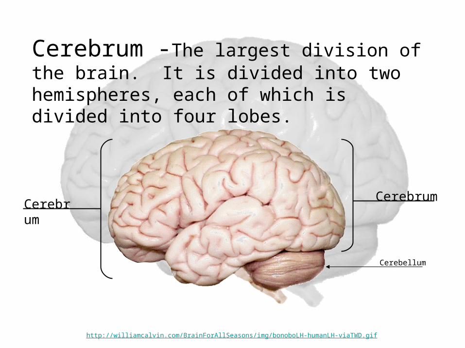
Cerebrum -The largest division of the brain. It is divided into two hemispheres, each of which is divided into four lobes.
CerebrumCerebrum
Cerebellum
http://williamcalvin.com/BrainForAllSeasons/img/bonoboLH-humanLH-viaTWD.gif

Cerebral Cortex
Cerebral Cortex
Cerebral Cortex - The outermost layer of gray matter making up the superficial aspect of the cerebrum.
http://www.bioon.com/book/biology/whole/image/1/1-6.tif.jpg

Nerve bodies
AxonsDendrites
CerebrumCerebrum
CerebellumCerebellum
Axons that have myelin sheaths (insulating covering which allows for faster impulse transmission)
Unmyleinated dendrites and cell bodies (nerve bodies)
•Gyri – Elevated ridges “winding” around the brain. •Sulci – Small grooves dividing the gyri

Basic Structure of a Nerve Cell (Neuron)Basic Structure of a Nerve Cell (Neuron)
Grey matter – closely packed neuron cell bodies form the grey matter of the brain.
White Matter - neuronal tissue containing mainly long, myelinated axons (insulation).

Main Differences of Sheep Brain and Human Brain:
– Temporal lobe of a sheep is proportionately much smaller, and does not extend forward, overlapping the frontal lobe as it does in a human brain. (Auditory Processing – rely less on sounds and more on smell)
– The brain stem of a sheep extends along the dorsal surface (straight back instead of straight down) because sheep walk on all fours.
– A sheep brain has an enlarged smooth area along the ventral aspect of the temporal lobe that is the primary olfactory center of the cerebrum. This indicates how much sheep rely on olfactory senses compared to humans.
– Additionally, sheep have proportionally larger olfactory bulbs

Gloves on, place brain on tray
Posterior or CaudalAnteri
or
Exterior contains Dura mater
Medial

student.ccbcmd.edu/c_anatomy/sheep_brain/sheep_brain.PPT
Dorsal View – Whole Brain
Cerebrum
Longitudinal Fissure
GyriSulciCerebellum
Medulla oblongataSpinal
cord

student.ccbcmd.edu/c_anatomy/sheep_brain/sheep_brain.PPT
Ventral View - Anterior
Pituitary gland
Olfactory bulb
Olfactory tract
Cerebrum

student.ccbcmd.edu/c_anatomy/sheep_brain/sheep_brain.PPT
Ventral View – Optic Nerve and Chiasma
Optic chiasma
Optic nerves
(dura mater intact)(dura mater intact)(dura mater (dura mater partially partially removed)removed)

Lobes of the Cerebrum:
Frontal Lobe
Parietal Lobe
Temporal Lobe
Occiptal Lobe

Midsagittal Section: Cerebrum and Cerebellum
Cerebellum
Occiptal Lobe
Parietal Lobe
Cerebrum
Frontal Lobe
Corpus Callosum
Medulla oblongata Pons
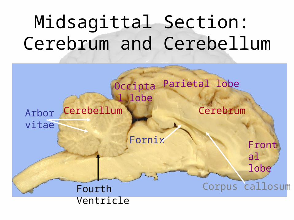
Midsagittal Section: Cerebrum and Cerebellum
CerebellumArbor vitae
Cerebrum
Corpus callosum
Occiptal lobe
Fornix
Parietal lobe
Frontal lobe
Fourth Ventricle

Thalamus and Hypothalamus

Circadian Rhythm• 24 hour cycle regulated by the suprachiasmatic
nucleus of the hypothalamus• Regulates• Sleep cycles• Feeding patterns• Hormone Release• Sex Drive
• “Thrown out of rhythm”• Jet Lag• Seasonal Affective Disorder• Obesity• Depression

Clean up your station prior to leaving lab.
• Throw brain material in the white garbage cans in the lab rooms.
• Clean off dissecting tray.
• Wipe down lab station with Clorox wipes before leaving lab.