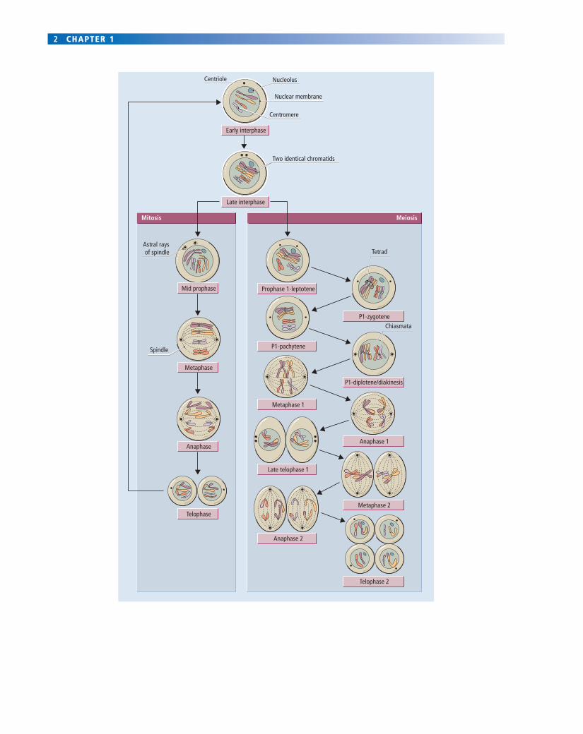
sex - john wiley & sons...sex 3 fig. 1.1 mitosis and meiosis in human cells. each human cell...
TRANSCRIPT

1
1CHAPTER 1
The genesis of two sexes depends on genetic differences 3The genetic determinant of sex is on the Y chromosome 3The two gonads develop from a bipotential precursor plus three waves of ingressing cells 5Primary hermaphrodites have both ovarian and testicular tissues 8
The differentiation of two sexes depends on the endocrine activity of the fetal testis 9
The male and female internal genitalia develop from different unipotential precursors through the actions of androgens and MIH 9The male and female external genitalia develop from a single bipotential precursor through the actions of androgens 9
Secondary hermaphrodites have genitalia that are not of the sex expected from their gonads 9
Pre- and postnatal growth of the gonads is slow until puberty 12
The testes migrate to a scrotal position 12Testicular growth and activity are important for male development 14Most ovarian germ cells die before puberty and all of them enter meiosis 15The ovary is not essential for prepubertal development 17
Summary 17Key learning points 17Further reading 18
Sex
The reproduction of mammals involves sex. Sex is defi ned formally in biology as a process whereby a genetically novel individual is formed as a result of the mixing of genes from two or more individuals. So the essential feature of mammalian sexual reproduction is that the new individual receives its chromosomes in two equal portions: half carried in a male gamete, the spermatozoon, and half carried in a female gamete, the oocyte. These gametes come together at fertilization to form the genetically novel zygote. In order to reproduce itself subsequently, the individual must trans-mit only half its own chromosomes to the new zygotes of the next generation. In sexually reproducing species, there-fore, a special population of germ cells is set aside. These cells undergo a reduction division known as meiosis, in which the chromosomal content of the germ cells is reduced by half and the genetic composition of each chromosome is modi-fi ed as a result of the exchange of pieces of homologous chromosomes (Fig. 1.1). The increased genetic diversity that is generated within a sexually reproducing population may offer a richer and more varied source of material on which natural selection can operate. The population would therefore be expected to show greater resilience in the face of environmental challenge.
However, sex is not by any means an essential compo-nent of reproductive processes. Thus, asexual (or vegetative) reproduction occurs continuously within the tissues of our
own bodies as individual cells grow, divide mitotically (Fig. 1.1) and generate two offspring that are genetically identi-cal to each other and to their single parent. Many unicellu-lar organisms reproduce themselves mitotically just like the individual cells of the body. Among multicellular organisms, including some complex vertebrates such as lizards, several reproduce themselves by setting aside a population of oocytes that can differentiate into embryos in the absence of a fertilizing spermatozoon to generate a complete new organism that is genetically identical or very similar to its parent. This asexual process of reproduction, often called parthenogenetic development, is simply not available to mammals. Although it is possible to stimulate a mammalian oocyte (including a human oocyte) in the complete absence of a spermatozoon, such that it under-goes the early processes of development and may even implant in the uterus, these parthenogenetic embryos always fail and die eventually. It seems that a complete set of chromosomes from a father and a complete set from a mother are an absolute requirement for normal and com-plete development to occur in mammals (see Chapter 9 for discussion as to why this is).
The consequences of obligatory sexual reproduction per-meate all aspects of mammalian life. At the core of the process lies the creation and fusion of the two types of gamete. This occurs in mammals in two distinct types of
COPYRIG
HTED M
ATERIAL
2 CHAPTER 1
Mitosis MeiosisMeiosis
Centriole Nucleolus
Two identical chromatids
Nuclear membrane
Astral raysof spindle Tetrad
Centromere
Chiasmata
Spindle
Prophase 1-leptotene
P1-zygotene
P1-diplotene/diakinesis
Anaphase 1
Metaphase 2
Telophase 2
P1-pachytene
Metaphase 1
Late telophase 1
Anaphase 2
Metaphase
Mid prophase
Late interphase
Early interphase
Anaphase
Telophase
SEX 3
Fig. 1.1 Mitosis and meiosis in human cells. Each human cell contains 23 pairs of homologous chromosomes, making 46 chromosomes in total (see Fig. 1.2). Each set of 23 chromosomes is called a haploid set. When a cell has two complete sets, it is described as being diploid. In this fi gure, we show at the top a single schematized human cell with just 2 of the 23 homologous pairs of chromosomes illustrated, each being colour coded. Before division, the cell is in interphase, during which it grows and duplicates both its centriole and the DNA in each of its chromosomes. As a result, each chromosome consists of two identical chromatids joined at the centromere. Interphase chromosomes are not readily visible, being long, thin and decondensed (but are shown in this fi gure in a more condensed form for simplicity of representation).
In mitotic prophase (left-hand side), the two chromatids become distinctly visible under the light microscope as each shortens and thickens by a spiralling contraction; at the end of prophase the nucleoli and nuclear membrane break down. In mitotic metaphase, microtubules form a mitotic spindle between the two centrioles and the chromosomes lie on its equator. In mitotic anaphase, the centromere of each chromosome splits and the two chromatids in each chromosome each migrate to opposite poles of the spindle (karyokinesis). Mitotic telophase sees: the reformation of nuclear membranes and nucleoli; division of the cytoplasm into two daughters (known as cytokinesis); breakdown of the spindle; and decondensation of chromosomes so that they are no longer visible under the light microscope. Two genetically identical daughter cells now exist where one existed before. Mitosis is a non-sexual or vegetative form of reproduction.
Meiosis involves two sequential divisions (right-hand side). The fi rst meiotic prophase (prophase 1) is lengthy and can be divided into several sequential steps: (1) leptotene chromosomes are long and thin; (2) during zygotene, homologous pairs of chromosomes from each haploid set come to lie side by side along parts of their length; (3) in pachytene, chromosomes start to thicken and shorten and become more closely associated in pairs along their entire length at which time synapsis, crossing over and chromatid exchange take place and nucleoli disappear; (4) in diplotene and diakinesis, chromosomes shorten further and show evidence of being closely linked to their homologue at the chiasmata where crossing over and the reciprocal exchange of DNA sequences has occurred, giving a looped or cross-shaped appearance. In meiotic metaphase 1, the nuclear membrane breaks down, and homologous pairs of chromosomes align on the equator of the spindle. In meiotic anaphase 1, homologous chromosomes move in opposite directions. In meiotic telophase 1, cytokinesis occurs; the nuclear membrane may re-form temporarily, although this does not always happen, yielding two daughter cells each with half the number of chromosomes (only one member of each homologous pair), but each chromosome consisting of two genetically unique chromatids (because of the crossing over at chiasmata). In the second meiotic division, these chromatids then separate much as in mitosis, to yield a total of four haploid offspring from the original cell, each one containing only one complete set of chromosomes. Due to chromatid exchange and the random segregation of homologous chromosomes, each haploid cell is genetically unique. At fertilization, two haploid cells will come together to yield a new diploid zygote.
individual, known as the two sexes: male and female. The gametes themselves take distinctive male or female forms (to prevent self-fertilization) and are made in distinctive male and female gonads: the testis and ovary, respectively. In addition, each gonad elaborates a distinctive group of hormones, notably the sex steroid hormones, which modify the tissues of the body to generate distinctive male and female somatic phenotypes suited to maturing and trans-porting their respective gametes. In most mammals, the sex steroids also affect the behaviour and physiology of the individuals of each sex to ensure that mating will only occur between different sexes at times of maximum fecun-dity. Finally, in mammals, not only do the steroids provide conditions to facilitate the creation of new individuals, they also prepare the female to carry the growing embryo for a prolonged period of pregnancy (viviparity), and to nurture it after birth through an extended period of maternal lacta-tion and parental care.
Thus, the genetic mixing inherent in sexual reproduction has ramifying consequences for mammalian biology, shaping not just anatomy and physiology, but also aspects of behaviour and social structure. This ramifi cation of sex throughout a whole range of biological and social aspects
of mammalian life is mediated largely through the actions of the gonadal hormones. However, in humans and in other higher primates, social learning also plays an impor-tant role in generating sex differences. Children are taught how to behave as women or men, what is feminine and what is masculine. In this way they acquire a sense of their gender. Thus, although studies on mammals in general are relevant to humans, they are not in themselves suffi cient. In order fully to understand human reproduction and sexuality, humans must be studied too. In this chapter, we examine how two sexes arise, differentiate and mature physically. In Chapter 2 we examine the related but distinct issues of gender development and sexuality.
The genesis of two sexes depends on genetic differences
The genetic determinant of sex is on the Y chromosome
In mammals, the genesis of two sexes has a genetic basis. Examination of human chromosomes reveals a consistent difference between the sexes in karyotype (or pattern of chromosomal morphologies). Thus, the human has 46

4 CHAPTER 1
chromosomes, 22 pairs of autosomes and one pair of sex chromosomes (Fig. 1.2). Human females, and indeed all female mammals, are known as the homogametic sex because the sex chromosomes are both X chromosomes and all the gametes (oocytes) are similar to one another in that they each possess one X chromosome. Conversely, the male is termed the heterogametic sex, as his pair of sex chromosomes consists of one X and one Y, so producing two distinct populations of spermatozoa, one bearing an X and the other a Y chromosome (Fig. 1.2). Examination of a range of human patients with chromosomal abnormalities has shown that if a Y chromosome is present then the individ-ual develops the male gonads (testes). If the Y chromosome is absent the female gonads develop (ovaries). The number of X chromosomes or autosomes present does not affect the primary determination of gonadal sex (Table 1.1). Similar
studies on a whole range of other mammals show that Y-chromosome activity alone is suffi cient to determine gonadal sex. Thus the fi rst step towards sexual dimorphism in mammals is the issuing of an instruction by the Y chro-mosome saying: ‘make a testis’.
The Y chromosome itself is small. Moreover, most of its DNA is heterochromatic (that is, very condensed and inca-pable of synthesizing RNA). Therefore, the many structural genes required to make an organ as complex as the testis cannot be located on the Y chromosome alone. Indeed, these genes are known to lie on other autosomal chromo-somes, and some even lie on the X chromosome. What the Y chromosome contains is a ‘switching’ or controller gene, which then somehow regulates the expression of all these other structural genes by determining whether and when they should become activated. The identity and location on
Female Diploid
1–3 4–5
6–12
13–15 16–18
19–20 X–X21–22
Meiosis
Male Diploid
1–3 4–5
6–12
13–15 16–18
19–20 X–Y21–22
Meiosis
Haploid and HeterogameticHaploid and Homogametic
Fig. 1.2 Karyotypes of two mitotic human cells: one male and one female. Each cell was placed in colchicine, a drug that arrested them in mitotic metaphase when the chromosomes were condensed and clearly visible (see Fig. 1.1). The chromosomes were stained and then classifi ed according to the so-called ‘Denver’ system. The 44 autosomes (22 pairs of homologues) are grossly similar in size in each sex, but the pair of sex chromosomes are distinguishable by size, being XX (both large) in the female and XY (one large, one small) in the male. After meiotic division, all four female cells (only two shown) contain one X chromosome: the homogametic sex. In contrast, two of the male cells each contain an X chromosome and two contain a Y chromosome: the heterogametic sex. An arrow indicates the position of the SRY gene on the short arm of the human Y chromosome.

SEX 5
the Y chromosome of this ‘make a testis’ gene was dis-covered initially by the study of some rare and atypical individuals.
Clinicians identifi ed a few men with an XX sex chromo-somal constitution and women with an XY chromosomal constitution—a situation called sex reversal At fi rst sight, these sex-reversed people appear to contradict all that has been said above (see Table 1.1). However, careful examina-tion of the DNA sequences on the short arm of the Y chromo-some of many XY females has revealed either that short pieces of DNA are missing (chromosomal deletions) or that there are mutations of one or more nucleic acid bases. By comparing the DNA sequences in a large number of such patients, it is possible to fi nd one region of the Y chromo-some common to all of them that is affected by deletion or mutation. This region is a likely locus for a testis-determining gene. Supportive evidence comes from many of the XX males, who are found to have translocations of small pieces of the Y chromosome to one of their autosomes or X chromosomes. Again, the critical piece of Y chromo-some that must be translocated to yield an XX male seems to come from the same region as is damaged in the XY females. This region contains a gene called SRY (in humans), which stands for ‘sex-determining region of the Y gene’. The gene is located close to the end of the short arm of the human Y chromosome (see arrow in Fig. 1.2). Genes that code for a common sequence of 88 amino acids (the Sry box) have been found in other mammals, and are also associated with the development of a testis. In the mouse the gene is called Sry and also lies on the short arm but nearer to the centromere.
The identifi cation of the mouse homologue was impor-tant, because it enabled a critical experimental test of the
function of this region of the Y chromosome to be per-formed. Thus, a region of DNA containing only the Sry gene was excised from the Y chromosome and injected into the nuclei of one-cell XX mouse embryos. The excised mate-rial can integrate into the chromosomal material of the XX recipient mouse, which now has an extra piece of DNA. If this piece of DNA is functional in issuing the instruction ‘make a testis’, the XX mouse should develop as a male. This is what happened, strongly supporting the idea that the region containing the controller gene had been identi-fi ed. This gene encodes a protein that binds DNA and localizes to the nucleus (Box 1.1). These features might be expected in a controller gene that infl uences other down-stream genes. But when and where does SRY act to cause a testis to be generated?
The two gonads develop from a bipotential precursor plus three waves of ingressing cells
The early development of the gonad is indistinguishable in males and females. In both sexes the gonads are derived from common somatic mesenchymal tissue precursors called the genital ridge primordia. These primordia develop at about 3.5–4.5 weeks in human embryos, on either side of the central dorsal aorta, on the posterior wall of the lower thoracic and upper lumbar region (Fig. 1.3b,c). These two knots of mesenchyme form the basic matrices of the two gonads. Three waves of ingressing cells expand this matrix and do so in sex-specifi c ways to give the fi nal forms of the ovary and testis.
One wave of migration consists of the gamete precursors called the primordial germ cells (PGCs). These are fi rst iden-tifi able in the human embryo at about 3 weeks in the epi-thelium of the yolk sac near the base of the developing allantois (Fig. 1.3a). By the 13–20-somite stage, the PGC population, expanded by mitosis, can be observed migrat-ing to the connective tissue of the hind gut and from there into the gut mesentery (Fig. 1.3b). From about the 25-somite stage onwards, 30 days or so after fertilization, the majority of cells have passed into the region of the developing kidneys, and thence into the adjacent genital ridge primor-dia. This migration of PGCs is completed by 6 weeks and occurs primarily by amoeboid movement. The genital ridges may produce a chemotactic substance to attract the PGCs, as PGCs co-cultured in a dish with a genital ridge move towards it. Moreover, gonad primordial tissue grafted into abnormal sites within the embryo attracts germ cells to colonize it.
At about the same time as the PGCs are entering the genital ridges, a second group of cells also migrates in. These cells are derived from the columnar coelomic (or germinal) epithelium that overlies the genital ridge mesenchyme. They migrate in as columns called the primitive sex cords (Fig.
Table 1.1 Effect of human chromosome constitution on the development of the gonad.
Chromosomal number
Autosomes Sex chromosomes Gonad Syndrome
44 XO Ovary Turner’s44 XX Ovary Normal female44 XXX Ovary Super female44 XY Testis Normal male44 XXY Testis Klinefelter’s44 XYY Testis Super male66 XXX Ovary { Triploids66 XXY Testis (nonviable)44 XXsxr Testis Sex reversed*
*An Xsxr chromosome carries a small piece of Y chromosome translocated onto the X: see text.
6 CHAPTER 1
BOX 1.1 The molecular biology of SRY* action
There is uncertainty as to how SRY protein actsIn some mammals it binds DNA and localizes in the nucleus, which may suggest an action as a conventional transcription factor by binding to target gene promoter sites. However, few genes have been identifi ed that it activates or represses directly in this way. It also has the property of opening up or remodelling chromatin (so-called DNA bending), thereby making genes accessible to conventional transcription factors, which has suggested a possible action as an ‘architectural’ transcription factor. There is also some evidence that it can affect RNA stability and/or pre-RNA splicing.
Sry may not be quite the master gene that we fi rst thoughtStudies of naturally occurring or induced mutations in humans and mice have implicated a number of other genes in the ‘make a testis’ instruction. These include genes called Sox9, Dax1 and Wnt4. Deletions or mutations of Sox9 lead to XY human and mouse females, while deletions or mutations of Dax1 and Wnt4 lead to XX males. These fi ndings have led to the suggestion that Sox9 enhances and Dax1 and Wnt4 oppose Sry activity. In support of this idea, Sox9 expression rises in males shortly after Sry, while Dax1 and Wnt4 expression decline in males over the same period of embryogenesis.
Interestingly, over-expression of Sox9 in XX embryos leads to XX males and over-expression of Dax1 and Wnt4 in XY embryos to XY females. These dosage effects suggest that it may not be the absolute amount of Sry protein that is important for the instruction ‘make a testis’ so much as the ratio of Sry and/or Sox9 to Dax1 and/or Wnt4. In normal development, perhaps Sry expression promotes Sox9 and depresses Dax1 and Wnt4 expression, but disturbances in the expression levels of these down-stream genes can override the original Sry push to ‘make a testis’.
Finally, downstream of all these ‘make a testis’ genes there seem to be at least two ‘confi rm a testis’ genes. One of these, encoding fi broblast growth factor 9 (Fgf9) is discussed in the main text; in embryos genetically lacking Fgf9 genes, mesonephric cell invasion fails, myoid cells do not develop, the emergent seminiferous cords collapse and
the gonad reorganizes as an XY ovary. The second gene is prostaglandin D synthase (Ptgds), which is produced by both pre-Sertoli and primordial germ cells and catalyses synthesis of prostaglandin D (PGD). Exogenous PGD can convert female gonads at least partially to XX male gonads, and endogenous PGD is thought to have a testicular reinforce-ment role in the developing testis.
Overall, the developing testis seems to use multiple genes in a ‘belt and braces’ approach triggered by Sry expression (the belt). However, this approach leaves testis development vulnerable to rare genetic mutations in the downstream ‘braces’ genes, which, helpfully, are also facilitating elucida-tion of the molecular web of male testis formation.
Advanced readingAdams IR, McLaren A (2002) Sexually dimorphic development of
mouse primordial germ cells: switching from oogenesis to spermatogenesis. Development 129, 1155–1164 (prostaglandin D synthase).
Chaboissier M-C et al. (2004) Functional analysis of Sox8 and Sox9 during sex determination in the mouse. Development 131, 1891–1901 (Sox 9).
Colvin JS et al. (2001).Male-to-female sex reversal in mice lacking fi broblast growth factor 9. Cell 104, 875–889 (Fgf9 mutants).
Grosschedl R et al. (1994) HMG domain proteins: architectural elements in the assembly of nucleoprotein structures. Trends in Genetics 10, 94–100. (DNA bending properties of the Sry box).
Ohe K et al. (2002) A direct role of SRY and SOX proteins in pre-mRNA splicing. Proceedings of the National Academy of Sciences of the USA 99, 1146–1151 (evidence about the molecular mechanism of action of Sry).
Swain A et al. (1998) Dax1 antagonizes Sry action in mammalian sex determination. Nature 391, 761–767.
Vainio S et al. (1999). Female development in mammals is regulated by Wnt-4 signalling. Nature 397, 405–409.
Vidal VPI et al. (2001) Sox9 induces testis development in XX transgenic mice. Nature Genetics 28, 216–217 (Sox9 dosage effect).
*Gene/Protein notationsThroughout the book uses the following gene/protein notation (Sry
as example):Human genes/mRNAs – SRY; Human protein SRY.Mouse genes/mRNAs – Sry; mouse protein Sry.Where a generic statement about mammals is made, the mouse
notation is used.
1.3d). The further development of these cells depends on whether the Sry gene is expressed or not. In the developing males, Sry expression is restricted to the cells of the sex cords. These cells proliferate vigorously and penetrate deep into the medullary mesenchyme, surrounding most of the PGCs to form testis cords (Fig. 1.4a). They will eventually become Sertoli cells, the main supporting cell for spermatogenesis. Because Sry expression is limited to the precursor Sertoli cells (pre-Sertolic cells), it has been suggested that the Sry gene actually issues the instruction: ‘make a Sertoli cell’. Now enclosed within the cords, the PGCs are known as prosper-matogonia and will later give rise to spermatozoa.
In contrast, females lack Sry expression, and their sex cords are ill-defi ned and do not penetrate deeply into the ridge. Instead, the cells condense cortically as small clusters around the PGCs, now called oogonia. This clustering initi-ates formation of the primordial ovarian follicles (Fig. 1.4c,d). In these follicles the condensing cord cells will give rise to the granulosa cells of the primordial follicle, while the oogonia will give rise to oocytes (see Chapter 5).
The third wave of migratory cells comes from the mes-onephric primordia, which lie just lateral to the genital ridges (Fig. 1.3c), and, like the sex cords, they show major sex differences. In the male, mesonephric cells are thought

SEX 7
to contribute at least three major cell types to the testis. Some cells contribute the vasculature tissue of the testis. Other cells synthesize steroid hormones and cluster between the cords to form Leydig cells—the main source of androgens. The third group of mesonephric cells condenses on the developing testis cords and stimulates formation of a basement membrane on which they then sit as myoid cells, thereby forming the seminiferous cords, the forerunners of the adult seminiferous tubules (Fig. 1.4b). The inward migra-tion of this latter group of mesonephric cells giving rise to myoblasts is attributable to the chemotactic action of a growth factor called fi broblast growth factor 9 (Fg f9), which is produced by the developing Sertoli cells. Should this migration fail (for example, in mice lacking Fgf9 genes), the testis cords regress, emphasizing the important role of myoid cells in testis formation. The mesonephric tissue also forms the rete blastema or rete testis cords, later becoming the rete testis, which forms part of the male sperm-exporting duct system (Fig. 1.4a,b). In the female, no myoid cells migrate and the rete blastema is vestigial and transient, leaving only a vestigial rete ovarii in the adult (Fig. 1.4c,d). However, the mesonephric vascular and Leydig cell pre-cursors in males may be paralleled in females by equivalent cells that will eventually form respectively blood vessels and condensations around the developing follicles called thecal cells.
With these three waves of inward migration completed, the basic patterns of testis and ovary are established. However, although the initial decision as to whether to make an ovary or a testis depends on the presence or absence of the SRY activity in developing Sertoli cells, subsequent development of the gonad, particularly of the ovary and its follicles, is dependent on the presence of a population of normal germ cells. For example, women suf-fering from Turner’s syndrome (see Table 1.1), who have a normal autosomal complement but only one X chromo-some, develop an ovary. Subsequently, however, normal oocyte growth requires the activity of both X chromosomes, and the activity of only one X in individuals with Turner’s syndrome leads to death of the oocyte. Secondary loss of the follicle cells follows, leading to ovarian dysgenesis (abnor-mal development), and a highly regressed or streak ovary. Conversely, men with Klinefelter’s syndrome (see Table 1.1) have a normal autosomal complement of chromosomes but three sex chromosomes, two X and one Y. Testes form normally in these individuals as a result of the expression of SRY. However, most of the germ cells die much later in life when they enter meiosis and their death is the result of the activity of two X chromosomes rather than one. These syndromes provide us with two important pieces of clinical evidence. First, initiation of gonad formation can occur when sex cord cells have only one Y (testis) or one X (ovary)
Allantois Genital ridge
Hind gut
Primordialgerm cells
Yolk sac
Heart
Fore gut
Hind gut
Q
Glomerulus
Genital ridgeprimordium
Mesonephricridge
Mesonephric orWolffian duct
Mesonephric orWolffian duct
Primitive medullarycords
Paramesonephric orMüllerian duct
Excretory mesonephric tubule
Aorta
Mesenchyme
Coelomicepithelium
Dorsalmesentery
Genital ridge
Mesonephros
Primitivesex cords
Cloaca
(a) (b)
(c) (d)
Fig. 1.3 A 3-week human embryo showing: (a) the origin of the primordial germ cells; and (b) the route of their migration. Section Q is the plane of transverse section through the lumbar region shown at 4 weeks in (c). In (d) the same plane of section is shown at 5 weeks of development: the ‘indifferent gonad’ stage.

8 CHAPTER 1
(a) (c)
(b) (d)
Rete ovarii
Wolffian duct
Wolffian duct (vas deferens)
Mesonephric tubule
Mesonephric tubules
Rete testis cordsTunica albuginea
Horseshoe-shapedseminiferous cordscontaining Sertolicells andprospermatogonia
Excretory mesonephrictubules (vasa efferentia)
Mesenchymal matrix in whichare Leydig cells
Testis cords
Tunica albuginea
Cortical cordsMüllerian duct
Müllerian duct
Müllerian duct
Surface epithelium
Wolffian duct
Germ cell (oogonium)contained withinfollicular cells(primordial follicle)
Surface epithelium
Müllerian duct
Rete blastemaWolffian duct
Mesonephric tubule
Urogenital mesenteryDegeneratingmedullary cords
Degeneratingmedullary cords
Male Female
Fig. 1.4 Testicular development during (a) week 8 and (b) weeks 16–20 of human embryo-fetal life. (a) The primitive sex cords proliferate into the medulla, establish contact with the mesonephric medullary cords of the rete testis blastema and become separated from the coelomic epithelium by the tunica albuginea (fi brous connective tissue), which eventually forms the testicular capsule. (b) Note the horseshoe shape of the seminiferous cords and their continuity with the rete testis cords. The vasa efferentia, derived from the mesonephric tubules, connect the seminiferous cords with the Wolffi an duct.
Comparable diagrams of ovarian development around (c) week 7 and (d) the weeks 20–24 of development. (c) The primitive sex cords are less well organized and cortical, while medullary mesonephric cords are absent or degenerate. The cortical coelomic epithelial cells condense around the arriving primordial germ cells to yield primordial follicles shown in (d). In the absence of medullary cords and a true persistent rete ovarii, no communication is established with the mesonephric tubules. Hence, in the adult, oocytes are shed from the surface of the ovary, and are not transported by tubules to the oviduct (compare with the male, see Chapters 4 & 5).
chromosome. Second, completion of normal gonad develop-ment requires that the germ cells have two X chromosomes in an ovary but do not have more than one X chromosome in a testis.
Primary hermaphrodites have both ovarian and testicular tissues
We have established that Sry activity on the Y chromosome converts an indifferent gonad into a testis, whereas the absence of its activity results in an ovary. Genetic maleness
leads to gonadal maleness. This primary step in sexual dif-ferentiation is remarkably effi cient, and only rarely are individuals found to have both testicular and ovarian tissue. Such individuals are called primary (or true) hermaphrodites and arise in many cases because of the presence of a mixture of XY and XX (or XO) cells.
The main role of the Sry gene in sexual determination is completed with the establishment of the fetal gonad, and the gene is no longer expressed in the fetus. From this point onwards, the gonads themselves assume the pivotal role in directing sexual differentiation both pre- and postnatally.
SEX 9
Again, it is the male gonad, like the Y chromosome before it, which plays the most active role, taking over the ‘baton of masculinity’ in this sexual relay.
The differentiation of two sexes depends on the endocrine activity of the fetal testis
Endocrine activity in the ovaries is not essential for sexual differentiation during fetal life. In contrast, the testes actively secrete two essential hormones. The interstitial cells of Leydig secrete steroid hormones, the androgens, and the Sertoli cells within the seminiferous cords secrete a dimeric glycoprotein hormone called Müllerian inhibiting hormone (MIH; also called MIS for Müllerian inhibiting substance and AMH for anti-Müllerian hormone). These hormones, which are discussed in more detail in Chapter 3, are the messengers of male sexual differentiation sent out by the testis. In their absence, female sexual differentiation occurs. Thus, sexual differentiation must be actively diverted along the male line, whereas differentiation along the female line again seems to refl ect an inherent trend requiring no active intervention.
The male and female internal genitalia develop from different unipotential precursors through the actions of androgens and MIH
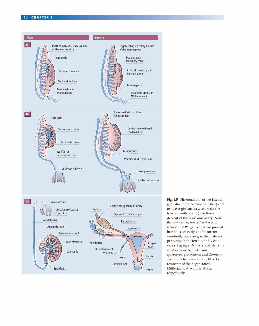
Examination of the primordia of the male and female inter-nal genitalia (see Figs 1.4 & 1.5) shows that instead of one indifferent but bipotential primordium, as was the case for the gonad, there are two separate sets of primordia, each of which is unipotential. These are both located in the mes-onephros adjacent to the developing gonad, and are called the Wolffi an or mesonephric (male) and Müllerian or parames-onephric (female) ducts. In the female, the Wolffi an ducts regress spontaneously and the Müllerian ducts persist and develop to give rise to the oviducts, uterus and cervix and upper vagina (Fig. 1.5). If a female fetus is castrated (its gonads removed), internal genitalia develop in a typical female pattern. This observation demonstrates that ovarian activity is not required for development of the female tract.
In the male, the two testicular hormones prevent this spontaneous development of female genitalia. Thus andro-gens, secreted in considerable amounts by the testis, actively maintain the Wolffi an ducts, which develop into the epidi-dymis, vas deferens and seminal vesicles. If androgen secretion by the testes should fail, or be blocked experimentally, then the Wolffi an duct system regresses and these organs fail to develop. Conversely, exposure of female fetuses to andro-gens causes the development of male internal genitalia.
Testicular androgens have no infl uence on the Müllerian duct system, however, and its regression in males is under
the control of the second testicular hormone, MIH. Thus, in vitro incubation of the primitive internal genitalia of female embryos with MIH provokes abnormal regression of the Müllerian ducts.
The male and female external genitalia develop from a single bipotential precursor through the actions of androgens
The primordia of the external genitalia, unlike those of the internal genitalia, are bipotential (Fig. 1.6). In the female, the urethral folds and genital swellings remain separate, thus forming the labia minora and majora, while the genital tuber-cle forms the clitoris (Fig. 1.6). If the ovary is removed, these changes still occur, indicating their independence of ovarian endocrine activity. In contrast, androgens secreted from the testes in the male cause the urethral folds to fuse (so enclos-ing the urethral tube and contributing, together with cells from the genital swelling, to the shaft of the penis); the genital swellings to fuse in the midline (so forming the scrotum); and the genital tubercle to expand (so forming the glans penis) (Fig. 1.6). Exposure of female fetuses to androgens will ‘masculinize’ their external genitalia, while castration, or suppression of endogenous androgens, in the male results in ‘feminized’ external genitalia.
Secondary hermaphrodites have genitalia that are not of the sex expected from their gonads
Failure of proper endocrine communication between the gonads and the internal and external genital primordia can lead to a dissociation of gonadal and genital sex. Such individuals are called secondary (or pseudo) hermaphrodites. For example, in the genetic syndrome of androgen insensitiv-ity syndrome (AIS; also called testicular feminization or Tfm) the genotype is XY (male), and testes develop normally and secrete androgens and MIH. However, the fetal genitalia are genetically insensitive to the action of androgens (see detailed discussion in Chapter 3), which results in complete regression of the androgen-dependent Wolffi an ducts and in the development of female external genitalia. Mean-while, the MIH secreted from the testes exerts its action fully on the Müllerian ducts, which regress. Thus, this genetically male individual, bearing testes and having androgens circulating, nonetheless appears female with labia, a clitoris and a vagina, but totally lacks other compo-nents of the internal genitalia (Fig. 1.7a).
A naturally occurring counterpart to testicular feminiza-tion is the genetically based adrenogenital syndrome (AGS; also called congenital adrenal hyperplasia or CAH) in female fetuses, in which the XX female develops ovaries as usual. However, as a result of genetic defects in the corticosteroid synthesizing enzymes, the fetal adrenal glands become
10 CHAPTER 1
Male Female
(a)
(b)
(c)
Degenerating excretory tubulesof the mesonephros
Rete testis
Seminal vesicle
Utriculus prostaticusof prostate
Vas deferens
Appendix testis
Seminiferous cords
Seminiferous cords
Rete testis
Seminiferous cord
Fimbria
Epoophoron
Gartner's cyst
Cervix
Round ligamentof uterus
Paroophoron
Suspensory ligament of ovary
Ligament of ovary proper
Mesovarium
Vasa efferentia
Rete testis
Epididymis
Tunica albuginea
Tunica albuginea
Mesonephric orWolffian duct
Wolffian duct (regresses)
Uterovaginal canal
Wolffian ormesonephric duct
Abdominal ostium of theFallopian duct
Degenerating medullary cords
Cortical mesenchymalcondensations
Cortical mesenchymalcondensations
Mesonephros
Mesonephros
Fornix
Corpusuteri
Vagina
Paramesonephric orMüllerian duct
Müllerian tubercle
Müllerian tubercle
Degenerating excretory tubulesof the mesonephros
Fig. 1.5 Differentiation of the internal genitalia in the human male (left) and female (right) at: (a) week 6; (b) the fourth month; and (c) the time of descent of the testis and ovary. Note the paramesonephric Müllerian and mesonephric Wolffi an ducts are present in both sexes early on, the former eventually regressing in the male and persisting in the female, and vice versa. The appendix testis and utriculus prostaticus in the male, and epoophoron, paroophoron and Gartner’s cyst in the female are thought to be remnants of the degenerated Müllerian and Wolffi an ducts, respectively.

SEX 11
Female Male
Genital tubercle
Genital swelling
Urethral fold
Cloacal membrane
Genital tubercle
Genital swelling
Urethral fold
Urogenital membrane
Anal fold
Anal membrane
Genital tubercle
Clitoris
Urethra
Vagina
Hymen
Labium minus
Labium majus
PeriniumPerineum
AnusAnus
Phallus
Urethral groove
Urethralplate
Lumen of penile urethra
Solid epithelialcord
Penileurethra
Glandularpart ofurethra
Scrotal swellings
Perineum
Anal folds
Urethralfold
Urethral fold
Urethral outlet
Glans penis
Line of fusion ofurethral folds
Line of fusion ofscrotal swellings(scrotal septum)
Perineum
Anus
Urogenital groove
Genital swelling
(a) (b)
(c) (d)
(e) (f)
Fig. 1.6 Differentiation of the external genitalia in the human female (left) and male (right) from common primordia shown at: (a) 4 weeks; and (b) 6 weeks. (c) In the female, the labia minora form from the urethral folds and the genital tubercle elongates to form the clitoris. (d) Subsequent changes by the fi fth month are more pronounced in the male, with enlargement of the genital tubercle to form the glans penis and fusion of the urethral folds to enclose the urethral tube and form the shaft of the penis (the genital swellings probably also contribute cells to the shaft). (e) The defi nitive external genitalia of the female at birth. (f) The defi nitive external genitalia of the male at birth.
hyperactive in an attempt to overcome the lack of corticos-teroids, and secrete large quantities of precursor steroids, some with strong androgenic activity (see Chapter 3 for details of steroid biosynthetic pathways). These androgens stimulate development of the Wolffi an ducts, and also cause the external genitalia to develop along the male pattern. The Müllerian system remains, as no MIH has been secreted. Thus, the individual appears partially or even wholly masculinized with a penis and scrotum, but is genetically and gonadally female and possesses the internal genitalia of both sexes (Fig. 1.7b).
Individuals with persistent Müllerian duct syndrome present as genetic males in whom either MIH production, or responsiveness to it, is inadequate. They therefore have testicular androgens that stimulate external genitalia and Wolffi an ducts, but retain Müllerian duct structures. These men are thus genetically and gonadally male but possess the internal genitalia of both sexes.
Apart from the problems of immediate clinical manage-ment raised by diagnosis of these syndromes, abnormali-ties of development of the external genitalia may have important long-term consequences. The single, most

12 CHAPTER 1
(a)
(b)
important event in the identifi cation of sex of the newborn human is examination of the external genitalia. These may be unambiguously male or female, regardless of whether the genetic and gonadal constitutions correspond. They may also be ambiguous as a result of partial masculiniza-tion during fetal life. Sex assignment at birth is one impor-tant step that contributes to the development of an individual’s gender identity, so uncertainty or error at this early stage can have major consequences for an indivi-dual’s self-perception later in life as a man or a woman. This issue is discussed in more detail in Chapter 2.
Pre- and postnatal growth of the gonads is slow until puberty
We have seen how phenotypic features of the male and female develop. The female path of development is taken
unless there is intervention via genetic (SRY) and then endocrine (androgens and MIH) activities, when a male develops. During the remainder of prenatal life and during postnatal life up to puberty, further sexual divergence of physical phenotypes occurs only at a very slow pace and both internal and external genitalia remain immature, growing slowly in line with general body growth. In the male (but not the female) this process is dependent on low and variable levels of gonadal hormones. Despite the rela-tive quiescence, some important reproductive changes do occur over this period.
The testes migrate to a scrotal position
The gonad develops in the upper lumbar region of the embryo, yet by adulthood in most mammalian species, including humans, the testes have descended through the
Fig. 1.7 (a) External genitalia of an XY adult with complete androgen insensitivity syndrome (testicular feminization). Although an androgen-secreting testis is present internally, the external genitalia are indistinguishable from those of a female, and at birth the child would be classifi ed as a girl (see Chapters 2 & 3 for more details). (b) The external genitalia from XX girls with adrenogenital syndrome show varying degrees of masculinization, from an enlarged clitoris to development of a small penis and (empty) scrotum. Ovaries are present internally. The adrenal cortex has inappropriately secreted androgens at the expense of glucocorticoids during fetal life and directed development of the genitalia along the male line. Clearly, the more severe cases could lead to sex assignment as a boy, or to indecision (see Chapter 2).

SEX 13
abdominal cavity, and over the pelvic brim through an inguinal canal to arrive in the scrotum (Fig. 1.8). Evidence of this extraordinary migration is found in the nerve and blood supplies to the testis, which retain their lumbar origins and pass on an extended course through the abdomen to reach their target organ. The transabdominal descent of the testis towards the inguinal canal involves two ligamentous struc-tures: at the superior pole of the testis is the suspensory liga-ment while inferiorly is the gubernaculum, which attaches the testis to the posterior abdominal wall (Fig. 1.8). As the fetal
male body grows, the suspensory ligament elongates but the gubernaculum does not, and thus in the male the relative position of the testis becomes increasingly caudal or pelvic. Two hormones, both secreted by the developing Leydig cells, are responsible for these male-specifi c effects. Andro-gens act on the suspensory ligament, allowing its elonga-tion, while insulin-like growth factor 3 (Insl3) acts on the gubernaculum to mature and stabilize it.
Testicular migration in humans may be arrested devel-opmentally at some point on the migratory route resulting
KidneyKidney
Testicularartery and vein
Testis
GubernaculumPubic bone
Tunica vaginalis
Spermaticcord
Ureter
Vas deferens
Gubernaculum
Penis
Scrotal swelling
Epididymis
Suspensory ligamentAorta
Aorta
Testicularartery
Testis
Bladder
Peritoneum
Fascia
Scrotum
(a) (d)
(e)
(b)
(c)
Fig. 1.8 (a–c) Parasagittal sections through a developing male abdomen. The initial retroperitoneal, abdominal position of the testis shifts pelvically between 10 and 15 weeks, extending the blood supply (and Wolffi an duct derivatives, not shown) as the gubernaculum shortens and the suspensory ligament (d) (connecting the testis to the posterior abdominal wall) lengthens and regresses. A musculofascial layer evaginates into the scrotal swelling accompanied by peritoneal membrane, which forms the processus vaginalis. Between weeks 25 and 28 of pregnancy in the human, the testis migrates over the pubic bone behind the processus vaginalis (which wraps around it forming a double-layered sac), reaching the scrotum by weeks 35–40. The fascia and peritoneum become closely apposed above the testis, obliterating the peritoneal cavity leaving only a tunica vaginalis around the testis below. The fascial layers, obliterated stem of the processus vaginalis, vas deferens and testicular vessels and nerves form the spermatic cord. (d, e) Front view of the migration, showing the extended course ultimately taken by the testicular vessels and vas deferens.
14 CHAPTER 1
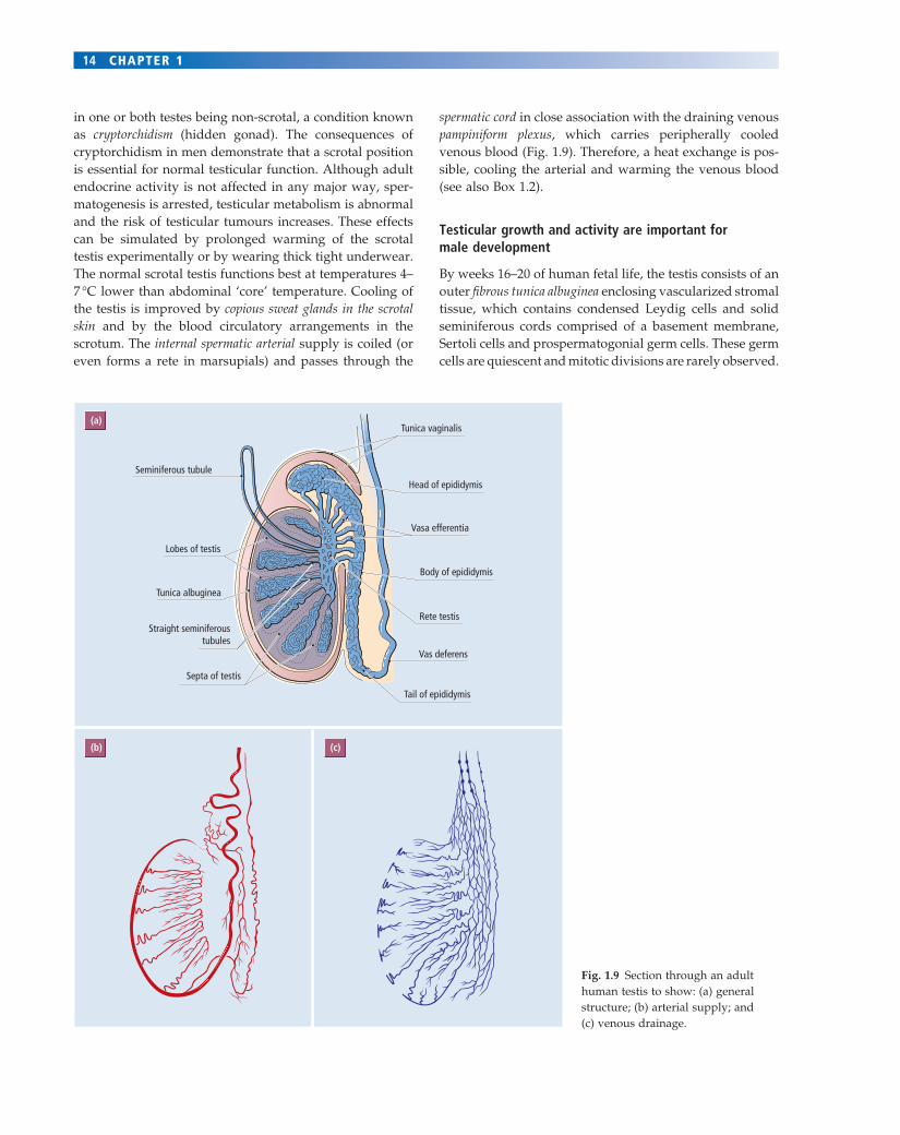
in one or both testes being non-scrotal, a condition known as cryptorchidism (hidden gonad). The consequences of cryptorchidism in men demonstrate that a scrotal position is essential for normal testicular function. Although adult endocrine activity is not affected in any major way, sper-matogenesis is arrested, testicular metabolism is abnormal and the risk of testicular tumours increases. These effects can be simulated by prolonged warming of the scrotal testis experimentally or by wearing thick tight underwear. The normal scrotal testis functions best at temperatures 4–7 °C lower than abdominal ‘core’ temperature. Cooling of the testis is improved by copious sweat glands in the scrotal skin and by the blood circulatory arrangements in the scrotum. The internal spermatic arterial supply is coiled (or even forms a rete in marsupials) and passes through the
spermatic cord in close association with the draining venous pampiniform plexus, which carries peripherally cooled venous blood (Fig. 1.9). Therefore, a heat exchange is pos-sible, cooling the arterial and warming the venous blood (see also Box 1.2).
Testicular growth and activity are important for male development
By weeks 16–20 of human fetal life, the testis consists of an outer fi brous tunica albuginea enclosing vascularized stromal tissue, which contains condensed Leydig cells and solid seminiferous cords comprised of a basement membrane, Sertoli cells and prospermatogonial germ cells. These germ cells are quiescent and mitotic divisions are rarely observed.
(a)
(b) (c)
Tunica vaginalis
Seminiferous tubule
Lobes of testis
Tunica albuginea
Straight seminiferoustubules
Septa of testis
Head of epididymis
Vasa efferentia
Body of epididymis
Rete testis
Vas deferens
Tail of epididymis
Fig. 1.9 Section through an adult human testis to show: (a) general structure; (b) arterial supply; and (c) venous drainage.

SEX 15
The seminiferous cords connect to the cords of the rete testis, the vasa efferentia and thereby to the epididymis.
The Leydig cells in the human testis actively secrete tes-tosterone from at least weeks 8–10 of fetal life onwards, with blood levels peaking at 2 ng/ml at around weeks 13–15. Thereafter, blood levels decline and plateau by 5–6 months at a level of 0.8 ng/ml. This transient prenatal peak of blood testosterone is a feature of many species, although in some, for example the rat and sheep, the peak may approach, or span, the period of parturition, and only begin its decline postnatally. The males of some primate species, including humans, show a postnatal peak in plasma testo-sterone, concentrations reaching 2–3 ng/ml by 3 months postpartum, but declining to around 0.5 ng/ml by 3–4 months. A second modest infantile rise in androgens occurs at 1 year and extends to puberty, when a prepuber-tal peak in androgen output occurs, reaching levels of
about 9 ng/ml. The capacity to secrete testosterone is, as we have seen, essential for establishment of the male pheno-type. It is also important for the continuing development of the male phenotype and, in many if not all species, can also infl uence the development of masculine behaviour patterns (see Chapter 2). The Sertoli cells continue to produce MIH throughout fetal life up until puberty, when levels drop sharply.
Throughout fetal and early postnatal life, testis size increases slowly but steadily. The prospermatogonial germ cells undergo only limited mitotic proliferation and con-tribute little to this growth. At puberty there is a sudden increase in testicular size to which all parts of the testis contribute: the solid seminiferous cords canalize to give rise to tubules; the intratubular Sertoli cells increase in size and activity; the germ cells resume mitotic activity and begin the process of spermatozoal formation; and endo-crine secretion by the intertubular Leydig cells increases sharply. These changes herald the onset of sexual maturity and the development of fertility. The causes and conse-quences of this sudden growth at puberty will be discussed in detail in Chapter 7. The details of how the mature testis functions are described in Chapter 4.
Most ovarian germ cells die before puberty and all of them enter meiosis
The ovary, unlike the testis, retains its position within the abdominal cavity, shifting slightly in some species, such as the human, to assume a pelvic location. It is attached to the posterior abdominal wall by the ovarian mesentery or meso-varium. The ovary, like the testis, grows slowly but steadily in size during early life. As in the testis, little of this growth is due to the germ cells themselves. However, quite unlike the situation in the testis, the ovarian germ cells undergo three major changes.• First, whereas in the male the prospermatogonial germ cells remain in a mitotic cell cycle, albeit rarely dividing, in the female all the oogonial germ cells cease dividing mitotically either before birth (human, cow, sheep, goat, mouse), or shortly thereafter (rat, pig, cat, rabbit, hamster), to enter into their fi rst meiotic division, thereby becoming primary oocytes. The termination of mitosis and entry into meiosis seems to be programmed into all PGCs, since even XY-bearing PGCs enter meiosis spontaneously when cultured in female genital ridges or within the extragonadal parts of the male embryo itself should they have gone astray and not reached the male genital ridge. It seems that the Sry-driven enclo-sure of the XY germ cells within the seminiferous cords suppresses meiotic onset and maintains the PGCs as mitotic cells. There is a major consequence for women of this early termination of mitosis in that by the time of birth a woman has all the oocytes within her ovaries that she will ever have. If
BOX 1.2 Evolutionary evidence of testis migration
Comparative biological study of the male testis provides evidence of ‘evolutionary cryptorchidism’. Thus:• in elephants, hyraxes and the monotremes (platypus
and echidna), the testes normally do not descend at all from the lumbar site
• in armadillos, whales and dolphins, the testes migrate only part of the route to the rear of the lower abdomen
• in hedgehogs, moles and some seals, they lodge in the inguinal canal
• in most rodents and wild ungulates, they retain mobility in the adult, migrating in and out of the scrotum to and from inguinal or abdominal retreats.In animals, such as humans, that have scrotal testes,
a null mutation of the Insl3 gene results in no Insl3 expression and failure of gubernacular maturation and testicular descent. It would be interesting to look at Insl3 gene expression in the above non-scrotal species!
The human scrotal testis clearly requires a lower ambient temperature for normal function, but this requirement may be a secondary consequence of its scrotal position rather than the original evolutionary cause of its migration. Thus, those species in which testes remain in the abdomen survive, fl ourish and reproduce despite the high testicular temperature. It remains unclear as to why testes are scrotal in so many species, given their greater physical vulnerability!
Advanced readingNef S, Prada LF (1999) Cryptorchidism in mice mutant for Insl3.
Nature Genetics 22, 295–299.Zimmermann S et al. (1999) Targeted disruption of the Insl3 gene
causes bilateral cryptorchidism. Molecular Endocrinology 13, 681–691.
16 CHAPTER 1
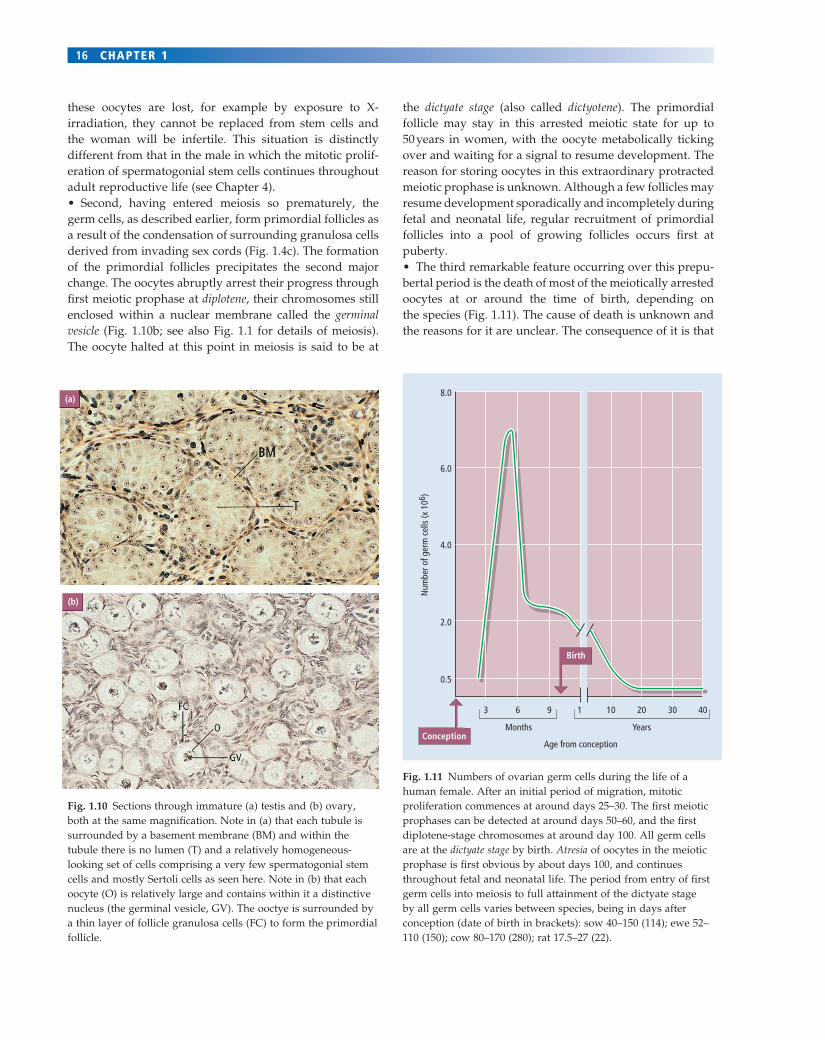
these oocytes are lost, for example by exposure to X-irradiation, they cannot be replaced from stem cells and the woman will be infertile. This situation is distinctly different from that in the male in which the mitotic prolif-eration of spermatogonial stem cells continues throughout adult reproductive life (see Chapter 4).• Second, having entered meiosis so prematurely, the germ cells, as described earlier, form primordial follicles as a result of the condensation of surrounding granulosa cells derived from invading sex cords (Fig. 1.4c). The formation of the primordial follicles precipitates the second major change. The oocytes abruptly arrest their progress through fi rst meiotic prophase at diplotene, their chromosomes still enclosed within a nuclear membrane called the germinal vesicle (Fig. 1.10b; see also Fig. 1.1 for details of meiosis). The oocyte halted at this point in meiosis is said to be at
the dictyate stage (also called dictyotene). The primordial follicle may stay in this arrested meiotic state for up to 50 years in women, with the oocyte metabolically ticking over and waiting for a signal to resume development. The reason for storing oocytes in this extraordinary protracted meiotic prophase is unknown. Although a few follicles may resume development sporadically and incompletely during fetal and neonatal life, regular recruitment of primordial follicles into a pool of growing follicles occurs fi rst at puberty.• The third remarkable feature occurring over this prepu-bertal period is the death of most of the meiotically arrested oocytes at or around the time of birth, depending on the species (Fig. 1.11). The cause of death is unknown and the reasons for it are unclear. The consequence of it is that
(a)
(b)
Fig. 1.10 Sections through immature (a) testis and (b) ovary, both at the same magnifi cation. Note in (a) that each tubule is surrounded by a basement membrane (BM) and within the tubule there is no lumen (T) and a relatively homogeneous-looking set of cells comprising a very few spermatogonial stem cells and mostly Sertoli cells as seen here. Note in (b) that each oocyte (O) is relatively large and contains within it a distinctive nucleus (the germinal vesicle, GV). The ooctye is surrounded by a thin layer of follicle granulosa cells (FC) to form the primordial follicle.
8.0
6.0
4.0
Num
ber o
f ger
m c
ells
(x 1
06)
2.0
0.5
3 6 9 1 10 20
YearsMonths
30 40
Age from conception
Birth
Conception
Fig. 1.11 Numbers of ovarian germ cells during the life of a human female. After an initial period of migration, mitotic proliferation commences at around days 25–30. The fi rst meiotic prophases can be detected at around days 50–60, and the fi rst diplotene-stage chromosomes at around day 100. All germ cells are at the dictyate stage by birth. Atresia of oocytes in the meiotic prophase is fi rst obvious by about days 100, and continues throughout fetal and neonatal life. The period from entry of fi rst germ cells into meiosis to full attainment of the dictyate stage by all germ cells varies between species, being in days after conception (date of birth in brackets): sow 40–150 (114); ewe 52–110 (150); cow 80–170 (280); rat 17.5–27 (22).
SEX 17
KEY LEARNING POINTS
• Sexual reproduction in mammals involves the creation of a genetically novel individual by the contribution of equal numbers of chromosomes from two parents of different sexes.
• Sexual reproduction requires the production of male and female gametes (spermatozoa and oocytes) by the process of meiosis during which the chromosome number of somatic cells is halved and genetic recombination occurs.
• The male and female gametes are made in male and female gonads (testis and ovary) in male and female individuals (men and women).
• Males and females are distinguished simply by the presence or absence of a Y chromosome.
• A single gene called Sry on the Y chromosome acts by issuing the instruction ‘make a testis’.
• The developing gonad arises from a unipotential genital primordium of mesenchymal tissue and three invading cell populations: the primordial germ cells, the germinal epithelial cells, and mesonephric cells.
• Sertoli cell precursors in the testis derive from the male germinal epithelial cells and are the sole site of Sry expression: so ‘make a testis’ may be expressed as ‘make a Sertoli cell’.
• The granulosa cells of the follicle derive from the female germinal epithelial cells.
• Mesonephric-derived myoid cells migrate into the male genital ridge under the infl uence of fi broblastic growth factor 9 and are essential for stabilizing testis tubule development.
• Leydig cells in the male and thecal cells in the female are also derived from ingressing mesonephric cells, as are the vascular cells of the gonads.
• The embryonic testis makes two main hormones (androgens in Leydig cells and Müllerian inhibiting hormone, MIH, in Sertoli cells).
• The androgens stimulate the Wolffi an ducts to make the epididymis, vas deferens and prostate.
• MIH causes the Müllerian ducts to regress.
• In the absence of MIH, the Müllerian duct becomes the oviduct, uterus, cervix and upper vagina.
• Common bipotential precursors of the external genitalia are stimulated to become the scrotum and penis by the testicular androgens, but become the labia and clitoris in the absence of androgens.
• The ovary is not required for the development of the prepubertal female, but the testis is required for the development of the prepubertal male.
• A sex reversed individual has an XX testis or an XY ovary.
• A primary hermaphrodite has both ovarian and testicular tissue.
• A secondary hermaphrodite has internal and/or external genitalia at variance with the sex of their gonads.
• In the fetal/neonatal ovary, the germ cells enter meiosis and then arrest in prophase of fi rst meiosis at the germinal vesicle stage.
• Most female germ cells die around the time of birth.
• The testes migrate caudally under the combined infl uence of androgens and insulin-like growth factor 3 (Insl3), and, in most male mammals, assume a scrotal position.
the stock of female germ cells available for use in adult life is reduced even further.
The ovary is not essential for prepubertal development
Over the prepubertal period the output of steroids by the ovary is minimal and, indeed, removal of the ovary does not affect prepubertal development. In some species, including humans, there may be a transitory stimulation of ovarian endocrine activity spanning the period of birth, but this does not appear to be important for female develop-ment. However, at puberty marked changes in both the structure and endocrine activity of the ovary occur, and for the fi rst time the ovary becomes an essential and positive feminizing infl uence on the developing individual. How it does so is discussed further in Chapters 5, 7 and 8.
Summary
In this chapter we have seen that sexual differentiation is an enduring process of divergence, which begins with the expression of a genetic message that establishes the struc-ture and nature of the fetal gonad, and then extends from the gonad via its hormonal secretions to many tissues of the body. Thus, sex may be defi ned at several levels and by several parameters. Concordance at all levels may be incomplete, and the medical, social and legal consequences of this ‘blurring’ of a clear, discrete sexual boundary may pose problems. However, in this chapter we have been able to defi ne, by a broad set of criteria, how the two sexes are established. In the next chapter, we examine the issues of gender and sexuality and how they are related to the estab-lishment of the two sexes.

18 CHAPTER 1
FURTHER READING
General reading
Brennan J, Capel B (2004) One tissue, two fates: molecular genetic events that underlie testis versus ovary development. Nature Reviews in Genetics 5, 509–521.
Capel B (2000) The battle of the sexes. Mechanisms in Development 92, 89–103.
Carrillo AA, Berkovitz GD (2004) Genetic mechanisms that regu-late testis determination. Reviews in Endocrine and Metabolic Disorders 5, 77–82.
Crow JF (1994) Advantages of sexual reproduction. Developmental Genetics 15, 205–213.
Josso N (1994) Anti-Müllerian hormone: a masculinizing relative of TGF-β. Oxford Reviews of Reproductive Biology 16, 139–164.
Ostrer H (2001) Identifying genes for male sex determination in humans. Journal of Experimental Zoology 290, 567–573 (a general review of strategies for identifying genes of interest).
Ostrer H (2001) Sex determination: lessons from families and embryos. Clinical Genetics 59, 207–215 (a human-focused review on genetics of sex determination).
Ramkisson Y, Goodfellow PN (1996) Early steps in mammalian sex determination. Current Opinions in Genetics & Development 6, 316–321.
Schafer AJ, Goodfellow PN (1996) Sex determination in humans. BioEssays 18, 955–963.
Sutton KA (2000) Molecular mechanisms involved in the differen-tiation of spermatogenic stem cells. Reviews of Reproduction 5, 93–98.
More advanced reading (see also Boxes)
Albrecht KH, Eicher EM (2001) Evidence that Sry is expressed in pre-Sertoli cells, and Sertoli cells and granulosa cells have a common precursor. Developmental Biology 240, 92–107.
Berta P et al. (1990) Genetic evidence equating Sry and the testis-determining factor. Nature 348, 448–450 (an early mutation study confi rming role of Sry in testis formation).
Jeans A et al. (2005) Evaluation of candidate markers for the per-itubular myoid cell lineage in the developing mouse testis. Reproduction 130, 509–516.
Koopman P et al. (1990) Expression of a candidate sex-determining gene during mouse testis differentiation. Nature 348, 450–452 (the original mouse evidence supporting Sry identifi cation as testis-determining gene).
Koopman P et al. (1991) Male development of chromosomally female mice transgenic for Sry. Nature 351, 117–121 (the trans-genic experiment).
Lovell-Badge R, Robertson E (1990) XY female mice resulting from a heritable mutation in the murine primary testis determining gene, Tdy. Development 109, 635–646 (an early mutation study confi rming role of Sry in testis formation).
McLaren A (2000) Germ and somatic cell lineages in the develop-ing gonad. Molecular and Cellular Endocrinology 163, 3–9.
Sinclair AH et al. (1990) A gene from the human sex-determining region encodes a protein with homology to a conserved DNA-binding motif. Nature 346, 240–244 (the original paper describing SRY as the testis-determining gene).