serveur academique lausannois serval …bib_22245425c64d...geiser et al. 2 1 abstract 2 3 patients...
TRANSCRIPT

Serveur Academique Lausannois SERVAL serval.unil.ch
Author ManuscriptFaculty of Biology and Medicine Publication
This paper has been peer-reviewed but does not include the final publisherproof-corrections or journal pagination.
Published in final edited form as:
Title: The coupling of low-level auditory dysfunction and oxidative stress
in psychosis patients.
Authors: Geiser E, Retsa C, Knebel JF, Ferrari C, Jenni R, Fournier M,
Alameda L, Baumann PS, Clarke S, Conus P, Do KQ, Murray MM
Journal: Schizophrenia research
Year: 2017 Feb 9
DOI: 10.1016/j.schres.2017.02.002
In the absence of a copyright statement, users should assume that standard copyright protection applies, unless the article containsan explicit statement to the contrary. In case of doubt, contact the journal publisher to verify the copyright status of an article.

The coupling of low-level auditory dysfunction and oxidative stress in psychosis 1
patients2
3 4
Eveline Geiser1,2,*, Chrysa Retsa1,*, Jean-François Knebel1-3, Carina Ferrari4,5, Raoul Jenni4,5, Margot 5 Fournier4, Luis Alameda4,5,6, Philipp S. Baumann4,5, Stephanie Clarke1, Philippe Conus5, Kim Q. Do4, and 6 Micah M. Murray1-3,6,7† 7
8 The LINE (Laboratory for Investigative Neurophysiology), 1Neuropsychology and Neurorehabilitation 9 Service and 2Radiodiagnostic Service, University Hospital Center and University of Lausanne, 1011 10 Lausanne, Switzerland 11 3The EEG Brain Mapping Core, Center for Biomedical Imaging (CIBM), University Hospital Center and 12 University of Lausanne, 1011 Lausanne, Switzerland 13 4Center for Psychiatric Neuroscience, Department of Psychiatry, University Hospital Center and 14 University of Lausanne, Prilly-Lausanne, Switzerland 15 5Service of General Psychiatry, Department of Psychiatry, University Hospital Center and University of 16 Lausanne, Prilly-Lausanne, Switzerland 17 6Psychiatric Liaison Service, Lausanne University Hospital (CHUV), Lausanne, Switzerland 18 7Department of Ophthalmology, University of Lausanne, Jules-Gonin Eye Hospital, Lausanne, Switzerland 19 8Department of Hearing and Speech Sciences, Vanderbilt University, Nashville, TN, USA 20
21
22 23 24 25 26
Running title: Auditory dysfunction coupled with oxidative stress 27 28
*These authors made equal contributions to this work.29 30
†Corresponding author 31 Professor Micah Murray, Ph.D. 32 University Hospital Center and University of Lausanne 33 Department of Radiology, CIBM, BH08.078 34 Rue du Bugnon 46, Lausanne 1011, Switzerland 35 Tel: +41213141321; Fax: +41213141319; email: [email protected] 36
37 38 39 40

Geiser et al.
2
Abstract 1 2
Patients diagnosed with schizophrenia often present with low-level sensory deficits. It is an open 3
question whether there is a functional link between these deficits and the pathophysiology of the 4
disease, e.g. oxidative stress and glutathione (GSH) metabolism dysregulation. Auditory evoked 5
potentials (AEPs) were recorded from 21 psychosis disorder patients and 30 healthy controls performing 6
an active, auditory oddball task. AEPs to standard sounds were analyzed within an electrical 7
neuroimaging framework. A peripheral measure of participants’ redox balance, the ratio of glutathione 8
peroxidase and glutathione reductase activities (GPx/GR), was correlated with the AEP data. Patients 9
displayed significantly decreased AEPs over the time window of the P50/N100 complex resulting from 10
significantly weaker responses in the left temporo-parietal lobe. The GPx/GR ratio significantly correlated 11
with patients’ brain activity during the time window of the P50/N100 in the medial frontal lobe. We 12
show for the first time a direct coupling between electrophysiological indices of AEPs and peripheral 13
redox dysregulation in psychosis patients. This coupling is limited to stages of auditory processing that 14
are impaired relative to healthy controls and suggests a link between biochemical and sensory 15
dysfunction. The data highlight the potential of low-level sensory processing as a trait-marker of 16
psychosis. 17
18
19 20

Geiser et al.
3
Introduction1
Low-level sensory deficits are increasingly recognized as core dysfunctions in patients with 2
chronic schizophrenia (Ethridge et al., 2015; Javitt, 2009; Turetsky et al., 2008). However, it is largely 3
uninvestigated whether there is a link between disease-related pathology of schizophrenia and low-level 4
sensory deficits. We investigated low-level auditory processing in a group of psychosis disorder patients 5
in the early phases of the disease, and correlated it with blood measures of oxidant state. This should 6
reveal whether low-level auditory processing is linked to a key pathological hub in schizophrenia and 7
could serve as a biomarker for psychosis and schizophrenia. 8
Sensory processing deficits have been observed in patients with chronic schizophrenia (Doniger 9
et al., 2002; Foxe et al., 2005, 2001; Javitt, 2015; Knebel et al., 2011; Oribe et al., 2013; Rosburg et al., 10
2008) as well as in patients at early stages of the disease (Foxe et al., 2011; Hall et al., 2011; Hong et al., 11
2009; Oranje et al., 2013; Salisbury et al., 2010). In the auditory modality, the P50 and the N100 12
components of the auditory evoked potential (AEP) reflect this sensory impairment. Differences in the 13
P50 between patients and controls have been found in isolated auditory stimulus processing (Clementz 14
and Blumenfeld, 2001; Jin et al., 1997) as well as in gating paradigms identifying a less than typically-15
reduced P50 amplitude in the second of a pair of tones in patients (Onitsuka et al., 2013; Potter et al., 16
2006). Compared to controls, patients also show reduced amplitude of the N100 components 17
(Ahveninen et al., 2006; Anokhin et al., 2007; Ethridge et al., 2015; Force et al., 2008; Rihs et al., 2013; 18
Turetsky et al., 2008; Wu et al., 2013), on the basis of which schizophrenic patients and controls can be 19
distinguished on the group level (del Re et al., 2015) and on a single-subject level (Neuhaus et al., 2014). 20
Moreover, there is evidence that low-level sensory deficits might be a characteristic trait marker of 21
individuals with a genetic risk for developing schizophrenia (Light and Makeig, 2015; Light and Swerdlow, 22
2015) as indicated by the P50 in healthy subjects carrying a hereditary risk for the development of 23
schizophrenia (Turetsky et al., 2012) and the N100 in first degree relatives or in subjects carrying a 24
Geiser et al.
4
genetic risk (Ahveninen et al., 2006; Anokhin et al., 2007; Foxe et al., 2011; Frangou et al., 1997; Rihs et 1
al., 2013). 2
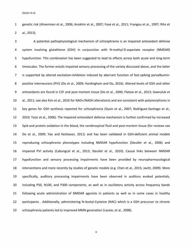
A potential pathophysiological mechanism of schizophrenia is an impaired antioxidant defense 3
system involving glutathione (GSH) in conjunction with N-methyl-D-aspartate receptor (NMDAR) 4
hypofunction. This combination has been suggested to lead to effects across both acute and long-term 5
timescales. The former entails impaired sensory processing of the variety discussed above, and the latter 6
is supported by altered excitation-inhibition induced by aberrant function of fast-spiking parvalbumin-7
positive interneurons (PVI) (Do et al., 2009; Hardingham and Do, 2016). Altered levels of GSH and other 8
antioxidants are found in CSF and post-mortem tissue (Do et al., 2000; Flatow et al., 2013; Gawryluk et 9
al., 2011; see also Kim et al., 2016 for NAD+/NADH alterations) and are consistent with polymorphisms in 10
key genes for GSH synthesis reported for schizophrenia (Gysin et al., 2007; Rodriguez-Santiago et al., 11
2010; Tosic et al., 2006). The impaired antioxidant defense mechanism is further confirmed by increased 12
lipid and protein oxidation in the blood, the cerebrospinal fluid and post-mortem tissue (for reviews see 13
Do et al., 2009; Yao and Keshavan, 2011) and has been validated in GSH-deficient animal models 14
reproducing schizophrenia phenotypes including NMDAR hypofunction (Steullet et al., 2006) and 15
impaired PVI activity (Cabungcal et al., 2013; Steullet et al., 2010). Causal links between NMDAR 16
hypofunction and sensory processing impairments have been provided by neuropharmacological 17
interventions and more recently by studies of genetic models (e.g. Chen et al., 2015; Javitt, 2009). More 18
specifically, auditory processing impairments have been observed in auditory evoked potentials, 19
including P50, N100, and P300 components, as well as in oscillatory activity across frequency bands 20
following acute administration of NMDAR agonists in patients as well as in some cases in healthy 21
participants . Additionally, administering N-Acetyl-Cysteine (NAC) which is a GSH precursor to chronic 22
schizophrenia patients led to improved MMN generation (Lavoie, et al., 2008). 23

Geiser et al.
5
Given this collective evidence, we hypothesized that early sensory processing deficits will be 1
linked to GSH dysregulation measures in psychosis. A peripheral measure of brain GSH levels is a high 2
oxidative status of blood redox indices, namely the ratio of GPx/GR which correlates negatively with 3
brain GSH levels in early-psychosis patients (Xin et al., 2016). The present study investigated for the first 4
time the link between the sensory deficits and potential pathophysiological characteristics of psychosis. 5
AEPs and peripheral GPX/GR was measured and correlated in psychosis disorder patients and healthy 6
controls and reveal a potential association between sensory deficits and oxidative stress in patients. 7
8
Methods and Materials 9
Participants 10
A total of 51 individuals participated in this study. There were 21 psychosis disorder patients (19 11
men, 17 right-handed) aged 18-35 years (mean ± SD = 24±4 years) at the time of EEG recording. The 12
patients were recruited from the TIPP program (Treatment and Early Intervention in Psychosis Program, 13
University Hospital, Lausanne) (Baumann et al., 2013), which is a 3 year program specialized in the 14
treatment of the early phase of psychosis that included only patients that had not received more than 6 15
months of previous treatment (Table 1). The diagnosis was confirmed 3 years after the data acquisition 16
(Table 2). The control population included 30 individuals (19 men, 26 right-handed) aged 18-37 years 17
(mean ± SD = 25±5 years). There was no reliable age difference between the two groups [t (49) = 0.73; p 18
= 0.46]. All patients met threshold criteria for psychosis, as defined by the “Psychosis threshold” subscale 19
of the CAARMS at the point of measurement (Comprehensive Assessment of at Risk Mental States scale 20
(Yung et al., 2005). This threshold is based on a combination of intensity frequency and duration of 21
psychotic symptoms. Details regarding clinical evaluation and medication are provided in Table 1. In a 22
follow-up diagnostic 3 years later, all patients met criteria either for affective psychosis (n=3) or non-23
affective psychosis (n=18) according to DSM-IV criteria (Table 2). Healthy controls, recruited from similar 24

Geiser et al.
6
geographic and socio-demographic areas, were assessed by the Diagnostic Interview for Genetic Studies 1
(Preisig et al., 1999) and matched on gender, age and handedness. Major mood, psychotic, or substance-2
use disorder and having a first-degree relative with a psychotic disorder were exclusion criteria for 3
controls. All participants reported normal hearing. All participants provided their written, informed 4
consent and the procedures were approved by the local Ethics Committee. 5
Stimuli and Task 6
The task entailed an active oddball detection paradigm. Participants were instructed to press a button on 7
a response pad as fast as possible when they heard infrequent stimuli. The frequent stimulus (70% of 8
trials) was a 1000Hz centrally-presented tone of 100ms duration. The infrequent stimuli (30% of trials 9
each) varied in pitch (1200 Hz), perceived lateralization (700µs inter-aural timing difference), or duration 10
(150ms) (1:1:1). The remaining parameters matched the frequent stimulus. A central visual fixation cross 11
was present throughout the experiment. All tones were presented with an average inter-stimulus-onset-12
interval of 916ms jittered with a maximum of ±67 ms. Stimulus delivery and response recordings were 13
controlled by E-Prime (Psychology Software Tools Inc., Pittsburgh, USA; www.pstnet.com/eprime). 14
15
EEG Acquisition and Pre-processing 16
Continuous EEG was acquired at 1024Hz through a 64-channel Biosemi ActiveTwo system 17
(http://www.biosemi.com). EEG data pre-processing and analyses were performed with the Cartool 18
software (Brunet et al., 2011). Data were filtered (second order Butterworth with -12db/octave roll-off; 19
0.1Hz high-pass; 60Hz low-pass; 50Hz notch). In light of the literature summarized in the Introduction 20
demonstrating both sensory processing impairments as well as impairments in processes subserving 21
MMN and P300 generation, we considered it prudent to focus first on AEPs to frequent stimuli so as to 22
establish to what extent sensory processing is intact in EPP and linked to redox dysregulation. Analyses 23
of MMN and P300 will appear in a separate forthcoming article. AEPs were calculated in response to the 24

Geiser et al.
7
frequent stimulus spanning 100ms pre-stimulus to 500ms post-stimulus and pre-stimulus baseline 1
corrected. Epochs with amplitude deviations ±80µV at any channel, and trials with blinks or other 2
transients were excluded. Channels with poor electrode-skin contact were interpolated using 3D splines. 3
Data were then recalculated against the average reference. The average number of accepted epochs was 4
1520 for controls (min./max. = 656/2020) and 1543 for patients (min./max. = 954/2002). These values 5
did not significantly differ [t (49) = 0.23; p = 0.81]. 6
7
AEP Analyses 8
The AEP data were analyzed within an electrical neuroimaging framework (Michel and Murray, 9
2012; Murray et al., 2008). Modulations in AEP strength (quantified by Global Field Power, GFP) and 10
topography (quantified by global dissimilarity) were analyzed separately as a function of time (Lehmann 11
and Skrandies, 1980). We likewise include an analysis of electrode Cz as a function of time. Except where 12
otherwise noted, comparisons of AEPs between groups were performed using independent samples t-13
tests. To correct for temporal autocorrelation, only effects meeting the criterion for at least 15 14
consecutive significant time points are reported (Guthrie and Buchwald, 1991). In the distribution of 106 15
randomizations of all time-points, 99.89% of consecutive significant time points fall below this length. 16
17
Source Estimations and Analyses 18
Source estimations were performed using a distributed linear inverse solution, applying the local 19
autoregressive average (LAURA) regularization approach (Grave de Peralta Menendez et al., 2004, 2001; 20
Michel et al., 2004). The solution space implemented here included 5013 nodes, selected from a 6 × 6 × 6 21
mm grid equally distributed within the gray matter of the Montreal Neurological Institute’s average brain 22
(courtesy of Grave de Peralta Menendez and Gonzalez Andino). The head model and lead field matrix 23
were generated with the Spherical Model with Anatomical Constraints (Spinelli et al., 2000). As an 24

Geiser et al.
8
output, LAURA provides current density measures; the scalar values of which were evaluated at each 1
node. The time periods used for source estimations were determined from AEP analyses. Data from each 2
subject and condition were first averaged as a function of time to generate a single data point. The 3
inverse solution was then calculated for each of the nodes in the solution space for each participant. 4
These data matrices were then submitted to statistical analyses comparing source estimations from each 5
group. A cluster size of 10 contiguous nodes was determined using the AlphaSim program (available at 6
http://afni.nimh.nih.gov) and assuming a spatial smoothing of 2mm FWHM and cluster connection 7
radius of 8.5mm. As measure of effect size, the coefficient of determination is indicated. 8
9
Peripheral ratio of GPx/GR enzymatic activity 10
The ratio of enzymatic activities of glutathione peroxidase relative to glutathione reductase (GPx/GR) 11
was measured in the blood of patients. Blood was collected by venipuncture in Vacutainer-tubes coated 12
with Li-heparinate (Becton Dickinson), between 7 and 8:30 AM under restricted activity conditions and 13
fasting from the previous midnight. GPx and GR enzymatic activities were assessed in blood cells (Xin et 14
al., 2016).The GPx/GR ratio per subject was used as a measure for GSH redox homeostasis, compared 15
between patients and controls with an independent samples t-test and used as a covariate in an 16
ANCOVA on the GFP with one between subject factor ‘group’. The analysis was performed using STEN 17
(http://www.unil.ch/line/home/menuinst/about-the-line/software--analysis-tools.html). 18
19
Results 20
21
Impaired early-latency responses 22
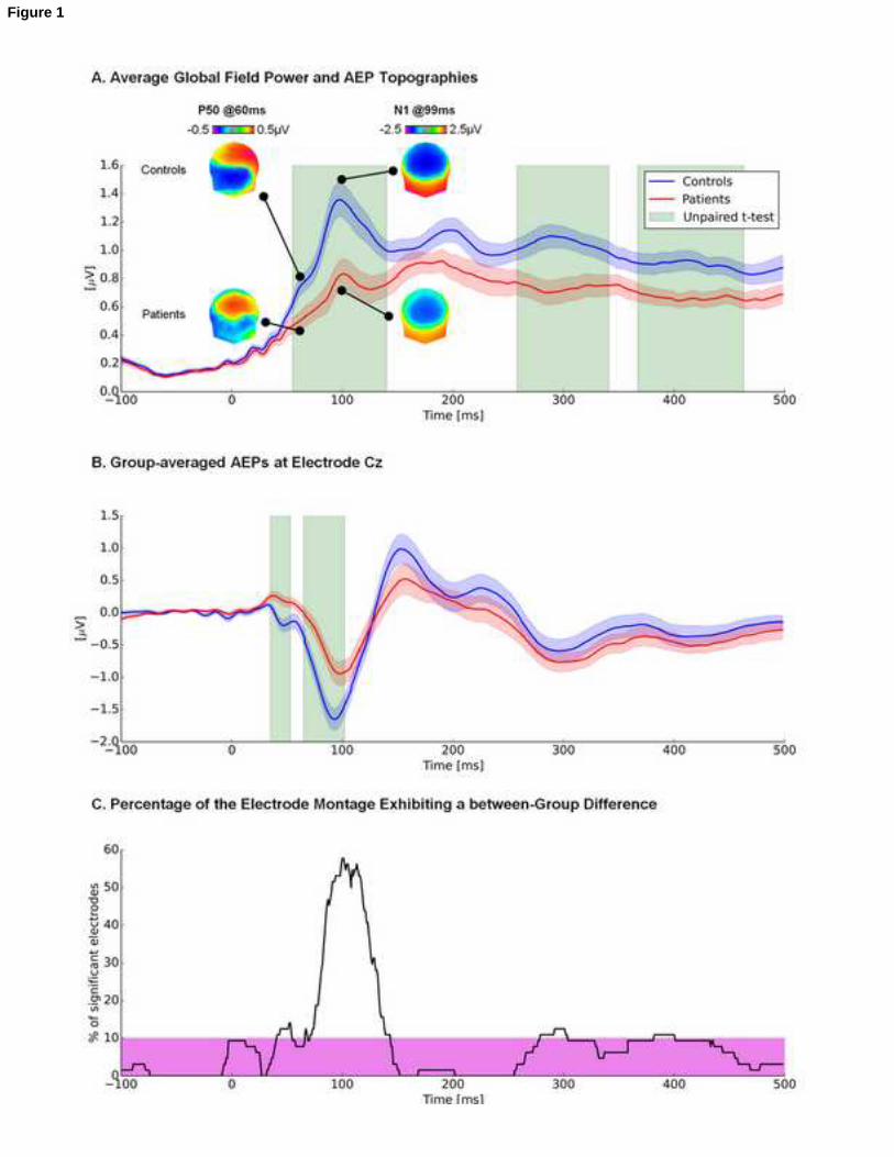
Patients compared to controls showed a weaker auditory response to frequent stimuli at 23
P50/N100 and at a later time windows as measured by GFP (mean r2 for = 0.08, min/max = 0.14/0.22) 24

Geiser et al.
9
(Figure 1A) as exemplary also shown by Cz (mean r2 = 0.15, min/max = 0.08/0.22) in Figure 1B. There was 1
no evidence for significant topographic differences between groups as quantified by global dissimilarity 2
(maximum number of consecutive and significant time points: 12; max/min = 1.48/0.22). No violation of 3
normality over this period of significant GFP modulations (i.e. 56-137ms) was found with the Shapiro-4
Wilk test. Correlations between GFP and the dose of medication (CPZ, chlorpromazine daily equivalent) 5
were below the significance threshold of r = ±0.44 (p > 0.05 for all time points (max/min = 0.41/-0.29). 6
Statistical analysis of source estimations indicated that the difference between controls and patients in 7
the time-window of 56-137ms was a result of differential cortical activity in the left temporo-parietal 8
lobe. The maximum t-value (t(49)max=5.71; average r2=0.22) was located in Brodmann’s Area 38 (i.e. 9
Superior Temporal Gyrus; Figure 2). 10
11
AEP covariance with peripheral redox homeostasis 12
Patients and healthy controls did not significantly differ in their ratio of peripheral GPx/GR 13
(patients: 6.02±0.68, controls: 7.48±0.7, t(47)=1.46;p=0.14); average r2=0.17 (Fig. 3A). However, the 14
GPx/GR ratio significantly covaried with GFP in a time-frame by time-frame regression over the 15
P50/N100 period in EPP (i.e. 66-97ms post-stimulus onset (Figure 3B-C) as revealed by an interaction in 16
the ANCOVA and post-hoc testing (p (max/min) = 0.049/0.02, F (min/max) = 4.43/6.42). No significant 17
correlations we observed at other post-stimulus latencies, and no such relationship was observed in 18
healthy controls. 19
Results of a 2 (controls vs. patients) X GPx/GR ANCOVA on each node within the solution space 20
showed a significant interaction within the frontal lobe (F (1,45)max= 13.5). The maximum F value was 21
located in Brodmann’s Area 9 (medial prefrontal cortex) (Figure 4A). Post-hoc regression performed in 22
each group separately showed a significant correlation between the source estimation in the time-23
Geiser et al.
10
window 56-137ms post stimulus onset for the EPP only. The maximum correlation value was located in 1
Brodmann Area 10 (Anterior prefrontal cortex) (Figure 4B). 2
3
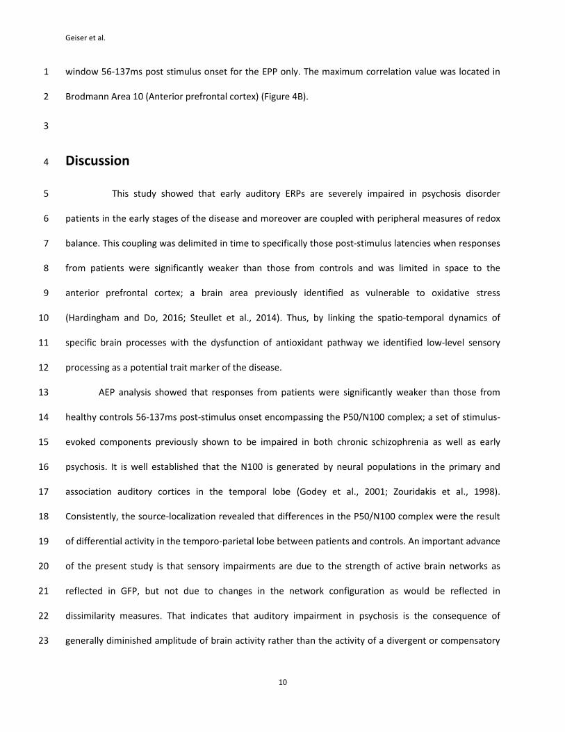
Discussion4
This study showed that early auditory ERPs are severely impaired in psychosis disorder 5
patients in the early stages of the disease and moreover are coupled with peripheral measures of redox 6
balance. This coupling was delimited in time to specifically those post-stimulus latencies when responses 7
from patients were significantly weaker than those from controls and was limited in space to the 8
anterior prefrontal cortex; a brain area previously identified as vulnerable to oxidative stress 9
(Hardingham and Do, 2016; Steullet et al., 2014). Thus, by linking the spatio-temporal dynamics of 10
specific brain processes with the dysfunction of antioxidant pathway we identified low-level sensory 11
processing as a potential trait marker of the disease. 12
AEP analysis showed that responses from patients were significantly weaker than those from 13
healthy controls 56-137ms post-stimulus onset encompassing the P50/N100 complex; a set of stimulus-14
evoked components previously shown to be impaired in both chronic schizophrenia as well as early 15
psychosis. It is well established that the N100 is generated by neural populations in the primary and 16
association auditory cortices in the temporal lobe (Godey et al., 2001; Zouridakis et al., 1998). 17
Consistently, the source-localization revealed that differences in the P50/N100 complex were the result 18
of differential activity in the temporo-parietal lobe between patients and controls. An important advance 19
of the present study is that sensory impairments are due to the strength of active brain networks as 20
reflected in GFP, but not due to changes in the network configuration as would be reflected in 21
dissimilarity measures. That indicates that auditory impairment in psychosis is the consequence of 22
generally diminished amplitude of brain activity rather than the activity of a divergent or compensatory 23

Geiser et al.
11
network. 1
The smaller the ratio of GPx/GR in patients, the weaker was their GFP over the specific time 2
window of the sensory deficit. It is important to note that the correlation was driven by the participants 3
that had reduced ratios of GPx/GR (Figure 3C). One interpretation of the findings is that the redox state 4
influences the responsiveness of NMDAR and any dysregulation on that redox state reflected in the 5
GPx/GR ratio can lead to generally reduced electrophysiological responses to tones. It is well established 6
that oxidative stress depresses NMDAR function and, inversely, that prolonged NMDAR hypofunction 7
induces an oxidative state, particularly in cortical parvalbumin interneurons (PVI) in patients 8
(Hardingham and Do, 2016). Moreover, in a rodent knockout model affecting NMDAR selective to PVI, 9
the AEP is decreased at N40 (Barnes et al., 2015), which is thought to be homologous to the human N100 10
response (Amann et al., 2010). It should be noted that a straightforward connection between N100 11
deficits and PVI dysfunction cannot be demonstrated in the current study, given evidence that acute 12
administration of NMDAR agonists are themselves sufficient to affect such responses. In this vein, 13
Lakatos et al. (2013) have shown that N100 activity occurs primarily in the theta frequency band rather 14
than in the gamma band whose dysfunctions are more closely related to PVI dysregulation (Jadi et al., 15
2016). Future studies focusing on oscillatory patterns and redox measures in psychosis could help 16
elucidate potential contributions of NMDAR and PVI dysfunction to specific brain responses. 17
Our results clearly establish a link between oxidative state and sensory processing in patients. 18
Interestingly and in keeping with Xin et al. (2016), we did not observe any difference in peripheral 19
oxidation status between patients and controls, as measured by GPx/GR. Furthermore, a recent meta-20
analysis indicated no changes in GPx activity in first episode psychosis or stable medicated outpatients 21
(Flatow et al., 2013). One speculative possibility is that patients with particularly low ratios, which appear 22
to be driven by GPx, may be those who are particularly susceptible to functional impairments; a 23
speculation borne out in results (cf. Figure 3). Thus, we show for the first time a potential link between 24

Geiser et al.
12
the pathophysiology in schizophrenic patients and low-level sensory deficits. 1
The GPx/GR ratio measured in patients correlated significantly with activity in the anterior and 2
medial prefrontal cortex, indicating that functional activity is affected by oxidative stress in this brain 3
area. Already in the early stages of psychosis the medial prefrontal cortex shows impaired white matter 4
integrity (Stark et al., 2004), correlating significantly with prefrontal GSH levels in these patients (Monin 5
et al., 2015). Moreover, medial prefrontal white matter impairments were observed in animal models 6
with GSH deficiency (Monin et al., 2015). Our findings complement these results by showing that 7
oxidative stress not only influences frontal cortex integrity but, potentially, also the functional activity in 8
this brain area. Importantly, our results support parallel functional impairments both of which manifest 9
during early post-stimulus processing stages. On the one hand, sensory processing is impaired within 10
low-level auditory cortices extending throughout the left temporo-parietal lobe. On the other hand and 11
contemporaneously, the positive coupling between oxidative stress (viz. GPx/GR) and activity within the 12
anterior medial prefrontal cortex may result from increased top-down control over sensory processing in 13
patients that is not required by controls. This further supports the notion that the low-level sensory 14
deficit is not simply an epiphenomenon of the disease, but could be directly linked to a potential 15
pathophysiology. 16
We report a reduced auditory P50/N100 in this sample of psychosis disorder patients indicating a 17
low-level sensory processing deficit in early stage psychosis and provide evidence of a direct link 18
between these deficits and peripheral measures of oxidative stress in patients. This supports the notion 19
that auditory deficits are a trait marker of the disease and provide insights on the functional 20
consequences of oxidative stress on brain function. 21
22
23
24

Geiser et al.
13
References 1
Ahveninen, J., Jaaskelainen, I.P., Osipova, D., Huttunen, M.O., Ilmoniemi, R.J., Kaprio, J., Lonnqvist, J., 2 Manninen, M., Pakarinen, S., Therman, S., Naatanen, R., Cannon, T.D., 2006. Inherited auditory-3 cortical dysfunction in twin pairs discordant for schizophrenia. Biol. Psychiatry 60, 612–620. 4 doi:10.1016/j.biopsych.2006.04.015 5
Amann, L.C., Gandal, M.J., Halene, T.B., Ehrlichman, R.S., White, S.L., McCarren, H.S., Siegel, S.J., 2010. 6 Mouse behavioral endophenotypes for schizophrenia. Brain Res. Bull. 83, 147–161. 7 doi:10.1016/j.brainresbull.2010.04.008 8
Anokhin, A.P., Vedeniapin, A.B., Heath, A.C., Korzyukov, O., Boutros, N.N., 2007. Genetic and 9 environmental influences on sensory gating of mid-latency auditory evoked responses: a twin 10 study. Schizophr. Res. 89, 312–319. doi:10.1016/j.schres.2006.08.009 11
Barnes, S.A., Pinto-Duarte, A., Kappe, A., Zembrzycki, A., Metzler, A., Mukamel, E.A., Lucero, J., Wang, X., 12 Sejnowski, T.J., Markou, A., Behrens, M.M., 2015. Disruption of mGluR5 in parvalbumin-positive 13 interneurons induces core features of neurodevelopmental disorders. Mol. Psychiatry 20, 1161–14 1172. doi:10.1038/mp.2015.113 15
Baumann, P., Crespi, S., Marion/Vezron, R., Solida, A., Thonney, J., Favrod, J., et al., 2013. Treatment and 16 early intervention in psychosis program (TIPP-Lausanne): implementation of an early 17 intervention programme for psychosis in Switzerland. Early Interv. Psychiatry 7, 322–8. 18
Baumann, P., Fournier, M., Griffa, A., Ferrari, C., Alameda, L., Thiran, J.-P., Cuenod, M., Hagmann, P., Do, 19 K.Q., Conus, P., 2016. Peripheral redox index as a maerker of abnormal fornix-hippocampus 20 circuit in the early phase of psychosis. Transl. Psychiatry accepted. 21
Baumann, P.S., Griffa, A., Ferrari, C., Luis, A., Thiran, J.-P., Do, K.Q., Conus, P., 2014. Integrity of the fornix 22 and hippocampus and relationship with peripheral oxidative stress markers in early phase 23 psychosis. Biol. Psychiatry 75, 109S. 24
Brunet, D., Murray, M.M., Michel, C.M., 2011. Spatiotemporal analysis of multichannel EEG: CARTOOL. 25 Comput. Intell. Neurosci. 2011. doi:10.1155/2011/813870 26
Cabungcal, J.-H., Steullet, P., Kraftsik, R., Cuenod, M., Do, K.Q., 2013. Early-Life Insults Impair 27 Parvalbumin Interneurons via Oxidative Stress: Reversal by N-Acetylcysteine. Biol. Psychiatry 73, 28 574–582. doi:10.1016/j.biopsych.2012.09.020 29
Chen, I.W., Helmchen, F., Lutcke, H, 2015. Specific early and late oddball-evoked responses in excitatory 30
and inhibotory neurons of mouse auditory cortex. J. Neurosci. 35(36), 12560-12573. doi: 31
10.1523?JNEUROSCI.2240-15.2015 32
Clementz, B.A., Blumenfeld, L.D., 2001. Multichannel electroencephalographic assessment of auditory 33 evoked response suppression in schizophrenia. Exp. Brain Res. 139, 377–390. 34
del Re, E.C., Spencer, K.M., Oribe, N., Mesholam-Gately, R.I., Goldstein, J., Shenton, M.E., Petryshen, T., 35 Seidman, L.J., McCarley, R.W., Niznikiewicz, M.A., 2015. Clinical high risk and first episode 36 schizophrenia: auditory event-related potentials. Psychiatry Res. 231, 126–133. 37 doi:10.1016/j.pscychresns.2014.11.012 38
Do, K.Q., Cabungcal, J.H., Frank, A., Steullet, P., Cuenod, M., 2009. Redox dysregulation, 39 neurodevelopment, and schizophrenia. Curr. Opin. Neurobiol. 19, 220–230. 40 doi:10.1016/j.conb.2009.05.001 41
Do, K.Q., Trabesinger, A.H., Kirsten-Kruger, M., Lauer, C.J., Dydak, U., Hell, D., Holsboer, F., Boesiger, P., 42 Cuenod, M., 2000. Schizophrenia: glutathione deficit in cerebrospinal fluid and prefrontal cortex 43 in vivo. Eur. J. Neurosci. 12, 3721–3728. 44

Geiser et al.
14
Doniger, G.M., Foxe, J.J., Murray, M.M., Higgins, B.A., Javitt, D.C., 2002. Impaired visual object 1 recognition and dorsal/ventral stream interaction in schizophrenia. Arch. Gen. Psychiatry 59, 2 1011–1020. 3
Ethridge, L.E., Hamm, J.P., Pearlson, G.D., Tamminga, C.A., Sweeney, J.A., Keshavan, M.S., Clementz, B.A., 4 2015. Event-related potential and time-frequency endophenotypes for schizophrenia and 5 psychotic bipolar disorder. Biol. Psychiatry 77, 127–136. doi:10.1016/j.biopsych.2014.03.032 6
Flatow, J., Buckley, P., Miller, B.J., 2013. Meta-analysis of oxidative stress in schizophrenia. Biol. 7 Psychiatry 74, 400–409. doi:10.1016/j.biopsych.2013.03.018 8
Force, R.B., Venables, N.C., Sponheim, S.R., 2008. An auditory processing abnormality specific to liability 9 for schizophrenia. Schizophr. Res. 103. doi:10.1016/j.schres.2008.04.038 10
Foxe, J.J., Doniger, G.M., Javitt, D.C., 2001. Early visual processing deficits in schizophrenia: impaired P1 11 generation revealed by high-density electrical mapping. Neuroreport 12, 3815–3820. 12
Foxe, J.J., Murray, M.M., Javitt, D.C., 2005. Filling-in in schizophrenia: a high-density electrical mapping 13 and source-analysis investigation of illusory contour processing. Cereb. Cortex N. Y. N 1991 15, 14 1914–1927. doi:10.1093/cercor/bhi069 15
Foxe, J.J., Yeap, S., Snyder, A.C., Kelly, S.P., Thakore, J.H., Molholm, S., 2011. The N1 auditory evoked 16 potential component as an endophenotype for schizophrenia: high-density electrical mapping in 17 clinically unaffected first-degree relatives, first-episode, and chronic schizophrenia patients. Eur. 18 Arch. Psychiatry Clin. Neurosci. 261, 331–339. doi:10.1007/s00406-010-0176-0 19
Frangou, S., Sharma, T., Alarcon, G., Sigmudsson, T., Takei, N., Binnie, C., Murray, R.M., 1997. The 20 Maudsley Family Study, II: Endogenous event-related potentials in familial schizophrenia. 21 Schizophr. Res. 23. doi:10.1016/S0920-9964(96)00089-8 22
Gawryluk, J.W., Wang, J.-F., Andreazza, A.C., Shao, L., Young, L.T., 2011. Decreased levels of glutathione, 23 the major brain antioxidant, in post-mortem prefrontal cortex from patients with psychiatric 24 disorders. Int. J. Neuropsychopharmacol. 14, 123–130. doi:10.1017/S1461145710000805 25
Godey, B., Schwartz, D., de Graaf, J.B., Chauvel, P., Liegeois-Chauvel, C., 2001. Neuromagnetic source 26 localization of auditory evoked fields and intracerebral evoked potentials: a comparison of data 27 in the same patients. Clin. Neurophysiol. Off. J. Int. Fed. Clin. Neurophysiol. 112, 1850–1859. 28
Grave de Peralta Menendez, R., Gonzalez Andino, S., Lantz, G., Michel, C.M., Landis, T., 2001. 29 Noninvasive localization of electromagnetic epileptic activity. I. Method descriptions and 30 simulations. Brain Topogr. 14, 131–137. 31
Grave de Peralta Menendez, R., Murray, M.M., Michel, C.M., Martuzzi, R., Gonzalez Andino, S.L., 2004. 32 Electrical neuroimaging based on biophysical constraints. NeuroImage 21, 527–539. 33 doi:10.1016/j.neuroimage.2003.09.051 34
Guthrie, D., Buchwald, J.S., 1991. Significance testing of difference potentials. Psychophysiology 28, 240–35 244. 36
Gysin, R., Kraftsik, R., Sandell, J., Bovet, P., Chappuis, C., Conus, P., Deppen, P., Preisig, M., Ruiz, V., 37 Steullet, P., Tosic, M., Werge, T., Cuenod, M., Do, K.Q., 2007. Impaired glutathione synthesis in 38 schizophrenia: Convergent genetic and functional evidence. Proc. Natl. Acad. Sci. U. S. A. 104, 39 16621–16626. doi:10.1073/pnas.0706778104 40
Hall, M.-H., Taylor, G., Salisbury, D.F., Levy, D.L., 2011. Sensory gating event-related potentials and 41 oscillations in schizophrenia patients and their unaffected relatives. Schizophr. Bull. 37, 1187–42 1199. doi:10.1093/schbul/sbq027 43
Hardingham, G.E., Do, K.Q., 2016. Linking early-life NMDAR hypofunction and oxidative stress in 44 schizophrenia pathogenesis. Nat. Rev. Neurosci. 17, 125–34. doi:10.1038/nrn.2015.19 45
Hong, X., Chan, R.C.K., Zhuang, X., Jiang, T., Wan, X., Wang, J., Xiao, B., Zhou, H., Jiang, L., Weng, B., 2009. 46 Neuroleptic effects on P50 sensory gating in patients with first-episode never-medicated 47 schizophrenia. Schizophr. Res. 108, 151–157. doi:10.1016/j.schres.2008.11.016 48

Geiser et al.
15
Javitt, D.C., 2015. Neurophysiological models for new treatment development in schizophrenia: early 1 sensory approaches. Ann. N. Y. Acad. Sci. 1344. doi:10.1111/nyas.12689 2
Javitt, D.C., 2009. Sensory processing in schizophrenia: neither simple nor intact. Schiz. Bul. 35, 1059-3
1064. doi:10.1093/schbul/sbp110 4
Jadi, M.P., Behrend, M.M., Sejnowski, T.J. 2016. Abnormal gamma oscillations in N-methyl-D-Aspartate 5 receptor hypofunction models of schizophrenia. Biological Psychiatry. 79, 716-726 6
Jin, Y., Potkin, S.G., Patterson, J.V., Sandman, C.A., Hetrick, W.P., Bunney, W.E.J., 1997. Effects of P50 7 temporal variability on sensory gating in schizophrenia. Psychiatry Res. 70. 8
Kim, S.Y., Cohen, B.M., Chen, X., Lukas, S.E., Shinn, A.K., Yuksel, A.C., Li, T., Du, F., Öngür, D., 2016. Redox 9 Dysregulation in schizophrenia revealed by in vivo NAD+/NADH measurement. Schizophrenia 10 Bulletin. 43, 197-204. 11
Knebel, J.-F., Javitt, D.C., Murray, M.M., 2011. Impaired early visual response modulations to spatial 12 information in chronic schizophrenia. Psychiatry Res. 193, 168–176. 13 doi:10.1016/j.pscychresns.2011.02.006 14
Lakatos, P., Schroeder, C.E., Leitman, D.I., Javitt, D.C. 2013. Predictive suppression of cortical excitability 15 and its deficit in schizophrenia. Journal of Neuroscience. 33, 11692-11702. 16
Lavoie, S., Murray, M.M., Deppen, P., Knyazeva, M.G., Berk, M., Boulat, O., Bovet, P., Bush, A.I., Conus, 17 P., Copolov, D., Fornari, E., Meuli, R., Solida, A., Vianin, P., Cuenod, M., Buclin, T. , Do, K.Q. 2008. 18 Glutathione precursor, N-Acetyl-Cysteine, improves mismatch negativity in schizoprhenia 19 pateints. Neuropsychopharmacology. 33, 2187-2199. 20
Lehmann, D., Skrandies, W., 1980. Reference-free identification of components of checkerboard-evoked 21 multichannel potential fields. Electroencephalogr. Clin. Neurophysiol. 48, 609–621. 22
Light, G.A., Makeig, S., 2015. Electroencephalographic biomarkers of psychosis: present and future. Biol. 23 Psychiatry 77, 87–89. doi:10.1016/j.biopsych.2014.11.002 24
Light, G.A., Swerdlow, N.R., 2015. Future clinical uses of neurophysiological biomarkers to predict and 25 monitor treatment response for schizophrenia. Ann. N. Y. Acad. Sci. 1344, 105–119. 26 doi:10.1111/nyas.12730 27
Michel, C.M., Murray, M.M., 2012. Towards the utilization of EEG as a brain imaging tool. NeuroImage 28 61, 371–385. doi:10.1016/j.neuroimage.2011.12.039 29
Michel, C.M., Murray, M.M., Lantz, G., Gonzalez, S., Spinelli, L., Grave de Peralta, R., 2004. EEG source 30 imaging. Clin. Neurophysiol. Off. J. Int. Fed. Clin. Neurophysiol. 115, 2195–2222. 31 doi:10.1016/j.clinph.2004.06.001 32
Monin, A., Baumann, P.S., Griffa, A., Xin, L., Mekle, R., Fournier, M., Butticaz, C., Klaey, M., Cabungcal, 33 J.H., Steullet, P., Ferrari, C., Cuenod, M., Gruetter, R., Thiran, J.P., Hagmann, P., Conus, P., Do, 34 K.Q., 2015. Glutathione deficit impairs myelin maturation: relevance for white matter integrity in 35 schizophrenia patients. Mol. Psychiatry 20, 827–838. doi:10.1038/mp.2014.88 36
Murray, M.M., Brunet, D., Michel, C.M., 2008. Topographic ERP analyses: a step-by-step tutorial review. 37 Brain Topogr. 20, 249–264. doi:10.1007/s10548-008-0054-5 38
Neuhaus, A.H., Popescu, F.C., Rentzsch, J., Gallinat, J., 2014. Critical evaluation of auditory event-related 39 potential deficits in schizophrenia: evidence from large-scale single-subject pattern classification. 40 Schizophr. Bull. 40, 1062–1071. doi:10.1093/schbul/sbt151 41
Onitsuka, T., Oribe, N., Nakamura, I., Kanba, S., 2013. Review of neurophysiological findings in patients 42 with schizophrenia. Psychiatry Clin. Neurosci. 67, 461–470. doi:10.1111/pcn.12090 43

Geiser et al.
16
Oranje, B., Aggernaes, B., Rasmussen, H., Ebdrup, B.H., Glenthoj, B.Y., 2013. P50 suppression and its 1 neural generators in antipsychotic-naive first-episode schizophrenia before and after 6 months 2 of quetiapine treatment. Schizophr. Bull. 39, 472–480. doi:10.1093/schbul/sbr183 3
Oribe, N., Hirano, Y., Kanba, S., del Re, E.C., Seidman, L.J., Mesholam-Gately, R., Spencer, K.M., McCarley, 4 R.W., Niznikiewicz, M.A., 2013. Early and late stages of visual processing in individuals in 5 prodromal state and first episode schizophrenia: an ERP study. Schizophr. Res. 146. 6 doi:10.1016/j.schres.2013.01.015 7
Potter, D., Summerfelt, A., Gold, J., Buchanan, R.W., 2006. Review of clinical correlates of P50 sensory 8 gating abnormalities in patients with schizophrenia. Schizophr. Bull. 32. 9 doi:10.1093/schbul/sbj050 10
Preisig, M., Fenton, B.T., Matthey, M.L., Berney, A., Ferrero, F., 1999. Diagnostic interview for genetic 11 studies (DIGS): inter-rater and test-retest reliability of the French version. Eur. Arch. Psychiatry 12 Clin. Neurosci. 249, 174–179. doi:92490174.406 [pii] 13
Rihs, T.A., Tomescu, M.I., Britz, J., Rochas, V., Custo, A., Schneider, M., Debbane, M., Eliez, S., Michel, 14 C.M., 2013. Altered auditory processing in frontal and left temporal cortex in 22q11.2 deletion 15 syndrome: a group at high genetic risk for schizophrenia. Psychiatry Res. 212, 141–149. 16 doi:10.1016/j.pscychresns.2012.09.002 17
Rodriguez-Santiago, B., Brunet, A., Sobrino, B., Serra-Juhe, C., Flores, R., Armengol, L.I., Vilella, E., Gabau, 18 E., Guitart, M., Guillamat, R., Martorell, L., Valero, J., Gutierrez-Zotes, A., Labad, A., Carracedo, 19 A., Estivill, X., Perez-Jurado, L.A., 2010. Association of common copy number variants at the 20 glutathione S-transferase genes and rare novel genomic changes with schizophrenia. Mol. 21 Psychiatry 15, 1023–1033. doi:10.1038/mp.2009.53 22
Rosburg, T., Boutros, N.N., Ford, J.M., 2008. Reduced auditory evoked potential component N100 in 23 schizophrenia--a critical review. Psychiatry Res. 161, 259–274. 24 doi:10.1016/j.psychres.2008.03.017 25
Salisbury, D.F., Collins, K.C., McCarley, R.W., 2010. Reductions in the N1 and P2 auditory event-related 26 potentials in first-hospitalized and chronic schizophrenia. Schizophr. Bull. 36. 27 doi:10.1093/schbul/sbp003 28
Spinelli, L., Andino, S.G., Lantz, G., Seeck, M., Michel, C.M., 2000. Electromagnetic inverse solutions in 29 anatomically constrained spherical head models. Brain Topogr. 13, 115–125. 30
Stark, A.K., Uylings, H.B.M., Sanz-Arigita, E., Pakkenberg, B., 2004. Glial cell loss in the anterior cingulate 31 cortex, a subregion of the prefrontal cortex, in subjects with schizophrenia. Am. J. Psychiatry 32 161, 882–888. doi:10.1176/appi.ajp.161.5.882 33
Steullet, P., Cabungcal, J.-H., Cuenod, M., Do, K.Q., 2014. Fast oscillatory activity in the anterior cingulate 34 cortex: dopaminergic modulation and effect of perineuronal net loss. Front. Cell. Neurosci. 8, 35 244. doi:10.3389/fncel.2014.00244 36
Steullet, P., Cabungcal, J.-H., Kulak, A., Kraftsik, R., Chen, Y., Dalton, T.P., Cuenod, M., Do, K.Q., 2010. 37 Redox Dysregulation Affects the Ventral But Not Dorsal Hippocampus: Impairment of 38 Parvalbumin Neurons, Gamma Oscillations, and Related Behaviors. J. Neurosci. 30, 2547–2558. 39 doi:10.1523/JNEUROSCI.3857-09.2010 40
Steullet, P., Neijt, H.C., Cuenod, M., Do, K.Q., 2006. Synaptic plasticity impairment and hypofunction of 41 NMDA receptors induced by glutathione deficit: relevance to schizophrenia. Neuroscience 137, 42 807–819. doi:10.1016/j.neuroscience.2005.10.014 43
Tosic, M., Ott, J., Barral, S., Bovet, P., Deppen, P., Gheorghita, F., Matthey, M.-L., Parnas, J., Preisig, M., 44 Saraga, M., Solida, A., Timm, S., Wang, A.G., Werge, T., Cuenod, M., Do, K.Q., 2006. 45 Schizophrenia and oxidative stress: Glutamate cysteine ligase modifier as a susceptibility gene. 46 Am. J. Hum. Genet. 79, 586–592. doi:10.1086/507566 47

Geiser et al.
17
Turetsky, B.I., Dent, G., Jaeger, J., Zukin, S.R., 2012. P50 amplitude reduction: a nicotinic receptor-1 mediated deficit in first-degree relatives of schizophrenia patients. Psychopharmacology (Berl.) 2 221. doi:10.1007/s00213-011-2544-5 3
Turetsky, B.I., Greenwood, T.A., Olincy, A., Radant, A.D., Braff, D.L., Cadenhead, K.S., Dobie, D.J., 4 Freedman, R., Green, M.F., Gur, R.E., Gur, R.C., Light, G.A., Mintz, J., Nuechterlein, K.H., Schork, 5 N.J., Seidman, L.J., Siever, L.J., Silverman, J.M., Stone, W.S., Swerdlow, N.R., Tsuang, D.W., 6 Tsuang, M.T., Calkins, M.E., 2008. Abnormal auditory N100 amplitude: a heritable 7 endophenotype in first-degree relatives of schizophrenia probands. Biol. Psychiatry 64, 1051–8 1059. doi:10.1016/j.biopsych.2008.06.018 9
Wu, K.-Y., Chao, C.-W., Hung, C.-I., Chen, W.-H., Chen, Y.-T., Liang, S.-F., 2013. Functional abnormalities in 10 the cortical processing of sound complexity and musical consonance in schizophrenia: evidence 11 from an evoked potential study. BMC Psychiatry 13. doi:10.1186/1471-244X-13-158 12
Xin, L., Mekle, R., Fournier, M., Baumann, P.S., Ferrari, C., Alameda, L., Jenni, R., Lu, H., Schaller, B., 13 Cuenod, M., Conus, P., Gruetter, R., Do, K.Q., 2016. Genetic Polymorphism Associated Prefrontal 14 Glutathione and Its Coupling With Brain Glutamate and Peripheral Redox Status in Early 15 Psychosis. Schizophr. Bull. Epub ahead of print. 16
Yao, J.K., Keshavan, M.S., 2011. Antioxidants, Redox Signaling, and Pathophysiology in Schizophrenia: An 17 Integrative View. Antioxid. Redox Signal. 15, 2011–2035. doi:10.1089/ars.2010.3603 18
Yung, A.R., Yuen, H.P., McGorry, P.D., Phillips, L.J., Kelly, D., Dell’Olio, M., Francey, S.M., Cosgrave, E.M., 19 Killackey, E., Stanford, C., Godfrey, K., Buckby, J., 2005. Mapping the onset of psychosis: the 20 Comprehensive Assessment of At-Risk Mental States. Aust. N. Z. J. Psychiatry 39, 964–971. 21 doi:10.1111/j.1440-1614.2005.01714.x 22
Zouridakis, G., Simos, P.G., Papanicolaou, A.C., 1998. Multiple bilaterally asymmetric cortical sources 23 account for the auditory N1m component. Brain Topogr. 10, 183–189. 24
25
26
27

Geiser et al.
18
Figure captions 1
2
Figure 1. A) Group-averaged global field power waveforms show that responses from early-phase 3
psychosis patients (red) were significantly weaker than those from healthy controls (blue). Shadows 4
depict standard errors of the mean. Periods shaded in green indicate significant differences (p < 0.05; 5
>15 consecutive time frames). Insets display topographic voltage maps at 60ms and 99ms post-stimulus 6
onset to illustrate the P50 and N100 components, respectively (top -view shown). B) Group-averaged 7
AEPs are shown at an exemplar midline scalp site (Cz) indicating a differential response between early-8
phase psychosis patients compared to controls in the P50/N100 time window Conventions are otherwise 9
as in panel A. C) The percentage of electrodes exhibiting a significant difference as a function of time is 10
displayed. The magenta bar indicates a threshold of 10% of the electrode montage. 11
12
Figure 2: Statistical comparison between early-phase psychosis patients and controls on distributed 13
source estimations (56-137ms post-stimulus onset). Controls exhibited significantly stronger activity 14
within the left temporo-parietal cortices. The color bar depicts t-values. Statistical results were 15
thresholded at p<0.01 at the single-node level in conjunction with a 10-node spatial extent criterion to 16
correct for multiple comparisons (i.e. KE>10 points). 17
18
Figure 3: A) The histogram depicts the average GPx/GR ratio for patients (red) and controls (blue). No 19
significant differences between groups were observed. B) Correlation between GPx/GR and GFP as 20
calculated by the Pearson correlation coefficient, R, as a function of time. Early-phase psychosis patients 21
(red) and healthy control subjects (blue) show pronounced differences between these two curves. 22
Indicated in green are time-frames displaying a significant interaction between the factor group and the 23
correlation coefficient R. Dotted lines indicated the significance threshold for the regression between 24
GPx/GR ratio and GFP, separately for each group. Early-phase psychosis patients display significantly 25
stronger correlations between the GPx/GR ratio and GFP (p<0.05) in the time-window of the N1. Only 26
differences including at least 15 consecutive time frames are reported. C) Illustrates the relation 27
between the GPx/GR and GFP for both groups of participants at the time-point of maximal correlation in 28
the patient group (i.e. 57ms) and the linear correlation for the early-phase psychosis patients (95% 29
confidence interval shown in the shaded area). 30
31

Geiser et al.
19
Figure 4: Statistical correlation between GPx/GR ratio and distributed source estimation in the time-1
window of 56-137 ms post stimulus-onset. There was a significant interaction between group and 2
GPx/GR in the anterior medial prefrontal cortex (A) which was driven by a significant correlation 3
between the GPx/Gr ratio and the distributed source estimation in the early-phase psychosis patients 4
only (B). Color bars in (A) depict F-values, and statistical results were thresholded at p<0.01 at the single-5
node level in conjunction with a 10-node spatial extent criterion to correct for multiple comparisons. 6

Tables
Table 1: Demographic and clinical characteristics of early-phase psychosis patients (n = 21). Mean and standard error are indicated.
Daily Chlorpromazine (CPZ)-equivalent [mg/day] (n = 18) 451.78 ± 57.41
Education of patients in years 12.090.56
PANSS: Positive Symptoms 15.521.06
PANSS: Negative Symptoms 16.721.25
PANSS: General 35.912.33
Time lapse between psychosis diagnosis and EEG recordings in days 613±104
(range; 116 -1439)
Table 2: Specific diagnosis of patients after 3 years and antipsychotic medication at the time of the experiment
Antipsychotic medication Number of patients Amisulpride 3 Aripiprazole 3 Olanzapine 4 Quetiapine 6 Risperidone 2 None 3
Diagnoses Number of patients Schizophrenia undifferentiated 3 Schizophrenia, paranoid type 9 Bipolar disorder 1 Unspecific psychosis disorder 1 Schizophrenia, disorganized type 2 Brief psychotic episode 1 Schizotypical personality disorder 1 Recurrent depressive disorder with psychotic features 1 Major depressive disorder with psychotic features (comborbidity with borderline personality disorder)
1
Schizo-affective disorder, depressive type 1
Table(s)


Acknowledgements
Cartool was programmed by Denis Brunet
(https://sites.google.com/site/fbmlab/cartool/cartooldownload), and the STEN toolbox by Jean-François
Knebel (http://www.unil.ch/line/home/menuinst/about-the-line/software--analysis-tools.html). Both are
freely available and are supported by the EEG Brain Mapping Core of the Center for Biomedical Imaging
(CIBM; www.cibm.ch). Lucas Spierer, Fosco Bernasconi, and Aurelie Manuel assisted with the EEG
acquisition.
*Acknowledgement

Conflict of Interest
Authors report no conflict of interest.
*Conflict of Interest

Author Contributions
Conceived and designed the study: MMM, SC, PC, KQD
Acquired the data: JFK, CR, CF, RJ, MF, LA, PSB
Analyzed and interpreted the data: EG, JFK, CR, MMM
Drafted the manuscript: EG, JFK, MMM
Critically revised the manuscript: all authors
Supervised the study: MMM, PC, KQD
All authors contributed to and have approved the final manuscript.
*Contributors

Role of the Funding Sources
Financial support was provided by the Swiss National Science Foundation (grants: PY00P1_148184/1 to
E.G., 320030_149982 to M.M.M.; 320030_122419 to P.C. and KQ.D, and the National Centre of
Competence in Research project ‘‘SYNAPSY, The Synaptic Bases of Mental Disease’’ [project 51AU40-
125759]), the Damm-Etienne and Alamaya foundations and the Swiss Brain League (2014 Research Prize
to M.M.M.). P.S.B. is supported by the Leenaards foundation. These sources had no further role in this
study design, in the data collection and analysis, in the writing of the report, and in the decision to
submit the paper for publication.
*Role of the Funding Source