serum leptin hormone as an indicator of bad … such as serum gherlin and adiponectin. subjects and...
TRANSCRIPT
The Egyptian Journal of Hospital Medicine (April 2014) Vol. 55, Page 184–196
184
DOI: 10.12816/0004504
Serum Leptin Hormone as an Indicator of Bad Prognosis in Colon Cancer
Patients Tawfik M.S.*
*Department of radiation health research, National centre for radiation research and technology
(NCRRT), Egyptian Atomic Energy Authority (EAEA). Correspondence: [email protected]
Abstract Background: Leptin has been linked to the pathology of several types of cancers related to obesity,
particularly colon cancer. This could be related to leptin’ s influence on the equilibrium of specific
intracellular mechanisms that control cellular growth, differentiation, apoptosis, neovascularization and invasiveness thus participating in the pathophysiology of colon cancer growth and metastasis.
Additionally, ghrelin is a gut peptide secreted from the fundus of gastric mucosa and adiponectin is an
adipocytokine released from adipose tissue and their low levels in obese subjects have been linked to
an increased risk of development of colon cancer. Subjects and methods: Forty (40) patients were enrolled from Cairo University hospitals and
included in this study beside the control group which comprised 20 age and sex-matched healthy
subjects. Patients were divided into two groups: Group 1: Included 20 patients suffering from colon cancer (stage II-A) without lymph node involvement or distant metastasis. Group 2: Included 20
patients suffering from colon cancer (stage III-C) with lymph node involvement but no distant
metastasis. Serum Leptin, ghrelin and adiponectin were measured in all patients using a
radioimmunoassy technique. Results: Serum leptin levels were significantly higher in colon cancer patients compared to that of
control subjects (p<0.001). Serum leptin levels were also significantly higher in stage II-A patients as
compared to stage III-C (p<0.001). Serum ghrelin and adiponectin levels were found to be significantly lower in colon cancer patients compared to the control subjects (p<0.001). Moreover,
serum ghrelin and adiponectin levels were found to be significantly lower in patients belonging to
stage III-C compared to stage II-A (p<0.001). A negative correlation was noted between seum leptin levels and both serum ghrelin and adiponectin levels in colon cancer patients enrolled. Conclution:
Serum leptin levels could serve as a good prognostic marker in colon cancer patients in addition to
serum ghrelin and adiponectin levels to predict the severity and the development of colon cancer
metastasis.
Keywords : Serum leptin, serum ghrelin, serum adiponectin, colon cancer, lymph node,
matastasis.
Introduction During the past ten years, adipose
tissue has been considered not only as a tissue responsible for calorie storage, but as one of
the vital endocrine tissues in the body as well (1)
. Many cytokines called adipocytokines are produced and secreted by adipose tissue.
Leptin is the most available adipocytokine . It
is a 16-kDa hormone that plays a key role in
regulating energy intake and expenditure, including appetite and hunger thus playing a
major part in regulating appetite and satiety
and modifying food consumption, energy storage and body mass index
(2). Leptin also
makes a significant contribution to lipid and
glucose metabolism, reproductive, adrenal, and thyroid functions, cardiovascular system,
immunity and brain functionality (3)
.
Leptin has been incriminated in the
pathology of several types of cancers linked to
obesity, particularly colon cancer (4)
. This could be related to leptin’s influence on the
equilibrium of specific intracellular
mechanisms that control cellular growth,
differentiation, apoptosis and neovascularization, thus participating in the
pathophysiology of cancer (5)
.
Leptin promotes the production and secretion of several pro-inflammatory cytokines such as
interleukin-6 (IL-6), interleukin-1 β (IL-1β)
and chemokine (C-X-C motif) ligand 1
(CXCL1) in humans, which have been linked to colon carcinogenesis
(6). Leptin (mediated
by the effect of CXCL1) promotes vascular
endothelial growth factor (VEGF) activity by epithelial cells, and thus provides a tool for
tumor-associated angiogenesis, nurturing

Serum Leptin Hormone …
185
tumor existence and proliferation (7)
.
Therefore, by enhancing angiogenesis, leptin accelerates tumor growth and metastasis of
adjacent organs (8)
.
Leptin affects many cellular signal
transduction pathways, and can act as an important gene expression modulator. The
genes AKT1, STAT3 and MCL1 are up-
regulated at the early stage of colon cancer, and on the other hand the gene STAT5B is
quelled (9)
.
Additionally, differences in the gene expression profile between early and late
stages of colon cancer suggest that leptin plays
a role in the dynamic and variable structure of
the malignant tumor. Leptin may consequently be partly accountable for tumor
progression by means of transcription
activation, repression or silencing (8)
. Ghrelin is a growth-hormone-
releasing acylated peptide. It is a 28 amino
acid hormone produced by P/D1 cells lining the fundus of the human stomach. It is known
to have a variety of metabolic functions that
range from stimulation of gastric acid and
regulation of digestive tract motility to regulation of energy storage and modulation of
appetite (10)
. In some respects, it can be
considered as the opposite of the hormone leptin; leptin is known to suppress the appetite.
In rats and mice, systemic or central
application of ghrelin increases food intake
and increases fat mass as a result of its action at the hypothalamus
(11). In contrast to leptin,
the satiety hormone, ghrelin plays a role at the
beginning of a meal with ghrelin blood levels rising before and falling after food
consumption (10).
The physiological role of ghrelin is
understood to be greater than simply its effect on metabolism; studies advocate that ghrelin is
expressed in human T lymphocytes and
monocytes and acts through the growth
hormone secretagogue receptor (type 1a) to inhibit the expression of the pro-inflammatory
cytokines interleukin 1β, interluekin 6, and
tumor necrosis factor-α (12)
thus having the opposite effect to leptin on the risk of
development of gastrointestinal tumors such as
colon cancer and subsequent metastasis to distant organs.
Colon cancer has been proven to be an
obesity-related disease, and adiponectin is one
of the important adipocytokines that is also important to be considered in colon cancer
patients. Adiponectin is secreted by adipocytes
and consists of 244 amino acids that represent a full 30 kDa-long protein hormone. The gene
for Adiponectin is located at chromosomal
band 3q27 (13)
. There is little information in
literature about adiponectin and all of it has been obtained in the past ten years. A study
carried out in 2005 suggested that a decrease
in plasma adiponectin concentration was associated with the development of colon
adenoma in Japanese patients (14)
. Later in a
prospective case control study it was suggested that the risk of colorectal cancer is higher in
patients with low adiponectin plasma levels (15)
. A third study on a mouse model, showed
that adiponectin depressed colorectal carcinogenesis and leads the way for further
investigations (16)
.
Aim of the work The aim of the present study is to
determine if measuring serum leptin could be
of any prognostic value in patients with stage II-A colon cancer (a model of non-metastatic
colon cancer) and patients with stage III-C
colon cancer (a model of colon cancer
spreading to adjacent lymph nodes just before the occurrence of distant metastasis). The
present study also aims at investigating the
inter-relationship between serum leptin in colon cancer patients enlisted and other
adipocytokines suspected of having an
influence on colon cancer development and
metastasis such as serum gherlin and adiponectin.
Subjects and Methods
Subjects: Forty (40) colon cancer patients were enrolled
from Cairo University Hospitals and included in this study beside the control group which
comprised 20 age and sex-matched healthy
subjects not complaining from any disease
related to the present study. Mean patient age was 55.6 ± 14 years. The patient group
comprised 20 males and 20 females. Patients
were divided into two groups: Group 1 :Included 20 patients suffering from
colon cancer (stage II-A) without lymph node
involvement or distant metastasis selected according to the TNM staging system from the
American Joint Committee on Cancer (AJCC) (17)
.
Group 2: Included 20 patients suffering from colon cancer (stage III-C) with metastasis to 4
or more regional lymph nodes but with no

Tawfik M.S.
186
distant metastasis selected according to the
TNM staging system from the AJCC.
Methods:
All patients and controls were subjected to the
following:
Clinical Evaluation: full history recording included age, sex, any medications
taken including the standard chemotherapeutic
regimens followed in the patient groups. Pulse monitoring and blood pressure measurement
were carried out as well as a meticulous
clinical examination for each subject. Weight and height were measured using the standard
technique. BMI was calculated as body weight
(kg) divided by height in meters squared
(Kg/m 2). The patients enrolled (20 colon
cancer patients in each group) were all obese
with a mean BMI of 33.5 ± 3.2.
Staging of colon cancer was done using the TNM (for tumours/nodes/metastases)
system, from the AJCC. The TNM system (T=
tumour, N= lymph node, M= distant metastasis) assigns a number based on three
categories.
T categories for colorectal cancer:
Tx: `No description of the tumour’s extent is possible because of incomplete information.
Tis: The cancer is in the earliest stage (in situ).
It involves only the mucosa. It has not grown beyond the muscularis mucosa (inner muscle
layer).
T1: The cancer has grown through the
muscularis mucosa and extends into the submucosa.
T2: The cancer has grown through the
submucosa and extends into the muscularispropria (thick outer muscle layer).
T3: The cancer has grown through the
muscularis propria and into the outermost layers of the colon or rectum but not through
them. It has not reached any nearby organs or
tissues.
T4a: The cancer has grown through the serosa (also known as the visceral peritoneum), the
outermost lining of the intestines.
T4b: The cancer has grown through the wall of the colon or rectum and is attached to or
invades into nearby tissues or organs.
N categories for colorectal cancer
N categories indicate whether or not the cancer
has spread to nearby lymph nodes and, if so,
how many lymph nodes are involved. To get an accurate idea about lymph node
involvement, most doctors recommend that at
least 12 lymph nodes be removed during
surgery and looked at under a microscope. Nx: No description of lymph node
involvement is possible due to incomplete
information.
N0: No cancer in nearby lymph nodes. N1: Cancer cells are found in or near 1 to 3
nearby lymph nodes.
N1a: Cancer cells are found in 1 nearby lymph node.
N1b: Cancer cells are found in 2 to 3
nearby lymph nodes. N1c: Small deposits of cancer cells are
found in areas of fat near lymph nodes,
but not in the lymph nodes themselves.
N2: Cancer cells are found in 4 or more nearby lymph nodes.
N2a: Cancer cells are found in 4 to 6
nearby lymph nodes. N2b: Cancer cells are found in 7 or more
nearby lymph nodes.
M categories for colorectal cancer M categories indicate whether or not the cancer has spread (metastasized) to distant
organs, such as the liver, lungs, or distant
lymph nodes. M0: No distant spread is seen.
M1a: The cancer has spread to 1 distant organ
or set of distant lymph nodes. M1b: The cancer has spread to more than 1
distant organ or set of distant lymph nodes, or
it has spread to distant parts of the peritoneum
(the lining of the abdominal cavity).
Stage grouping Once a person's T, N, and M categories have been determined, usually after surgery, this
information is combined in a process
called stage grouping. The stage is expressed
in Roman numerals from stage I (the least advanced) to stage IV (the most advanced).
Some stages are subdivided with letters.
Stage 0 Tis, N0, M0: The cancer is in the earliest
stage. It has not grown beyond the inner layer
(mucosa) of the colon or rectum. This stage is also known as carcinoma in
situ or intramucosal carcinoma.
Stage I T1-T2, N0, M0: The cancer has grown through the muscularis mucosa into the
submucosa (T1) or it may also have grown
into the muscularis propria (T2). It has not spread to nearby lymph nodes or distant sites.
Serum Leptin Hormone …
187
Stage II-A T3, N0, M0: The cancer has grown into the outermost layers of the colon or rectum but has
not gone through them (T3). It has not reached
nearby organs. It has not yet spread to the
nearby lymph nodes or distant sites.
Stage II-B T4a, N0, M0: The cancer has grown through
the wall of the colon or rectum but has not grown into other nearby tissues or organs
(T4a). It has not yet spread to the nearby
lymph nodes or distant sites.
Stage II-C T4b, N0, M0: The cancer has grown through
the wall of the colon or rectum and is attached
to or has grown into other nearby tissues or organs (T4b). It has not yet spread to the
nearby lymph nodes or distant sites.
Stage III-A One of the following applies.
T1-T2, N1, M0: The cancer has grown
through the mucosa into the submucosa (T1)
and it may also have grown into the muscularis propria (T2). It has spread to 1 to 3 nearby
lymph nodes (N1a/N1b) or into areas of fat
near the lymph nodes but not the nodes themselves (N1c). It has not spread to distant
sites.
T1, N2a, M0: The cancer has grown through
the mucosa into the submucosa (T1). It has spread to 4 to 6 nearby lymph nodes (N2a). It
has not spread to distant sites.
Stage III-B One of the following applies:
T3-T4a, N1, M0: The cancer has grown into
the outermost layers of the colon or rectum (T3) or through the visceral peritoneum (T4a)
but has not reached nearby organs. It has
spread to 1 to 3 nearby lymph nodes
(N1a/N1b) or into areas of fat near the lymph nodes but not the nodes themselves (N1c). It
has not spread to distant sites.
T2-T3, N2a, M0: The cancer has grown into the muscularis propria (T2) or into the
outermost layers of the colon or rectum (T3).
It has spread to 4 to 6 nearby lymph nodes
(N2a). It has not spread to distant sites. T1-T2, N2b, M0: The cancer has grown
through the mucosa into the submucosa (T1)
or it may also have grown into the muscularis propria (T2). It has spread to 7 or more nearby
lymph nodes (N2b). It has not spread to distant
sites.
Stage III-C One of the following applies:
T4a, N2a, M0: The cancer has grown through
the wall of the colon or rectum (including the visceral peritoneum) but has not reached
nearby organs (T4a). It has spread to 4 to 6
nearby lymph nodes (N2a). It has not spread to
distant sites. T3-T4a, N2b, M0: The cancer has grown into
the outermost layers of the colon or rectum
(T3) or through the visceral peritoneum (T4a) but has not reached nearby organs. It has
spread to 7 or more nearby lymph nodes
(N2b). It has not spread to distant sites. T4b, N1-N2, M0: The cancer has grown
through the wall of the colon or rectum and is
attached to or has grown into other nearby
tissues or organs (T4b). It has spread to at least one nearby lymph node or into areas of fat
near the lymph nodes (N1 or N2). It has not
spread to distant sites.
Stage IV-A Any T, Any N, M1a: The cancer may or may
not have grown through the wall of the colon
or rectum, and it may or may not have spread to nearby lymph nodes. It has spread to 1
distant organ (such as the liver or lung) or set
of lymph nodes (M1a).
Stage IV-B Any T, Any N, M1b: The cancer may or may
not have grown through the wall of the colon or rectum, and it may or may not have spread
to nearby lymph nodes. It has spread to more
than 1 distant organ (such as the liver or lung)
or set of lymph nodes, or it has spread to distant parts of the peritoneum (the lining of
the abdominal cavity) (M1b).
All patients involved in the study were staged after a resection-anastomosis surgery of
the colon including a safety margin and biopsy
followed by histopathology of the resected
portion of the colon affected by the tumour as well as lymph nodes removed during the
operation.
Laboratory assessment: Venous blood sampling was performed after a 12h fasting
then patients were subjected to the following:
1. Routine complete blood picture: was carried out using an electronic Coulter
counter (Cobas, Roch Inc. Switzerland)
according to the standards specified by
Cheng et al (18)
. 2. Routine complete liver and kidney
function tests were also undertaken for
each participant in this work. 3. Serum Leptin assessment: The level of
serum leptin was analysed in all subjects
Tawfik M.S.
188
enrolled by radio- immune assessment using
recombinant human leptin (Leptin- Human Ria-CT) according to the method described
by Feldkamp and Smith (19)
.
4- Serum Gherlin assessment:
The level of serum gherlin was evaluated in all subjects joining in the study by using serum
immune-reactive ghrelin that was measured in
duplicates by a radioimmunoassay involving
125 I-labelled Ghrelin and a Ghrelin antiserum
to determine the level of active Ghrelin in
serum by the double antibody/PEG technique. This was done according to the method
described by Feldkamp and Smith (19)
.The
lower and upper limits of detection were 24
and 740 pmol per liter.
5-Serum adiponectin assessment:
The level of serum adiponectin was estimated
in all subjects joining in the study by using serum immune-reactive adiponectin that was
measured in duplicates by a
radioimmunoassay that utilizes 125I-labeled murine adiponectin and a multispecies
adiponectin rabbit antiserum to determine the
level of adiponectin in serum by the double
antibody/PEG technique. The adiponectin standards were prepared using recombinant
human adiponectin and were used to determine
the circulating levels of adiponectin in human
serum samples. This was done according to the method described by Feldkamp and Smith
(19).
Statistical analysis Statistical analysis was performed using
Statistica v. 10 software. Quantitative results were expressed as mean ± SD. T-test or the
Mann-Whitney U test was used to compare
quantitative variables in the two qualitative groups and ANOVA or the Kruskal Wallis test
was used in more than two groups. Pearson
test and Spearman's rho test were used to assess the correlation of two quantitative
variables. P value <0.05 was considered as
statistically significant. P value <0.01 was
considered as statistically highly significant. All statistical analysis were performed
according to Snedcor and Cochran (20)
.
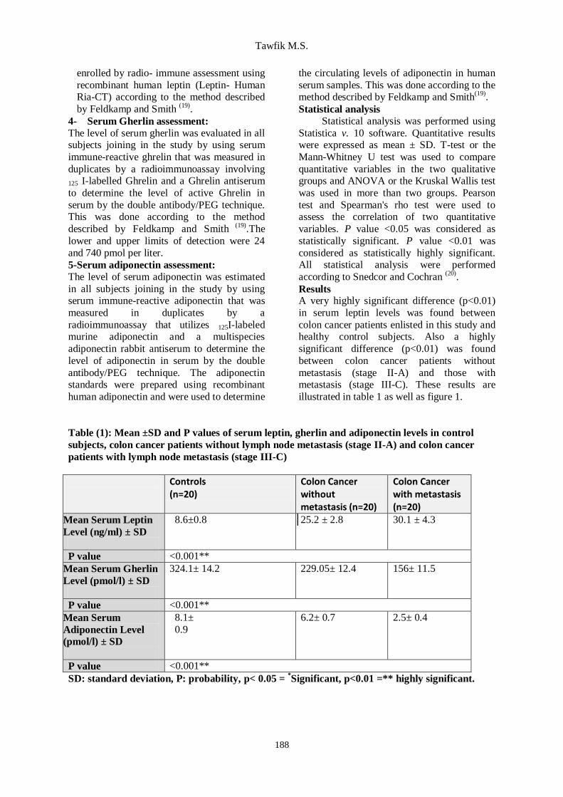
Results A very highly significant difference (p<0.01)
in serum leptin levels was found between
colon cancer patients enlisted in this study and healthy control subjects. Also a highly
significant difference (p<0.01) was found
between colon cancer patients without
metastasis (stage II-A) and those with metastasis (stage III-C). These results are
illustrated in table 1 as well as figure 1.
Table (1): Mean ±SD and P values of serum leptin, gherlin and adiponectin levels in control
subjects, colon cancer patients without lymph node metastasis (stage II-A) and colon cancer
patients with lymph node metastasis (stage III-C)
Controls (n=20)
Colon Cancer without metastasis (n=20)
Colon Cancer with metastasis (n=20)
Mean Serum Leptin
Level (ng/ml) ± SD
8.6±0.8
25.2 ± 2.8 30.1 ± 4.3
P value <0.001**
Mean Serum Gherlin
Level (pmol/l) ± SD
324.1± 14.2
229.05± 12.4
156± 11.5
P value <0.001**
Mean Serum
Adiponectin Level
(pmol/l) ± SD
8.1±
0.9
6.2± 0.7
2.5± 0.4
P value <0.001**
SD: standard deviation, P: probability, p< 0.05 = *Significant, p<0.01 =** highly significant.

Serum Leptin Hormone …
189
Figure 1: Mean ±SD serum leptin levels (ng/ml) in controls, colon cancer patients without lymph
node metastasis (stage II-A) and those with lymph node metastasis (stage III-C).
In addition, a very highly significant difference (p<0.01) in serum gherlin levels was observed
between colon cancer patients enrolled in this study and healthy control subjects. A highly significant
difference (p<0.01) was also found between colon cancer patients without metastasis (stage II-A) and
those with metastasis (stage III-C). These results are illustrated in table 1 as well as figure 2.
Figure 2 : Mean ±SD serum gherlin levels (pmol/l) in controls, colon cancer patients without
lymph node metastasis (stage II-A) and those with metastasis (stage III-C) respectively.
A negative correlation was observed between seum leptin and serum gherlin levels in the two groups of colon cancer patients enrolled in the study. (r = - 0.50, r
2 = 0.25). This correlation is shown
in figure 3.
Controls Without Metastasis
With Metastasis
0
50
100
150
200
250
300
350
400
0
5
10
15
20
25
30
35
40
Without Metastasis With Metastasis
Serum Gherlin Level (pmol/l)
Controls
Serum Leptin Levels (ng/ml)
Controls Without Metastasis
Tawfik M.S.
190
18 20 22 24 26 28 30 32 34 36 38
Serum Leptin (ng/ml)
80
100
120
140
160
180
200
220
240
260
280
300
320
Seru
m G
herl
in (
pm
ol/
l)
Figure 3: Correlation between serum leptin (ng/ml) and ghrelin levels (pmol/l) in colon cancer
patients.
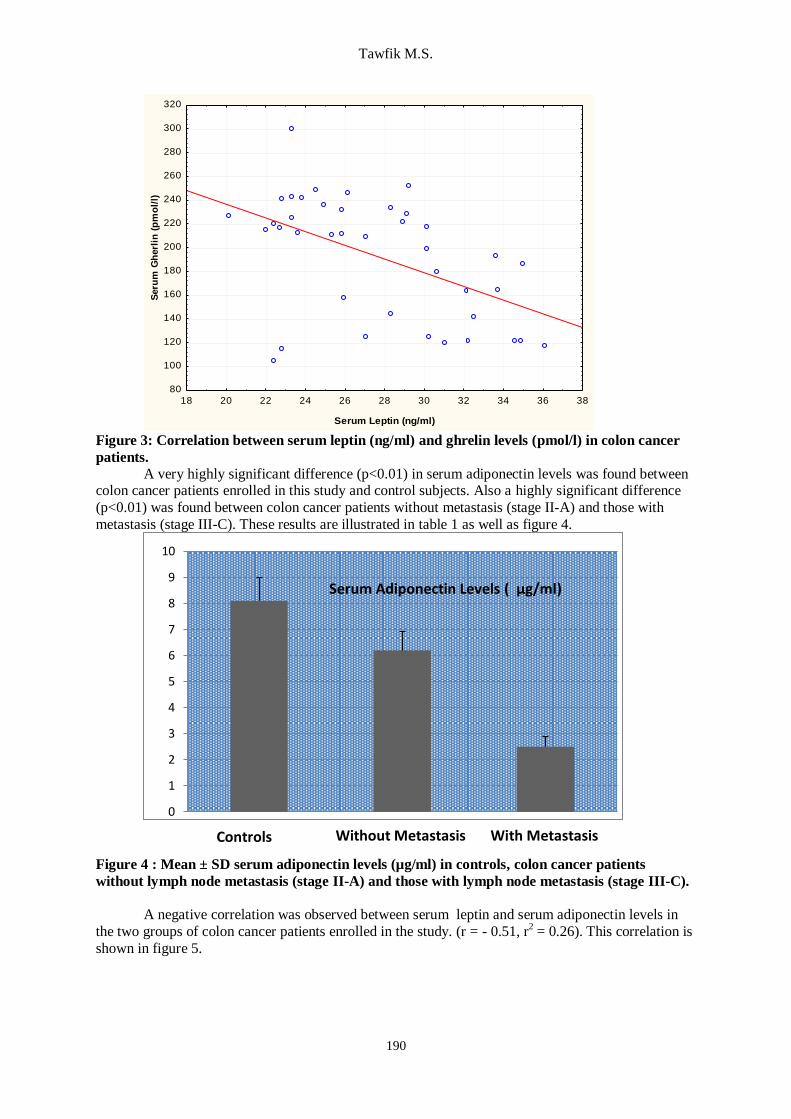
A very highly significant difference (p<0.01) in serum adiponectin levels was found between colon cancer patients enrolled in this study and control subjects. Also a highly significant difference
(p<0.01) was found between colon cancer patients without metastasis (stage II-A) and those with
metastasis (stage III-C). These results are illustrated in table 1 as well as figure 4.
Figure 4 : Mean ± SD serum adiponectin levels (µg/ml) in controls, colon cancer patients
without lymph node metastasis (stage II-A) and those with lymph node metastasis (stage III-C).
A negative correlation was observed between serum leptin and serum adiponectin levels in
the two groups of colon cancer patients enrolled in the study. (r = - 0.51, r2 = 0.26). This correlation is
shown in figure 5.
0
1
2
3
4
5
6
7
8
9
10
Controls Without Metastasis
Serum Adiponectin Levels ( µg/ml)
With Metastasis

Serum Leptin Hormone …
191
18 20 22 24 26 28 30 32 34 36 38
Serum Leptin (ng/ml)
0
1
2
3
4
5
6
7
8
9
Seru
m A
dip
onecti
n (
µg/m
l)
Figure 5: Correlation between serum leptin (ng/ml) and adiponectin levels (µg/ml) in colon
cancer patients.
Discussion Colorectal cancer is considered the
second leading cause of cancer-related deaths
in many countries worldwide including the United States. Early diagnosis,
nevertheless, could in many cases lead to a
better prognosis. Almost all colon cancers start in glands in the lining of the colon and rectum.
However; colon cancer is usually diagnosed at
advanced stages, when it is usually fatal (21)
. Excess body weight, as defined by the
body mass index (BMI), has been linked to
several diseases and includes subjects who are
overweight (BMI≥25-29.9 kg/m2) or obese
(BMI≥30 kg/m2). Around 11% of colorectal
cancer (CRC) cases have been attributed to
overweight and obesity in Europe. Epidemiological data suggest that obesity is
associated with a 30-70% increased risk of
colon cancer in men, whereas the association is less consistent in women. The relative risk
of colorectal cancer of obese patients is about
1.5 times higher than in normal-weight
individuals and obesity is also associated with premalignant colorectal adenoma. Visceral fat,
or abdominal obesity, seems to be of greater
concern than subcutaneous fat obesity, and any1 kg/m
2 increase in BMI confers additional
risk. Obesity might be associated with worse
cancer outcomes, such as recurrence of the
primary cancer or mortality. Several factors, including reduced sensitivity to anti-
angiogenic-therapeutic regimens, might
explain these differences (22)
. Comstock et al (23)
documented that obesity is a key risk factor
for the development of colon cancer in white
obese males.
In the present study, the patients
belonging to the two groups selected (20 colon
cancer patients each) were all obese with a mean BMI of 33.5 ± 3.2.
Aleksandrova et al (24)
estimated the
extent to which biomarkers with inflammatory
and metabolic actions mediate the association of adiposity measures, waist circumference
(WC) and body mass index (BMI), with colon
cancer in men and women. It is hypothesized that obesity is a
chronic low grade inflammatory process that
results in increased secretion of products from adipose tissue that include leptin, interleukin-
6, interleukin-17, tumor necrosis factor-alpha,
and associated decreased blood levels of
ghrelin and adiponectin that appear to have a shielding effect against the development of
several types of cancer, including colon
cancer. These products induce malignancy-related metabolic alterations in colon cancer
cells leading to metabolic syndrome, insulin
resistance and modifications in levels of
adipocytokines that seem to be of great importance
(25).
The leptin hormone can modulate
several important functions of the gastrointestinal tract. It interacts with the
vagus nerve and cholecystokinin to delay
gastric emptying and has a complex effect on motility of the small bowel. Leptin modulates
absorption of macronutrients in the
gastrointestinal tract differentially in
physiologic and pathologic states. In physiologic states, exogenous leptin has been
shown to decrease carbohydrate absorption

Tawfik M.S.
192
and to increase the absorption of small
peptides by the PepT1 di-/tripeptide transporter. In certain pathologic
states, leptin has been shown to increase
absorption of carbohydrates, proteins, and
fat. The hormone has been shown to be upregulated in the colonic mucosa in patients
with inflammatory bowel disease. Leptin
stimulates gut mucosal cell proliferation and inhibits apoptosis. These functions have led to
speculation about the role of leptin in
tumorigenesis in the gastrointestinal tract, which is complicated by the multiple
immunoregulatory effects of this hormone (26)
.
Kemik et al (27)
investigated the inter-
relationship between leptin, associated cytokines and the development of colon cancer
to elaborate these suspected links. The study
found significantly higher serum C- reactive protein (CRP), interleukin 1α (IL-1α), IL-1β,
IL-6, IL-8, IL-10, tumour necrosis factor α
(TNF-α), midkine, vascular endothelial growth factor-A (VEGF-A), VEGF-C, VEGF receptor
1 (VEGFR1) and leptin in colon cancer
patients. These factors are known to promote
chronic inflammatory processes and enhanced cell growth and proliferation together with
increased angiogenesis culminating into
enhanced carcinogenesis. Miyoshi et al
(28) also hypothesized
that leptin plays a pivotal role in the
pathogenesis of colorectal cancer (CRC).
The present study showed a very highly significant statistical increase in serum
leptin levels in colon cancer patients compared
to normal healthy controls (p< 0.001). These results were very similar to those of Guadagni
et al (29)
and Chia et al (30)
who noted that leptin
levels were significantly higher in colon cancer patients compared to healthy control
subjects. Yet, these results were contradictory
to those of Arpaci et al (31)
who found
significantly lower serum levels of leptin in patients with colorectal cancer. They explained
this discrepancy with other studies by
suggesting that the increased weight of colon cancer patients in the earlier stages of the
disease was not the sole factor in determining
their serum leptin levels and that other factors could be involved. Similarly, Kosovva et al
(32)
also speculated that leptin levels in colon
cancer patients were not statistically
significantly different from those in the benign group.
Elevated serum leptin levels have
been incriminated in colon cancer growth and development of metastasis to lymph nodes or
distant metastasis (33)
. Tutino et al (34)
documented that high circulating levels
of leptin receptor occur in patients with advanced stage of colon cancer, suggesting a
role of leptin in cancer progression and
aggressiveness. The results of a study done by Erkasap et al
(35) suggested a role of leptin on
the progression of colon carcinoma to
metastatic disease without weight loss. The present study included two groups
of patients. Group 1 included 20 patients who
were diagnosed as stage II-A colon cancer
(without lymph node involvement or distant metastasis) while group 2 included 20 patients
with stage III-C colon cancer with 4 or more
lymph nodes involved. The selection of these patients was based on the study’s aim to
compare colon cancer patients who did not
have lymph node involvement and those who did as regards serum leptin levels in order to
predict the increased risk of lymph node
metastasis and eventual distant metastasis
using this hormone as a prognostic marker. The results showed that serum leptin levels
were significantly higher in patients with stage
III-C colon cancer compared to stage II-A colon cancer patients (p<0.001). These results
are in agreement with those of Wang et al (36)
who found significantly higher serum leptin
levels in stage III colon cancer patients compared to those belonging to stage II.
Ghrelin is a metabolism-regulating
hormone recently investigated for its role in cancer survival and progression. Moreover,
low ghrelin levels observed in obese people
may be implicated in cancer development and progression. Limited data are currently
available on the effects exerted by ghrelin on
intracellular proteolytic pathways in cancer.
Both the lysosomal and the proteasomal systems are fundamental in cellular
proliferation and apoptosis regulation.
Preliminary in vitro fluorimetric assays have evidenced for the first time a direct inhibition
of 20S proteasomes by ghrelin, particularly
evident for the trypsin-like activity (37)
. Many studies have pointed out a
possible role of gut peptides, such as ghrelin,
in the pathogenesis of gastrointestinal
malignancies including colon cancer which is one of the most common death causes in the
western world (38)
.
Serum Leptin Hormone …
193
The present study showed that there
was a statistically significant difference in serum ghrelin levels of the patients vs. the
controls, with the patients having much lower
levels (p<0.001). These results are confirmed
Legakis et al.(39)
who found that colon cancer patients had significantly lower circulating
levels of ghrelin than healthy controls.
The role of low serum levels of ghrelin in enhancing cell proliferation and apoptosis
has been illustrated, however; studies also
implicate that low levels of ghrelin may influence the cancer cells motility or ability to
metastasize. D'Onghia et al (38)
found that
ghrelin plays a role in protecting against colon
cancer metastasis.
The results of the present study
showed that serum ghrelin levels were significantly lower in patients with stage III-C
colon cancer compared to stage II-A colon
cancer patients (p<0.001) showing a possible protective role for ghrelin against colon cancer
lymph node affection and metastasis.
A negative correlation was observed between serum leptin levels and serum gherlin
levels in the patients enrolled in this study. A
similar study done by Kemik et al (40)
also established a negative correlation between
serum leptin and ghrelin in colon cancer
patients. On the other hand, such a negative
correlation could not be observed in another study carried out by Wolf et al.
(41) who
investigated serum leptin and ghrelin levels in
colon cancer patients. This discrepancy may be attributed to the fact that the patients
enrolled in the latter study were belonging to a
more advanced stage of colon cancer and were suffering from cachexia with disturbed
leptin/ghrelin balance.
Another adipocytokine investigated in
this study was adiponectin. This hormone has some known effects on the metabolic process
such as gluconeogenesis, glucose uptake, lipid
β-oxidation, triglyceride clearance, protection from endothelial dysfunction, insulin
sensitivity and weight loss (42)
.
Recent studies have indicated a significant correlation between the reduced
plasma adiponectin levels (associated with
obesity) and the increased risk of various
cancers. Additionally, these studies have also demonstrated some of the antiangiogenic and
antitumoral effects of adiponectin (43)
.
Endometrial, breast, prostate, colon, pancreatic
cancer, and more recently non-small cell lung cancer and esophageal cancer have been found
to be correlated with low serum adiponectin
levels.
Adiponectin exerts its effects via 5′ adenosine monophosphate-activated protein
kinase (AMPK). Increased concentrations of
adenosine monophosphate (AMP), calcium-dependent kinases and Ser/Thr liver kinase B1
(LKB1) contribute to AMPK activation which
in turn interferes with cellular growth signaling through mammalian target of
rapamycin (mTOR) thus inhibiting the
promotion of carcinogenesis. AMPK also
promotes growth arrest and apoptosis via increased p53 and p21 expression, respectively (44)
. In contrast, the findings of a study done by
Song et al (2013) (45)
supported a positive role for adiponectin in colorectal carcinogenesis in
men. They documented an association of
plasma adiponectin and soluble leptin receptor (sOB-R) with colorectal cancer .
The present study showed that there
was a highly significant statistical difference in
serum adiponectin levels of the colon cancer patients vs. the controls with the patients
having much lower levels (p<0.001). These
results are in accordance with other studies by Gulcelik et al
(46) and Guadagni et al
(9) that
demonstrated significantly lower levels of
adiponectin in colon cancer patients compared
to control subjects. In addition, adiponectin via AMPK
pathway causes inhibition of tumor cell
adhesion and migration in general thus inhibiting the metastasis of many types of
tumours (47)
. The results of the present study
showed that serum adiponectin levels were significantly lower in patients with stage III-C
colon cancer compared to stage II-A colon
cancer patients (p<0.001) showing a possible
sheilding role for adiponectin against colon cancer lymph node affection or metastasis.
These results are in agreement with another
study by Gialamas et al (48)
that demonstrated that serum adiponectin levels and tissue
expression of adiponectin receptors are
associated with risk, stage, and grade of colorectal cancer.
Data of the present work speculated a
negative correlation between serum leptin
levels and serum adiponectin levels in the patients joining in this study. This result is in
accordance with that of Gauadagni et al (7)
who
Tawfik M.S.
194
demonstrated that leptin inversely correlated
with adiponectin in colon cancer patients. The results of the present study
highlighted important alterations occurring in
serum adipocytokines, (leptin and adiponectin)
in addition to the gut peptide ghrelin in colon cancer patients. It could be concluded from the
striking features of this study that serum leptin
levels could serve as a good prognostic marker to predict the clinical outcome of colon cancer
and subsequent occurrence of metastasis in
patients especially as it is the most readily available adipocytokine secreted by
adipocytes. In addition to this, measuring
serum levels of this hormone could also help
clinicians decide whether or not to start aggressive therapy at an earlier stage before
the occurrence of lymph node involvement or
metastasis depending on the predictive value of leptin hormone. Serum ghrelin and
adiponectin hormones are the counterparts of
leptin and their determination in serum could also be used in conjunction with serum leptin
level determination to predict clinical outcome
and prognosis in these patients. However;
further investigations are required to add more information on the relationship between serum
leptin levels and colon cancer staging.
References 1) Kershaw EE and Flier JS (2004): Adipose
tissue as an endocrine organ. J Clin Endocrinol
Metab, 89:2548-2556.
2) Boguszewski CL, Paz-Filho G and Velloso
LA(2010): Neuroendocrine body weight regulation: integration between fat tissue,
gastrointestinal tract, and the brain.
Endokrynol Pol, 61:194-206.
3) Paz-Filho G, Wong ML and Licinio J
(2011): Ten years of leptin replacement
therapy. Obes Rev, 12: e315-23.
4) Paz-Filho G, Lim EL, Wong ML, et al
(2011): Associations between adipocytokines
and obesity-related cancer. Front Biosci
(Landmark Ed), 16:1634-50.
5) Drew JE (2012): Symposium 3: obesity-
related cancers molecular mechanisms linking adipocytokines to obesity-related colon cancer:
focus on leptin. Proc Nutr Soc, 71:175.
6) Padidar S, Farquharson AJ, Williams LM,
et al (2011): Leptin up-regulates pro-
inflammatory cytokines in discrete cells within
mouse colon. J Cell Physiol, 226:2123-30.
7) Cascio S, Ferla R, D’Andrea A, et al (2009): Expression of angiogenic regulators, VEGF
and leptin, is regulated by the
EGF/PI3K/STAT3 pathway in colorectal
cancer cells. J Cell Physiol, 221:189-94.
8) Koda M, Sulkowska M, Kanczuga-Koda L,
et al (2007): Expression of the obesity
hormone leptin and its receptor correlates with
hypoxia-inducible factor-1 alpha in human colorectal cancer. Ann Oncol,18 (6):116-119.
9) Uchiyama T, Takahashi H, Endo H,
Sugiyama M, Sakai E, Hosono K,
Nagashima Y, Inayama Y, Wada K, Hippo
Y and Nakajima A (2011): Role of the long
form leptin receptor and of the STAT3
signaling pathway in colorectal cancer
progression. Int J Oncol, 39(4):935-40.
10) Higgins SC, Gueorguiev M and Korbonits
M (2007): Ghrelin, the peripheral hunger
hormone. Ann Med, 39(2):116-136.
11) Tschöp M, Smiley Dl and Heiman ML (2000): Ghrelin induces adiposity in rodents.
Nature, 407: 908-913.
12) Dixit VD, Schaffer EM, Pyle RS, et al.
(2004): Ghrelin inhibits leptin- and activation-
induced proinflammatory cytokine expression
by human monocytes and T cells. J Clin Invest,
114(1):57-66.
13) Yamauchi T, Kamon J, Ito Y, et al (2003): Cloning of Adiponectin receptors that mediate
antidiabetic metabolic effects. Nature, 423,
762-769.
14) Otake S, Takeda H, Suzuki Y, et al (2005):
Association of visceral fat accumulation and
plasma adiponectin with colorectal adenoma:
evidence for participation of insulin resistance.
Clin Cancer Res, 11, 3642-3646.
15) Wei EK, Giovannucci E, Fuchs CS, et al
(2005): Low plasma adiponectin levels and
risk of colorectal cancer in men: a prospective
study. J Natl Cancer Inst, 97: 1688-1694.
16) Fujisawa T, Endo H, Tomimoto A, et al
(2008): Adiponectin suppresses colorectal
carcinogenesis under the high-fat diet condition. Gut, 57: 1531-1538.
17) American Joint Committee on Cancer
(2010): Colon and rectum. In: Edge SB, Byrd
DR, Comptom CC et al. (Eds.) AJCC Cancer
Staging Handbook, 7th edition, 173-206.
Chicago, IL: Springer.
18) Cheng CK, Chan J, Cembrowski GS and
van Assendelft OW (2004): Complete blood
count reference interval diagrams derived from
NHANES III: stratification by age, sex, and
race. Lab Hematol, 10(1):42-53.
19) Feldkamp CS and Smith SW (1987): Practical guide to immunoassay evaluation. In:
Immunoassay: A Practical Guide, Chan DW,
editor. Orlando, FL, Academic Press, 49-95.
20) Snedecor, GW and Cochran, WG (1994). Statistical Methods. 8th Edn. Iowa State
University Press. Ames, Iowa.

Serum Leptin Hormone …
195
21) Smith RA, Cokkinides V and Brawley
OW(2012): Cancer screening in the United
States, a review of current American Cancer
Society guidelines and current issues in cancer
screening. CA Cancer J Clin, 62:129-142.
22) Bardou M1, Barkun AN, Martel M (2013) : Obesity and colorectal cancer. Postgrad Med J,
89(1055):519-33.
23) Comstock SS, Hortos K, Kovan B,
McCaskey S, Pathak DR, Fenton JI (2014): Adipocytokines and obesity are associated
with colorectal polyps in adult males: a cross-
sectional study. PLoS One, 9(1 ):e85939.
24) Aleksandrova K, Drogan D, Boeing H, et al
(2014): Adiposity, mediating biomarkers and
risk of colon cancer in the European
prospective investigation into cancer and nutrition study. Int J Cancer, 134(3):612-621.
25) Chen J and Iverson D (2012): Estrogen in
obesity-associated colon cancer: friend or foe?
Protecting postmenopausal women but
promoting late-stage colon cancer. Cancer
Causes Control, 23(11):1767-1773.
26) Yarandi SS, Hebbar G, Sauer CG, Cole CR
and Ziegler TR(2011): Diverse roles of
leptin in the gastrointestinal tract: modulation
of motility, absorption, growth, and
inflammation. Nutrition, 2011 Mar;27(3):269-275.
27) Kemik O, Kemik AS, Begenik H, Erdur
FM, Emre H, Sumer A, Purisa S, Tuzun S
and Kotan C (2012): The relationship among
acute-phase response proteins, cytokines, and
hormones in various gastrointestinal cancer
types patients with cachectic. Hum Exp
Toxicol, 31(2):117-25
28) Miyoshi H, Morishita A, Tani J, Sakamoto
T, Fujita K, Katsura A, Tatsuta M,Nomura
T, Yoneyama H, Iwama H, Suzuki Y and
Masaki T (2014): Expression profiles of 507 proteins from a biotin label-based antibody
array in human colorectal cancer. Oncol
Rep,31(3):1277-1281.
29) Guadagni F, Roselli M, Martini F, Spila A,
Riondino S, D'Alessandro R, Del Monte G,
Formica V, Laudisi A, Portarena I,
Palmirotta R and Ferroni P (2009): Prognostic significance of serum
adipocytokine levels in colorectal cancer
patients. Anticancer Res, 29(8):3321-3327.
30) Chia VM, Newcomb PA, Lampe JW, White
E, Mandelson MT, McTiernan A and Potter
JD (2007): Leptin concentrations, leptin
receptor polymorphisms, and colorectal
adenoma risk. Cancer Epidemiol Biomarkers
Prev, 16(12):2697-2703.
31) Arpaci F, Yilmaz MI, Ozet A, Ayta H,
Ozturk B, Komurcu S, Ozata M (2002):
Low serum leptin level in colon cancer patients
without significant weight loss. Tumori,
88(2):147-9.
32) Kosova F, Coskun T, Kaya Y, Kara E and
Ari Z (2013): Adipocytokine levels of colon
cancer patients before and after treatment.
Bratisl Lek Listy,114(7):394-397.
33) Wang D, Chen J, Guo F, Chen H, Duan Z,
Wei MY, Xu QM, Wang LH and Zhong
MZ (2011): Clinical significance of mTOR
and p-mTOR protein expression in human
colorectal carcinomas. Asian Pac J Cancer
Prev, 12(10):2581-2584.
34) Tutino V, Notarnicola M, Guerra V,
Lorusso D, Caruso MG(2012): Increased
soluble leptin receptor levels are associated
with advanced tumor stage in colorectal cancer
patients. Anticancer Res, 31(10):3381-3383.
35) Erkasap N, Ozkurt M, Erkasap S, Yasar F,
Uzuner K, Ihtiyar E, Uslu S, Kara M and
Bolluk O(2013): Leptin receptor (Ob-R)
mRNA expression and serum leptin
concentration in patients with colorectal and
metastatic colorectal cancer. Braz J Med Biol
Res, 46(3):306-310.
36) Wang D, Chen J, Chen H, Duan Z, Xu Q,
Wei M, Wang L, Zhong M (2012): Leptin
regulates proliferation and apoptosis of
colorectal carcinoma through PI3K/Akt/mTOR
signalling pathway. J Biosci, 37(1):91-101.
37) Bonfili L, Cuccioloni M, Cecarini V,
Mozzicafreddo M, Palermo FA, Cocci P,
Angeletti M and Eleuteri AM (2013): Ghrelin induces apoptosis in colon
adenocarcinoma cells via proteasome
inhibition and autophagy induction. Apoptosis,
18(10):1188-1200.
38) D'Onghia V, Leoncini R, Carli R, Santoro
A, Giglioni S, Sorbellini F, Marzocca G,
Bernini A, Campagna S, Marinello E and
Vannoni D (2007): Circulating gastrin and
ghrelin levels in patients with colorectal cancer: correlation with tumour stage,
Helicobacter pylori infection and BMI.
Biomed Pharmacother, 61(2-3):137-141.
39) Legakis I, Stathopoulos J, Matzouridis T
and Stathopoulos GP (2009): Decreased
plasma ghrelin levels in patients with advanced
cancer and weight loss in comparison to
healthy individuals. Anticancer Res,
29(10):3949-52.
40) Kemik O, Sumer A, Kemik AS, Hasirci I,
Purisa S, Dulger AC, Demiriz B and Tuzun S (2010): The relationship among acute-phase
response proteins, cytokines and hormones in
cachectic patients with colon cancer. World J
Surg Oncol, 8:85.
41) Wolf I, Sadetzki S, Kanety H, Kundel Y,
Pariente C, Epstein N, Oberman B, Catane
R, Kaufman B and Shimon I (2006): Adiponectin, ghrelin, and leptin in cancer
Tawfik M.S.
196
42) cachexia in breast and colon cancer patients.
Cancer, 106(4): 966-973.
43) Takahashi M, Arita Y, Yamagata K, et al
(2000): Genomic structure and mutations in
adipose-specific gene, Adiponectin. Int J Obes
Relat Metab Disord, 24(7):861-868.
44) Kumor A, Daniel P, Pietruczuk M, et al
(2009): Serum leptin, Adiponectin, and resistin
concentration in colorectal adenoma and
carcinoma (CC) patients. Int J Colorectal Dis,
24: 275–278.
45) Shackelford DB and Shaw RJ (2009): The
LKB1-AMPK pathway: metabolism and
growth control in tumour suppression. Nat Rev
Cancer, 9(8):563-575.
46) Song M, Zhang X, Wu K, Ogino S, Fuchs
CS, Giovannucci EL, Chan AT. Plasma
adiponectin and soluble leptin receptor and risk
of colorectal cancer: a prospective study.
Cancer Prev Res (Phila), 6(9):875-85.
47) Gulcelik MA, Colakoglu K, Dincer H,
Dogan L, Yenidogan E, and Gulcelik NE
(2012): Associations between Adiponectin and
two different cancers: breast and colon. Asian Pac J Cancer Prev, 13(1):395-398.
48) Luo Z, Saha AK, Xiang X and Ruderman
NB (2005): AMPK, the metabolic syndrome
and cancer. Trends Pharmacol Sci, 26(2):69-
76.
49) Gialamas SP, Petridou ET, Tseleni-
Balafouta S, Spyridopoulos TN, Matsoukis
IL, Kondi-Pafiti A, Zografos G and
Mantzoros CS (2011): Serum Adiponectin
levels and tissue expression of adiponectin
receptors are associated with risk, stage, and
grade of colorectal cancer. Metabolism, 60(11):1530-1538.