serologic and urinary characteristics of laboratory
TRANSCRIPT

So and Villanueva BMC Urol (2021) 21:125 https://doi.org/10.1186/s12894-021-00888-3
RESEARCH
Serologic and urinary characteristics of laboratory-confirmed genitourinary tuberculosis at a tertiary hospital in the PhilippinesPaolo Nikolai H. So* and Anthony Russell T. Villanueva
Abstract
Background: Genitourinary tuberculosis (GUTB) is known to cause high rates of structural organ damage, however, literature on its biochemical manifestations is limited. Additionally, local studies in the Philippine setting, where cases are rampant, are few and dated. This study aimed to determine the serologic and urinary profile of patients with GUTB admitted at a tertiary hospital within January 2009 to March 2020 and their association with short-term outcomes.
Methods: This retrospective study included 112 patients with laboratory-confirmed GUTB (i.e., positivity in acid-fast smear, polymerase chain reaction, culture, or histology). Demographic data, clinical characteristics, laboratory and radiologic findings, histopathology reports, treatment, and short-term outcomes were recorded.
Results: Bladder (54.5%) and kidney (36.4%) were the most affected organs. The male:female ratio was 1:1.15, and the mean age was 35.79 ± 18.29 years. Weakness (14.29%) was the most common chief complaint. A majority pre-sented with anemia (83.04%), while several had leukocytosis (41.96%) and thrombocytosis (26.79%). Hypoalbumine-mia (58.10%), impairment of renal function (36.94%), and electrolyte abnormalities such as hyponatremia (50.93%), hypercalcemia (20.19%), and hypokalemia (21.82%) were common. Proteinuria (67.96%) and pyuria (67.96%) were the most frequent abnormal findings, followed by hematuria (51.46%), acidic urine (45.63%) and low specific grav-ity (31.07%). Age, leukocytosis, and the need for pressors were all significantly associated with mortality (p values of <0.001, 0.010, and <0.001, respectively).
Conclusions: The young age at presentation with severe clinical and laboratory manifestations may reflect local epidemiology as TB continues to be widespread in the country. Apart from the more commonly cited abnormali-ties in literature, multiple electrolyte imbalances and urinary concentration defects were also observed in many cases, possibly indicating tubulointerstitial involvement—a complication increasingly mentioned in case reports. As several patient characteristics were found to be associated with the high mortality rates observed in the study, further research is recommended to explore predictive modeling.
Keywords: Genitourinary tuberculosis, Electrolytes, Urinalysis, Association
© The Author(s) 2021. Open Access This article is licensed under a Creative Commons Attribution 4.0 International License, which permits use, sharing, adaptation, distribution and reproduction in any medium or format, as long as you give appropriate credit to the original author(s) and the source, provide a link to the Creative Commons licence, and indicate if changes were made. The images or other third party material in this article are included in the article’s Creative Commons licence, unless indicated otherwise in a credit line to the material. If material is not included in the article’s Creative Commons licence and your intended use is not permitted by statutory regulation or exceeds the permitted use, you will need to obtain permission directly from the copyright holder. To view a copy of this licence, visit http:// creat iveco mmons. org/ licen ses/ by/4. 0/. The Creative Commons Public Domain Dedication waiver (http:// creat iveco mmons. org/ publi cdoma in/ zero/1. 0/) applies to the data made available in this article, unless otherwise stated in a credit line to the data.
IntroductionTuberculosis (TB) remains an important global epi-demic, with latest estimates of disease burden amount-ing to 10.0 (range, 9.0–11.1) million people in 2018 [1]. Extrapulmonary TB (EPTB) is reported to comprise
Open Access
*Correspondence: [email protected] of Nephrology, Department of Medicine, University of the Philippines Manila – Philippine General Hospital, Taft Avenue, Ermita, 1000 Manila, Philippines

Page 2 of 34So and Villanueva BMC Urol (2021) 21:125
around 16.5–25% of all cases, attributing 4.5–27% to genitourinary TB (GUTB) [2–4]. GUTB histori-cally pertains to the infection of the urogenital system organs in any combination by Mycobacterium tuber-culosis (MTB) or Mycobacterium bovis [5–8]. It pre-sents with insidious and late-onset symptoms, making its diagnosis and treatment difficult and delayed, and consequently leading to high rates of structural organ damage and kidney failure [9, 10]. Some physicians advocate the term urogenital TB (UGTB) as kidney TB is the most relevant infection and is more frequently diagnosed than genital TB [5, 6, 11].
Philippines ranked 4th among the countries with high TB burden in 2018, accounting 6% of the global total. It has a TB incidence rate of 554 (311–866) per 100,000 population, and a mortality rate of 24.57 (20–32) per 100,000 population [1]. This high disease prevalence in the country is multifactorial, attributing to high poverty rate, marked social inequities, and rise in slum housing and crowded living conditions from rapid urbanization [12]. In the Filipino pediatric population, GUTB is found to cause 3% of extrapulmonary TB cases admitted in a tertiary hospital [13].
Despite these figures, there is paucity in literature regarding local experience on GUTB, especially with regards to serologic and urinary findings [14–16]. The last available reference was published 25 years ago, which also needs to be updated. The study aims to determine the serologic and urinary profile of patients with geni-tourinary TB admitted at a tertiary government hospi-tal in Philippines and their association with short-term outcomes.
MethodsStudy design and populationThis is a single-center, retrospective study performed at the Philippine General Hospital (PGH; 1500 beds). This study included GUTB patients diagnosed from January 2009 to March 2020 through positivity in at least one of the following: (1) urine acid-fast bacilli (AFB) staining, (2) urine or tissue polymerase chain reaction (PCR) for Mycobacterium tuberculosis, (3) urine or tissue M. tuber-culosis culture, and (4) histologic findings of granuloma-tous inflammation (granulomas composed of epithelioid cells and Langhans giant cells with or without case-ous necrosis) [14, 15, 17–19]. Culture-positive samples solely involving the female genital tract without urologic involvement, out-patients, in-patients without base-line serologic and urinary tests, and those who got dis-charged against medical advice were excluded. This study
was approved by the University of the Philippines Manila Research Ethics Board and the requirement for informed consent was waived since the investigators evaluated anonymized data.
Data collection and definition of variablesData collection was done through chart review of patient medical records. This included patient demograph-ics; comorbidities; clinical symptoms; complete blood counts; serum chemistries; urinalysis; results of microbial smears and cultures, diagnostics, and histopathology; treatment; and short-term outcomes. Organ involvement was distinguished in those with tissue samples obtained from biopsies or operations.
Serologic abnormalities were defined as follows: (1) anemia as hemoglobin of < 150 g/L in neonates 0–30 days old, < 105 g/L in 1–23 months of age, < 115 g/L in chil-dren 2–9 years of age, < 125 g/L in males 10–17 years of age, < 120 g/L in non-pregnant females 10 years of age and above, < 110 g/L in pregnant women, and < 130 g/L in men 15 years of age and above [20, 21]; (2) thrombo-cytopenia as platelets < 84,000/µL in newborns ≤ 1 week old, and < 150,000/µL for the rest of the age groups [21, 22]; (3) thrombocytosis as platelets > 450,000/µL [22]; (4) leukocytosis as white blood cells > 34,000/µL in neo-nates 0–30 days, > 14,000/µL in infants 1–23 months of age, > 12,000/µL in 2–9 years of age, > 10,500/µL in 10–18 years of age, and > 11,000/µL in adults [21, 23]; (5) leukopenia as white blood cells < 9100/µL in neo-nates 0–30 days, < 6000/µL in infants 1–23 months of age, < 4000/µL in 2–18 years of age, and < 4400/µL in adults [21, 23]; (6) hypoalbuminemia as serum albumin < 18 g/L in premature neonates 1 day old, < 25 g/L in full term neonates < 6 days old, < 19 g/L in 8 days-1 year old, < 34 g/L in 1–3 years of age, < 35 g/L in 4–19 years of age, and < 34 g/L in adults [21]; (7) impaired renal function as estimated glomerular filtration rate < 60 mL/min/1.73 m [2]; (8) hyperkalemia as plasma K+ concen-tration≥ 5.5 mM [22], (2) hypokalemia as plasma K+ con-centration < 3.5 mM [22], (9) hyponatremia as plasma Na+ concentration < 135 mM [22], and (10) hypercalcemia as total serum calcium concentration ≥ 10.4 mg/dL [24].
Urinary findings were defined as: (1) acidic urine as urine pH ≤ 5.5 [24], (2) low specific gravity as urine specific gravity ≤ 1.010, (3) hematuria as three or more erythrocytes per high-power field [24], (4) proteinuria as detection of proteinuria by dipstick examination [22], and (5) pyuria as detection of more than 5 white blood cells per high-power field in urine microscopy or positive leukocyte esterase dipstick testing [15, 24].

Page 3 of 34So and Villanueva BMC Urol (2021) 21:125
Fig. 1 Flow diagram of study selection. DAMA discharged against medical advice
Table 1 Proportion of patients with GUTB
Diagnosis No. of patients (%) (n = 112)
Urine AFB smear-positive 56 (50.00%)
Urine PCR-positive 16 (14.29%)
Urine culture-confirmed 18 (16.07%)
Histopathology 22 (19.64%)
Bladder 12
Kidney 6
Kidney and ureter 2
Ureter 2
Table 2 Laterality of organ involvement in GUTB patients confirmed by histopathology
Organ No. of patients (%) (n = 10)
Right Left Unspecified
Kidney 1 4 1
Kidney and ureter 0 2 0
Ureter 1 0 1
Total 2 (20.0%) 6 (60.0%) 2 (20.0%)
Table 3 Clinical characteristics of patients with GUTB
Clinical characteristics No. of patients (%) (n = 112)
Gender
M 52 (46.43%)
F 60 (53.57%)
Age
0 months–1 year 1 (0.89%)
1 year–5 years 2 (1.79%)
6 years–10 years 4 (3.57%)
11 years–18 years 18 (16.07%)
19 years–29 years 21 (18.75%)
30 years–49 years 35 (31.25%)
50 years–69 years 27 (24.11%)
70 years or older 4 (3.57%)
Marital status
Single/widowed 69 (61.61%)
Married 43 (38.39%)
Occupation
Employed 19 (16.96%)
Unemployed 58 (51.79%)
Not applicable (i.e., pediatric) 26 (23.21%)
Unspecified 9 (8.04%)
Location
Urban 60 (53.57%)
Rural 48 (42.86%)
Unspecified 4 (3.57%)
Co-morbidity
Previous TB 14 (12.50%)
Diabetes mellitus 5 (4.46%)
HIV/AIDS 12 (10.71%)
Steroid use (e.g., SLE, NS) 9 (8.04%)
Malignancy 1 (0.89%)
Chronic kidney disease 5 (4.46%)
History of urolithiasis 8 (7.14%)
RTA Type 1 1 (0.89%)
COPD 2 (1.79%)
Bronchial asthma 2 (1.79%)
Hypertension 13 (11.61%)
Heart failure 2 (1.79%)
Cerebrovascular disease 1 (0.89%)
Other organ involvement 64 (57.14%)
Pulmonary 57 (50.89%)
Gastrointestinal 28 (25.00%)
Abdominopelvic 11 (9.82%)
Central nervous system 7 (6.25%)
Bone 4 (3.57%)
Lymph node 8 (7.14%)
Ear 2 (1.79%)
Psoas 2 (1.79%)
Cutaneous/wound 3 (2.68%)
Statistical analysisDescriptive statistics were used in the analysis of this study. Frequency and percentage were used to describe categorical variables and proportions of patients who improved, expired, or developed short-term outcomes such as the need for pressors or renal replacement ther-apy. Continuous variables were expressed as median.
Associations were determined by bivariate analy-sis using Fisher’s exact test for characteristics involv-ing 2 categories or Chi-square test for those with > 2

Page 4 of 34So and Villanueva BMC Urol (2021) 21:125
and kidney (n = 8, 36.4%) were the most involved geni-tourinary organs. Among those with kidney and ureter involvement, left laterality was observed in 60% (Table 2).
Patient characteristicsBaseline clinical characteristics of the patients with GUTB are shown in Table 3. The mean age (± SD) was 35.79 ± 18.29 years (range, 1–82 years) and the male-to-female ratio was 1:1.15 (52:60). Most patients were single or widowed (61.61%) and lived in urban areas (53.57%). Fourteen patients (12.5%) had a previous history of tuberculosis, while 64 patients (57.14%) had present
categories. Mean lengths of hospital stay between those with and without the identified clinical, serologic, and urinary characteristics were compared using Mann–Whitney U-test for characteristics involving 2 categories or Kruskal–Wallis test for those with > 2 categories. For all tests, p value of at most 0.05 indicate significance.
ResultsA total of 228 patients with laboratory-confirmed GUTB were identified. Ninety-six charts were irretrievable due to institutional limitations in records retention, while 20 cases met the exclusion criteria (Fig. 1). Among the 112 patients included in the study, half (50.0%) had posi-tive smears for urine AFB (Table 1). In those with histo-pathologic evidence of infection, bladder (n = 12, 54.5%)
Table 4 Serologic characteristics of patients with GUTB
Serologic abnormalities No. of patients (%)
Anemia 93 (83.04%, n = 112)
Thrombocytopenia 6 (5.36%, n = 112)
Thrombocytosis 30 (26.79%, n = 112)
Leukocytosis 47 (41.96%, n = 112)
Leukopenia 6 (5.36%, n = 112)
Hypoalbuminemia 61 (58.10%, n = 105)
Renal function impairment 41 (36.94%, n = 111)
Hyperkalemia 9 (8.18%, n = 110)
Hypokalemia 24 (21.82%, n = 110)
Hyponatremia 55 (50.93%, n = 108)
Hypercalcemia 21 (20.19%, n = 104)
COPD chronic obstructive pulmonary disease, F female, HIV/AIDS human immunodeficiency virus or acquired immunodeficiency syndrome, M male, NS nephrotic syndrome, RTA Type 1 renal tubular acidosis Type 1, SLE systemic lupus erythematosus, TB tuberculosis
Table 3 (continued)
Clinical characteristics No. of patients (%) (n = 112)
Chief complaint
Weakness 16 (14.29%)
Difficulty of breathing 14 (12.50%)
Flank pain 13 (11.61%)
Abdominal pain 11(9.82%)
Hematuria 9 (8.04%)
Dysuria 9 (8.04%)
Fever 8 (7.14%)
Abdominal/pelvic mass on diagnostic 5 (4.46%)
Pedal edema 4 (3.57%)
Umbilical discharge 3 (2.68%)
Gluteal pain 2 (1.79%)
Vaginal bleeding 2 (1.79%)
Cough 2 (1.79%)
Seizure 2 (1.79%)
Urinary retention 1 (0.89%)
Inguinal pain 1 (0.89%)
Fistula formation (ureterocutaneous) 1 (0.89%)
Scrotal discharge 1 (0.89%)
Double J stent reinsertion 1 (0.89%)
Decrease in sensorium 1 (0.89%)
Vomiting 1 (0.89%)
Others 5 (4.46%)
Table 5 Urinary characteristics of patients with GUTB
Urinary abnormalities No. of patients (%) (n = 103)
Acidic pH 47 (45.63%)
Low specific gravity 32 (31.07%)
Proteinuria 70 (67.96%)
Negative 33 (32.04%)
Trace 15 (14.56%)
1+ 36 (34.95%)
2+ 16 (15.53%)
3+ 3 (2.91%)
Hematuria 53 (51.46%)
Pyuria 70 (67.96%)
Pyuria + hematuria 50 (48.54%)
Casts 17 (16.50%)
Crystals 4 (3.88%)

Page 5 of 34So and Villanueva BMC Urol (2021) 21:125
Table 6 Imaging findings associated with GUTB
Intravenous pyelography
Unilateral renal parenchymal diseaseNon-functioning kidneyCalcification of the urinary tract: medullary nephrocalcinosis, nephrolithiasis, ureterolithiasis, cystolithiasisBladder wall thickening
CT scan
Hypodense renal foci with or without internal septations or peripheral calcificationsRenal cysts, Bosniak I and IIRenal massCalcification of the urinary tract: nephrolithiasis, ureteropelvic junction lithiasesUrinary tract dilatation: hydronephrosis, ureteropelvocaliectasia with possible distal ureteral strictureUreteral wall thickeningBladder wall thickeningVesicocutaneous fistulous tractEvidence of extra-renal TB infection: Pulmonary tuberculosis with or without endobrochial spread Distal ileal and ileocecal wall thickening with multiple abscess formation (intraabdominal, pelvic, and prostatic regions) and lymphadenopathies Multilevel vertebral lesions with disc destruction (Pott’s disease) with abscess formation involving adjacent muscles (psoas, iliopsoas and gluteus maximus)
Ultrasound
Unilateral or bilateral renal parenchymal disease with or without signs of chronicityEchogenic renal walls with or without internal echoes suggestive of pyelitis or pyelonephritisPyonephrosisRenal cysts or massCalcification of the urinary tract: nephrocalcinosis, nonspecific parenchymal/perinephric/periureteral calcifications, nephrolithiasis, urolithiases,Urinary tract dilatation: hydronephrosis, focal caliectasia, pelvocaliectasia, ureteropelvocaliectasiaIrregular, diffuse, or heterogeneous bladder wall thickeningBladder wall foci or massEvidence of abdominopelvic Koch’s infection: tobacco pouch appearance of fallopian tube, thickening of uterine serosa and peritoneum, palisading
bowel loops, and massive ascites
Table 7 Management of admitted patients with GUTB
Intervention No. of patients (%) (n = 112)
Anti-Koch’s therapy 71 (63.39%)
Operation 29 (25.89%)
Percutaneous tube nephrostomy 10 (8.93%)
Double J stent insertion 11 (9.82%)
Nephrectomy 2 (1.79%)
Subcapsular nephrectomy 5 (4.46%)
Cytoreductive nephrectomy 1 (0.89%)
Aspiration of renal abscess 1 (0.89%)
Radial nephrolithotomy 1 (0.89%)
Pelvolithotomy 1 (0.89%)
Ureterotomy 1 (0.89%)
Ureteroneocystostomy 1 (0.89%)
Bladder mass excision 3 (2.68%)
Transurethral resection of bladder tumor 1 (0.89%)
Table 8 Short-term outcomes of admitted patients with GUTB
Length of hospital stay in days, median (min–max) 11 (0.67–90)
Improved, n (%) 103 (91.96%)
Expired, n (%) 9 (8.04%)
Need for pressors, n (%) 15 (13.39%)
Need for renal replacement therapy, n (%) 1 (0.89%)
evidence of other organ involvement, with lungs (50.89%) being the most concomitantly involved organ. Twenty-four patients (21.43%) exhibited systemic symptoms such as weakness (14.29%) and fever (7.14%), while 57 patients (50.89%) had genitourinary manifestations as their chief complaint. Flank or abdominal pain was the most com-mon presenting genitourinary symptom (21.43%).

Page 6 of 34So and Villanueva BMC Urol (2021) 21:125
Table 9 Bivariate analysis: clinical characteristics and mortality
Characteristic Category Mortality p value
Survived Expired
Count Row % Count Row %
Gender Male 48 92.3 4 7.7 1.000
Female 55 91.7 5 8.3
Age 0 months to 1 year 0 0.0 1 100.0 <0.001
1–5 years 0 0.0 2 100.0
6–10 years 4 100.0 0 0.0
11–18 years 17 94.4 1 5.6
19–29 years 19 90.5 2 9.5
30–49 years 33 94.3 2 5.7
50–69 years 26 96.3 1 3.7
≥ 70 years 4 100.0 0 0.0
Marital status Single/widowed 63 91.3 6 8.7 1.000
Married 40 93.0 3 7.0
Occupation Employed 18 94.7 1 5.3 0.394
Unemployed 54 93.1 4 6.9
Not applicable 22 84.6 4 15.4
Unspecified 9 100.0 0 0.0
Location City 55 91.7 5 8.3 0.834
Province 44 91.7 4 8.3
Unspecified 4 100.0 0 0.0
Co-morbidity Yes 50 94.3 3 5.7 0.495
No 53 89.8 6 10.2
Diabetes mellitus Yes 5 100.0 0 0.0 1.000
No 98 91.6 9 8.4
Hypertension Yes 13 100.0 0 0.0 0.595
No 90 90.9 9 9.1
Chronic kidney disease Yes 4 80.0 1 20.0 0.347
No 99 92.5 8 7.5
History of urolithiasis Yes 8 100.0 0 0.0 1.000
No 95 91.3 9 8.7
Malignancy Yes 1 100.0 0 0.0 1.000
No 102 91.9 9 8.1
HIV/AIDS Yes 10 83.3 2 16.7 0.247
No 93 93.0 7 7.0
Steroid use Yes 9 100.0 0 0.0 1.000
No 94 91.3 9 8.7
Systemic lupus erythematosus Yes 8 100.0 0 0.0 1.000
No 95 91.3 9 8.7
Nephrotic syndrome Yes 1 100.0 0 0.0 1.000
No 102 91.9 9 8.1
Cerebrovascular disease Yes 1 100.0 0 0.0 1.000
No 102 91.9 9 8.1
Coronary artery disease No 103 92.0 9 8.0 No test*
Heart failure Yes 2 100.0 0 0.0 1.000
No 101 91.8 9 8.2
COPD Yes 1 100.0 0 0.0 1.000
No 102 91.9 9 8.1
Bronchial asthma Yes 2 100.0 0 0.0 1.000
No 101 91.8 9 8.2

Page 7 of 34So and Villanueva BMC Urol (2021) 21:125
Table 9 (continued)
Characteristic Category Mortality p value
Survived Expired
Count Row % Count Row %
Spina bifida No 103 92.0 9 8.0 No test*
RTA Type 1 Yes 1 100.0 0 0.0 1.000
No 102 91.9 9 8.1
Previous TB Yes 14 100.0 0 0.0 0.599
No 89 90.8 9 9.2
Other organ involvement of TB Yes 57 89.1 7 10.9 0.296
No 46 95.8 2 4.2
Pulmonary TB Yes 50 87.7 7 12.3 0.162
No 53 96.4 2 3.6
Gastrointestinal TB Yes 24 85.7 4 14.3 0.224
No 79 94.0 5 6.0
Abdominopelvic TB Yes 11 100.0 0 0.0 0.595
No 92 91.1 9 8.9
CNS TB Yes 6 85.7 1 14.3 0.453
No 97 92.4 8 7.6
Bone TB Yes 3 75.0 1 25.0 0.288
No 100 92.6 8 7.4
Cutaneous/Wound TB Yes 3 100.0 0 0.0 1.000
No 100 91.7 9 8.3
TB Adenitis Yes 7 87.5 1 12.5 0.500
No 96 92.3 8 7.7
Ear TB Yes 1 50.0 1 50.0 0.155
No 102 92.7 8 7.3
Psoas TB Yes 1 50.0 1 50.0 0.155
No 102 92.7 8 7.3
Dysuria Yes 7 100.0 0 0.0 1.000
No 82 90.1 9 9.9
Hematuria Yes 7 77.8 2 22.2 0.192
No 82 92.1 7 7.9
Urinary retention Yes 1 100.0 0 0.0 1.000
No 88 90.7 9 9.3
Flank pain Yes 11 100.0 0 0.0 0.592
No 78 89.7 9 10.3
Inguinal pain Yes 1 100.0 0 0.0 1.000
No 88 90.7 9 9.3
Umbilical discharge Yes 3 100.0 0 0.0 1.000
No 86 90.5 9 9.5
Fistula (uretero-cutaneous fistula) Yes 1 100.0 0 0.0 1.000
No 88 90.7 9 9.3
Scrotal discharge Yes 1 100.0 0 0.0 1.000
No 88 90.7 9 9.3
Double J stent reinsertion Yes 1 100.0 0 0.0 1.000
No 88 90.7 9 9.3
Pedal edema Yes 4 100.0 0 0.0 1.000
No 85 90.4 9 9.6
Fever Yes 7 100.0 0 0.0 1.000
No 82 90.1 9 9.9

Page 8 of 34So and Villanueva BMC Urol (2021) 21:125
Table 10 Bivariate analysis: serologic characteristics and mortality
Bold value indicates statistically significant differences
Characteristics Category Mortality p value
Survived Expired
Count Row % Count Row %
Anemia Yes 84 90.3 9 9.7 0.353
No 19 100.0 0 0.0
Thrombocytopenia Yes 6 100.0 0 0.0 1.000
No 97 91.5 9 8.5
Thrombocytosis Yes 28 93.3 2 6.7 1.000
No 75 91.5 7 8.5
Leukocytosis Yes 47 100.0 0 0.0 0.010
No 56 86.2 9 13.8
Leukopenia Yes 5 83.3 1 16.7 0.402
No 98 92.5 8 7.5
Hypoalbuminemia Yes 54 88.5 7 11.5 0.298
No 42 95.5 2 4.5
Renal function impairment Yes 39 95.1 2 4.9 0.481
No 63 90.0 7 10.0
Hyperkalemia Yes 8 88.9 1 11.1 0.550
No 93 92.1 8 7.9
Hypokalemia Yes 23 95.8 1 4.2 0.681
No 78 90.7 8 9.3
Hyponatremia Yes 48 87.3 7 12.7 0.162
No 51 96.2 2 3.8
Hypercalcemia Yes 19 90.5 2 9.5 1.000
No 76 91.6 7 8.4
Table 9 (continued)
Characteristic Category Mortality p value
Survived Expired
Count Row % Count Row %
Weakness Yes 14 93.3 1 6.7 1.000
No 75 90.4 8 9.6
Abdominal pain Yes 6 75.0 2 25.0 0.157
No 83 92.2 7 7.8
Abdominal/pelvic mass on diagnostic Yes 4 100.0 0 0.0 1.000
No 85 90.4 9 9.6
Vaginal bleeding Yes 2 100.0 0 0.0 1.000
No 87 90.6 9 9.4
Difficulty of breathing Yes 10 83.3 2 16.7 0.303
No 79 91.9 7 8.1
Others Yes 9 81.8 2 18.2 0.266
No 80 92.0 7 8.0
Bold values indicate statistically significant differences
*No test was done since all patients were classified under the No category
COPD chronic obstructive pulmonary disease, HIV/AIDS human immunodeficiency virus or acquired immunodeficiency syndrome, RTA Type 1 renal tubular acidosis Type 1, TB tuberculosis, y year
Page 9 of 34So and Villanueva BMC Urol (2021) 21:125
Table 11 Bivariate analysis: urinary characteristics and mortality
Characteristics Category Mortality p value
Survived Expired
Count Row % Count Row %
Acidic pH Yes 44 93.6 3 6.4 0.504
No 50 89.3 6 10.7
Low specific gravity Yes 31 96.9 1 3.1 0.268
No 63 88.7 8 11.3
Proteinuria None 32 97.0 1 3.0 0.355
Trace 12 80.0 3 20.0
1+ 32 88.9 4 11.1
2+ 15 93.8 1 6.3
3+ 3 100.0 0 0.0
Hematuria Yes 50 94.3 3 5.7 0.310
No 44 88.0 6 12.0
Pyuria Yes 64 91.4 6 8.6 1.000
No 30 90.9 3 9.1
Both hematuria and pyuria Yes 48 96.0 2 4.0 0.162
No 46 86.8 7 13.2
Casts Yes 15 88.2 2 11.8 0.640
No 79 91.9 7 8.1
Crystals Yes 4 100.0 0 0.0 1.000
No 90 90.9 9 9.1
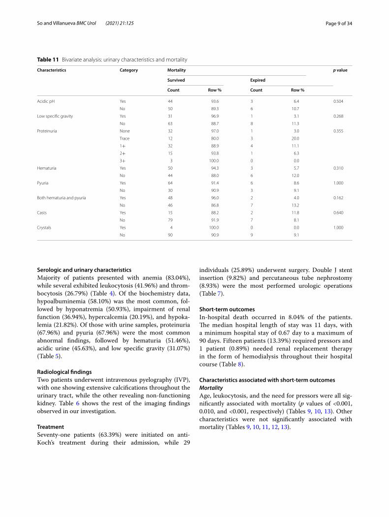
Serologic and urinary characteristicsMajority of patients presented with anemia (83.04%), while several exhibited leukocytosis (41.96%) and throm-bocytosis (26.79%) (Table 4). Of the biochemistry data, hypoalbuminemia (58.10%) was the most common, fol-lowed by hyponatremia (50.93%), impairment of renal function (36.94%), hypercalcemia (20.19%), and hypoka-lemia (21.82%). Of those with urine samples, proteinuria (67.96%) and pyuria (67.96%) were the most common abnormal findings, followed by hematuria (51.46%), acidic urine (45.63%), and low specific gravity (31.07%) (Table 5).
Radiological findingsTwo patients underwent intravenous pyelography (IVP), with one showing extensive calcifications throughout the urinary tract, while the other revealing non-functioning kidney. Table 6 shows the rest of the imaging findings observed in our investigation.
TreatmentSeventy-one patients (63.39%) were initiated on anti-Koch’s treatment during their admission, while 29
individuals (25.89%) underwent surgery. Double J stent insertion (9.82%) and percutaneous tube nephrostomy (8.93%) were the most performed urologic operations (Table 7).
Short‑term outcomesIn-hospital death occurred in 8.04% of the patients. The median hospital length of stay was 11 days, with a minimum hospital stay of 0.67 day to a maximum of 90 days. Fifteen patients (13.39%) required pressors and 1 patient (0.89%) needed renal replacement therapy in the form of hemodialysis throughout their hospital course (Table 8).
Characteristics associated with short‑term outcomesMortalityAge, leukocytosis, and the need for pressors were all sig-nificantly associated with mortality (p values of <0.001, 0.010, and <0.001, respectively) (Tables 9, 10, 13). Other characteristics were not significantly associated with mortality (Tables 9, 10, 11, 12, 13).

Page 10 of 34So and Villanueva BMC Urol (2021) 21:125
Table 13 Bivariate analysis: other outcomes and mortality
Bold value indicates statistically significant differences
Characteristics Category Mortality p value
Survived Expired
Count Row % Count Row %
Need for renal replacement therapy
Yes 1 100.0 0 0.0 1.000
No 102 91.9 9 8.1
Need for pressors Yes 8 53.3 7 46.7 <0.001No 95 97.9 2 2.1
Table 12 Bivariate analysis: treatments and mortality
Characteristics Category Mortality p value
Survived Expired
Count Row % Count Row %
Anti-Kochs treatment Yes 64 90.1 7 9.9 0.482
No 39 95.1 2 4.9
Underwent operation Yes 29 100.0 0 0.0 0.167
No 74 89.0 9 11.0
Percutaneous tube nephrostomy Yes 10 100.0 0 0.0 1.000
No 93 91.2 9 8.8
DJS insertion Yes 11 100.0 0 0.0 0.595
No 92 91.1 9 8.9
Transurethralresection of bladder tumor Yes 1 100.0 0 0.0 1.000
No 102 91.9 9 8.1
Bladder mass excision Yes 3 100.0 0 0.0 1.000
No 100 91.7 9 8.3
Aspiration of abscess Yes 1 100.0 0 0.0 1.000
No 102 91.9 9 8.1
Ureteroneocystostomy Yes 1 100.0 0 0.0 1.000
No 102 91.9 9 8.1
Ureterotomy Yes 1 100.0 0 0.0 1.000
No 102 91.9 9 8.1
Pelvolithotomy Yes 1 100.0 0 0.0 1.000
No 102 91.9 9 8.1
Radial nephrolithotomy Yes 1 100.0 0 0.0 1.000
No 102 91.9 9 8.1
Subcapsular nephrectomy Yes 5 100.0 0 0.0 1.000
No 98 91.6 9 8.4
Cytoreductive nephrectomy Yes 1 100.0 0 0.0 1.000
No 102 91.9 9 8.1
Nephrectomy Yes 2 100.0 0 0.0 1.000
No 101 91.8 9 8.2

Page 11 of 34So and Villanueva BMC Urol (2021) 21:125
Table 14 Bivariate analysis: clinical characteristics and need for pressors
Characteristic Category Mortality p value
Survived Expired
Count Row % Count Row %
Gender Male 8 15.1 45 84.9 1.000
Female 10 15.4 55 84.6
Age 0 months to 1 year 1 100.0 0 0.0 0.019
1–5 years 2 66.7 1 33.3
6–10 years 0 0.0 4 100.0
11–18 years 4 21.1 15 78.9
19–29 years 5 21.7 18 78.3
30–49 years 3 8.1 34 91.9
50–69 years 2 7.4 25 92.6
≥ 70 years 1 25.0 3 75.0
Marital status Single/widowed 13 17.6 61 82.4 0.436
Married 5 11.4 39 88.6
Occupation Employed 3 15.0 17 85.0 0.762
Unemployed 8 13.1 53 86.9
Not applicable 6 21.4 22 78.6
Unspecified 1 11.1 8 88.9
Location City 10 16.1 52 83.9 0.898
Province 7 13.7 44 86.3
Unspecified 1 20.0 4 80.0
Co-morbidity Yes 8 14.5 47 85.5 1.000
No 10 15.9 53 84.1
Diabetes mellitus Yes 0 0.0 5 100.0 1.000
No 18 15.9 95 84.1
Hypertension Yes 0 0.0 13 100.0 0.214
No 18 17.1 87 82.9
Chronic kidney disease Yes 2 33.3 4 66.7 0.227
No 16 14.3 96 85.7
History of urolithiasis Yes 2 25.0 6 75.0 0.352
No 16 14.5 94 85.5
Malignancy Yes 0 0.0 1 100.0 1.000
No 18 15.4 99 84.6
HIV/AIDS Yes 2 15.4 11 84.6 1.000
No 16 15.2 89 84.8
Steroid use Yes 3 33.3 6 66.7 0.139
No 15 13.8 94 86.2
Systemic lupus erythematosus Yes 2 25.0 6 75.0 0.352
No 16 14.5 94 85.5
Nephrotic syndrome Yes 1 100.0 0 0.0 0.153
No 17 14.5 100 85.5
Cerebrovascular disease Yes 0 0.0 1 100.0 1.000
No 18 15.4 99 84.6
Coronary artery disease No 18 15.3 100 84.7 No test*
Heart failure Yes 0 0.0 2 100.0 1.000
No 18 15.5 98 84.5
COPD Yes 0 0.0 1 100.0 1.000
No 18 15.4 99 84.6
Bronchial asthma Yes 0 0.0 2 100.0 1.000
No 18 15.5 98 84.5

Page 12 of 34So and Villanueva BMC Urol (2021) 21:125
Table 14 (continued)
Characteristic Category Mortality p value
Survived Expired
Count Row % Count Row %
Spina bifida No 18 15.3 100 84.7 No test*
RTA Type 1 Yes 0 0.0 1 100.0 1.000
No 18 15.4 99 84.6
Previous TB Yes 0 0.0 14 100.0 0.124
No 18 17.3 86 82.7
Other organ involvement of TB Yes 15 21.4 55 78.6 0.035
No 3 6.3 45 93.8
Pulmonary TB Yes 13 21.0 49 79.0 0.079
No 5 8.9 51 91.1
Gastrointestinal TB Yes 10 32.3 21 67.7 0.006
No 8 9.2 79 90.8
Abdominopelvic TB Yes 3 23.1 10 76.9 0.417
No 15 14.3 90 85.7
CNS TB Yes 3 37.5 5 62.5 0.102
No 15 13.6 95 86.4
Bone TB Yes 1 25.0 3 75.0 0.489
No 17 14.9 97 85.1
Cutaneous/Wound TB Yes 3 100.0 0 0.0 0.003
No 15 13.0 100 87.0
TB Adenitis Yes 1 10.0 9 90.0 1.000
No 17 15.7 91 84.3
Ear TB Yes 1 50.0 1 50.0 0.283
No 17 14.7 99 85.3
Psoas TB Yes 1 50.0 1 50.0 0.283
No 17 14.7 99 85.3
Dysuria Yes 2 28.6 5 71.4 0.603
No 16 16.7 80 83.3
Hematuria Yes 1 11.1 8 88.9 1.000
No 17 18.1 77 81.9
Urinary retention Yes 0 0.0 1 100.0 1.000
No 18 17.6 84 82.4
Flank pain Yes 1 8.3 11 91.7 0.687
No 17 18.7 74 81.3
Inguinal pain Yes 0 0.0 1 100.0 1.000
No 18 17.6 84 82.4
Umbilical discharge Yes 0 0.0 3 100.0 1.000
No 18 18.0 82 82.0
Fistula (uretero-cutaneous fistula) Yes 0 0.0 1 100.0 1.000
No 18 17.6 84 82.4
Scrotal discharge Yes 0 0.0 1 100.0 1.000
No 18 17.6 84 82.4
Double J stent reinsertion Yes 0 0.0 1 100.0 1.000
No 18 17.6 84 82.4
Pedal edema Yes 1 25.0 3 75.0 0.542
No 17 17.2 82 82.8
Fever Yes 0 0.0 7 100.0 0.349
No 18 18.8 78 81.3
Page 13 of 34So and Villanueva BMC Urol (2021) 21:125
Table 14 (continued)
Characteristic Category Mortality p value
Survived Expired
Count Row % Count Row %
Weakness Yes 2 13.3 13 86.7 1.000
No 16 18.2 72 81.8
Abdominal pain Yes 3 27.3 8 72.7 0.402
No 15 16.3 77 83.7
Abdominal/pelvic mass on diagnostic Yes 0 0.0 4 100.0 1.000
No 18 18.2 81 81.8
Vaginal bleeding Yes 0 0.0 2 100.0 1.000
No 18 17.8 83 82.2
Difficulty of breathing Yes 4 30.8 9 69.2 0.235
No 14 15.6 76 84.4
Others Yes 4 36.4 7 63.6 0.098
No 14 15.2 78 84.8
Bold values indicate statistically significant differences
*No test was done since all patients were classified under the No category
COPD chronic obstructive pulmonary disease, HIV/AIDS human immunodeficiency virus or acquired immunodeficiency syndrome, RTA Type 1 renal tubular acidosis Type 1, TB tuberculosis, y year
Table 15 Bivariate analysis: serologic characteristics and need for pressors
Characteristics Category Mortality p value
Survived Expired
Count Row % Count Row %
Anemia Yes 16 16.2 83 83.8 0.734
No 2 10.5 17 89.5
Thrombocytopenia Yes 1 16.7 5 83.3 1.000
No 17 15.2 95 84.8
Thrombocytosis Yes 4 13.3 26 86.7 1.000
No 14 15.9 74 84.1
Leukocytosis Yes 8 15.4 44 84.6 1.000
No 10 15.2 56 84.8
Leukopenia Yes 1 16.7 5 83.3 1.000
No 17 15.2 95 84.8
Hypoalbuminemia Yes 14 21.2 52 78.8 0.116
No 4 8.9 41 91.1
Renal function impairment Yes 7 16.7 35 83.3 0.794
No 11 14.7 64 85.3
Hyperkalemia Yes 2 22.2 7 77.8 0.628
No 16 15.0 91 85.0
Hypokalemia Yes 4 16.7 20 83.3 1.000
No 14 15.2 78 84.8
Hyponatremia Yes 12 20.3 47 79.7 0.204
No 6 10.9 49 89.1
Hypercalcemia Yes 4 16.7 20 83.3 1.000
No 13 15.1 73 84.9

Page 14 of 34So and Villanueva BMC Urol (2021) 21:125
Tabl
e 16
Biv
aria
te a
naly
sis:
urin
ary
char
acte
ristic
s an
d ne
ed fo
r pre
ssor
s
Char
acte
rist
ics
Cate
gory
Mor
talit
yp
valu
e
Surv
ived
Expi
red
Coun
tRo
w %
Coun
tRo
w %
Aci
dic
pHYe
s9
18.0
4182
.00.
798
No
915
.350
84.7
Low
spe
cific
gra
vity
Yes
25.
932
94.1
0.05
3
No
1621
.359
78.7
Prot
einu
riaN
one
411
.431
88.6
0.72
7
Trac
e2
13.3
1386
.7
1+7
17.9
3282
.1
2+4
23.5
1376
.5
3+1
33.3
266
.7
Hem
atur
iaYe
s8
14.8
4685
.20.
797
No
1018
.245
81.8
Pyur
iaYe
s11
15.3
6184
.70.
786
No
718
.930
81.1
Both
hem
atur
ia a
nd p
yuria
Yes
714
.043
86.0
0.60
9
No
1118
.648
81.4
Cast
sYe
s4
22.2
1477
.80.
493
No
1415
.477
84.6
Cry
stal
sYe
s0
0.0
410
0.0
1.00
0
No
1817
.187
82.9

Page 15 of 34So and Villanueva BMC Urol (2021) 21:125
Table 17 Bivariate analysis: treatments and need for pressors
Characteristics Category Mortality p value
Survived Expired
Count Row % Count Row %
Anti-Kochs treatment Yes 14 18.7 61 81.3 0.196
No 4 9.3 39 90.7
Underwent operation Yes 1 3.3 29 96.7 0.095
No 17 19.3 71 80.7
Percutaneous tube nephrostomy Yes 1 10.0 9 90.0 1.000
No 17 15.7 91 84.3
DJS insertion Yes 0 0.0 12 100.0 0.209
No 18 17.0 88 83.0
Transurethral resection of bladder tumor
Yes 0 0.0 1 100.0 1.000
No 18 15.4 99 84.6
Bladder mass excision Yes 0 0.0 3 100.0 1.000
No 18 15.7 97 84.3
Aspiration of abscess Yes 0 0.0 1 100.0 1.000
No 18 15.4 99 84.6
Ureteroneocystostomy Yes 0 0.0 1 100.0 1.000
No 18 15.4 99 84.6
Ureterotomy Yes 0 0.0 1 100.0 1.000
No 18 15.4 99 84.6
Pelvolithotomy Yes 0 0.0 1 100.0 1.000
No 18 15.4 99 84.6
Radial nephrolithotomy Yes 0 0.0 1 100.0 1.000
No 18 15.4 99 84.6
Subcapsular nephrectomy Yes 0 0.0 5 100.0 1.000
No 18 15.9 95 84.1
Cytoreductive nephrectomy Yes 0 0.0 1 100.0 1.000
No 18 15.4 99 84.6
Nephrectomy Yes 0 0.0 2 100.0 1.000
No 18 15.5 98 84.5
Table 18 Bivariate analysis: other outcomes and need for pressors
Characteristics Category Need for pressors p value
Yes No
Count Row % Count Row %
Need for renal replacement therapy Yes 0 0.0 1 100.0 1.000
No 18 15.4 99 84.6

Page 16 of 34So and Villanueva BMC Urol (2021) 21:125
Table 19 Comparison of mean hospital stay by clinical characteristics
Characteristic Category Hospital length of stay (in days)
p value
Mean SD
Gender Male 15.5 16.1 0.914
Female 13.5 9.5
Age 0 months to 1 years 23.0 0.444
1–5 years 35.0 47.9
6–10 years 9.5 9.4
11–18 years 19.6 16.7
19–29 years 13.6 9.5
30–49 years 13.2 9.9
50–69 years 12.1 7.6
≥ 70 years 7.0 4.2
Marital status Single/widowed 17.2 14.8 0.001Married 9.6 6.2
Occupation Employed 12.2 11.7 0.506
Unemployed 13.6 9.0
Not applicable 18.4 20.1
Unspecified 12.4 6.8
Location City 15.3 15.4 0.915
Province 13.3 9.3
Unspecified 15.0 10.8
Co-morbidity Yes 15.3 12.4 0.338
No 13.6 13.3
Diabetes mellitus Yes 7.8 4.8 0.143
No 14.7 13.0
Hypertension Yes 13.3 9.4 0.897
No 14.5 13.3
Chronic kidney disease Yes 16.5 9.7 0.333
No 14.3 13.0
History of urolithiasis Yes 9.4 5.1 0.297
No 14.8 13.2
Malignancy Yes 17.0 0.427
No 14.4 12.9
HIV/AIDS Yes 14.5 10.9 0.740
No 14.4 13.1
Steroid use Yes 28.7 18.5 0.002No 13.2 11.6
Systemic lupus erythematosus Yes 23.5 10.9 0.008No 13.7 12.8
Nephrotic syndrome Yes 70.0 0.091
No 13.9 11.8
Cerebrovascular disease Yes 5.0 0.270
No 14.5 12.9
Coronary artery disease Yes No test*
No 14.4 12.9
Heart failure Yes 19.0 18.4 0.669
No 14.3 12.8

Page 17 of 34So and Villanueva BMC Urol (2021) 21:125
Table 19 (continued)
Characteristic Category Hospital length of stay (in days)
p value
Mean SD
COPD Yes 11.0 0.953
No 14.4 12.9
Bronchial asthma Yes 5.5 2.1 0.150
No 14.5 12.9
Spina bifida Yes No test*
No 14.4 12.9
RTA Type 1 Yes 11.0 0.953
No 14.4 12.9
Previous TB Yes 9.9 5.6 0.204
No 15.0 13.4
Other organ involvement of TB Yes 16.4 14.6 0.030No 11.5 9.2
Pulmonary TB Yes 17.1 15.3 0.021No 11.4 8.6
Gastrointestinal TB Yes 17.7 19.1 0.489
No 13.2 9.6
Abdominopelvic TB Yes 16.0 10.4 0.273
No 14.2 13.2
CNS TB Yes 22.5 27.7 0.309
No 13.8 11.1
Bone TB Yes 34.5 37.8 0.157
No 13.7 10.9
Cutaneous/Wound TB Yes 19.7 7.1 0.161
No 14.3 13.0
TB Adenitis Yes 13.8 12.8 0.642
No 14.4 12.9
Ear TB Yes 51.5 54.4 0.159
No 13.8 10.9
Psoas TB Yes 61.0 41.0 0.022No 13.6 10.7
Bold values indicate statistically significant differences
*No test was done since all patients were classified under the No category
COPD chronic obstructive pulmonary disease, HIV/AIDS human immunodeficiency virus or acquired immunodeficiency syndrome, RTA Type 1 renal tubular acidosis Type 1, TB tuberculosis, y year
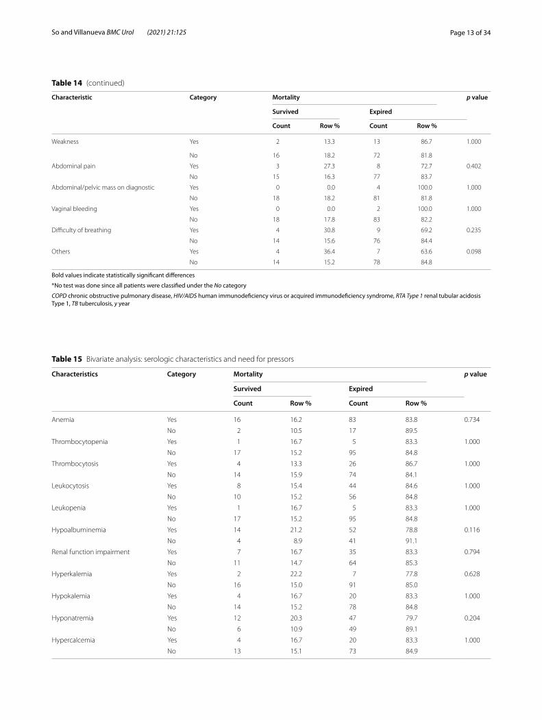
Need for pressorsAge, other organ involvement of MTB, gastrointesti-nal TB, and cutaneous or wound TB, were significantly associated with the need for pressors (p-values of 0.019, 0.035, 0.006, and 0.003, respectively) (Table 14). Other characteristics were not significantly associated with the need for pressors (Tables 14, 15, 16, 17, 18).
Mean hospital stayMarital status, steroid use, systemic lupus erythematosus (SLE), other organ involvement of MTB, pulmonary TB,
psoas TB, the presence of anemia, leukocytosis, hypoal-buminemia, hyponatremia, hypercalcemia, and anti-Koch’s treatment had a statistically longer mean length of hospital stay compared to those without these character-istics (Tables 19, 20, 22). Other characteristics were not significantly associated with a longer mean length of hos-pital stay (Tables 19, 20, 21, 22, 23).

Page 18 of 34So and Villanueva BMC Urol (2021) 21:125
Table 21 Comparison of mean hospital stay by urinary characteristics
Characteristic Category Hospital length of stay (in days) p value
Mean Standard Deviation
Acidic pH Yes 14.3 11.8 0.745
No 14.3 13.5
Low specific gravity Yes 13.3 9.9 0.724
No 14.8 13.8
Proteinuria None 14.6 8.0 0.145
Trace 8.5 5.5
1+ 15.3 15.9
2+ 14.4 8.7
3+ 26.0 38.3
Hematuria Yes 12.9 8.2 0.709
No 15.7 15.8
Pyuria Yes 14.0 12.7 0.679
No 14.9 12.8
Both hematuria and pyuria Yes 12.7 8.2 0.609
No 15.7 15.4
Casts Yes 15.6 16.7 0.722
No 14.1 11.8
Crystals Yes 10.3 3.2 0.693
No 14.5 12.9
Table 20 Comparison of mean hospital stay by serologic characteristics
Bold values indicate statistically significant differences
Characteristic Category Hospital length of stay (in days) p value
Mean SD
Anemia Yes 15.7 13.5 0.001
No 7.6 4.6
Thrombocytopenia Yes 14.2 14.3 0.708
No 14.4 12.8
Thrombocytosis Yes 17.9 19.2 0.315
No 13.2 9.7
Leukocytosis Yes 15.6 11.3 0.027
No 13.4 14.0
Leukopenia Yes 16.7 14.1 0.690
No 14.3 12.8
Hypoalbuminemia Yes 17.0 15.3 0.029
No 11.3 8.0
Renal function impairment Yes 15.5 10.3 0.123
No 13.9 14.1
Hyperkalemia Yes 10.2 5.2 0.411
No 14.9 13.3
Hypokalemia Yes 11.4 6.8 0.373
No 15.4 14.0
Hyponatremia Yes 16.0 13.5 0.046
No 13.1 12.4
Hypercalcemia Yes 16.6 7.6 0.015
No 14.3 14.3

Page 19 of 34So and Villanueva BMC Urol (2021) 21:125
DiscussionGenitourinary TB is the second and third most com-mon form of EPTB in countries with high and low TB burden, respectively [9, 19, 25]. According to other reg-isters, however, GUTB is only seen in 1.7–6.5% of the
total TB cases reported [26]. In the Philippines, a 5-year retrospective study reported GUTB to have caused 3% of pediatric EPTB cases admitted in a tertiary government hospital [13]. It is important to emphasize that this infec-tion is underdiagnosed in most health care centers, as GUTB remains a diagnostic challenge [9, 19, 25–27].
Diagnosis of GUTB is often delayed due to the insidi-ous nature of the disease, non-specificity of symptoms, poor health-seeking behavior of patients, and lack of clinician awareness [28, 29]. In autopsy studies, only half of patients with renal involvement had symptoms, while only 18% were diagnosed clinically [30]. The four pillars to GUTB diagnosis are bacteriology, pathomor-phology, radiology, and provocative test with therapy ex juvantibus [6, 19, 31], with culture as the gold standard [25, 29, 32]. In world literature, most cases of GUTB (64.2%) were diagnosed through identification of MTB
Table 22 Comparison of mean hospital stay by treatments
Bold value indicates statistically significant differences
Characteristic Category Hospital length of stay (in days) p value
Mean Standard Deviation
Anti-Kochs treatment Yes 17.1 13.9 <0.001No 9.7 9.1
Underwent operation Yes 13.0 9.8 0.666
No 14.9 13.8
Percutaneous tube nephrostomy Yes 16.1 12.0 0.436
No 14.2 13.0
DJS insertion Yes 14.4 9.8 0.762
No 14.4 13.2
Transurethral resection of bladder tumor Yes 6.0 0.370
No 14.5 12.9
Bladder mass excision Yes 5.3 3.1 0.074
No 14.6 12.9
Aspiration of abscess Yes 18.0 0.347
No 14.4 12.9
Ureteroneocystostomy Yes 9.0 0.724
No 14.4 12.9
Ureterotomy Yes 5.0 0.270
No 14.5 12.9
Pelvolithotomy Yes 11.0 0.953
No 14.4 12.9
Radial nephrolithotomy Yes 11.0 0.953
No 14.4 12.9
Subcapsular nephrectomy Yes 11.6 3.6 0.936
No 14.5 13.1
Cytoreductive nephrectomy Yes 7.0 0.481
No 14.5 12.9
Nephrectomy Yes 17.0 12.7 0.602
No 14.3 12.9
Table 23 Comparison of mean hospital stay by other outcomes
Characteristics Category Hospital length of stay (in days)
p value
Mean Standard Deviation
Need for renal replacement therapy
Yes 23.0 0.246
No 14.3 12.9
Need for pressors Yes 20.2 24.0 0.790
No 13.3 9.4

Page 20 of 34So and Villanueva BMC Urol (2021) 21:125
Tabl
e 24
Stu
dies
in th
e A
sia–
Paci
fic re
gion
invo
lvin
g pa
tient
s w
ith G
UTB
Stud
y (p
ublic
atio
n ye
ar)
Size
Sett
ing
(cou
ntry
)Po
pula
tion
Out
com
esM
etho
d
Mis
hra
[33]
(202
0)53
Dep
artm
ent o
f Uro
logy
, Ind
ira G
andh
i In
stitu
te o
f Med
ical
Sci
ence
s (In
dia)
Patie
nts
with
con
firm
ed G
UTB
Dem
ogra
phic
, clin
ical
pre
sent
atio
nU
rinar
y pr
ofile
, rou
tine
bloo
d ex
ams
Urin
e A
FB s
mea
r tes
t, ur
ine
MTB
cul
ture
Radi
olog
ical
exa
min
atio
ns, c
ysto
scop
ic
exam
inat
ion,
his
topa
thol
ogic
al e
xam
i-na
tions
4-ye
ar p
rosp
ectiv
e ob
ser-
vatio
nal c
ase
serie
s
Hua
ng [1
0] (2
019)
57C
hang
Gun
g M
emor
ial H
ospi
tal-C
hiay
i (T
aiw
an)
Patie
nts
with
dia
gnos
is o
f GU
TB w
ith a
t le
ast o
ne o
f the
follo
win
g: p
ositi
ve M
TB
cultu
re o
r his
tolo
gic
evid
ence
Dem
ogra
phic
s, co
mor
bidi
ties,
sym
ptom
s an
d si
gns
Resu
lts o
f myc
obac
teria
l sm
ears
and
cu
lture
s, hi
stop
atho
logy
CBC
s, se
rum
bio
chem
istr
y pr
ofile
Che
st ra
diog
raph
yG
U tr
act o
pera
tions
, ant
i-TB
ther
apy,
com
-pl
icat
ions
, clin
ical
out
com
es
15-y
ear r
etro
spec
tive
stud
y
Kim
[35]
(201
8)56
Seve
ranc
e H
ospi
tal,
Seou
l(S
outh
Kor
ea)
Part
icip
ants
old
er th
an 1
8 ye
ars
diag
nose
d w
ith G
UTB
bas
ed o
n pr
esen
ce o
f any
cl
inic
al fi
ndin
g pl
us a
pos
itive
resu
lt fo
r one
of t
he ff
: (1)
urin
e A
FB, (
2) u
rine
MTB
cul
ture
, (3)
urin
e M
TB P
CR,
or (
4)
hist
opat
holo
gy
Clin
ical
and
labo
rato
ry d
ata
Dia
gnos
tic m
etho
ds, t
reat
men
t mod
aliti
es
and
outc
omes
11-y
ear r
etro
spec
tive
stud
y
Cao
[36]
(201
7)41
9Pe
king
Uni
vers
ity F
irst H
ospi
tal (
Chi
na)
All
patie
nts
with
clin
ical
rena
l TB
with
m
icro
biol
ogic
or h
isto
logi
c co
nfirm
atio
nD
emog
raph
ics,
clin
ical
dat
a, c
ompl
ica-
tions
, tre
atm
ent
Labo
rato
ry fi
ndin
gsIm
agin
g fin
ding
sPa
thol
ogic
feat
ures
15-y
ear r
etro
spec
tive
stud
y
Kris
hnam
oort
hy [8
] (20
17)
110
Che
nnai
, Tam
il N
adu
(Indi
a)Pa
tient
s w
ith e
ither
(1) p
rove
n G
UTB
ba
sed
on u
rine
AFB
sm
ear,
AFB
cul
ture
, hi
stop
atho
logi
cal e
vide
nce
of T
B, a
nd/o
r by
ser
olog
ical
met
hods
; or (
2) p
resu
med
G
UTB
who
had
≥ 2
con
sist
ent f
eatu
res
on u
rolo
gica
l im
agin
g or
end
osco
pic
eval
uatio
n
Clin
ical
his
tory
and
exa
min
atio
nSe
rum
bio
chem
istr
yU
rine
cultu
reIm
agin
g fin
ding
s
3-ye
ar re
tros
pect
ive
stud
y
Ye [3
7] (2
016)
193
Wes
t Chi
na H
ospi
tal,
Sich
uan
Uni
vers
ity
(Chi
na)
Case
s w
ith d
efini
te U
TB b
ased
on
resu
lts
of c
ompr
ehen
sive
dia
gnos
is, i
nclu
d-in
g cl
inic
al fe
atur
es, l
abor
ator
y re
sults
(i.
e., s
mea
r mic
rosc
opy,
MTB
cul
ture
, re
al-t
ime
PCR,
and
his
tolo
gica
l pat
tern
s),
radi
olog
ical
find
ings
, and
resp
onse
to
anti-
TB th
erap
y
Dem
ogra
phic
dat
a, c
linic
al h
isto
ry,
prog
nosi
sRa
diol
ogic
al fi
ndin
gsSe
lect
ed la
bora
tory
resu
lts
5-ye
ar c
ross
-sec
tiona
l stu
dy
Sing
h [3
8] (2
013)
117
Uro
logy
Dep
artm
ent o
f Ins
titut
e of
Po
st G
radu
ate
Med
ical
Edu
catio
n an
d Re
sear
ch a
nd S
SKM
Hos
pita
l (In
dia)
All
case
s cl
inic
ally
dia
gnos
ed a
s G
UTB
Clin
ical
pre
sent
atio
nU
rine
AFB
sm
ear,
urin
e M
TB c
ultu
re, u
rine
PCR
for M
TBRa
diol
ogic
al a
nd h
isto
path
olog
ical
ex
amin
atio
ns
13-y
ear r
etro
spec
tive
stud
y

Page 21 of 34So and Villanueva BMC Urol (2021) 21:125
Tabl
e 24
(co
ntin
ued)
Stud
y (p
ublic
atio
n ye
ar)
Size
Sett
ing
(cou
ntry
)Po
pula
tion
Out
com
esM
etho
d
Cha
ndra
[39]
(201
2)25
Him
alay
an In
stitu
te o
f Med
ical
Sci
ence
s, U
ttar
khan
d St
ate
(Indi
a)M
ale
patie
nts
with
his
topa
thol
ogic
ally
co
nfirm
ed G
UTB
Occ
upat
ion,
soc
ioec
onom
icst
atus
Clin
ical
his
tory
Rele
vant
radi
olog
ical
, lab
orat
ory
and
hist
opat
holo
gy fi
ndin
gsTr
eatm
ent
13-y
ear r
etro
spec
tive
stud
y
Hsu
[40]
(201
1)64
Nat
iona
l Tai
wan
Uni
vers
ity H
ospi
tal a
nd
Taip
ei M
edic
al U
nive
rsity
– W
an F
ang
Hos
pita
l (Ta
iwan
)
All
patie
nts
with
urin
e cu
lture
-con
firm
ed
GU
TBC
linic
al fe
atur
esLa
bora
tory
cha
ract
eris
tics
Trea
tmen
t out
com
esG
enot
ypic
cha
ract
eris
tics
of M
TB is
olat
es
12-y
ear r
etro
spec
tive
stud
y
Lee
[17]
(201
1)10
1D
epar
tmen
t of U
rolo
gy, H
anya
ng U
nive
r-si
ty C
olle
ge o
f Med
icin
e (K
orea
)Pa
tient
s di
agno
sed
with
GU
TB b
ased
on
the
pres
ence
of o
ne o
r mor
e po
sitiv
ities
in
term
s of
his
topa
thol
ogic
al fi
ndin
gs,
urin
e A
FB s
mea
r, ur
ine
MTB
cul
ture
, and
ur
ine
PCR
for M
TB
Year
ly p
ropo
rtio
n, g
ende
r, pa
tient
dis
tri-
butio
n ac
cord
ing
to a
ge, h
isto
ry o
f TB,
an
d pr
esen
ce o
f oth
er o
rgan
TB
Urin
alys
is fi
ndin
gs
10-y
ear r
etro
spec
tive
stud
y
Karn
jana
wan
ichk
ul [4
1] (2
010)
35Pr
ince
of S
ongk
la U
nive
rsity
, Hat
Yai
, Son
g-kh
la (T
haila
nd)
Patie
nts
diag
nose
d w
ith u
rinar
y tr
act T
B by
dem
onst
ratio
n of
AFB
in u
rine
smea
r, gr
owth
from
urin
e M
TB c
ultu
re, o
r con
-si
sten
t his
topa
thol
ogic
find
ings
Dem
ogra
phic
dat
a, c
linic
al fe
atur
esLa
bora
tory
dat
aC
hest
x-r
ay, i
ntra
veno
us u
rogr
aphy
, ultr
a-so
nogr
aphy
, or e
ndos
copi
c fin
ding
s
10-y
ear r
etro
spec
tive
stud
y
Taka
hash
i [42
] (20
07)
12U
rolo
gy c
linic
s of
six
med
ical
cen
ters
, Hok
-ka
ido
(Jap
an)
Patie
nts
diag
nose
d w
ith u
rinar
y TB
bas
ed
on N
AAT
or h
isto
path
olog
yD
emog
raph
ic d
ata,
clin
ical
feat
ures
Det
ectio
n m
etho
d fo
r MTB
Dia
gnos
tic fi
ndin
gsTr
eatm
ent,
outc
omes
, and
med
icat
ion-
rela
ted
adve
rse
even
ts
5-ye
ar re
tros
pect
ive
stud
y
Hsi
eh [1
8] (2
006)
31Ka
ohsi
unng
Med
ical
Uni
vers
ity H
ospi
tal,
Kaoh
siun
g (T
aiw
an)
Patie
nts
diag
nose
d w
ith G
UTB
bas
ed o
n m
icro
biol
ogic
al o
r his
tolo
gica
l find
ings
pl
us c
ompa
tible
clin
ical
and
roen
tgen
o-gr
aphi
c fin
ding
s
Base
line
char
acte
ristic
s, un
derly
ing
dise
ases
, tre
atm
ent r
espo
nses
, and
ou
tcom
es
11-y
ear r
etro
spec
tive
stud
y
Bucc
holz
[43]
(200
0)55
Aga
Kha
n U
nive
rsity
Hos
pita
l (Pa
kist
an)
In-p
atie
nts
with
GU
TB p
rove
n ei
ther
by
urin
e cu
lture
pos
itivi
ty fo
r MTB
, or
hist
opat
holo
gy
Age
, sex
, con
com
itant
dis
ease
s, m
edic
al
hist
ory,
sym
ptom
s, di
agno
sis,
trea
tmen
t an
d fo
llow
-up
13-y
ear r
etro
spec
tive
stud
y
Ram
anat
han
[34]
(199
8)38
Sanj
ay G
andh
i Pos
t Gra
duat
e In
stitu
te o
f M
edic
al S
cien
ces,
Luck
now
(Ind
ia)
All
patie
nts
with
eith
er: (
1) u
rinar
y TB
ba
sed
on p
ositi
ve u
rine
or p
us c
ultu
res
for M
TB o
r his
topa
thol
ogy,
or (
2)
pres
umed
urin
ary
TB w
ith ≥
3 c
onsi
st-
ent f
eatu
res
on u
rolo
gica
l im
agin
g or
en
dosc
opy
His
tory
and
phy
sica
l exa
min
atio
nSe
rum
che
mis
try
Urin
e cu
lture
Che
st x
-ray
and
ultr
ason
ogra
phy
8-ye
ar re
tros
pect
ive
stud
y
Dy
[16]
(199
5)61
Sant
o To
mas
Uni
vers
ity H
ospi
tal (
Phili
p-pi
nes)
In-p
atie
nts
clin
ical
ly d
iagn
osed
with
GU
TBD
emog
raph
ic fe
atur
esPr
esen
ting
man
ifest
atio
ns, h
isto
ry o
f pr
evio
us T
BD
iagn
ostic
mod
aliti
es (r
adio
grap
hic,
ba
cter
iolo
gic,
his
topa
thol
ogic
)Th
erap
eutic
mod
aliti
es
Case
ser
ies

Page 22 of 34So and Villanueva BMC Urol (2021) 21:125
Tabl
e 24
(co
ntin
ued)
Stud
y (p
ublic
atio
n ye
ar)
Size
Sett
ing
(cou
ntry
)Po
pula
tion
Out
com
esM
etho
d
Tanc
huco
[15]
(198
7)42
Phili
ppin
e G
ener
al H
ospi
tal a
nd N
atio
nal
Kidn
ey In
stitu
te (P
hilip
pine
s)Pa
tient
s w
ith d
isch
arge
dia
gnos
is o
f ur
inar
y tr
act T
B ba
sed
on th
e pr
esen
ce
of o
ne o
f the
follo
win
g: p
ositi
ve u
rine
AFB
sm
ear,
posi
tive
urin
e A
FB c
ultu
re, o
r co
nsis
tent
his
topa
thol
ogic
find
ings
Clin
ical
and
labo
rato
ry p
aram
eter
s6-
year
retr
ospe
ctiv
e st
udy
GU
gen
itour
inar
y, M
TB M
ycob
acte
rium
tube
rcul
osis
, NAA
T nu
clei
c ac
id a
mpl
ifica
tion
test
, PTB
pul
mon
ary
tube
rcul
osis
, UTB
urin
ary
tube
rcul
osis
Page 23 of 34So and Villanueva BMC Urol (2021) 21:125
Tabl
e 25
Stu
dies
in th
e A
sia–
Paci
fic re
gion
des
crib
ing
the
dem
ogra
phic
feat
ures
of p
atie
nts
with
GU
TB
Stud
y (p
ublic
atio
n ye
ar)
Age
Mal
e‑to
‑fem
ale
ratio
Dem
ogra
phic
Gen
itour
inar
y or
gans
in
volv
ed (n
)A
ssoc
iate
d co
mor
bidi
ties
(%)
Mis
hra
[33]
(202
0)M
ean,
39.
15 ±
12.
62 y
1:1.
21 (2
4:29
)So
cioe
cono
mic
cla
ss: l
ower
(8
8.7%
), m
iddl
e (9
.4%
), up
per
(1.9
%)
Kidn
ey (3
3; 1
8 un
ilate
ral,
15
bila
tera
l inv
olve
men
t), u
rete
r (1
6; 1
4 lo
wer
ure
tera
l str
ictu
re,
1 m
iddl
e ur
eter
al s
tric
ture
, 1
mul
tiple
str
ictu
res)
, bla
dder
(1
3)
His
tory
of P
TB (2
0.8%
)
Hua
ng [1
0] (2
019)
Med
ian,
71
year
s (ra
nge,
33–
89
year
s)1.
85:1
(37:
20)
Kidn
eys
(8),
kidn
ey a
nd u
rete
r (4
), ep
idid
ymis
(3),
epid
idym
is
and
test
is (3
), ki
dney
and
pr
osta
te (2
), pr
osta
te (2
), ur
eter
(1),
uret
er a
nd b
ladd
er
(1),
test
is (1
), ep
idid
ymis
an
d te
stis
and
pro
stat
e (1
), sc
rotu
m a
nd p
enis
(1),
uter
us
and
cerv
ix (1
)
DM
type
II (3
5.1%
), ch
roni
c re
nal
dise
ase
(33.
3%),
unde
rlyin
g m
alig
nanc
ies
(hep
atoc
ellu
lar,
pros
tate
, bla
dder
, cer
vix,
rec-
tum
, thy
roid
gla
nd, l
ymph
oma,
an
d sk
in) (
24.6
%),
adre
nal i
nsuf
-fic
ienc
y (2
4.6%
), co
rtic
oste
roid
us
e (2
1.1%
), ch
roni
c ai
rway
di
seas
e (1
9.3%
), liv
er c
irrho
sis
(17.
5%),
past
his
tory
of T
B (1
5.8%
), al
coho
lism
(8.8
%),
and
auto
imm
une
dise
ase
(3.5
%)
Kim
[35]
(201
8)M
ean,
52.
8 y
1:1.
15 (2
6:30
)Ki
dney
or u
rete
r (39
, 69.
6%),
blad
der (
16, 2
8.6%
), ep
idid
ymis
or t
estis
(13,
23
.2%
), ut
erus
or f
allo
pian
tu
bes
(5, 8
.9%
), pr
osta
te (4
, 7.
1%)
His
tory
of T
B (4
2.9%
, PTB
37.
5%),
CVD
(28.
6%),
imm
unoc
ompr
o-m
ised
sta
te (2
1.4%
), pu
lmon
ary
dise
ase
(10.
7%),
liver
dis
ease
(7
.1%
), D
M (5
.4%
), hi
stor
y of
ga
stre
ctom
y (3
.6%
)
Cao
[36]
(201
7)M
ean,
42.
7 ±
13.
4 ye
ars
(rang
e,
12–7
8 ye
ars)
1:1.
29 (1
83:2
36)
Une
mpl
oyed
(24.
6%),
farm
er
(21%
), ci
vil s
erva
nt (1
5.5%
), w
orke
r (10
.7%
), re
tiree
9.1
%),
stud
ent (
4.3%
), ot
her o
ccup
a-tio
ns (1
4.6%
)
Left
kid
ney
(210
, 50.
1%),
right
ki
dney
(171
, 40.
8%),
both
(38,
9.
1%)
His
tory
of P
TB (2
0.3%
)
Kris
hnam
oort
hy [8
] (20
17)
Mea
n, 3
5.4
year
s (ra
nge,
11–
67
year
s)1.
4:1
(65:
45)
Kidn
ey (7
0), u
rete
rs (3
0), b
ladd
er
(18)
, tes
tis a
nd e
pidi
dym
is (6
), pr
osta
te (4
), pe
nis
(1)
His
tory
of P
TB (2
2.7%
), ga
stro
in-
test
inal
TB
(2.7
%)
Ye [3
7] (2
016)
Mea
n, 4
2.8 ±
14.
95 y
1.64
:1 (1
20:7
3)Ex
tra-
urin
ary
TB (3
6.3%
)
Sing
h [3
8] (2
013)
Third
dec
ade
of li
fe (6
3.2%
)1:
1.51
(47:
70)
Kidn
ey (7
6; 5
6 un
ilate
ral,
20
bila
tera
l inv
olve
men
t), u
rete
r (3
2), b
ladd
er (2
0), p
rost
ate
(4),
scro
tal s
wel
ling
(6)
Past
his
tory
of P
TB (1
8.9%
)
Page 24 of 34So and Villanueva BMC Urol (2021) 21:125
Tabl
e 25
(co
ntin
ued)
Stud
y (p
ublic
atio
n ye
ar)
Age
Mal
e‑to
‑fem
ale
ratio
Dem
ogra
phic
Gen
itour
inar
y or
gans
in
volv
ed (n
)A
ssoc
iate
d co
mor
bidi
ties
(%)
Cha
ndra
[39]
(201
2)M
ean,
37.
7 y
NA
; onl
y m
ales
wer
e in
clud
edLo
catio
n: h
illy
regi
on o
f sta
te
(68%
), no
n-hi
lly re
gion
of
stat
e (3
2%)
Occ
upat
ion:
farm
er (5
6%),
labo
rer (
20%
), sh
opke
eper
(8
%),
stud
ent (
8%),
unkn
own
(8%
)So
cioe
cono
mic
sta
tus:
low
(8
0%)
Urin
ary
blad
der (
7, 2
8%),
pros
-ta
te (6
, 24%
), ep
idid
ymis
(3,
12%
), te
stes
(3, 1
2%),
kidn
ey
(2, 8
%),
uret
er (2
, 8%
), sc
rotu
m
(1, 4
%)
Prev
ious
his
tory
of T
B (3
6%),
alco
-ho
lism
(28%
), di
abet
es (1
2%)
Hsu
[40]
(201
1)M
ean,
60.
3 ±
16.
1 y
1.46
:1 (3
8:26
)Bl
adde
r (5)
, ure
ter (
4), k
idne
y (2
), ki
dney
/ure
ter (
1), k
idne
y/ur
e-te
r/bl
adde
r (1)
, epi
didy
mis
(3),
test
is/e
pidi
dym
is (2
), te
stis
/ep
idid
ymis
/pro
stat
e gl
and
(2),
epid
idym
is/p
rost
ate
glan
d (1
), te
stis
(1),
pros
tate
gla
nd (1
)
57.8
%D
isse
min
ated
TB
(48.
4%),
PTB
(43.
8%),
DM
(23.
4%),
mal
ig-
nanc
y (1
4.1%
), CO
PD (1
4.1%
), pr
evio
us T
B (1
2.5%
), C
VD
(12.
5%),
rece
ivin
g st
eroi
ds
(12.
5%),
ESRD
(6.3
%),
liver
cir-
rhos
is (4
.7%
), al
coho
lism
(4.7
%)
Lee
[17]
(201
1)M
ean,
45.
57 ±
12.
55 y
ears
(ra
nge,
19–
81 y
ears
)1:
1.53
(40:
61)
Kidn
ey a
nd/o
r ure
ter (
80.2
0%),
epid
idym
is a
nd/o
r tes
tis
(14.
85%
), bl
adde
r (3.
96%
), pr
osta
te(0
.99%
)
Past
his
tory
of P
TB (2
1.8%
), in
test
inal
TB
(0.9
9%),
spin
e TB
(0
.99%
)
Karn
jana
wan
ichk
ul [4
1] (2
010)
Mod
e, 3
1–40
yea
rs (r
ange
,10–
76)
1.3:
1 (2
0:15
)O
ccup
atio
n: fa
rmer
(34.
3%),
hous
ewife
(20.
0%),
busi
-ne
sspe
rson
(14.
3%),
gove
rnm
ent s
ervi
ce (1
4.3%
), bl
ue-c
olla
r wor
ker (
14.3
%),
and
stud
ent (
2.9%
)
Kidn
ey (7
; 3 b
ilate
ral,
3 le
ft, 1
rig
ht),
uret
er (7
; 3 b
ilate
ral,
1 le
ft, 3
righ
t), b
ladd
er (4
), te
stis
(3
; 1 le
ft, 2
righ
t), k
idne
ys to
ur
ethr
a (2
), ki
dney
+ b
ladd
er
(2),
uret
er +
bla
dder
(1),
blad
-de
r + u
reth
ra (1
), ur
ethr
a (1
)
Act
ive
or p
ast h
isto
ry o
f PTB
(3
4.3%
)
Taka
hash
i [42
] (20
07)
Med
ian,
68.
5 ye
ars
(rang
e,
40–9
0 ye
ars)
1:1
(6:6
)Ki
dney
(7),
blad
der (
6), u
rete
r (2)
Act
ive
PTB
(16.
7%)
Hsi
eh [1
8] (2
006)
Mea
n, M
: 54.
4 ye
ars
(rang
e,
32–7
5 ye
ars)
, F: 6
1.8
year
s (ra
nge,
31–
81 y
ears
)
1:1.
21 (1
4:17
)H
isto
ry o
f PTB
(25.
8%)
Bucc
holz
[43]
(200
0)M
ean,
39.
9 ±
17.
1 ye
ars
(7–8
1 ye
ars)
3:1
(41:
14)
Kidn
ey (2
8), b
ladd
er (1
5), u
rete
r (1
3), t
este
s (5
), ur
ethr
a (1
)A
ctiv
e PT
B or
EPT
B on
Cat
egor
y I
trea
tmen
t (93
%),
on C
ateg
ory
II (5
.4%
), on
Cat
egor
y III
(1.8
%),
hist
ory
of E
PTB
(11%
), D
M (3
3%)
Ram
anat
han
[34]
(199
8)M
ean,
38.
8 ye
ars
1:3.
22 (9
:29)
His
tory
of P
TB (4
3.2%
)

Page 25 of 34So and Villanueva BMC Urol (2021) 21:125
Tabl
e 25
(co
ntin
ued)
Stud
y (p
ublic
atio
n ye
ar)
Age
Mal
e‑to
‑fem
ale
ratio
Dem
ogra
phic
Gen
itour
inar
y or
gans
in
volv
ed (n
)A
ssoc
iate
d co
mor
bidi
ties
(%)
Dy
[16]
(199
5)M
ean,
M: 4
8.4 ±
17.
01 y
ears
(ra
nge,
21–
72),
F: 4
3.3 ±
17.
58
year
s (2
1–78
)
1:2.
2 (1
9:42
)Ki
dney
s (5
0.8%
), ki
d-ne
ys +
ure
ter (
4.9%
), ki
dney
s + u
rete
r + b
ladd
er
(1.6
%),
kidn
eys +
pro
stat
e (1
.6%
), pe
lvis
(8.2
%),
blad
der
(1.6
%),
blad
der +
ure
ter
(1.6
%),
epid
idym
is (3
.3%
), ep
idid
ymis
+ te
stis
+ v
as
defe
rens
(1.6
%),
fallo
pian
tu
be +
per
itone
um (1
.6%
)
Act
ive
PTB
(47.
5%),
past
his
tory
of
TB
(32.
8%),
hype
ruric
emia
(1
3.1%
), D
M (1
.6%
), H
TN (1
,6%
), D
M +
HTN
(1.6
%),
hype
rten
sive
re
nal f
ailu
re (1
.6%
), liv
er c
ir-rh
osis
(1.6
%),
cong
estiv
e he
art
failu
re (1
.6%
), m
yelo
fibro
sis
(1.6
%),
oste
oart
hriti
s an
d pr
os-
tatic
can
cer (
1.6%
), rh
eum
atoi
d ar
thrit
is (1
.6%
)
Tanc
huco
[15]
(198
7)M
ean,
39
year
s (ra
nge,
2 to
64)
1.3:
124
:18
Past
his
tory
of T
B or
exp
osur
e (2
8.6%
), m
alnu
triti
on (4
.8%
), D
M (2
.4%
)
COPD
chr
onic
obs
truc
tive
pulm
onar
y di
seas
e, C
VD c
ardi
ovas
cula
r dis
ease
, DM
dia
bete
s m
ellit
us, E
SRD
end
-sta
ge re
nal d
isea
se, F
fem
ale,
HTN
hyp
erte
nsio
n, M
mal
e, N
A no
t app
licab
le, P
TB p
ulm
onar
y tu
berc
ulos
is

Page 26 of 34So and Villanueva BMC Urol (2021) 21:125
Tabl
e 26
Stu
dies
in th
e A
sia–
Paci
fic re
gion
illu
stra
ting
the
clin
ical
pre
sent
atio
n of
pat
ient
s w
ith G
UTB
Stud
y (p
ublic
atio
n ye
ar)
Tim
e fr
om s
ympt
om o
nset
to d
iagn
osis
Syst
emic
sym
ptom
sG
enito
urin
ary
man
ifest
atio
ns
Mis
hra
[33]
(202
0)Co
nstit
utio
nal s
ympt
oms
(28.
3%)
Irrita
tive
void
ing
sym
ptom
s (6
9.8%
), he
mat
uria
(56.
6%),
flank
pai
n (5
6.6%
), as
soci
ated
rena
l fai
lure
(13.
2%),
infe
rtili
ty a
nd h
emat
ospe
rmia
(5.6
%),
scro
tal m
ass
(1.9
%)
Hua
ng [1
0] (2
019)
Med
ian,
4 m
(ran
ge, 0
.5–5
0 m
)75
.4%
Feve
r (56
.1%
), m
alai
se/f
atig
ue (3
6.8)
, wei
ght l
oss
(31.
6%),
nigh
t sw
eats
(8.8
%)
71.9
%G
ross
hem
atur
ia (4
0.4%
), fre
quen
cy/u
rgen
cy (3
3.3%
), dy
suria
(29.
8%),
flank
pai
n (2
6.3%
)
Kim
[35]
(201
8)N
onsp
ecifi
c sy
mpt
oms
(feve
r, an
orex
ia, w
eigh
t los
s, sw
eatin
g, w
eakn
ess,
perip
hera
l lym
phad
enop
athy
) (1
2.5%
)
Urin
ary
frequ
ency
or d
ysur
ia, u
reth
ral p
ain,
or i
rrita
ble
void
ing
sym
ptom
s (5
5.4%
); lo
in o
r abd
omin
al p
ain
(42.
9%);
gros
s he
mat
uria
(33.
9%);
scro
tal p
ain/
mas
s (1
9.6%
); ab
sces
s or
fist
ula
(5.4
%);
vagi
nal b
leed
ing
(3.6
%)
Cao
[36]
(201
7)Co
nstit
utio
nal s
ympt
oms
incl
udin
g w
eigh
t los
s, fe
ver,
nigh
t sw
eats
, and
/or f
atig
ue (3
8.9%
)Lo
wer
urin
ary
trac
t sym
ptom
s in
clud
ing
frequ
ency
, ur
genc
y, a
nd o
dynu
ria (6
5.2%
); fla
nk p
ain
(37.
9%);
gros
s he
mat
uria
(26.
3%)
Kris
hnam
oort
hy [8
] (20
17)
Loin
pai
n (2
7.0%
), st
orag
e sy
mpt
oms
(25.
5%),
hem
a-tu
ria (1
2.0%
), st
one
dise
ase
(9.1
%),
palp
able
mas
s (8
.2%
), sc
rota
l sin
us (5
.5%
), in
fert
ility
(2.7
%),
gast
ro-
inte
stin
al s
ympt
oms
(2.7
%),
uros
epsi
s (1
.8%
), re
nal
failu
re (1
.8%
), ca
lcifi
ed k
idne
y (0
.9%
), ur
inom
a (0
.9%
)
Ye [3
7] (2
016)
Feve
r (26
.4%
), ni
ght s
wea
t (13
.0%
), w
eigh
t los
s (1
0.9%
)U
rinar
y irr
itatio
n (6
1.1%
), lu
mba
go (4
9.2%
)
Sing
h [3
8] (2
013)
32.6
%Irr
itativ
e vo
idin
g sy
mpt
oms
(66.
47%
), he
mat
uria
(4
7.6%
), fla
nk p
ain
(33.
8%),
recu
rren
t urin
ary
trac
t sy
mpt
oms
(18.
9%),
scro
tal m
ass
(5.1
%),
colo
cuta
ne-
ous
fistu
la (0
.8%
), ne
phro
cuta
neou
s fis
tula
(1.8
%),
asso
ciat
ed re
nal f
ailu
re (1
4.7%
), in
fert
ility
or h
emat
o-sp
erm
ia (3
.4%
)
Cha
ndra
[39]
(201
2)Fe
ver a
nd m
alai
se (3
2.0%
)U
rgen
cy a
nd in
crea
sed
frequ
ency
of m
ictu
ritio
n (5
6%),
lum
bar p
ain
(56%
), dy
suria
(52%
), he
mat
uria
(44%
), py
uria
(40%
), in
fert
ility
(12%
), re
nal f
ailu
re (8
%),
recu
r-re
nt a
bsce
ss (8
%),
scro
tal l
ump
(8%
), sc
rota
l sin
us (4
%)
Hsu
[40]
(201
1)18
2.0 ±
311
.1 d
(ran
ge, 5
to 1
245
d)51
.6%
Feve
r (43
.8%
), fa
tigue
(37.
5%),
body
wei
ght l
oss
(12.
5%)
62.5
%D
ysur
ia (3
1.3%
), fre
quen
cy (3
1.3%
), fla
nk p
ain
(28.
1%),
hem
atur
ia (1
7.2%
), sc
rota
l pai
n or
mas
s (1
0.9%
)
Lee
[17]
(201
1)Fe
ver (
3.0%
)Fr
eque
ncy
(40.
6%),
hem
atur
ia (3
3.7%
), dy
suria
(16.
8%),
flank
pai
n (1
6.8%
), sc
rota
l sw
ellin
g (3
%)
Karn
jana
wan
ichk
ul [4
1] (2
010)
< 6
m (6
5.7%
), 6–
12 m
(17.
1%), >
1 y
ears
(8.6
%),
unce
r-ta
in d
ata
(8.6
%)
Freq
uenc
y (4
8.6%
), dy
suria
(42.
9%),
hem
atur
ia (3
1.4%
), ab
dom
inal
pai
n or
mas
s (2
5.7%
), ur
ethr
al p
ain
(20.
0%),
rete
ntio
n (1
4.3%
), cu
tane
ous
fistu
la (1
4.3%
), re
nal f
ailu
re (5
.7%
)
Taka
hash
i [42
] (20
07)
Med
ian
dura
tion;
bet
wee
n in
itial
sym
ptom
s an
d cl
inic
vi
sit:
120
d (ra
nge,
3–3
60 d
); be
twee
n cl
inic
vis
it an
d di
agno
sis:
14 d
(7–1
50 d
)
Chi
ef c
ompl
aint
: fre
quen
cy (5
8.3%
), he
mat
uria
(25.
0%),
posi
tive
nucl
ear m
atrix
pro
tein
22
on s
cree
ning
test
fo
r bla
dder
can
cer (
8.3%
), in
cide
ntal
righ
t ren
al tu
mor
(8
.3%
)

Page 27 of 34So and Villanueva BMC Urol (2021) 21:125
Tabl
e 26
(co
ntin
ued)
Stud
y (p
ublic
atio
n ye
ar)
Tim
e fr
om s
ympt
om o
nset
to d
iagn
osis
Syst
emic
sym
ptom
sG
enito
urin
ary
man
ifest
atio
ns
Hsi
eh [1
8] (2
006)
Mea
n, 2
m (r
ange
, 5 d
to 1
8 m
)Fe
ver (
29.0
%),
mal
aise
/fat
igue
(12.
9%),
nigh
t sw
eats
(3
.2%
), bo
dy w
eigh
t los
s (3
.2%
)Fr
eque
ncy/
urge
ncy
(61.
3%),
dysu
ria (5
8.1%
), fla
nk p
ain
(35.
5%),
gros
s he
mat
uria
(32.
2%),
scro
tal m
ass/
pain
(1
6.1%
)
Bucc
holz
[43]
(200
0)Fe
ver (
36.0
%),
lass
itude
(13.
0%),
wei
ght l
oss
(13.
0%)
Dys
uria
(49%
), fre
quen
cy (4
0%),
flank
pai
n (3
6%),
gros
s he
mat
uria
(31%
), ur
genc
y (1
5%),
test
icul
ar s
wel
ling
(13%
), su
prap
ubic
pai
n (9
%),
rena
l col
ic (1
%)
Ram
anat
han
[34]
(199
8)Pa
in (6
3.6%
), he
mat
uria
(61.
3%),
lum
p (1
8.2%
)
Dy
[16]
(199
5)M
ean,
M: 3
0.4 ±
42.
09 m
(ran
ge, 0
.25–
180)
, F:
27.8
± 3
5.2
m (0
.5–1
68)
Feve
r (29
.5%
), w
eigh
t los
s (1
8.0%
), ch
ills
(9.8
%),
nau-
sea/
vom
iting
(6.6
%),
anor
exia
(6.6
%)
Dys
uria
(32.
8%),
flank
pai
n (2
7.9%
), he
mat
uria
(19.
6%),
hypo
gast
ric p
ain
(19.
6%),
noct
uria
(19.
6%),
frequ
ency
(1
8%),
edem
a (1
4.8%
), va
gina
l spo
ttin
g (1
3.1%
), co
s-to
vert
ebra
l ang
le te
nder
ness
(11.
4%),
urge
ncy
(9.8
%),
hesi
tanc
y (6
.6%
)
Tanc
huco
[15]
(198
7)Fe
ver (
52.4
%),
wei
ght l
oss
(26.
2%),
chill
s (2
1.4%
), m
alai
se (1
1.9%
), ni
ght s
wea
ts (2
.4%
)D
ysur
ia (7
1.5%
), he
mat
uria
(62.
0%),
flank
pai
n (4
4.5%
), tu
rbid
urin
e (4
7.6%
), fre
quen
cy (4
0.5%
), hy
poga
stric
pa
in 2
3.8%
), ed
ema
(4.8
%)

Page 28 of 34So and Villanueva BMC Urol (2021) 21:125
Tabl
e 27
Stu
dies
in th
e A
sia–
Paci
fic re
gion
sho
win
g se
rolo
gic
and
urin
ary
profi
les
of p
atie
nts
with
GU
TB
ALT
alan
ine
amin
otra
nsfe
rase
, Cr s
erum
cre
atin
ine,
Hb
hem
oglo
bin,
HPF
hig
h-po
wer
fiel
d, R
BC re
d bl
ood
cell,
WBC
whi
te b
lood
cel
l
Stud
y (p
ublic
atio
n ye
ar)
Hem
atol
ogic
dat
aBi
oche
mis
try
Uri
naly
sis
Mis
hra
[33]
(202
0)A
cidi
c ur
ine
(98.
1%),
ster
ile p
yuria
(81.
1%),
pyur
ia
(69.
8%),
hem
atur
ia (5
8.4%
), al
kalin
e ur
ine
(1.9
%)
Hua
ng [1
0] (2
019)
Ane
mia
(< 1
0 g/
dL) (
28.1
%),
thro
mbo
cyto
peni
a (<
150
× 1
012/L
) (26
.3%
)H
ypoa
lbum
inem
ia (<
2.5
g/d
L) (4
0.4%
)Py
uria
+ h
emat
uria
(29.
8%),
isol
ated
hem
atur
ia (>
30/
HPF
) (17
.5%
), is
olat
ed p
yuria
(> 2
0/H
PF) (
12.3
%)
Kim
[35]
(201
8)Py
uria
(> 5
WBC
s/H
PF) (
66.1
%),
hem
atur
ia (>
2 R
BCs/
HPF
) (50
.0%
), pr
otei
nuria
(19.
6%)
Cao
[36]
(201
7)Py
uria
(> 5
WBC
s/H
PF) (
56.3
%),
hem
atur
ia (>
3 R
BCs/
HPF
) (48
.8%
)
Ye [3
7] (2
016)
Ane
mia
(15.
6%),
leuk
ocyt
osis
(13.
0%)
Incr
ease
d BU
N (2
3.3%
), in
crea
sed
crea
tinin
e (2
0.2%
)H
emat
uria
(63.
2%),
prot
einu
ria (4
5.6%
), py
uria
(19.
2%)
Sing
h [3
8] (2
013)
Ster
ile p
yuria
(62.
4%),
hem
atur
ia (6
1.5%
), pr
otei
nuria
(5
7.4%
)
Hsu
[40]
(201
1)A
nem
ia (H
b <
12
g/dL
) (46
.9%
), le
ukoc
ytos
is
(WBC
> 1
0,00
0/µL
) (17
.2%
)H
ypoa
lbum
inem
ia (a
lbum
in <
3.5
g/d
L) (3
7.5%
), re
nal
func
tion
impa
irmen
t (C
r > 1
.5 m
g/dL
) (18
.8%
), liv
er
func
tion
impa
irmen
t (A
LT >
40
IU/L
) (17
.2%
)
Pyur
ia o
r hem
atur
ia (W
BC >
10/
HPF
, × 4
00; R
BC >
5/
HPF
, × 4
00) (
64.1
%),
asep
tic p
yuria
(53.
1%)
Lee
[17]
(201
1)Pr
otei
nuria
(57.
4%),
hem
atur
ia (5
1.5%
), py
uria
(42.
4%)
Karn
jana
wan
ichk
ul [4
1] (2
010)
Aci
dic
urin
e w
ith p
yuria
(80.
0%)
Hsi
eh [1
8] (2
006)
Ane
mia
(Hb
< 1
2 g/
dL) (
25.8
%),
leuk
ocyt
osis
(W
BC >
10,
000/
µL) (
25.8
%),
leuk
open
ia (W
BC <
400
0/µL
) (0%
)
Poor
rena
l fun
ctio
n (C
r > 1
.5 m
g/dL
) (58
.1%
), po
or
liver
func
tion
(ALT
> 4
0 IU
/L) (
16.1
%),
hype
rkal
emia
(K
+ >
5.5
meq
/L) (
3.2%
)
Pyur
ia +
hem
atur
ia (5
1.6%
), py
uria
(WBC
> 1
0/H
PF;
400x
) (25
.8%
), he
mat
uria
(RBC
> 5
/HPF
; 400
x) (1
2.9%
)
Bucc
holz
[43]
(200
0)Py
uria
(56%
), he
mat
uria
(36%
), st
erile
pyu
ria (6
%)
Dy
[16]
(199
5)H
emat
uria
(37.
7%),
prot
einu
ria (3
2.8%
), py
uria
+ b
ac-
teriu
ria (3
2.8%
), st
erile
pyu
ria (2
6.2%
), py
uria
(8.2
%),
ster
ile p
yuria
+ b
acte
riuria
(3.3
%),
cylin
drur
ia (1
3.1%
)
Tanc
huco
[15]
(198
7)A
nem
ia H
b <
12
g/dL
(60.
0%),
leuk
ocyt
osis
(W
BC >
10,
000/
µL) (
37.0
%)
Cre
atin
ine
clea
ranc
e <
30
ml/m
in (7
5.0%
), el
evat
ed
BUN
(> 0
.53
mm
ol/L
) (38
.9%
), el
evat
ed s
erum
Cr
(> 1
76.8
mm
ol/L
) (35
.1%
)
Alk
alin
e ur
ine
(pH
≥ 6
) (88
.9%
), py
uria
(WBC
≥ 6
/HPF
) (8
2.4%
), he
mat
uria
(RBC
≥ 6
/HPF
) (61
.8%
), al
bum
inu-
ria o
f 2 +
or m
ore
(55.
8%)

Page 29 of 34So and Villanueva BMC Urol (2021) 21:125
in the urine, mostly establishing the diagnosis via posi-tive urine culture [9]. Similarly, most retrospective stud-ies in the Asia–Pacific region diagnosed infection based on bacteriologic and histologic findings with consistent clinical history, while a few depended mainly on clinico-radiologic evaluation (Table 24). In our study, the lack of clinical registries of GUTB in our hospital prompted us to instead use laboratory registries for case finding, not-ing majority of cases being diagnosed based on positiv-ity for urine AFB smear (50.00%). In contrast, one local study observed mycobacterial culture being sent in only 22.2% of urologic cases and in 11.1% of gynecology cases, relying more heavily on clinical, radiographic, and histo-pathologic assessment [16]. Despite being a recognized tool for diagnosis of GUTB, imaging is particularly useful only during the later stages of the disease when calcifi-cations or cavernous forms have already developed [19, 33]. Nonetheless, this diversity in practice standards is expected in developing countries because of the dispro-portionate availability of medical facilities and services [34].
Many reviews of the world literature [28, 29] noted kidneys to be the most frequently organ, which is con-sistent with most studies done in the Asia–Pacific region (Table 25). In contrast, some reports observed the bladder to be the most frequently affected genitourinary organ [38, 40], similar to our investigation. Renal involvement of TB infection can either be a localized urogenital disease or a part of a disseminated infection [6]. In literature, up to 10% of affected individuals have concomitant active pulmonary TB, suggesting hematogenous or lymphatic spread to this highly vascularized organ [28]. The latent period between pulmonary infection and development of clinical GUTB is described to range from 1 to 46 years, averaging around 22 years [29]. Infection may also be acquired hematog-enously from the gut [28]. Other genitourinary organs may become affected through ascent or descent of MTB from a source elsewhere in the genitourinary tract, or from con-tact with the bacilli shed into the urine [26]. They may also get involved from descending spread from the lymphatics [29] or from sexual intercourse [28].
Unilateral organ involvement is commonly shown in retrospective, clinical, and autopsy studies [28, 33, 36, 38, 41]. In our investigation, left laterality was observed in 60% of those with kidney and ureter involvement, consistent with other reports [36, 41]. Renal lesions are initially described to be bilateral attributing to hema-togenous spread. They generally undergo a period of cicatrization then enter a latent phase of infection, only reactivating the moment an individual becomes immu-nocompromised. From a single focus, infection eventu-ally progresses, affecting one kidney while sparing the
other. This phenomenon accounts for the greater fre-quency of unilateral renal TB [9].
Patient characteristicsOur investigation noted several patients having chronic diseases (e.g., diabetes mellitus, hypertension, chronic kidney disease), immunocompromised conditions (e.g., HIV/AIDS, steroid use), history of tuberculosis, or pres-ence of TB in other organs. Traditionally, risk factors for developing TB include malnutrition, immunosuppres-sion, HIV infection, diabetes, chronic kidney or liver disease, smoking, and low socioeconomic status [25, 28]. Factors considered high-risk for GUTB include past or present TB infection, recurrent or resistant urinary tract infection, and fistulas involving the scrotum, peri-neum, or lumbar area [5]. Emergence of drug-resistant strains of TB as well as anatomical abnormalities of the urogenital tract from congenital conditions, renal cysts, and urolithiasis also predispose to its development [19]. In our study, a substantial number of patients were single or widowed (61.61%), lived in urban areas (53.57%), and were unemployed (51.79%). In comparison, several stud-ies reported most patients to be living in the hilly region of the state (68%), working as farmers (21–56%) or unem-ployed (20.0–24.6%), and having low socioeconomic sta-tus (80.0–88.7%) [33, 36, 39, 41].
Worldwide, cases encountered between developed and developing countries exhibit different patterns. In devel-oped countries, GUTB mainly affects the elderly, ethnic minorities, and immigrants [6, 9]. On the other hand, patients from developing countries are younger due to higher incidence and severity of TB disease. They present with more specific symptoms and complications which are further exacerbated by delays in diagnosis [9]. Our data demonstrated a mean age of 35.79 ± 18.29 years, with 22.32% belonging to the pediatric population. These findings are consistent with most studies in the region (Table 25). GUTB generally has the propensity to infect both men and women of child-bearing age (20–40 years old), with a mean age of 40.7 years (range, 5–88 years) [25, 29]. We also report a male-to-female ratio of 1:1.15 (52:60). A proper estimate is controversial since there is a lack of controlled epidemiological and clinical studies [28]. While some report more men to be affected than women (2:1) [29], others report women to be affected twice as many as men [28]. Even among past local data, sex distribution was inconsistent [15, 16]. Variation between geographical regions might reflect local TB endemicity or study bias, thereby making accurate epide-miological and clinical data on GUTB difficult to obtain [28].

Page 30 of 34So and Villanueva BMC Urol (2021) 21:125
Clinical manifestationsGUTB does not present with any specific clinical feature and may in fact be asymptomatic [6]. Up to 50% of cases are incidentally diagnosed when patients undergo work-up for other genitourinary disorders [28]. In those with symptoms, storage symptoms (e.g., urinary frequency, urgency, incontinence, nocturia) were the most common presentation on admission, followed by dysuria, hematu-ria, and lumbar pain [9, 26, 29]. In our study, we recorded 50.89% of patients to have genitourinary manifestations as their chief complaint, with 21.43% having flank or abdominal pain. Several studies also mentioned abdomi-nal or hypogastric pain to be common (19.6%-42.9%) [15, 16, 35, 41].
We noted 21.43% of patients to have systemic symp-toms such as weakness and fever as their initial com-plaints, while 12.50% manifested with difficulty of breathing. Such findings might be explained by the high rates of TB infection in other organ systems (57.14%), with lungs being the most common extra-genitourinary site (50.89%). In some literature, constitutional symp-toms such as fever, weight loss, and night sweats are uncommon and, if present, are indicative of concomi-tant TB outside the genitourinary system. Some patients may initially present with a myriad of symptoms reflec-tive of other concomitant infections like PTB and hence GUTB symptoms and signs are not always defined by the anatomical site of disease [28]. Moreover, second-ary bacterial infections can concurrently occur in up to 50% of patients with GUTB [25, 28]. Our data is consist-ent with most studies in the Asia–Pacific region, with the exception of those done in South Korea where systemic symptoms are relatively uncommon (3–12.5%) [17, 35]. Systemic manifestations are otherwise present in many reports (28.3–75.4%), with fever being the most cited symptom (29–56.1%) (Table 26). Delays in diagnosis may result in disease progression and severe complications seen at presentation [28].
Hematologic abnormalitiesHematological and biochemical tests are considered non-specific and are instead utilized as adjuncts to GUTB management [28]. In our study, majority of patients pre-sented with anemia (83.04%), while several exhibited leu-kocytosis (41.96%) and thrombocytosis (26.79%). This is similar to past local data, where anemia and leukocytosis were found in 60.0% and 37.0% of patients, respectively [15]. These estimates are higher than what is recorded in literature, where 15.6–46.9% of patients exhibited ane-mia and 13.0–25.8% had leukocytosis (Table 27). Throm-bocytopenia (26.3%) occurred more frequently in some populations [10]. It is important to note that these studies
applied different definitions of hematologic abnormali-ties, making comparison difficult.
TB infection is traditionally known to affect various cell lines. It can cause anemia attributed to four mecha-nisms: chronic disease, nutritional deficiencies, autoim-mune hemolytic anemia, and marrow complications. It is described to commonly affect all subtypes of granulo-cytes during its course, predominantly affecting neutro-phils either quantitatively or qualitatively. It may result in transient neutrophilia or, in extreme cases, leukemoid reaction. TB may also cause leukopenia, especially in females, elderly, or those with recurrent infections. Neu-tropenia may occur from direct suppression of granu-lopoiesis by activated T cells [44]. Lymphopenia and lymphocytosis are also commonly reported in active TB [45]. Lastly, TB may result in various platelet abnormali-ties. Thrombocytosis is frequently reactive in nature and is related to the degree of inflammation. It is mediated by increased levels of endogenous thrombopoietin pro-duced as an acute-phase reactant. Thrombocytopenia, on the other hand, is usually from bone marrow infiltration, disseminated intravascular coagulation, immune throm-bocytopenic purpura, thrombotic thrombocytopenic purpura, or drug-induced [44].
Biochemical abnormalitiesAmong those with laboratory results, hypoalbuminemia (58.10%) and impairment of renal function (36.94%) were commonly found, similar to those reported in literature, with estimates at 37.5–40.4% and 18.8–75.0%, respec-tively (Table 27). As plasma creatinine concentration is frequently described to be normal in the setting of uni-lateral renal involvement, increased levels may indicate bilateral renal involvement or presence of concomitant disorders such as interstitial nephritis or glomerulone-phritis [46, 47]. In a review of 8961 cases, 5.7% of patients with GUTB were reported to develop end-stage renal disease (ESRD) [29]. Likewise, one retrospective study in South Korea reported occurrence of ESRD in 7.1% of GUTB patients, identifying acute renal failure and old age as independent risk factors for chronic kidney disease [35].
We also observed several electrolyte abnormali-ties, specifically hyponatremia (50.93%), hypercalce-mia (20.19%), hypokalemia (21.82%), and hyperkalemia (8.18%). Mild hyponatremia has been reported in the set-ting of active pulmonary or miliary TB, with incidences ranging from 11 to 51% [27, 48]. Most cases are due to syndrome of inappropriate antidiuretic hormone secre-tion (SIADH), with a third having reset osmostat wherein the plasma sodium stabilizes at lower concentration

Page 31 of 34So and Villanueva BMC Urol (2021) 21:125
levels. Mechanisms for such persistent ADH release are yet to be explored, but abnormalities in water handling are demonstrated to resolve following successful treat-ment of infection [27]. Hyponatremia in TB may also be attributed to adrenal insufficiency. Hyponatremia from this condition is accompanied by hyperkalemia and increased urinary potassium excretion. Cerebral salt wasting is another mechanism for hyponatremia, usually seen in patients with tuberculous meningitis [48]. Lastly, patients with kidney TB may also develop salt-losing nephropathy [49].
Tubulointerstitial nephritis (TIN) is one possible complication of TB which might contribute to the mul-tiple electrolyte abnormalities seen in patients with infec-tion. Several case reports described the development of chronic granulomatous TIN in patients with TB as evi-denced by renal biopsy [50–53]. Unlike the other studies, one case series in west London found only 18.7% of those with granulomatous inflammation on renal biopsy to exhibit caseation necrosis [53], whereas another study in France raised the possibility of severe TIN in TB despite the absence of renal granuloma [54]. Although reports on this tubulointerstitial disorder are increasing, mecha-nisms for its development are poorly understood [55].
Hypercalcemia is another electrolyte abnormality com-monly cited in patients with TB. Surveys from different countries show prevalence rates up to 11%-48%, not-ing that the actual estimates of prevalence are difficult to establish since concurrent serum albumin levels are not consistently reported [48]. Renal or extrarenal TB granulomas accounted for the non-physiologic synthesis of 1,25-dihydroxyvitamin D3 and the ensuing hypercal-cemia [56]. Calcification is unusual in the early stages of infection, however, in the advanced stages, nearly all kid-neys contains calcification [26].
Urinary abnormalitiesUrinalysis is abnormal in up to 90% of patients with GUTB, with findings ranging from mild changes to extreme pyuria [6, 29, 57]. Pyuria with or without micro-scopic hematuria is seen in majority of patients, whereas heavy proteinuria and cellular casts are not generally observed [6, 26]. Persistent sterile pyuria, pyuria in an acidic urine without growth on routine culture, or symp-tomatic UTI unresponsive to standard antibiotics should prompt suspicion of GUTB [6, 25, 28, 58]. Our study showed proteinuria (67.96%) and pyuria (67.96%) to be the most common urinary findings, consistent with those cited in literature (Table 27). Low specific gravity was also found to be common (31.07%), possibly reflecting poor urinary concentrating ability especially in the set-ting of chronic kidney disease or tubulopathy.
Radiological findingsVarious imaging modalities are used to support the diag-nosis of GUTB, with findings dependent upon the extent of disease progression. Despite traditionally being used to suggest, and not to confirm or exclude, the presence of the disease, they are still paramount in management and can still be utilized to confidently diagnose GUTB by those with sufficient experience [59].
Intravenous urogram (IVU) is considered the gold standard for imaging in early renal TB [33, 59]. There might be no abnormalities present during early disease, but there usually appear moderate to severe urinary tract changes once patients become symptomatic [59]. Early findings include infundibular narrowing, calyceal ero-sion or blunting, and papillary necrosis with associated parenchymal scarring and calcification [27]. GUTB is considered if there is simultaneous involvement of both the upper and the lower urinary tracts, especially the kidney and the bladder [27, 29]. Later stages of the dis-ease may demonstrate extensive cavitation, mass lesions, calyceal distortion, cortical scarring, calcification, auto-nephrectomy, perinephric abscess, fistula formation, ure-teral strictures, and bladder fibrosis [25, 59]. In our study, both patients who underwent intravenous pyelography showed advanced findings of extensive stone formation and non-functioning kidney.
Triphasic computed tomography (CT) scan remains the mainstay imaging technique for cross-sectional imag-ing in GUTB [6]. It is the most sensitive modality to detect calcification, and is superior to IVU in detecting multiple small urothelial lesions [25, 60]. Findings sug-gestive of GUTB include the presence of lesions in other organs beyond the urinary tract, such as liver, lymph nodes, and vertebrae [29], as seen in our study.
Although IVU and CT scan are reported to be the more frequently used imaging modalities in GUTB [29], some investigations still heavily rely on ultrasonography [18, 36]. Ultrasonography may only give indirect evidence of GUTB, but it avoids exposure to ionizing radiation and can be conveniently used to guide fine needle aspiration biopsies [5, 25, 59]. In literature, two patterns of GUTB have been described: (1) the infiltrative pattern show-ing increased echogenicity from calcifications, infected debris, or abscesses; and (2) hydronephrosis or pyone-phrosis, involving calyceal dilatation and a small renal pelvis. Another distinguishing feature for GUTB is the visualization of multiple abnormalities in different dis-ease stages with various organ involvement [59]. All these changes were observed in our investigation.
TreatmentMedical treatment of GUTB should be initiated promptly when clinical, laboratory, and radiological findings

Page 32 of 34So and Villanueva BMC Urol (2021) 21:125
suggest a presumptive diagnosis, even prior to the release of microbiologic and histopathologic results [26, 47]. Pharmacologic therapy for drug-sensitive TB consists of an intensive phase of quadruple therapy with first-line anti-TB agents (e.g., isoniazid, rifampicin, pyrazinamide and ethambutol) for 2 months, followed by a continua-tion phase with two drugs (e.g., isoniazid, rifampicin) for 4 months [28]. In our study, over sixty percent (63.39%) of patients were started on anti-TB therapy during admis-sion, reflecting diverse in-hospital practices wherein several cases were discharged upon resolution of their reasons for encounter, with eventual treatment initiated on an out-patient basis after microbiologic and histo-pathologic confirmation.
Around half (54.9%) of patients with GUTB require surgery [28, 47]. Indications for surgery include diversion of urologic obstruction, drainage of abscesses, nephrec-tomy of non-functioning kidneys, reconstruction of affected ureters, and dilation of contracted urinary blad-der [26, 28]. A quarter (25.89%) of patients in our study underwent an operation, lower than that observed in a previous local investigation (52%) [15]. This difference might be attributed to the higher proportion of younger individuals involved in our study, whose illness duration may not be long enough to develop complications that would warrant surgical therapy. This finding might sig-nify a changing pattern of disease in the country.
Short‑term outcomesDeaths from tuberculosis are higher in developing (2–3 million deaths per year) than developed countries (40 thousand deaths per year) [9]. In GUTB, mortality rates vary (1.2–28.1%) [10], with most patients succumb-ing from disseminated infection [15]. In our center, all-cause in-hospital mortality occurred in 8.04% of the included patients. Age, leukocytosis, and the need for pressors were all significantly associated with mortal-ity (p values of <0.001, 0.010, and <0.001, respectively). Several researches in Taiwan investigated risk factors for mortality in patients with GUTB. Fever was shown to be positively correlated with mortality (OR = 42.716; 95% CI 1.032–1767.569; p = 0.048), possibly attributed to its high prevalence in elderly patients, those with multiple co-morbidities, and those who had delays in diagnosis [10]. Other poor prognostic factors included genitou-rinary tract surgery (OR = 0.000; 95% CI 0.000–0.255; p = 0.020) [10], age older than 65 years old (HR = 4.03; 95% CI 1.27–12.76; p = 0.02), cardiovascular disease (HR = 5.96; 95% CI 1.98–17.92; p = 0.001), steroid use (HR = 10.16; 95% CI 2.27–45.47; p = 0.02), and no treat-ment (HR = 4.81; 95% CI 1.12–20.67; p = 0.04) [40].
It was observed that patients with longer mean lengths of hospital stay did not necessarily develop
unfavorable hospital outcomes. Instead, prolonged admission duration seemed to have been influenced by management of co-morbidities (e.g., SLE, pulmonary TB, psoas TB) and correction of various clinically sig-nificant laboratory abnormalities (e.g., anemia, leuko-cytosis, hyponatremia, and hypercalcemia). Awaiting results of TB workups during admission for diagno-sis of equivocal cases and for subsequent initiation of definitive therapy might have also contributed since anti-Koch’s treatment had a statistically longer mean length of hospital stay (p <0.001). These findings were reflective of the varying practices in our center, with many cases undergoing surgery for prompt treatment of symptoms and having medical therapy be initiated on an out-patient basis depending on the results of cul-tures and histopathology.
LimitationsOur study had several limitations. Its retrospective design which was affected by institutional limitations in recordkeeping made data collection restricted, as evi-denced by a low chart retrieval rate (57.89%, 132/228). Given the absence of a unified clinical registry and data recording, our case finding was laboratory-based and was subject to selection bias, thereby rendering our results nongeneralizable to a broader patient population. It also proved to be a major barrier preventing inclusion of pre-dictive modeling of study outcomes. Despite these limita-tions, our research is the largest study in the country to date, involving more accurate diagnosis of GUTB com-pared to past local studies since all reviewed cases had bacteriologic or histopathologic evidence of infection. It also involved more specific definitions of serologic find-ings adjusted per age and sex.
ConclusionOur investigation observed a high prevalence rate of serologic and urinary abnormalities among admit-ted GUTB patients in the study. Apart from the com-monly cited abnormalities in literature, multiple electrolyte abnormalities and urinary concentration defects were observed in many cases, possibly indi-cating tubulointerstitial involvement. Accordingly, we recommend future research be done on such compli-cation to determine its correlation with disease activ-ity and to possibly help with the diagnosis of infection particularly in low-resource settings. Mortality rate was also noted to be high among admitted patients with GUTB. Age, leukocytosis, and need for pressors were significantly associated with mortality. However, further research is recommended to explore predic-tive modeling.

Page 33 of 34So and Villanueva BMC Urol (2021) 21:125
AcknowledgementsWe would like to thank the Philippine General Hospital and the Division of Nephrology for supporting us in this work. We would also like to thank Prof. Francisco N. de los Reyes for his assistance with our data analysis.
Authors’ contributionsConception and design of study: PNHS and ARTV. Acquisition of data: PNHS. Data analysis and interpretation, drafting of manuscript and critical revision, and approval of final version of manuscript: PNHS and ARTV. All authors read and approved the final manuscript.
FundingThis work was self-funded by the authors.
Availability of data and materialsThe datasets used in this study are available from the corresponding author upon reasonable request.
Declarations
Ethics approval and consent to participateThe study was approved by the University of the Philippines Manila Research Ethics Board and the requirement for informed consent was waived because this was a retrospective study involving a review of anonymized data. All methods were carried out in accordance with the ethical standards of the Philippine Health Research Ethics Board’s 2017 National Ethical Guidelines for Health and Health-Related Research and with the Data Privacy Act of 2012.
Consent for publicationNot applicable.
Competing interestsThe authors declare that they have no competing interests in any form.
Received: 18 February 2021 Accepted: 23 August 2021
References 1. World Health Organization: Global tuberculosis report 2019. 2019. 2. Mehta JB, Dutt A, Harvill L, Mathews KM. Epidemiology of extrapul-
monary tuberculosis: a comparative analysis with pre-AIDS era. Chest. 1991;99(5):1134–8.
3. Das P, Ahuja A, Gupta SD. Incidence, etiopathogenesis and pathologi-cal aspects of genitourinary tuberculosis in India: a journey revisited. Indian J Urol. 2008;24(3):356–61.
4. Noertjojo K, Tam CM, Chan SL, Chan-Yeung MMW. Extra-pulmonary and pulmonary tuberculosis in Hong Kong. Int J Tuberc Lung Dis. 2002;6(10):879–86.
5. Kulchavenya E. Urogenital tuberculosis: definition and classification. Ther Adv Infect Dis. 2014;2(5–6):117–22.
6. Kulchavenya E, Yasuda M, Grabe M. Epidemiology, classification, clini-cal features, and diagnostics of urogenital tuberculosis. In: Bjerklund Johansen TE, Wagenlehner FME, Matsumoto T, Cho YH, Krieger JN, Shoskes D, Naber KG, editors. Urogenital infections and inflammations. Berlin: German Medical Science GMS Publishing House; 2019. https:// doi. org/ 10. 5680/ lhuii 000052.
7. Wildbolz H. Ueber urogenical tuberkulose. Schweiz Med Wochenschr. 1937;67:1125.
8. Krishnamoorthy S, Palaniyandi V, Kumaresan N, Govindaraju S, Rajasekaran J, Murugappan I, et al. Aspects of evolving genito urinary tuberculosis: a profile of genito urinary tuberculosis (GUTB) in 110 patients. J Clin Diagn Res. 2017;11(9):PC01–5.
9. Figueiredo AA, Lucon AM, Junior RF, Srougi M. Epidemiology of uro-genital tuberculosis worldwide. Int J Urol. 2008;15:827–32.
10. Huang TY, Hung CH, Hsu WH, Peng KT, Hung MS, Lai LJ, et al. Geni-tourinary tuberculosis in Taiwan: a 15-year experience at a teaching hospital. J Microbiol Immunol Infect. 2019;52:312–9.
11. Porter MF III. Uro-genital tuberculosis in the male. Ann Surg. 1894;20(4):396–405.
12. Lansang MAD, Alejandria MM, Law I, Juban NR, Amarillo MLE, Sison OT, et al. High TB burden and low notification rates in the Philippines: the 2016 national TB prevalence survey. PLoS ONE. 2021;16(6):e0252240.
13. Santos R. Clinical profile of extrapulmonary tuberculosis cases admitted and diagnosed in a tertiary government hospital from January 2006 to June 2010. Pediatr Infect Dis Soc Philippines J. 2013;14(2):77–84.
14. Lim RG, Caro HP, Dominguez-Mejia AE, Yuhico MJ. The clinical profile of patients with sterile pyuria and recurrent urinary tract infection. Phillip-pine J Intern Med. 1996;34(3):89–92.
15. Tanchuco JQ, Lazo PO, King B. Urinary tract tuberculosis: local experi-ence. Philippine J Nephrol. 1987;11:183–90.
16. Dy EER, Rosales LN, Lim EU. Genitourinary tuberculosis at the Santo Tomas University Hospital. Phillippine J Intern Med. 1995;33(2):47–52.
17. Lee JY, Park HY, Park SY, Lee SW, Moon HS, Kim YT, et al. Clinical charac-teristics of genitourinary tuberculosis during a recent 10-year period in one center. Korean J Urol. 2011;52:200–5.
18. Hsieh HC, Lu PL, Chen YH, Chen TC, Tsai JJ, Chang K, et al. Genitourinary tuberculosis in a medical center in southern Taiwan: an eleven-year experience. J Microbiol Immunol Infect. 2006;39:408–13.
19. Kulchavenya E. Best practice in the diagnosis and management of urogenital tuberculosis. Ther Adv Urol. 2013;5(3):143–51.
20. World Health Organization. Haemoglobin concentrations for the diag-nosis of anaemia and assessment of severity. Geneva: World Health Organization; 2011.
21. Lo SF. Reference intervals for laboratory tests and procedures. In: Klieg-man R, St Geme JW, Blum NJ, Tasker RC, Shah SS, Wilson KM, editors. Nelson textbook of pediatrics. 21st ed. Philadelphia: Elsevier; 2020. p. e6–7.
22. Longo DL, Fauci AS, Kasper DL, Hauser SL, Jameson J, Loscalzo J. Har-rison’s principles of internal medicine. 20th ed. New York: McGraw-Hill; 2018.
23. Berliner N. Approach to the adult with unexplained neutropenia. Upto-date, 2018. Accessed 3 Feb 2020.
24. Skorecki K, Chertow GM, Marsden PA, Brenner BM, Rector FC, et al. Brenner & Rector’s the kidney. 10th ed. Philadelphia: Elsevier; 2016.
25. Abbara A, Davidson RN. Etiology and management of genitourinary tuberculosis. Nat Rev Urol. 2011;8:678–88.
26. Amaya-Tapia G, Aguirre-Avalos G. Urinary tract tuberculosis. IntechO-pen. 2018. https:// doi. org/ 10. 5772/ intec hopen. 76063.
27. Venyo AK, Venyo LK, Maloney DJL, Khan AN. Tuberculosis of the kidney and the genitourinary tract: a review of the literature. Hamdan Med J. 2015;8:345–60.
28. Muneer A, Macrae B, Krishnamoorthy S, Zumla A. Urogenital tubercu-losis: epidemiology, pathogenesis and clinical features. Nat Rev Urol. 2019;16(10):573–98.
29. Figueiredo AA, Lucon AML. Urogenital tuberculosis: update and review of 8961 cases from the world literature. Rev Urol. 2008;10(3):207–17.
30. Medlar EM, Spain DM, Holliday RW. Post-mortem compared with clinical diagnosis of genitourinary tuberculosis in adult males. J Urol. 1949;61:1078–88.
31. Lewinsohn DM, Leonard MK, LoBue PA, Cohn DL, Daley CL, Desmond E, et al. Official American Thoracic Society/Infectious Diseases Society of America/Centers for Disease Control and Prevention clinical practice guidelines: diagnosis of tuberculosis in adults and children. Clin Infect Dis. 2017;64:1–33.
32. Mehta PK, Kamra E. Recent trends in diagnosis of urogenital tuberculosis. Future Microbiol. 2020;15(3):159–62.
33. Mishra KG, Ahmad A, Singh G, Tiwari R. Current status of genitourinary tuberculosis: presentation, diagnostic approach and management—single centre experience at IGIMS (Ptana, Bihar, India). Indian J Surg. 2020;82(817–823):2020.
34. Ramanathan R, Kumar A, Kapoor R, Bhandari M. Relief of urinary tract obstruction in tuberculosis to improve renal function. Analysis of predic-tive factors. Br J Urol. 1998;81:199–205.
35. Kim EJ, Lee W, Jeong WY, Choi H, Jung IY, Ahn JY, et al. Chronic kidney disease with genitourinary tuberculosis: old disease but ongoing compli-cation. BMC Nephrol. 2018;19:193.

Page 34 of 34So and Villanueva BMC Urol (2021) 21:125
• fast, convenient online submission
•
thorough peer review by experienced researchers in your field
• rapid publication on acceptance
• support for research data, including large and complex data types
•
gold Open Access which fosters wider collaboration and increased citations
maximum visibility for your research: over 100M website views per year •
At BMC, research is always in progress.
Learn more biomedcentral.com/submissions
Ready to submit your researchReady to submit your research ? Choose BMC and benefit from: ? Choose BMC and benefit from:
36. Cao Y, Fan Y, Chen Y, Zhao Z, Song Y, Shen C, et al. Gross hematuria is more common in male and older patients with renal tuberculosis in China: a single-center 15-year clinical experience. Urol Int. 2017;99:290–6.
37. Ye Y, Hu X, Shi Y, Zhou J, Zhou Y, Song X, et al. Clinical features and drug-resistance profile of urinary tuberculosis in south-western China. Medicine. 2016;95(19):1–8.
38. Singh JP, Priyadarshi V, Kundu AK, Vijay MK, Bera MK, Pal DK. Genito-uri-nary tuberculosis revisited: 13 years’ experience of a single centre. Indian J Tuberc. 2013;60:15–22.
39. Chandra S, Chandra H, Chauhan N, Gaur DS, Gupta H, Pathank VP, et al. Male genitourinary tuberculosis: 13 years experience at a ter-tiary care center in India. Southeast Asian J Trop Med Public Health. 2012;43(2):364–9.
40. Hsu H-L, Lai C-C, Yu M-C, Yu F-L, Lee J-C, Chou C-H, et al. Clinical and microbiological characteristics of urine culture-confirmed genitourinary tuberculosis at medical centers in Taiwan from 1995 to 2007. Eur J Clin Microbiol Infect Dis. 2011;30:319–26.
41. Karnjanawanichkul W, Pripatnanont C. Tuberculosis of the urinary tract in southern Thailand. J Med Assoc Thai. 2010;93(8):916–9.
42. Takahasi S, Takeyama K, Kunishima Y, Hashimoto K, Miyamoto S, Ichihara K, et al. Current survey of urinary tuberculosis in Hokkaido, Japan. J Infect Chemother. 2007;13:105–8.
43. Buccholz NP, Haque R, Salahuddin S. Genitourinary tuberculosis: a profile of 55 in-patients. J Pak Med Assoc. 2000;50(8):265–9.
44. Balepur SS, Schlossberg D. Hematologic complications of tubercu-losis. Microbiol Spectr. 2016. https:// doi. org/ 10. 1128/ micro biols pec. TNMI7- 0004- 2016.
45. Oyer RA, Schlossberg D. Hematologic changes in tuberculosis. In: Schloss-berg D, editor. Tuberculosis. New York: Springer; 1994.
46. Visweswaran RK, Pais VM, Dionne-Odom J. Urogenital tuberculosis. UpTo-Date, 2019. Accessed 9 Nov 2020.
47. Figueiredo AA, Lucon AM, Srougi M. Urogenital tuberculosis. Microbiol Spectr. 2017;5(1):1–16.
48. Vinnard C, Blumberg EA. Endocrine and metabolic aspects of tuberculo-sis. Microbiol Spectr. 2017;5(1):1–19.
49. Murphy FD, Settimi AL, Kozokoff NJ. Renal disease with the salt losing syndrome: a report of four cases of so-called “salt losing nephritis.” Ann Intern Med. 1953;38(6):1160–76.
50. Mallison WJ, Fuller RW, Levison DA, Baker LR, Cattell WR. Diffuse interstitial renal tuberculosis: an unusual cause of renal failure. Q J Med. 1981;50:137–48.
51. El-Reshaid KA, Madda JP, Al-Saleh MA. A case of progressive tubulo-inter-stitial nephritis due to culture-negative renal tuberculosis. Saudi J Kidney Dis Transpl. 2001;12:534–7.
52. Sampathkumar K, Sooraj YS, Mahaldar AR, Ramakrishnan M, Rjappannair A, Nalumakkal SV, et al. Granulomatous interstitial nephritis due to tuber-culosis: a rare presentation. Saudi J Kidney Dis Transpl. 2009;20:842–5.
53. Chapagain A, Dobbie H, Sheaff M, Yaqoob M. Presentation, diagnosis, and treatment outcome of tuberculous-mediated tubulointerstitial nephritis. Kidney Int. 2011;79(6):671–7.
54. Delafosse M, Teuma C, Miailhes P, Nouvier M, Rabeyrin M, Fouque D. Severe tubulointerstitial nephritis: tracking tuberculosis even in the absence of renal granuloma. Clin Kidney J. 2018;11(5):667–9.
55. Eastwood JB, Corbishley CM, Grange JM. Tuberculosis and tubulointersti-tial nephritis: an intriguing puzzle. Kidney Int. 2011;79:579–81.
56. Pouchot J, Dreyfuss D, Gardin JP, Mier L, Remy P, Esdaile JM, et al. Ectopic production of 1,25-dihydroxyvitamin D3 in tuberculosis. Nephrol Dial Transpl. 1993;8:560–2.
57. Daher EDF, da Silva GB, Barros EJG. Review: renal tuberculosis in the modern era. Am J Trop Med Hyg. 2013;88(1):54–64.
58. American Thoracic Society. Diagnostic standards and classification of tuberculosis in adults and children. Am J Respir Crit Care Med. 2000;161(4 Pt 1):1376–95.
59. Merchant S, Bharati A, Merchant N. Tuberculosis of the genitourinary sys-tem: urinary tract tuberculosis: renal tuberculosis—part I. Indian J Radiol Imaging. 2013;23(1):46–63.
60. Merchant S, Bharati A, Merchant N. Tuberculosis of the genitourinary sys-tem: urinary tract tuberculosis: renal tuberculosis—part II. Indian J Radiol Imaging. 2013;23(1):64–77.
Publisher’s NoteSpringer Nature remains neutral with regard to jurisdictional claims in pub-lished maps and institutional affiliations.