september 2016 vol.:7, issue:2 characterization of...
TRANSCRIPT

Human Journals
Research Article
September 2016 Vol.:7, Issue:2
© All rights are reserved by Baojun Xu et al.
Characterization of Monascus Metabolites Isolated from
Red Yeast Rice (Monascus ruber-fermented rice) and Their
Proliferation Inhibitory Effects against Cancer Cells
www.ijppr.humanjournals.com
Keywords: Monascus ruber; red yeast rice; monacolins;
pigments; anticancer
ABSTRACT
The objective of current study was to characterize the secondary
metabolites from red yeast rice and explore their anticancer
activities. Solvent fractionation methods combined with open
column chromatographic method were performed to isolate the
major compounds from red yeast rice. High-resolution mass
spectrum and 13
C and 1H-NMR spectral techniques were further
applied to elucidate the chemical structures of the isolated
compounds. The isolated compounds were subjected to cancer
cell proliferation inhibition analyzed by MTT method. Four
isolated compounds were identified as lactone-form monacolin
K, monacolin L, apigenin, and rubropunctatin based on the
spectral analyses. The red pigment, rubropunctatin, exhibited
strong anticancer effects against both SNU-1 cells and
SK-OV-3 cells at the concentration of 10 μg/mL, its anticancer
activities were close to or higher than the positive drugs. The
IC50 values of monacolin L and rubropunctatin against cancer
cells SNU-1 and SK-OV-3 were 9.8, 13.3, 28.9 and 8.2 μg/mL
respectively.
Qijun Wang 1, Changkeun Sung
2, Baojun Xu
3, *
1 College of Food and Bioengineering, South China
University of Technology, Guangzhou 510640, China.
2Department of Food Science and Technology,
Chungnam National University, Taejon 305-764, South
Korea.
3 Food Science and Technology Program, Beijing
Normal University-Hong Kong Baptist
University-United International College, Zhuhai
519085, China.
Submission: 5 September 2016
Accepted: 10 September 2016
Published: 25 September 2016

www.ijppr.humanjournals.com
Citation: Baojun Xu et al. Ijppr.Human, 2016; Vol. 7 (2): 302-317. 303
1. INTRODUCTION
Red yeast rice (RYR) produced by fermenting Monascus species on steamed rice is one
paradigm of traditional foods consumed in Asia. Clinical observations clearly showed that
functional RYR had the ability to lower blood lipid levels in humans (Heber, Yip, & Ashley,
1999), and these observations were partly due to the presence of cholesterol synthetase
(HMG-CoA reductase) inhibitors. As cholesterol synthetase inhibitors, more than ten
monacolin-related compounds (statins) were found in the metabolites of several fungal
cultures. A couple of research work had been done on isolation and structure elucidation of
monacolin compositions in the seventies and eighties of last century. On the other hand, more
than 58 Monascus strains have been deposited in the American Type Culture Collection
(ATCC, Bethesda, MD, U.S.A.) (Lin, Wang, Lee, & Su, 2008). The differences of Monascus
strains and fermentation technology may conduce to the occurrence of different metabolites.
In the past decade, some new pigments and metabolites have been isolated and characterized
from RYR (Wild, Toth, & Humpf, 2002; 2003; Akihisa et al., 2004; Campoy, Rumbero,
Martin, & Liras, 2006; Knecht, Cramer, & Humpf, 2006; Wu, Cheng, Yuan, Yech, & Chen,
2010; Zheng, Xin, Shi, & Guo, 2010; Cheng, Wu, Su, Chen, & Yuan, 2012; Hsu, Hsu, Liang,
Liao, Kuo, & Pan, 2012; Zhu et al., 2012). In addition, RYR also contains several categories
of bioactive compounds, such as unsaturated fatty acids, amino acids, phytosterols,
isoflavones, and saponins. Consequently, the beneficial effects of RYR may derive not only
from monacolins but also from some of other constituents.
Beyond cholesterol-lowering activities, the antibiotic activity (Cheng, Wu, Chen, Tseng, &
Yuan, 2011), immunosuppressive activity (Martinkova et al., 1999), hypertension alleviating
activity (Kohama, Matsumoto, Mimura, Tanabe, Inada, & Nakanishi, 1987), adipogenesis
suppression activity (Jeon et al., 2004), bone formation promotion activity (Gutierrez, Mundy,
Rossini, Garrett, Chen, & Mungy, 2006; Wong & Rabie, 2008), and anti-inflammatory
activity (Cheng, Wu, Su, Chen, Yuan, 2012) of RYR have been found. Continuous discovery
of new therapeutic activities of Monascus metabolites encourages more people to participate
in a further study on this precious mold.
In recent years, there are emerging interests in analogs of monacolins use as anticancer agents
based on preclinical evidence of their antiproliferative, pro-apoptosis, anti-invasive, and
radiosensitizing properties (Dimitroulakos et al., 2001; Chan, Oza, & Siu, 2003; Hong,
Seeram, Zhang, & Heber, 2008). In addition, the effect of Monascus pigment on tumor

www.ijppr.humanjournals.com
Citation: Baojun Xu et al. Ijppr.Human, 2016; Vol. 7 (2): 302-317. 304
promotion had been reported that Monascus pigment dose-dependent reduced the incidence
of skin tumor formation (Yasukawa, Takahashi, Yamanouchi, & Takido, 1996). The
pharmacological evidence also indicated that analogs of monacolins may inhibit colon cancer
cell growth and thereby reduces the incidence of colon cancer (Agarwal et al., 2002;
Ukomadu & Dutta, 2003; Lin, Song, & Pan, 2006). Hong, Seeram, Zhang, & Heber (2008)
reported the anticancer effects of RYR on colon cancer cells. The anticancer effects were
found in a pigment-rich fraction from RYR that showed anti-proliferation and pro-apoptotic
activities (Martinkova et al., 1999). However, there is no exact evidence that which
components are responsible the anticancer activity of RYR. Therefore, further studies have to
be performed to discover the anticancer effect of active constituents from RYR. In order to
illustrate its anticancer compositions and mechanisms, several monacolins compounds and
pigments were isolated from RYR; the anticancer activities were further investigated by
evaluating their anti-proliferation activity.
2. MATERIALS AND METHODS
2.1. Chemicals and materials
Trifluoroacetic acid (TFA), propidium iodide (PI), 3-(4, 5-dimethylthiazolyl-2)-2,
5-diphenyletrazolium bromide (MTT), 10-hydoxyl-camptothecin (HCPT),
cis-diammineplatinum (Cis-platinum) were purchased from Sigma-Aldrich Chemical Co. (St.
Louis, MO, U.S.A). Taxol was purchased from Xi’an Tiancheng Drug and Bioengineering
Co. (Xi’an, China). Column chromatographic material silica gel (silica gel 60, particle size
0.063-0.200 mm) was purchased from Merck Co. Ltd. (Germany), Sephadex LH-20 was
purchased from Amersham Biosciences, Sweden). Cell culture medium RPMI 1640 and fetal
bovine serum (FBS) were purchased from GIBCO (Carlsbad, CA, U.S.A).
Penicillin-streptomycin and Trypsin-Versene Mixture were purchased from BioWhittaker
(Walkersville Inc., MD, U.S.A). Sterilized cell culture materials were purchased from
Beckton Dickinson Labware (NJ, U.S.A). Polyvinylidene difluoride (PVDF) syringe filters
with pore size 0.2 µm were purchased from National Scientific Company (Duluth, GA,
U.S.A). HPLC grade water and cell culture water was prepared with a Compact Ultra-pure
water system (Compact Co. Ltd., Iowa, U.S.A). Chromatographic grade solvents were
purchased from Duksan (Dusan Pure Chemical Co. Ltd., Ansan, South Korea). Deuterated
solvents used for structural elucidation were purchased from Sigma-Aldrich (St. Louis, MO,
U.S.A).

www.ijppr.humanjournals.com
Citation: Baojun Xu et al. Ijppr.Human, 2016; Vol. 7 (2): 302-317. 305
2.2. Monascus strain and solid culture
The strain of Monascus employed in the production of functional RYR (producing monacolin
K) at Dbio Co. Inc., Korea was isolated from a RYR sample from China. The strain was
classified as Monascus ruber and proved to be 100% similarity with the type strain of
AS3.549 M. ruber (Type Culture Collection of Chinese Academy of Science) (Wang, 2003).
The solid-state fermentation on steamed rice was carried out in plastic mushroom bottle at
32°C for 15 days, solid medium consisted of 100 g rice, 14 g soybean powder, 2 g sucrose,
and 1 g yeast extract according to our previous publication (Xu, Wang, Jia, & Sung, 2005).
2.3. HPLC analysis of secondary metabolites from red yeast rice
The HPLC system was System Gold ® (Beckman, U.S.A.) equipped with 128 solvent module
and 168 photo-diode array (PDA) detector and a power supplier, a 7725 autosampler and an
on-line degassing instrument. ODS column (250 × 4.6 mm, 5 μm) from Sphenomenex was
used. GoldTM
nouveau chromatography station was used for system control, data collection,
and analysis.
The identification of monacolins was carried out by HPLC, using a Phenomenex reverse
phase column. The flow rate was set at 0.8 mL/min. Monacolin K exhibits UV absorption
(monacolin K: MeOH
max = 237 nm), so the detection was monitored at 237 nm, UV photodiode
array (PDA) detection range was set from 200 to 600 nm. The injection volume was 5 μL,
and run time was 40 min. The mobile phase consisted of acetonitrile¯ water contained 0.05%
TFA (62:38, v/v) in this study.
2.4. Extraction and separation of monacolins and pigments from solid cultures
The dried RYR solid cultures (1 kg) were soaked in 5 L of 60% acetonitrile aqueous solution
for 48 hr at room temperature and then extracted for 30 min under ultrasonication for three
times. The extract was concentrated under reduced pressure. The resultant pellet was
dissolved in 1 L H2O and the insoluble materials were removed by filtration. The filtrate was
extracted with petroleum ether. The water layer was extracted with ethyl acetate. The ethyl
acetate layer was evaporated to dryness under reduced pressure. The residue was subjected to
silica gel (63-200 mesh) column chromatography; the column was eluted with a gradient
solvent system of dichloromethane-ethyl acetate. The crude crystals were found in the
fraction eluted by dichloromethane-ethyl acetate (6:4), the compound was recrystallized from
aqueous acetone, colorless needle-like crystals (compound 1) was obtained. The fraction

www.ijppr.humanjournals.com
Citation: Baojun Xu et al. Ijppr.Human, 2016; Vol. 7 (2): 302-317. 306
eluted by dichloromethane-ethyl acetate (7:3) was subjected to silica gel column
chromatography, the column was eluted with a gradient solvent system of n-hexane-acetone
(9:1 and 8:2), the crude crystals were obtained from the fraction eluted by n-hexane-acetone
(9:1), the crystals (compound 2) were purified by solvents; the precipitation was found in the
fraction eluted by n-hexane-acetone (8:2), the crystals (compound 3) were obtained by
recrystallization from acetone aqueous solution. The red pigments contained a solution
infraction 4 from the first column chromatography was further gone through Sephadex HL-20
column chromatography; finally, a purified red pigment was yielded (compound 4).
2.5. Structural analyses of isolated compounds
13C-NMR and
1H-NMR spectra of isolated compounds were recorded in CDCl3 with a
Brucker NMR spectrometer (DRX 300-MHz, Japan). The high-resolution mass measurement
was performed by a Mariner mass spectrometer (Perseptive Biosystem, U.S.A.), an
electrospray ionization (ESI) probe was operated in the positive ion mode, the mobile phase
was 100% methanol (0.1% acetate acid), and sample solvent was 100% methanol. The
analysis was also introduced to MS detector by injecting 5 μL of the sample through an
HPLC system consisting of a 1100 separations module (Hewlett-Packard, U.S.A.) connected
to a Sphenomenex RP-C18 column (250 × 4.6 mm, 5 μm ).
Compound 1. 1H-NMR (CD3OD, 300 MHz) δ 5.38 (1H, H-1), 1.92 (2H, H-2), 2.41 (1H, H-3),
1.09 (3H, d, J = 7.2 Hz, 3-CH3), 5.53 (1H, H-4), 5.98 (1H, dd, J = 9.6 Hz, H-5), 5.79 (1H, dd, J
= 9.6, 5.7 Hz, H-6), 2.39 (1H, H-7), 0.91 (3H, d, J = 7.5 Hz, 7-CH3), 1.72 (1H, H-8), 2.25 (1H,
8α), 2.64 (1H, ddd, J = 17.7, 2.3, 1.4 Hz, 2´ eq), 2.71 (1H, dd, J = 17.74, 4.2 Hz, 2´ ax), 4.37
(1H, 3´), 1.66 (1H, 4´ ax), 1.99 (1H, 4´ eq), 4.63 (1H, 5´), 1.32 (1H, 6´), 1.39 (1H, 7´), 2.36 (1H,
2´´), 1.10 (3H, d, J = 6.9 Hz, 2´´-CH3), 1.42 (1H, 3´´), 0.88 (3H, t, J = 7.2 Hz, 4´´). 13
C-NMR
(75 MHz, CD3OD) δ 177.26 (C-1´´), 172.37 (C-1´), 132.97 (C-5), 132.12 (C-4α), 129.36 (C-6),
128.56 (C-4), 77.07 (C-5´), 68.56 (C-1), 62.30 (C-3´), 41.83 (C-8α), 38.16 (C-2´), 37.53
(C-2´´), 36.98 (C-8), 35.65 (C-4´), 33.05 (C-6´), 32.64 (C-2), 30.95 (C-7), 27.79 (C-3), 26.96
(C-3´´), 22.39 (3-CH3), 15.60 (2´´-CH3), 13.10 (7-CH3), 11.16 (C-4´´). ESI-MS (m/z): 427, [M
+ Na]+(calculated for [M + Na]
+, C24H36NaO5: 427).
Compound 2. 1H-NMR (600 MHz, CDCl3) δ 5.92 (d, H-5), δ 5.72 (dd, H-6), δ 5.43 (H-4), δ
4.71 (H-5΄), δ 4.40 (H-3΄), δ 2.72 (dd, H-2΄ax), δ 2.64 (ddd, H-2΄eg), δ 2.33 (H-3), δ 2.31 (H-7),
δ 2.05 (H-8a), δ 1.97 (H-4΄eg), δ 1.82, δ 1.80 (H-4΄ax), δ 1.58 (H-2), δ 1.47 (H-6΄), δ 1.42
(H-7΄), δ 1.38 (h-8), δ 1.18 (H-1), δ 0.98 (H-3-CH3), δ 0.89 (H-7-CH3). 13
C-NMR (125 MHz,

www.ijppr.humanjournals.com
Citation: Baojun Xu et al. Ijppr.Human, 2016; Vol. 7 (2): 302-317. 307
CDCl3) δ 170.69 (C-1΄), δ 132.82 (C-5), δ 131.85 (C-4α), δ 130.71 (C-6), δ 128.58 (C-4), δ
76.21 (C-5΄), δ 63.05 (C-3΄), δ 42.11 (C-8α), 38.87 (C-2´), 36.30 (C-8), 36.34 (C-4´), 33.40
(C-6´), 33.24 (C-2), 30.95 (C-7), 27.79 (C-3), 22.39 (3-CH3), 13.10 (7-CH3). ESI-MS (m/z):
324.27, [M + Na -3H]+ (calculated for [M + Na -3H]
+, C19H25NaO3: 324).
Compound 3. 1H-NMR (300 MHz, CDCl3) δ: 8.31 (1H, s, H-3), 7.37 (1H, d, J = 8.4 Hz,
H-2’, 6’), 6.81 (1H, d, J = 8.4 Hz, H-3’, 5’), 6.39 (1H, s, H-8), 6.23 (1H, s, H-6). 13
C-NMR
(75 MHz, CDCl3) δ: 181.08 (C-4), 165.12 (C-7), 162.86 (C-2), 159.53 (C-5), 158.45 (C-9),
158.27 (C-4’), 131.0 (C-3’, 5’), 123.16 (C-1’), 115.93 (C-2’, 6’), 105.34 (C-10), 99.82 (C-6),
94.52 (C-8). ESI-MS (m/z): 271.24, [M + H]+
(calculated for [M + H]+, C15H10O5: 271.24).
Compound 4. 1H-NMR (600 MHz, CDCl3) δ: 7.87 (1H, s, H-4), 6.89 (1H, s, H-5), 6.61 (1H,
dq, J = 7.5 Hz, H-12), 6.15 (1H, s, H-1), 6.05 (1H, dd, J = 15.6, 1.2 Hz, H-11), 2.94 (4H, m,
H-16, 17), 1.96 (3H, d, H-13), 1.71 (3H, s, H-10), 1.62 (2H, m, H-15), 1.32 (2H, m, H-18),
0.88 (3H, t, J = 6.6 Hz, H-19). 13
C-NMR (125 MHz, CDCl3) δ: 197.6 (C-14), 191.0 (C-9),
171.9 (C-7), 169.4 (C-8a), 156.6 (C-3), 153.0 (C-1), 141.9 (C-6), 136.6 (C-11), 122.6 (C-12),
116.5 (C-4a), 113.4 (C-5a), 109.8 (C-5), 10.4.4 (C-4), 85.9 (C-9a), 41.8 (C-15), 31.6 (C-16),
28.5 (C-13), 23.6 (C-17), 22.7 (C-18), 18.9 (C-10), 14.2 (C-19). ESI-MS (m/z): 354, [M + H]+
(calculated for [M + H]+, C21H22O5: 354).
2.6. Cell lines and cell culture
The human ovary adenocarcinoma cell line SK-OV-3 and human stomach adenocarcinoma
cell line SNU-1 were purchased from the Korea Cell Line Bank (Seoul, Korea) and
maintained in RPMI 1640 medium containing phenol red, supplemented with 10% fetal
bovine serum (FBS) and penicillin-streptomycin (100 U/mL) in 5% CO2, 95% air, at 37°C.
2.7. Preparation of test sample solutions
Cytotoxic screening was done with a fast-growth cell line SNU-1, according to evaluation
criteria of NCI (IC50 value of compound less than 10 μg/mL was considered as potential
anticancer agent), 100 μg/mL of working solutions (final concentration was 10 μg/mL in
MTT assay) were made by diluting 1 mg/mL of stocking solution. For plotting cell viability
curve and measuring IC50 value, the samples which cell viability less than 50% in the
preliminary screening were selected out (IC50 less than 10 μg/mL), gradient concentration
solutions were further made by diluting 1 ml stocking solution with PBS buffer.

www.ijppr.humanjournals.com
Citation: Baojun Xu et al. Ijppr.Human, 2016; Vol. 7 (2): 302-317. 308
2.8. Cancer cell proliferation inhibitory assay
The anti-proliferation determination was performed using MTT assay (Carmichael, Degraff,
Gazdar, Minna, & Mitchell, 1987) with minor modifications (Xu & Chang, 2012). Briefly,
cells were seeded into 96-wells culture plates at seeding density of 1×10⁴ SNU-1 cells per
well, 1.5×10⁴ SK-OV-3 cells per well in 180 μL RPMI 1640 medium. The cells were cultured
in an atmosphere of 95% air and 5% carbon dioxide at 37°C and 90% humidity for 24 hr
before sample-treatment. Subsequently, cells were exposed to samples for 48 hr. After the
samples treatment, 20 μL MTT (5 mg/mL) was co-cultured with cells for 4 hr. Suspension
cells (SNU-1) were centrifuged at 1760 rpm for 10 min, then the supernatant was removed,
the formation of yellow formazan (product of the reduction of tetrazolium by viable cells)
was dissolved in 150 μL DMSO, then gently shaken for 10-15 min under dark. Absorbance at
540 nm was measured by ELISA microplate reader (Molecular devices Emax, Sunnyvale,
CA, U.S.A). Software Softmax Pro 4.6 was used for data processing. Cytotoxicity was
evaluated by IC50 values (inhibitory concentration values, i.e. drug concentration required to
inhibit viability by 50%) and each assay was done in triplicate wells. Cell viability curve
against control was created by software SigmaPlot 7.0.
2.9. Statistical analysis
Data were expressed as mean ± standard deviation. The inhibitory concentration 50% (IC50)
was calculated from the concentration effect regression line. In each case, an appropriate
range of 4–5 concentrations was used.
3. RESULTS AND DISCUSSION
3.1. Typical HPLC chromatogram analysis of solid cultures
The analysis of secondary metabolic compositions of the solid culture was achieved by
separating the components of acetonitrile extract from solid state cultivation of M. rubber on
steamed rice. Several monacolins were isolated from the extract of red yeast rice in
sufficiently pure form for identification. The other compounds were presumed to be
monacolin-related compounds based on HPLC and UV-PDA profiles.
Reversed-phase HPLC system was carried out for separation of monacolins. After HPLC
separation, eluting substances were monitored with a photodiode array detector. A typical

www.ijppr.humanjournals.com
Citation: Baojun Xu et al. Ijppr.Human, 2016; Vol. 7 (2): 302-317. 309
HPLC profiles of the monacolins in red yeast rice was shown in Fig. 1. Nine peaks with full
baseline separation were achieved. Their retention time and UV absorption maxima of peaks
were peak 1, 6.8 min, UV 198, 237, 260 nm; peak 2, 7.1 min, 198, 237, 312, 415, 528 nm;
peak 3, 9.5 min, UV 198, 237 nm; peak 4, 12.4 min, UV 231, 237, 246 nm; peak 5, 14.5 min,
UV 237, 388 nm; peak 6, 16.2 min, UV 237 nm; peak 7, 18.4 min, UV 237 nm; peak 8, 21.9
min, UV 231, 237, 246 nm; peak 9, 24.5 min, UV 237, 388 nm; respectively. Peak 1, 3, 4, 6,
7 and 8 contained 237 nm UV absorption peak, so they were perhaps monacolin-related
compounds; peak 2 contained 415 nm and 528 nm visible area absorption peak, meanwhile
red visible spot (Rf value was 0.44) can be observed on TLC plate developed in
cyclohexane-chloroform- isopropanol = 6:3:1 (v/v/v), so it was one kind of red pigment
composition; peak 5 and 9 contained 237 nm UV area absorption peak and 388 nm visible
area absorption peak. Meanwhile, yellow visible clear spot (Rf value was 0.71) can be
observed on TLC plate developed by the same condition, so it was one kind of yellow
pigment composition. In addition, the compounds of peak 5 and 9 may contain some basic
structural unit of monacolins, due to the existence of a UV absorption peak at 237 nm; peak 4
and 8 contained 231 nm, 237 nm, 246 nm UV absorption peaks. The HPLC retention time of
peak 4 was identical with that of alkaline hydrolyzed substance (acid-form monacolin K) of
lactone form monacolin K standard, HPLC retention time of peak 8 was identical with that of
standard (lactone form monacolin K), so the compounds of peak 4 and 8 were one pair of
convertible structures between acid form and lactone form, and relationship and HPLC
characteristics of them have been reported in our previous paper (Moon, Wang, Xu, & Sung,
2001). It is confirmed in the further study of this report. Among them, peak 8 (compound I)
and peak 5 (compound II) were isolated from solid fermentation. Other two compounds
(compound III and IV) were not found in typical HPLC chromatogram of solid fermentation
of M. ruber, their spectral analyses in details will be further elucidated in the following
section.
3.2. Identification of monascus metabolites from red yeast rice
Compound 1 was obtained as colorless needle-like crystal, its melting point was 165-166°C.
The UV spectrum (in methanol) showed maxima peaks at 231, 237 and 246 nm. Its molecular
formula C24H36O5 was determined from the molecular ion peak and the pseudo molecular ion
MH+ peak at m/z 405 in the positive ESI-MS and its
13C and DEPT-NMR spectral data. The
molecular weight 404 was confirmed by elemental analysis (calculated: C 71.31, H 8.91, O

www.ijppr.humanjournals.com
Citation: Baojun Xu et al. Ijppr.Human, 2016; Vol. 7 (2): 302-317. 310
19.78%) and high-resolution mass spectrum. The 13
C-NMR spectrum (in CD3OD) indicated
the presence of 2 ester carbonyl carbons at δ 172.37 (C-1΄΄) and δ 177.26 (C-1΄), 4 methyl
carbons at δ 11.16 (C-4΄΄), δ 13.10 (7-CH3), δ 15.60 (2΄΄-CH3) and δ 22.39 (3-CH3), and 4
olefinic carbons at δ 128.56 (C-4), δ 132.12 (C-4α), δ 129.36 (C-6) and δ 132.97 (C-5). The
above-mentioned data were identical with those of reference (Endo, 1979), summed up all the
data, the compound 1 was identified as Monacolin K, its chemical structure was elucidated
in Fig. 2.
Compound 2 was obtained as a white amorphous powder, melting point: 146.8-147.8°C (in
ethyl acetate). Its molecular formula C19H28O3 was determined from its 13
C and DEPT-NMR
spectral data. The 1H-NMR spectrum showed proton signals at δ 5.92 (d, H-5) δ 5.72 (dd, H-6),
δ 5.43 (H-4), δ 4.71 (H-5΄), δ 4.40 (H-3΄), δ 2.72 (dd, H-2΄ax), δ 2.64 (ddd, H-2΄eg), δ 2.33
(H-3), δ 2.31 (H-7), δ 2.05 (H-8a), δ 1.97 (H-4΄eg), δ 1.80 (H-4΄ax), δ 1.58 (H-2), δ 1.47 (H-6΄),
δ 1.42 (H-7΄), δ 1.38 (h-8), δ 1.18 (H-1), δ 0.98 (H-3-CH3), δ 0.89 (H-7-CH3). The 13
C-NMR
spectrum showed 19 carbons, in which there is a carboxylic at δ 170.69 (C-1΄), 4 olefinic
carbons at δ 128.58 (C-4), δ 131.85 (C-4α), δ 130.71 (C-6) and δ 132.82 (C-5) in low -field
region, and other carbonic signals including two methyl, six methylene, and four methane
carbons in the high-field region. All spectral data of compound 2 were identical with those of
Monacolin L (Endo, Hasumi, & Nakamura, 1985). Therefore, the compound 2 from red yeast
rice was Monacolin L, its chemical structure was elucidated in Fig. 2.
Compound 3 was obtained as a yellow amorphous powder, melting point: 288-290°C. Its
molecular formula C15H10O5 was determined from its 13
C and DEPT-NMR spectral data. The
1H-NMR spectrum showed proton signals at δ 8.31 (1H, s, H-3), 7.37 (1H, d, J = 8.4 Hz,
H-2’, 6’), 6.81 (1H, d, J = 8.4 Hz, H-3’, 5’), 6.39 (1H, s, H-8), 6.23 (1H, s, H-6). The
13C-NMR spectrum showed 15 carbons, in which there is a carboxylic at δ 181.08 (C-4), and
other aromatic carbon signals at 165.12 (C-7), 162.86 (C-2), 159.53 (C-5), 158.45 (C-9),
158.27 (C-4’), 131.0 (C-3’, 5’), 123.16 (C-1’), 115.93 (C-2’, 6’), 105.34 (C-10), 99.82 (C-6),
94.52 (C-8), respectively. All spectral data of compound 3 were identical with those of
Apigenin (5,7,4’-trihydroxyl flavone) (Roh, Moon, & Zee, 2000). Therefore, compound 3
was identified as Apigenin, its chemical structure was elucidated in Fig. 2.
Compound 4 was obtained as a red amorphous powder, melting point: 152.5-153.5°C. Its
molecular formula C21H22O5 was determined from its 13
C and DEPT-NMR spectral data. The
1H-NMR spectrum showed three methyl signals at δ 1.71 (3H, s, H-10), δ 1.96 (3H, d, H-13),

www.ijppr.humanjournals.com
Citation: Baojun Xu et al. Ijppr.Human, 2016; Vol. 7 (2): 302-317. 311
δ 0.88 (3H, t, J = 6.6 Hz, H-19), and four methylene signals at δ 2.94 (4H, m, H-16, 17), δ
1.62 (2H, m, H-15), δ 1.32 (2H, m, H-18). The 13
C-NMR spectrum showed 21 carbons, in
which there are three carboxylic at δ 197.6 (C-14), δ 191.0 (C-2), δ 171.9 (C-4), and 10
olefinic carbons at δ 169.4 (C-8a), δ 156.6 (C-3), δ 153.0 (C-1), δ 141.9 (C-6), δ 136.6 (C-11),
δ 122.6 (C-12), δ 116.5 (C-4a), δ 113.4 (C-5a), δ 109.8 (C-5), δ 86.2 (C-9a), three methyl
carbonic signals at δ 28.5 (C-13), δ 18.9 (C-10), δ 14.2 (C-19), and four methylene carbons at
δ 41.8 (C-15), δ 31.6 (C-16), δ 23.6 (C-17), δ 22.7 (C-18), respectively. All spectral data of
compound 4 were identical with those of rubropunctatin (Martinkova et al., 1999). Therefore,
compound 4 was identified as Rubropunctatin, and its chemical structure was elucidated in
Fig. 2.
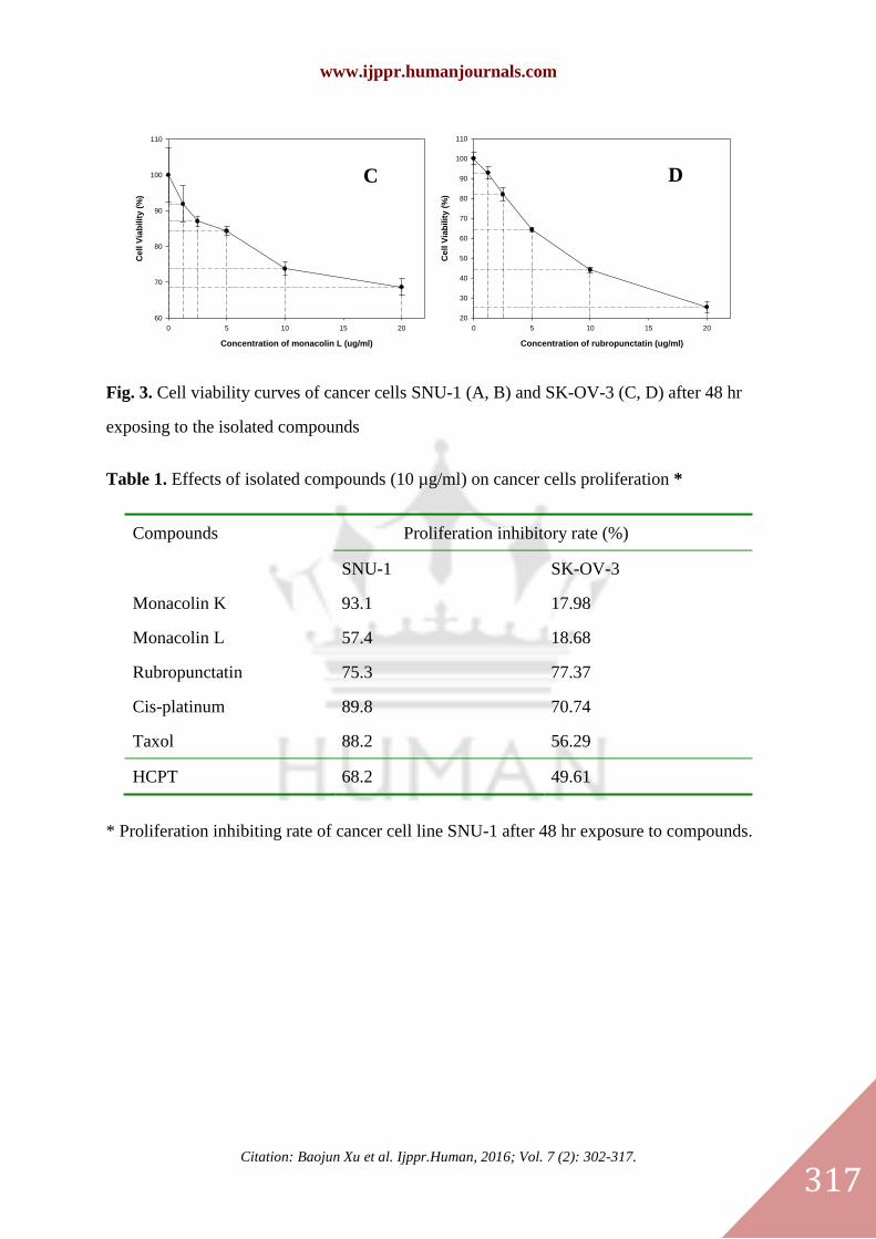
3.3. Anti-proliferation properties of the metabolites in red yeast rice
Anti-proliferation activities of isolated compounds (at the concentration of 10 μg/mL) from
red yeast rice against gastric cancer cell line SNU-1 and SK-OV-3 were done by MTT assay.
The percentage inhibitory rates of isolated compounds and positive anticancer drugs against
SNU-1 and SK-OV-3 cell proliferation were listed in Table 1. The results showed that
monacolin K, monacolin L, and rubropunctatin possessed strong anticancer activities against
gastric cancer cells SNU-1, the inhibition rates were 93.1%, 57.4%, and 75.3%, respectively.
These values were higher or close to that of the positive anticancer drugs (cis-platinum, taxol,
and HCPT) at a concentration of 10 μg/mL. Rubropunctatin also exhibited strong inhibition
activity against SK-OV-3; its inhibition rate (77.4%) was higher than that of positive
anti-cancer drugs at a concentration of 10 μg/mL.
Dose-dependent inhibitory effect and IC50 value of the isolated compounds were further
investigated using cell lines SNU-1 and SK-OV-3. After 48 hr exposing to the isolated
compounds with various concentrations, the cell viability curves (Fig. 3) of SNU-1 and
SK-OV-3 were created by software Sigma Plot 2001 at three parallel level treatments. The
IC50 values of the isolated compounds were calculated from 3rd-degree polynomial fit by
using software CurveExpert 1.3. The IC50 values of moma colin L against cancer cells SNU-1
and SK-OV-3 were 9.8 μg/mL and 28.9 μg/mL, respectively. The IC50 values of
rubropunctatin against cancer cells SNU-1 and SK-OV-3 were 13.3 μg/mL and 8.2 μg/mL,
respectively.

www.ijppr.humanjournals.com
Citation: Baojun Xu et al. Ijppr.Human, 2016; Vol. 7 (2): 302-317. 312
4. CONCLUSIONS
In summary, four compounds (Two statins, one flavonoid and one red pigment) were isolated
from solid fermentation products (red yeast rice) of M. ruber. The four compounds were
identified as monacolin K, monacolin L, apigenin, and rubropunctatin, respectively by mass
spectrum and 13
C and 1H-NMR spectral techniques. Cytotoxic assay indicated that monacolin
L and rubropunctatin possessed potent anticancer activities against cancer cells SNU-1 and
SK-OV-3. Therefore, as conventional cholesterol-lowering agents, red yeast rice, and its
chemical compositions have a great potential to be developed into anticancer candidate
agents.
Abbreviation used
RYR, red yeast rice; MTT, 3-(4, 5-dimethylthiazolyl-2)-2, 5-diphenyletrazolium bromide;
NMR, Nuclear Magnetic Resonance; HCPT, 10-hydroxy-camptothecin; HPLC,
High-Performance Liquid Chromatography.
Acknowledgments
This work was supported by venture companies developing funding in Chungnam National
University, South Korea. The authors thank Dr. Hae-Kap Cheong and Mi-Hee Kim in KBSI
(Korea Basic Science Institute, Korea) for recording NMR and MS respectively.
REFERENCES
1. Agarwal, B., Halmo, B., Feoktistov, A. S., Protiva, P., Ramey, W. G., Chen, M., Pothoulakis, C., Lamount,
J. T., & Holt, P. R. (2002). Mechanism of lovastatin -induced apoptosis in intestinal epithelial cells.
Carcinogenesis, 23, 521-528.
2. Akihisa T., Tokuda, H., Ukiya, M., Kiyota, A., Yasukawa, K., Sakamoto, N., Kimura, Y., Suzuki, T.,
Takayasu, J., & Nishino, H. (2007). Antitumor-initiating effects of monascin, an azaphilonoid pigment from
the extract of Monascus pilosus fermented rice (red-mold rice). Chemistry & Biodiversity, 2, 1305-1309.
3. Bach, T. J. (1986). Hydroxymethylglutary-CoA reductase, a key enzyme in phytosterol synthesis? Lipids,
21, 82-88.
4. Brown, A. G., Smale, T. C., & King, T. J. (1976). Crystal and molecular structure of compactin, a new
antifungal metabolite from Penicillium brevicompactum. Journal of the Chemical Society, Perkin
Transactions 1, 1165-1170.
5. Campoy, S, Rumbero, A., Martin, J. F., & Liras, P. (2006). Characterization of an hyperpigmenting mutant
of Monascus purpureus IB1: Identification of two novel pigment chemical structures. Applied Microbioloy
and Biotechnology, 70, 488-496.
6. Carmichael, J., Degraff, W. G., Gazdar, A. F., Minna, J. D., & Mitchell, J. B. (1987). Evaluation of a
tetrazolium-based semi-automated colorimetric assay: assessment of chemosensitivity testing. Cancer
Research, 47, 936–94

www.ijppr.humanjournals.com
Citation: Baojun Xu et al. Ijppr.Human, 2016; Vol. 7 (2): 302-317. 313
7. Chan, K. K. W., Oza, A. M., & Siu, L. L. (2003). The statins as anticancer agents. Clinical Cancer
Research, 9, 10-19.
8. Cheng, M. J., Wu, M. D., Chen, I. S., Tseng, M., & Yuan, G. F. (2011). Chemical constituents from the
fungus Monascus purpureus and their antifungal activity. Phytochemistry Letter, 4, 372-376.
9. Cheng, M. J., Wu, M. D., Su, Y. S., Chen, I. S., & Yuan, G. F. (2012). Anti-inflammatory compounds from
Monascus pilosus-fermented rice. Phytochemistry Letter, 5, 63-67.
10. Dimitroulakos, J., Ye, L. Y., Benzaquen, M., Moore, M. J., Kamel-Reid, S., Freedman, M. H., Yeger, H., &
Penn, L. Z. (2001). Differential sensitivity of various pediatric cancers and squamous cell carcinomas to
lovastatin-induced apoptosis: Therapeutic implications. Clinical Cancer Research, 7, 158-167.
11. Gutierrez, G. E., Mundy, B., Rossini, G., Garrett, R., Chen, S.T., & Mungy, G. R. (2006). Red yeast rice
stimulates bone formation in rats. Nutrition Research, 26, 124-129.
12. Heber, D., Yip, I., & Ashley, J. M. (1999). Cholesterol-lowering effects of a proprietary Chinese red yeast
rice dietary supplement. American Journal of Clinical Nutrition 69, 231-236.
13. Hong, M. Y., Seeram, N. P., Zhang, Y., & Heber, D. (2008). Anticancer effects of Chinese red yeast rice
versus monacolin K alone on colon cancer cells. The Journal of Nutritional Biochemistry, 19, 448-458.
14. Hsu, L. C., Hsu, Y. W., Liang, Y. H., Liaw, C. C., Kuo, Y. H., & Pan, T. M. (2012). Induction of apoptosis
in human breast adenocarcinoma cells MCF-7 by monapurpyridine A, a new azaphilone derivative from
Monascus purpureus NTU 568. Molecules, 17, 664-673.
15. Hsu, Y. W., Hsu, L. C., Liang, Y. H., Kuo, Y. H., & Pan, T. M. (2012). New bioactive orange pigments
with yellow fluorescence from Monascus-fermented dioscorea. Journal of Agricultural & Food Chemistry,
59, 4512-4518.
16. Jeon, T., Hwang, S. G., Hirai, S., Matsui, T., Yano, H., Kawada, T., Lim, B. O., & Park, D. K. (2004). Red
yeast rice extracts suppress adipogenesis by down-regulating adipogenic transcription factors and gene
expression in 3T3-L1 cells. Life Sciences, 75, 3195-3203.
17. Knecht, A., Cramer, B., & Humpf, H. U. (2006). New monascus metabolites: Structure elucidation and
toxicological properties studied with immortalized human kidney epithelial cells. Molecular Nutrition &
Food Research, 50, 314-321.
18. Kohama, Y., Matsumoto, S., Mimura, T., Tanabe, N., Inada, A., & Nakanishi, T. (1987). Isolation and
identification of hypotensive principle in red-mold rice. Chemical & Pharmaceutical Bulletin 35,
2484-2489.
19. Li, C., Zhu, Y., & Wang, Y. (1998). Monascus purpureus fermented rice (red yeast rice): a natural food
product that lowers blood cholesterol in animal models of hypercholesterolemia. Nutrition Research, 18,
71-78.
20. Lin, W. Y., Song, C. Y., & Pan, T. M. (2006). Proteomic analysis of Caco-2 cells treated with monacolin K.
Journal of Agricultural & Food Chemistry, 54, 6192-6200.
21. Lin, Y. L., Wang, T. H., Lee, M. H., & Su, N. W. (2008). Biological active components and nutraceuticals
in the Monascus-fermented rice: a review. Applied Microbiology and Biotechnology, 77, 965-973.
22. Ma, J. Y., Li, Y. G., & Ye, Q. (2000). Constituents of red yeast rice, a traditional food and medicine.
Journal of Agricultural & Food Chemistry, 48, 5220-5225.
23. Martinkova, L., Juzlova, P., Kren, V., Kucerova, Z., Havlicek, V., Olsovsky, P., Hovorka, O., Rihova, B., &
Vesely, D. (1999). Biological activities of oligoketide pigments of Monascus purpureus. Food Additive &
Contaminations, 16, 15-24.
24. Moon, Y. J., Wang, Q. J., Xu, B. J., & Sung, C. K. (2001). Simultaneous analysis of both latone form and
acid form monacolin K in red yeast rice by HPLC. Korean Journal of Food & Nutrition, 14, 521-526.
25. Roh, J. H., Moon, H. I., & Zee, O. P. (2000). Phytochemical constituents from Melampyrum roseum var.
hirsutum Beauv. Korean Journal of Pharmacognosy, 31, 157-162.
26. Schonberg, G. A., Joshua, H., & Lopez, M. B. (1981). Dihydromevinolin, a potent hypocholesterolemic
metabolite produced by Aspergillus terreus. The Journal of Antibiotics, 34, 507-512.
27. Tony, Y. K., Lam, V. P., & Gullo, R. T. (1981). Dihydrocompactin, a new potent inhibitor of
3-hydroxy-3-methylglutaryl from Penicillium citrinin. The Journal of Antibiotics, 34, 614-616.

www.ijppr.humanjournals.com
Citation: Baojun Xu et al. Ijppr.Human, 2016; Vol. 7 (2): 302-317. 314
28. Ukomadu, C., & Dutta, A. (2003). p21-dependent inhibition of colon cancer cell growth by mevastatin is
independent of inhibition of G1 cyclindependent kinases. Journal of Biological Chemistry, 278,
43586–43594.
29. Wang, Q. J. Fermentology. Ph.D. Thesis. Chungnam National University, Daejon. 2003. 76-77.
30. Wild, D., Toth, G., & Humpf, H. U. (2002). New Monascus metabolite isolated from red yeast rice
(Angkak, Red Koji). Journal of Agricultural & Food Chemistry, 50, 3999-4002.
31. Wild, D., Toth, G., & Humpf, H. U. (2003). New Monascus metabolites with a pyridine structure in red
fermented rice. Journal of Agricultural & Food Chemistry, 51, 5493-5496.
32. Wong, R., & Rabie, B. (2008). Chinese red yeast rice (Monascus purpureus-fermented rice) promotes bone
formation. Chinese Medicine, 3, 4.
33. Wu, M. D., Cheng, M. J., Yuan, G. F., Yech, Y. J., & Chen, I. S. (2010). A new pyrrole derivative from the
extracts of the fungus Monascus pilosus-fermented rice. Acta Chimica Slovenica, 57, 305-309.
34. Xu, B. J., & Chang, S. K. C. (2012). Comparative study on anti-proliferation properties and cellular
antioxidant activities of commonly consumed food legumes against nine human cancer cell lines. Food
Chemistry, 134, 1287-1296.
35. Xu, B. J., Wang, Q. J., Jia, X. Q., & Sung, C. K. (2005). Enhanced lovastatin production by solid state
fermentation of Monascus ruber. Biotechnology and Bioprocess Engineering, 10, 78-84.
36. Yasukawa, K. M., Takahashi, S., Yamanouchi, S., & Takido, M. (1996). Inhibitory effect of oral
administration of Monascus pigment on tumor promotion in two-stage carcinogenesis in mouse skin.
Oncology, 53, 247-249.
37. Zheng, Y. Q., Xin, Y. W., Shi, X. A., & Guo, Y. H. (2010). Cytotoxicity of Monascus pigments and their
derivatives to human cancer cells. Journal of Agricultural & Food Chemistry, 58, 9523-9528.
38. Zhu, L., Yau, L. F., Lu, J. G., Zhu, G.Y., Wang, J. R., Han, Q. B., Hsiao, W. L., & Jiang, Z. H. (2012).
Cytotoxic dehydromonacolins from red yeast rice. Journal of Agricultural & Food Chemistry, 60, 934-939.

www.ijppr.humanjournals.com
Citation: Baojun Xu et al. Ijppr.Human, 2016; Vol. 7 (2): 302-317.
315
Fig.1. Typical chromatogram and spectrums (from 190 to 600 nm) of 9 typical peaks of red
yeast rice by HPLC/UV/PDA.

www.ijppr.humanjournals.com
Citation: Baojun Xu et al. Ijppr.Human, 2016; Vol. 7 (2): 302-317.
316
CH3
CH3
O
O
OH O
CH3
CH3
O
H
12
3
44a
8a
5
6
78
7'
6'
5'4'
3'
2'1'
1''2''
3''
4''
CH3
CH3
O
OH O
H
12
3
44a
8a
5
6
78
7'
6'
5'4'
3'
2'1'
1 2
O
O
OH
OH
OH
1
2
345
6
7
8
9
10
1'
2'
3'
4'
5'
6'
O
O
O
O
O
CH3
H11
C5
H3C 1
2
3
3a
4
4a
8a 9a
9
8
711
12
13
6
5
10
14
15-19
3 4
Fig. 2. Chemical structures of isolated compounds from red yeast rice. 1. monacolin K; 2.
monacolin L; 3. apigenin; 4. rubropunctatin
Concentration of monacolin L (ug/ml)
0 5 10 15 20 25
Ce
ll V
iab
ilit
y (
%)
30
40
50
60
70
80
90
100
110
Concentration of rubropunctatin (ug/ml)
0 10 20 30 40 50
Ce
ll V
iab
ilit
y (
%)
10
20
30
40
50
60
70
80
90
100
110
A B
A
www.ijppr.humanjournals.com
Citation: Baojun Xu et al. Ijppr.Human, 2016; Vol. 7 (2): 302-317.
317
Concentration of monacolin L (ug/ml)
0 5 10 15 20
Ce
ll V
iab
ilit
y (
%)
60
70
80
90
100
110
Concentration of rubropunctatin (ug/ml)
0 5 10 15 20
Ce
ll V
iab
ilit
y (
%)
20
30
40
50
60
70
80
90
100
110
Fig. 3. Cell viability curves of cancer cells SNU-1 (A, B) and SK-OV-3 (C, D) after 48 hr
exposing to the isolated compounds
Table 1. Effects of isolated compounds (10 µg/ml) on cancer cells proliferation *
Compounds Proliferation inhibitory rate (%)
SNU-1 SK-OV-3
Monacolin K 93.1 17.98
Monacolin L 57.4 18.68
Rubropunctatin 75.3 77.37
Cis-platinum 89.8 70.74
Taxol 88.2 56.29
HCPT 68.2 49.61
* Proliferation inhibiting rate of cancer cell line SNU-1 after 48 hr exposure to compounds.
C
A
D
C
A