separation of peptides by high-performance liquid chromatography on a weak anion-exchange bonded...
TRANSCRIPT

Journal of Chro~rography. 237 (1982) 41742.8 Qscvier Sciei1t5c PubGstig Company, Amnerdam - F’rintcd in The NethcrlaEds
CHROM. 14,548
SEPARATION OF PEPTIDES BY HIGH-PERFORMANCE LIQUID CHRO- MATOGRAPHY ON A WEAK ANION-EXCHANGE BONDED PHASE
MIRAL DIZDAROGLU*
Cenrer for Radkiion Research, National Bureau of Sranbrds, Washington, DC 200234 (USA.)
HENRY C. KRUTZSCH
Laboratory of fmmmogenetics, National Institute of Allergy and Infectious Diseases, National Institutes of
Health, Bethesab, MD 20205 (U.S.A.)
and
MICHAEL G. SIMIC Cenrer for Radiation Research, National Bureau of Stanakds, Washingron, DC 20234 (U.S.A.)
(Received November ilth, 1981)
SUMMARY
Multicomponent peptide mixtures were separated by high-performance liquid chromatography on a MicroPak AX-10 column, a silica-based bonded-phase weak anion exchanger. A gradient of increasing 0.01 M triethylammonium acetate buffer (pH 6.0) into acetonitrile was usually used for elution. For peptides containing a number of acidic amino acids without compensating basic residues, such as delta sleep-inducing peptide, a dilute 0.04 M formic acid solution (pH 2.6) was employed as the eluent. Peptides of up to ahout 30 residues were successfully tested, including peptides such as somatostatin, neurotensin, ribonuclease s-peptide, cc-endorphin, glu- cagon, and various angiotensins and bradykinins. Tryptic digests of horse heart cytochrome c, cahnodulin and rtzluced and alkylated hen egg-white lysozyme were also successfully examin ed_ Because of the volatility of the eluents used, peptides can be readily isolated for further investigation. Recoveries of over 80 % were observed in those cases tested by comparative amino acid analysis_
INTRODUCX-ION
In the past several years, high-performance liquid chromatography (HPLC) has become one of the most important techniques for peptide separation. Reversed-
phase HPLC, utilizing a variety of solvent systems’-‘“, has been the type most widely used for this purpose. lMore recently, HPLC employing a macroreticular anion-ex- change column was used to separate tryptic digestion mixtures of peptides15.
We recently reported a method for the separation of dipeptides, including resolution of sequence isomeric and diastereoisomeric dipeptides, by HPLC on a weak anion-exchange bonded phase using mixtures of triethylammonium acetate (TEAA) buffer and acetonitrile for elution I6 . In this paper, we describe the separation
002b%73/8275 0 1982 Ekevicr Scie&xic Publishing Company

418 M. DIZDAROGLU, H. C. KRUTZSCH, M. G. SIMIC
of a large variety of peptides containing up to approximately 30 amino acid residues using a similar system. These peptides include somatosta+&, a-endorphin, ribonu- cIeasc s-peptide, glucagon. various angiotensins and bradykinins as well as uyptic digestion mixtures of horse heart cytochrome c, calmodulin and reduced and alkyl- ated hen egg-white lysozyme- This methodolo_gy has been developed for the study of the effects of ionizing radiation on peptides.
EXPERIMENTAL*
Apparatus Separations were performed on a Hewiett-Packard Model 10843 liquid chro-
matograph (HewIett-Packard, Avondale, PA, U.S.A.) equipped with a micropro- cessor, an automatic injector and a variable waveIength detector. The buffer solutions were filtered (Millipore, Bedford, MA, U.S.A.) prior to use. All separations were carried out on a 30 x 0.4 cm MicroPak AX-IO column (Varian, Walnut Creek, CA, U.S.A.), a difunctional weak anion-exchange bonded phase prepared on LiChrosorb Si-60 silica (10 m)“. Prior to chromatography of most peptide mixtures, the coiumn was first equilibrated with at least 50 ml of 0.01 M TEAA pH 6.0 buf%r, to insure that cohnnn pH was also 6.0. Then, after column equilibration with the starting soIution, usually aeetonitrile-buffer (75125), elution was begun with a gradient terminating at 100% buffer. Specific conditions are described in the Figures. For elution of acidic peptides, an isocratic flow of 0.04 M formic acid pH 2.6 buffer was employed. Again. the cohunn was first pre-equilibrated with at least 20 ml of this eluent to insure that cohmn pH was also 2.6.
Materials Peptides were obtained from Si_gma (St. Louis, MO, U.S.A.), Research Plus
(Bayonne, NJ, U.S.A.) and Beckman (Palo Alto, CA, U.S.A.)_ Triethylamine was purchased from Eastman Kodak (Rochester, NY, USA) and puri&cl by distil- lation_ Btier solutions were prepared by titrating 0.01 M acetic acid solutions with rriethylamine to pH 6.0. Glass-distilled water and acetonitriie were purchased from Burdick &Jackson Labs. (Muskegon, MI, U.S.A.). Trypsin was obtained from Boeh- ringer (Indianapolis, IN, U.S.A.).
Trytic digestion Tryptic digestions were carried out according to the procedure described by
Margoliash”_ 0.3 /unoI of peptide wnre dissolved in 0.1 ml of 0.03 M Tris-HCl buffer (pH 8.0). 0.1 mg of trypsin was added and the mixture was incubated at 40°C for 24 h.
* Certain commercial equipam& instruments. or material% are i&n&&d ia this pzper in order to - _ _ - adequately specify the experimental procedure. Such iden@cation does not inzply recommendation or endorsement by the National Bureau of Standards. nor does it imply that the materials or equipment iwtifigd zre necessarily the best avaihble for the purpose.

HPLC OF PEFl-IDES 419
Amino acid analysis For recovery studies, the eluted peptides were dried in VQCKO and hydrolyzed
with constant boiling HCI in evacuated and se&d tubes at 110°C for 24 h. The hydrolyzatcs were analyzed on a Durrum D-500 amino acid analyzer.
RESULTS AND DISCUSSION*
The separation of a multicomponent peptide mixture is shown in Fig. 1, and peak identifications are given in Table I. Excellent peak symmetry for all peptides in this mixture was obtained with the exception of somatostatin (peak I), which gave a tailing peak_ Glucagon (peak 9) gave two peaks; amino acid analysis of collected material showed the same amino acid composition for both components. Ah of the other peptides in this chromatogram gave a single peak when freshly prepared solu- tions were individually injected. The 0.01 A4 TEAA pH 6.0 buffer used with acetoni- trile in the ahove separations is the usual gradient system employed in peptide separa- tion. It buffers well at the pH employed and .is compatible with the pumps and coIumn_ It also allows sensitive detection of peptides at wavelengths in the range of 210-225 nm. Furthermore, this buffer is readily removed from eluted compounds by freeze-drying”, facilitating the easy recovery of the separated peptides. Some pep
20 40 60 60 TIHE IminI
Fig- I- Separation of various peptides Colums MicroPak AX-10 (10 m). 30 x 0.4 cm. Temperature, 3O’C. Eked: A, zcetonitriie; B, 0.01 M triethylammonium acetate (pH 6.0), gradient program: iinear starting from 25% B with a rate of 1 o/0 B per min. Ffow-rate, 1 ml/min. Peak idemifieation and sequences are @en in Table I. Amount of injecticn, OS-5 g.g per peptide.
* Abbreviations for amino xids follow IUPAC-IUB recommendations (see Biochem. J., 126 (1972) 773)_

Peak
fig_ J I
2 3
4
5
6 7
S 9
10
II
Fig_ 2 1 2 3
4 5 6 7
Eg. 3 1
2 3 4 5
Eg. J ! 2
3
Fig_ 5 I 2 3 4 5 6 7
420 M. DIZDAROGLU, I-L Cm KRUTZSCH, Mm G- SiMIC
TABLE I
PEAK IDENTIFIC4TION AND SEQUENCES IN FIGURES
Sornatostatin
Roctoiin Neurotcnsin
Met-Enkephalin Btadykinin p4Jtentiator c - a-Endorphin
EAE-peptide Glucagon -._
R.&onucIease s- peptide (RSP) IgE-peptide
FWn~nuckase s-peptide igE-peptide Glutathione o.xidized form
DSIP - -
AIlgioterl& III (A Iii) (Sar’-AIa*)-A II -4 II
&&* II
Bradykitin Me& Lys- Bmdy!&in Lys-Brady%
-
Ah-GI)4&.-L~S TrpLys-Thr-Fhe-Thr-Ser-Cjx Arg-Tyr-Lm-R*Thr pGIu-Lx-Tyr-GbAsn-Lys-R* .Qg-_R_T>T-Ik-I&U Tyr-Gly-Gly-Pk-Met pClu_Cly-Le~R~R~ly-~~~~~r~ _
Lys-GIu-Th-Tyr-Se-Lys - - Tyr-Gly~Iy-Pellet-ThrSer-GIu- L+!ier-Gln-T&r-Ro-ku-VaI-Thr Phe-Ser-TrpCly-AIa-Glu-Gly~ln-Arg Hk-Ser-GIn-GIy--ihr-Phe-Thr-Ser-&pTyr- ser-Lys_“r4r-lcu-AspSer-Arg-Ar%-Ala-Gln- Asp-Phc-Val-Gln-Trp-Leu&~et-Asn-Thr Lys-GIu-Thr-Ala-AIa-Ala-LpPhe-GIu-Arg- G~His-Met-~~-~-~-~-~~_~ Asp-Se-AspRc+Arg
see above see above pGll.l-C~s-Gl_V
I ~-Glu-Cys-Gly Phe-Leu-Glu-Glu-Ile Trp-~a~ly~iy-~~~~r~ly-Gfu j-GIL+Leu ~-GlU-GlU
Xr,-\ral-TJ;r-e-HRo-Phe
Sar-_4rg-Val-Tyr-Ile-I-ik-Pro-AIa Asp-Arg-Val-Tyr-11&-iiiRo-Phe Asp-Arg-Val-Tjcik-His-Pro-Phe-His&m Asp-AreVal-Tyr-Val-His-Pro-Phe
ArePro-Ro-GIy-Phe-Ser-Pr+Phe-Arg Met-Lys-Arg-Ply-Phe+Ser-Prc+PhcArg
Leu-Trp-Met-Ar@he-Ala ku-Trp-Mei-Arg-Phe Leu-Trp-Met-Arg Met-ArePIte-iUa Met--Phe Arg-Pi+Ala Leu-Trp-Met

HPLC OF PEPTiDEs
20 LO TIME Iminl
Fig_ 2- wtion of some acidic peptides. Column z+s in Fig. I_ Temperature. 6O’C. Euent. 0.04 M formic acid (pH 26). Fiow-rztc, 1 mlimio. Peak identification and sequences are given in Table I. Amount of iojecGon ss in Fig_ I_
tides which contain a number of acidic amino acids with no compensating basic residues, such as delta sleep-inducing peptides (DSIP) (see Table I) or the dipeptide y- Giu-Glu, have unaccqtably long retention times when the elution conditions de- scribed in Fig. I are used. These peptides were chromatographed using an isocratic fIow of dilute formic acid (pH 2.6) instead as the eluent, as is shown in Fig. 2. Both ribonuclease s-peptide (RSP) and IgE-peptide (peaks 1 and 2, respectively) could also be eluted with the solvent system described in Fig. 1. Although RSP contains three acidic amino acids (see TabIe I), it elutes much earlier than the other peptides in this mixture. This is probably due to the previously mentioned compensation of the acidic amino acids by the three basic amino acids or possibly steric hindrance to interaction
of the carboxy soups with the stationary phase With increasing chain length, con- formation of the peptide may also play a role. Thus, the pentapeptide with two Glu residues (peak 4) and DSIP (peak 5) elute later than RSP and IgE-peptide. As migth he eqected from these other results, ~&lu-Glu has the longest retention time. Dilute formic acid solutions are also compatible with the column and pumps used. In ad- dition, formic acid is aIso vo!atiIe, allowing recovery of peptides for further use. As with the TEAA btier, peptide recoveries are SO o/o or greater. The figures also demon- strate that sensitiue detection of eluted peptides is also obtained with this eluent
Sepertzrhz of closely -ieiated peptides The weak anionexchange column is also capable of separating closely related
peptides such as angiotensins or bradykinins. For example. Fig. 3 shows the separa-

0 lC! 20 30
TIME [mid
M. DIZDAROGLU, H. C. KRUTZSCH, M. G. SIhiIC
0 10 20 TIME [mini
Fig_ 3. Separation of some angiotensins. Column details as in Fig. 1. Peak identikation and wquences are
given in Table 1. Amount of injection CLL 1 pg per peptide-
Fig_ 4. Separation of several bradykinins. Column details as in Fig. 1 except gradient program: linear starting from 25 7; B with a rate of 1.7 7; B per min. Peak identification and sequences are given in Table 1. Amount of injection, co. 2 pg per pcptide-
tion of a mixture of five angiotensins with excellent resolution, and ah angiotensins gave symmetrical peaks. The peak representing angiotensin II showed a shoulder {peak 3) which corresponds to an impurity in this peptide. The small peak in front of (Va15)-angiotensin II peak also represents an impurity_
The separation of three bradykinins is given in Fig. 4. B_mdykinin gave a symmetrical peak whereas Met,Lys-bradykinin and Lys-bradykinin (peaks 2 and 3, respectively) yielded somewhat broad peaks.
Fig. 5 shows the separation of the hexapeptide Leu-Trp-Met-Arg-Phe-Ala (Research Plus Labs.) from the synthetic fragments that would be obtained by diges- tion with proteolytic enzymes or chemical cIeavage_ Excellent separation of these _peptides was achieved by isocratic elution at 50°C.
Separation of tryptic digestion mixtures Enzymatic digestion is frequently employed in sequence analysis of large pep-
tides and proteins_ For this reason, the weak anionexchange column methodolo_q was also employed to separate the peptides resulting from tryptic digestion of horse heart cytochrome c, cahnoduhn or reduced and alkylated egg-white Iysoqme.
Fig. 6 shows the separation by gradient elution of the peptides resulting from tryptic digestion of horse heart cytochrome c. The elution became isocratic at 100% buffer after 60 min, and another four peaks were observed with continued flow of buffer. According to the sequence of horse heart cytochrome c given by Margohash et al_“, sixteen fragments and free Iysine are expected upon digestion with trypsin.

HPLC OF PEPTLDES 423
10 20 TiME [mini
Fig. 5. Separation of Leu-‘Trp-Met-e-Phe-Ala and its simulated digestion fragments. Column and duent as in Fig_ 1. Isocratic elution with 21 y0 B. Flow-rate. 1 ml/min. Temperature, 5O’C. Peak identifi- cation and sequences are given in Table I_ Amount of injection, ca. 1 pg per peptide.
Fourteen major peaks and some other small peaks were observed. Further work would be needed to characterize the separated fragments_ The large peaks probably represent the fragments containing Tyr and Trp residues20. Since some digestion products contain a number of acidic ammo acids, the separation of the above diges- tion mixture employing conditions described in Fig. 2 was also undertaken (i”ig:- 7). As expected, the majority of the fragments have no or little retention using these
0 30 60 90
TIME [mini
Fig. 6. Separation of a tqptic digest of horse heart cytochrome c. Column details as in Fig. 1 e=xpt ~~~pro~rlinearstattlngfrom25%Bwitharateof0.6%Bperminto~%Bthen4.5%B~min to 100% B. Amount of injection, ca. 10 nmol of cytochrome c.

424 M. DIZDAROGLU. H_ C. KRUTZSCH. M. G. SIMIC
10 20 TIME [mid
Fig_ 7_ Spration of a uyptic digest of how heart cytochrome c. Column details as in Fig. 2. Amount of injection. cu. 10 nmol of qtochrome c_
0 20 LO 60 TIME Cminl
Fig 8. Separation of a tryptic diw of calmodulh Column detaik as in Fig. 1 exapt gradient program: linear starting from 25 % B with a rate of 2 % B per mh Amount of injection, ca. 15 mnol of cahqdnlin_
HPLC OF PEgnDES 425
0 10 20 30 TIME [mini
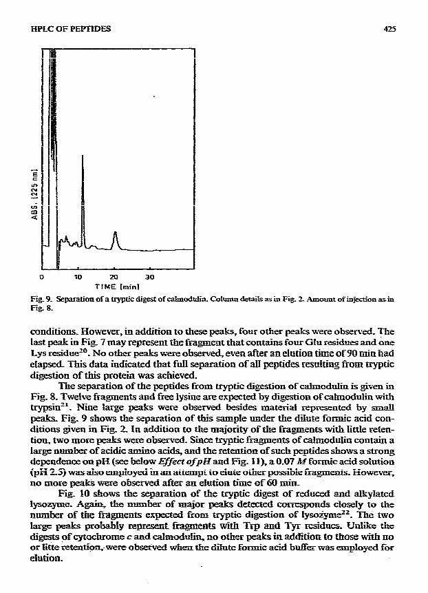
Fig_ 9. Sepxation of a tryptic digest of c&nodU. Co!umn details as in Fig Z Amount of injestion asin Fig. 8_
conditions_ However, in addition to these peaks, four other peaks were observed. The last peak in Fig_ 7 may represent the fngment that contains four GIu residues and bne Lys residuezO_ No other peaks were observed, even after an elution time of90 min had eIapsed_ This data indicated that full separation 0faJ.I peptides resulting from tryptic digestion of this protein was achieved.
The separation of the peptides from tqptic digestion of calmodulin is given in Fig. 8. Twelve fragments and free lysine are expected by digestion of cahnodulin with trypsin”_ Nine large peaks were observed besides material represented by SE&J peaks. Fig. 9 shows the separation of this sample under the d%te formic acid con- ditions given in Fig_ 2. Pn addition to the majority of the fragments with litt!e reten- tion, two more peaks were observed. Since tqptic fragments ofcalmoduiin contain a large numb of acidic amino acidq and the retention of such peptides shows a strong dependence on pH (see Mow Effect ofpH and Fig- I I), a 0.07 34 formic acid sohxtion (pH 2.5) was ako employed in awl attempt to elute other possibk fragments, However, no more pea& were observed a&x an eiution time of 60 ti
Fig_ 10 shows the separation of the tqptic digest of reduced and a&y?!ated lysoq-me- Again, the rmmber of major peaks detected con-esponds closeiy to the number of the fmgments expected from tryptic digestion of Iyso*ez2_ atre two large pe&s probably represetrk f&.gtnats with Trp and Tp residues. Unlike the digests of cytochnxne c and c&mod&in, so other peaks in ad$itioa to those with QO or litte ztentiQ%- were obsezked when the dikte formic acid b&k was tipluyed fw elution.

g.26 Si_ DIZDAROGLW. H. C_ KRUlZSCH, ht. G. SIhUC
0 15 30 L5 60 TIME Cminl
Fig. lo_ Sizparation of a tryptic digest of reduced and alkylated lysozyme- Column details as in Fig. 1 except temperature: aO’C_ Amount of injection. cu. 10 tmol of lysozyme.
Effect of temperature on retention and resolution
An increase in column temperature generally causes an increase in retention time. In some cases, resolution could be improved by increasing temperature to a certain limit starting from room temperature. For instance, the mixtures in Figs. 2 and 5 could be optimahy separated at 60°C and 50X, respectively. However, when gradient elution was used, an up-scale drift of the baseline was observed at tempera- tures above 40°C due to higher UV absorption of the buffer. Nevertheless, baseline drift is still acceptable, and temperatures up to 60°C can be used to improve resolu- tion of a given mixture if necessary_
Effect of pH In a recent paper, we described separation of dipeptides using the TEAA buffer-
acetomtriIe solvent system, except the pH of the TEAA buffer was 4.3 (ref. 16). In the present work with peptides, a pH value of 6.0 was more suitable in terms of retention, peak symmetry and resolution. The separation selectivity of the stationary phase thus shows a quite strong dependence on the pH value of the eluent. This means that any pH value between 4 and 6 with this btier system can be tried in order to improve resolution in a given separation using the chromatograpbic system de- scribed here.
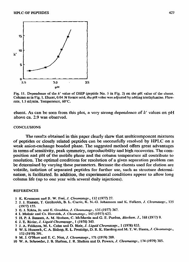
The retention of acidic peptides even more strongly depends on the pH value of the eluent, as can be seen by the conditions described in Fig. 2 for their Separation. This was further demonstrated in Fig. 11, where capacity factors (k’) of DSIP were plotted versus pH of the eluent_ In order to obtain a constant solvent strength, a 0.04 A4 formic acid solution was used and pH was changed by adding triethylamine to the
HPLC OF PEPTIDES 421
2.5 3-O 35
P”
Fig. il. Dependence of the k’ value of DSIP (peptide No. 5 in Fig. 2) on the pH value of the eluent. Column as in Fig. 1. Eluent, 0.04 .ci formic acid, the pH value was adjusted by adding triethylamine. Flow- rate, 1.5 ml~min. Temperature, 60°C.
eluent. As can be seen from this plot, a very strong dependence of k’ values on pH above ca. 2.9 was observed.
CONCLUSIONS
The rest&s obtained in this paper clearly show that multicomponent mixtures of peptides or closely related peptides can be successfully resolved by HPLC on a weak anionexchange bonded phase. The suggested method offers great advantages in terms of sensitivity, peak symmetry, reproducibility and high recoveries. The com- position and pH of the mobile phase and the column temperature all contribute to resolution. The optimal conditions for resolution of a given separation problem can be determined by varying these parameters_ Because the eluents used for elution are volatile, isolation of separated peptides for further use, such as structure determi- nation, is facilitated. in addition, the experimental conditions appear to allow long column life (up to one year with several daily injections).
REFERENCES
I
2 K. Krusmzen and R W. Frei, 1. Chrutnatogr_, 132 (1977) 27. J_ J. Hansen, T. Grceibrokk, B. L. Currie, K. N-G. Johansso n and K. Folkers, J_ Chromatogr., 135
(1977) 155. E. J. Rikta, Jr. and E. Grushka, J_ Chromtogr.. 135 (1977) 367. I. Moln5r and Cs. Hotiffi. .I_ Clrromtogr_. 142 (1977) 623_ H. P. J. Bennett, A. M. Hudson, C. McMartin and G. E. Purdon. biocizem. 3., 168 (1977) 9. J. E. Rivier, J_ Liquid Chrotnatogr., 1 (1978) 343. J_ k Feldman, M. L. Cohn and D. Blair, J_ Liquid Chromatogr., 1 (1978) 833. W_ S. Hancock, C. A Bishop, R L. Prcstidgc, D. R. K. Hardir?g aud M. T- W. Heam, J- Chronmtogr-, 153 (1978) 391_
9 M. J. O’Hare and E_ C. Nice, 1. Chrotnatogr., 171 (1979) 209. IO W. k Schroeder, J. B Shelton, J. R Shelton and D. Powars, J. Chrornatogr., 174 (1979) 3SS.
415 M DIZDAROGLU, He C KRUTZSCH. hf. G_ SIMIC
I 1 W_ C. Mahony and M_ A_ Hammdwn, X Bid_ Cfie, 255 (19%) II !39. 12 M_ Sc&nuh~fa and A_ Femer, J. CJzmmarogr_, 224 (i981) 472 13 Ed. N. Guy, G_ M. Roberson and L D_ Barnes, z4mzI_ t?iwhem. 112 (1981) 272. 14 S. Trrabc. Ii. xishi and -r_ hdo, L Ckromogr_. 212 (1981) 295. 15 N_ Tw T. isobt, H- EEzsai. K_ Seta and T_ Okuyamz, Ad Biochem, 115 (1981) ISL 16 Mm IXzdarodu and M. G_ simic, 1. Chmt~gr_ 195 (1980) 119_ 17 E H_ J5kisoi.1. 3. G. Lawkq C_ T. Wehr 2nd S_ R Abbott, J. Chomrogr, 174 (1979) 409. 1s E Mar@ia&. I_ Bid. C&m, 237 (f962) 2161. 19 J_ Pc%L-a& Nafure (Lo&n). 175 (1955) 478_ 20 E MaqoLiash E L Smith G. Krkl and H. Tuppy. Ahrue ~lotw5.m). 192 (i%l) 1125. 11 D_ M_ Wancrso~ F_ S&a&f znd T_ C_ V- J_ sini_ f&m__ 255 ( 1980) 962 22 It E C&d, 1 Pioi. Chem., 23s (19633 2698.