seminars in immunology - lambris · seminars in immunology 28 (2016) 285–291 contents lists...
TRANSCRIPT

Cp
GDa
b
1c
d
a
ARRAA
KCCTCPIP
1
motiitoota
in
S
h1
Seminars in Immunology 28 (2016) 285–291
Contents lists available at ScienceDirect
Seminars in Immunology
j ourna l ho me page: www.elsev ier .com/ locate /ysmim
omplement inhibition in pre-clinical models of periodontitis androspects for clinical application
eorge Hajishengallisa,∗, Evlambia Hajishengallisb, Tetsuhiro Kajikawaa, Baomei Wanga,espina Yancopoulouc, Daniel Ricklind, John D. Lambrisd
University of Pennsylvania, Penn Dental Medicine, Department of Microbiology, Philadelphia, PA 19104, USAUniversity of Pennsylvania, Penn Dental Medicine, Department of Preventive and Restorative Sciences, Division of Pediatric Dentistry, Philadelphia, PA9104, USAAmyndas Pharmaceuticals, Glyfada 16675, GreeceDepartment of Pathology and Laboratory Medicine, Perelman School of Medicine, University of Pennsylvania, Philadelphia, PA 19104, USA
r t i c l e i n f o
rticle history:eceived 8 February 2016eceived in revised form 12 March 2016ccepted 14 March 2016vailable online 24 March 2016
eywords:
a b s t r a c t
Periodontitis is a dysbiotic inflammatory disease leading to the destruction of the tooth-supporting tis-sues. Current therapies are not always effective and this prevalent oral disease continues to be a significanthealth and economic burden. Early clinical studies have associated periodontitis with elevated comple-ment activity. Consistently, subsequent genetic and pharmacological studies in rodents have implicatedthe central complement component C3 and downstream signaling pathways in periodontal host-microbeinteractions that promote dysbiosis and inflammatory bone loss. This review discusses these mechanis-
omplement3herapeuticsompstatin cp40rimate modelsnflammationeriodontitis
tic advances and moreover focuses on the compstatin family of C3 inhibitors as a novel approach totreat periodontitis. In this regard, local application of the current lead analog Cp40 was recently shownto block both inducible and naturally occurring periodontitis in non-human primates. These promisingresults from non-human primate studies and the parallel development of Cp40 for clinical use highlightthe feasibility for developing an adjunctive, C3-targeted therapy for human periodontitis.
© 2016 Elsevier Ltd. All rights reserved.
. Introduction
Periodontitis is an oral disease driven by dysregulated inflam-ation induced by polymicrobial dysbiotic communities that form
n subgingival tooth sites [1]. The disease can lead to the destruc-ion of the periodontium (i.e., the tooth-supporting structures,ncluding gingiva, periodontal ligament, and the alveolar bone) and,f untreated, can lead to tooth loss and possibly impaired mastica-ion [2]. Nearly half of adults in the U.S. are affected by some formf periodontal disease [3]. In its severe form that afflicts nearly 10%f adults [3,4], periodontitis is associated with increased risk of cer-ain systemic conditions, such as atherosclerosis and rheumatoidrthritis [5,6].
As alluded to above, periodontitis is not a bacterial infectionn the classical sense, i.e., it is not caused by specific exoge-ous pathogen(s) [7,8]. Moreover, the implicated resident dysbiotic
∗ Corresponding author at: University of Pennsylvania, Penn Dental Medicine, 240outh 40th Street, Philadelphia, PA 19104-6030, USA.
E-mail address: [email protected] (G. Hajishengallis).
ttp://dx.doi.org/10.1016/j.smim.2016.03.006044-5323/© 2016 Elsevier Ltd. All rights reserved.
communities (pathogenic dental plaque biofilm), while necessary,are not sufficient to precipitate periodontitis [9]. This is becauseperiodontal tissue destruction is predominantly mediated by thehost inflammatory response to the microbial challenge [10–12].This understanding has provided a strong rationale for developingstrategies that modulate the host microenvironment to treat peri-odontitis. Such novel approaches can be used in an adjunctive modeto enhance current therapies (e.g., mechanical removal of the dentalplaque biofilm), which are not always adequate to control peri-odontitis [13–15]. Indeed, periodontitis continues to be a seriouspublic health issue with a substantial economic burden [2,16,17].
Therefore, the identification of key inflammatory pathways thatdrive periodontal tissue destruction has important translationalimplications and hence received particular attention in the pastdecade. In this context, we discuss recent proof-of-concept studiesin preclinical models that have established a causal relationshipbetween periodontitis and complement, thereby supporting the
usefulness of complement-targeted therapies in this oral disease.
286 G. Hajishengallis et al. / Seminars in Immunology 28 (2016) 285–291
Fig. 1. Complement and its involvement in periodontal dysbiosis and inflammatory bone loss. (A) Complement cascade: The classic, lectin, and alternative pathways convergeto activate C3 leading to the generation of effector molecules. These include the inflammatory anaphylatoxins C3a and C5a, which respectively activate C3aR and C5aR1,which moreover cross-talk with TLRs. Intriguingly, the keystone periodontal pathogen P. gingivalis can directly activate C5aR1 through its arginine-specific gingipains thatcan cleave C5 to generate biologically active C5a. C3b is an opsonin that promotes microbial opsonization. The cleavage of C5 by its convertase (C3bBb3b) also generates C5bwhich in the terminal pathway initiates the assembly of the C5b-9 membrane attack complex (MAC), which induces lysis of susceptible targeted microbes. The alternativepathway C3 convertase, C3bBb, is also involved in an amplification loop for all complement pathways. Compstatin and derivative drugs, such as Cp40, block C3 activation,thus inhibiting all activities downstream of C3. (B) Dysbiotic inflammation: C5aR1 is involved a subversive crosstalk with Toll-like receptors (TLR) leading to the remodelingof a symbiotic microbiota into a dysbiotic one. This cross-talk is instigated by keystone pathogens (see text for details). The resulting dysbiotic microbial community causesi k. Inflap cerbaT is in n
2
arTpoititbtaawtF
nflammation that is largely dependent on complement (C3aR, C5aR1)-TLR crosstalroducts are used as nutrients by the dysbiotic microbiota, which thus further exaherapeutic blockade of C3 activation/cleavage using Cp40 has blocked periodontit
. Complement and periodontitis
Complement represents a network of interacting fluid-phasend cell surface-associated molecules that activate, amplify, andegulate immune and inflammatory response pathways [18].he integrated complement system includes the classic serumroteins (C1-9), pattern-recognition molecules, convertases andther proteases, regulators, and receptors for interactions withmmune mediators, which together coordinate the host responseo infection or tissue injury [18]. For instance, complement-basednteractions can amplify host immune and inflammatory responseshrough synergy with Toll-like receptors (TLRs) [19–21], provide aarrier against the spread of infections by potentiating local clot-ing [22,23], and regulate the activation and differentiation of B cellsnd T-cell subsets [24–28]. The complement cascade can be initi-ted by distinct mechanisms (classical, lectin, or alternative), all of
hich converge at C3, the third complement component, leading tohe generation of cleavage products that mediate various functions.or instance, convertase-mediated cleavage of C3 and C5 generates,
mmation and dysbiosis reinforce each other since inflammatory tissue breakdowntes inflammation and ultimately leads to bone loss, the hallmark of periodontitis.on-human primates.
respectively, the anaphylatoxins C3a and C5a which activate spe-cific G-protein-coupled receptors (C3aR and C5aR1 [CD88]) that notonly mediate recruitment and activation of inflammatory cells butalso cross-talk with TLR signaling pathways [20]. Moreover, othermolecules, such as C4b and C3b, act as opsonins promoting micro-bial opsonization and phagocytosis, whereas the so-called terminalpathway culminates in the C5b-9 membrane attack complex whichcan directly lyse susceptible targeted microbes [18] (Fig. 1A).
The possible involvement of complement in human periodonti-tis was first recognized in the 1970s and 1980s by clinical studiesthat associated the disease with the presence of complementactivation fragments [29–35]. Moreover, successful periodontaltherapy (i.e., that reduced clinical indices that measure periodon-tal inflammation and tissue destruction) resulted in decreased C3activation in the gingival crevicular fluid (GCF) [36]. Conversely,in an experimental gingivitis study in human volunteers, the pro-
gression of gingival inflammation was correlated with increased C3cleavage in the GCF [37]. These early studies therefore suggestedthat complement is involved in the pathogenesis of periodon-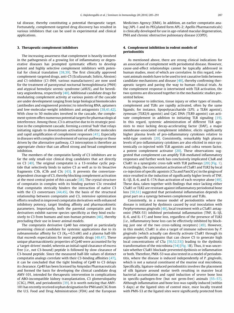
rs in Im
tFva
3
ieptcafatma(aWmitiaidai
fotfdaotwreipdsp
pstuafCciiaAo(1t
G. Hajishengallis et al. / Semina
al disease, thereby constituting a potential therapeutic target.ortunately, complement-targeted drug discovery has resulted inarious inhibitors that can be used in experimental and clinicalpplications.
. Therapeutic complement inhibitors
The increasing awareness that complement is heavily involvedn the pathogenesis of a growing list of inflammatory or degen-rative diseases has prompted systematic efforts to developotent and highly selective complement inhibitors with poten-ial for clinical translation [38,39]. The first clinically approvedomplement-targeted drugs, anti-C5 (Eculizumab; Soliris, Alexion)nd C1-inhibitor (C1-INH; various manufacturers) are now usedor the treatment of paroxysmal nocturnal hemoglobinuria (PNH)nd atypical hemolytic uremic syndrome (aHUS), and for heredi-ary angioedema, respectively [40]. Additional candidate drugs for
odulating complement activity at various points of the cascadere under development ranging from large biological biomoleculesantibodies and engineered proteins) to interfering RNA, aptamersnd low-molecular-weight inhibitors and antagonists [38,41,42].ith close to 50 molecules involved in the cascade, the comple-ent system offers numerous potential targets for pharmacological
nterference. Among those, C3 is attractive due to its strategic posi-ion in the complement cascade, forming a central ‘hub’ that relaysnitiating signals to downstream activation of effector moleculesnd rapid amplification of complement responses [41]. Especiallyn diseases with complex involvement of various pathways or thoseriven by the alternative pathway, C3 interception is therefore anppropriate choice that can afford strong and broad complementnhibition.
The members of the compstatin family of C3 inhibitors are soar the only small-size clinical drug candidates that act directlyn C3 [40]. The original compstatin is a 13-residue cyclic pep-ide that selectively binds to native C3 as well as to its bioactiveragments C3b, iC3b and C3c [43]. It prevents the convertase-ependent cleavage of C3, thereby blocking complement activationt the most central point of this cascade [38]. The crystal structuref compstatin in complex with its binding partner C3c revealedhat compstatin sterically hinders the interaction of native C3ith the C3 convertases [44,45]. On the basis of the structural
elationship between compstatin and C3, intensive optimizationfforts resulted in improved compstatin derivatives with enhancednhibitory potency, target binding affinity and pharmacokineticarameters. Importantly, both the parental compstatin and itserivatives exhibit narrow species specificity as they bind exclu-ively to C3 from humans and non-human primates [46], therebyrecluding their use in lower animal models.
The compstatin derivative Cp40 has emerged as particularlyromising clinical candidate for systemic applications due to itsubnanomolar affinity for C3 (KD = 0.5 nM) and a plasma half-lifehat exceeds expectations for most peptidic drugs [40,47]. Thesenique pharmacokinetic properties of Cp40 were accounted for by
‘target-driven’ model, wherein an initial rapid clearance of excessree (i.e., not C3-bound) peptide is followed by slow clearance of3-bound peptide. Since the measured half-life values of distinctompstatin analogs correlate with their C3-binding affinities [47],t can be concluded that the tight binding of Cp40 to C3 delaysts clearance. Cp40 has been licensed by Amyndas Pharmaceuticalsnd formed the basis for developing the clinical candidate drugMY-101, intended for therapeutic intervention in complications
f ABO-incompatible kidney transplantation, C3 glomerulopathyC3G), PNH, and periodontitis [39]. It is worth noticing that AMY-01 has recently received orphan designation for PNH and C3G fromhe U.S. Food and Drug Administration (FDA) and the Europeanmunology 28 (2016) 285–291 287
Medicines Agency (EMA). In addition, an earlier compstatin ana-log (APL-1 and its PEGylated form APL-2; Apellis Pharmaceuticals)is clinically developed for use in age-related macular degeneration,PNH and chronic obstructive pulmonary disease (COPD).
4. Complement inhibition in rodent models ofperiodontitis
As mentioned above, there are strong clinical indications foran association of complement with periodontal disease. However,cause-and-effect relationships cannot be typically addressed inhuman studies, most of which are correlative. In this regard, rele-vant animals models have to be used to test causative links betweencandidate mechanisms and disease [48], thereby confirming ther-apeutic targets and paving the way to human clinical trials. Asthe complement response is intertwined with TLR activation, thetwo systems are discussed together in the mechanistic studies pre-sented below.
In response to infection, tissue injury or other types of insults,complement and TLRs are rapidly activated, often by the sameligands; for instance, lipopolysaccharide (LPS; a TLR4 agonist),zymosan (TLR2/6 agonist) and CpG DNA (TLR9 agonist) can acti-vate complement in addition to initiating TLR signaling [19].In this regard, systemic administration of different TLR ago-nists to mice lacking decay-accelerating factor (DAF), a majormembrane-associated complement inhibitor, elicits significantlyhigher plasma levels of pro-inflammatory cytokines relative towild-type controls [20]. Consistently, remarkably high plasmalevels of pro-inflammatory cytokines are also elicited in mice sys-temically co-injected with TLR agonists and cobra venom factor,a potent complement activator [20]. These observations sug-gested that complement can amplify TLR-mediated inflammatoryresponses and further work has conclusively implicated C3aR andC5aR1 in a synergistic cross-talk with TLR pathways [20] (Fig. 1).Accordingly, the concomitant activation of C5aR1 and TLR2 by localco-injection of specific agonists (C5a and Pam3Cys) in the gingiva ofmice resulted in the induction of significantly higher levels of TNF,IL-1�, IL-6, and IL-17A than activation of each receptor alone [49].These data, taken together with findings that mice lacking eitherC5aR1 or TLR2 are resistant against inflammatory periodontal boneloss [50,51] suggested that periodontal inflammation depends ingreat part on a synergy between complement and TLRs.
Consistently, in a mouse model of periodontitis where thedisease is initiated by dysbiosis caused by oral inoculation withPorphyromonas gingivalis [48], local treatment with a C5aR1 antag-onist (PMX-53) inhibited periodontal inflammation (TNF, IL-1�,IL-6, and IL-17) and bone loss, regardless of the presence of TLR2(i.e., inflammatory bone loss can be effectively inhibited by block-ing just one of the two cross-talking receptors) [49]. However,in this model, C5aR1 is also a target of immune subversion by P.gingivalis (which actually can directly activate C5aR1 through itsarginine-specific gingipains that can cleave C5 to generate highlocal concentrations of C5a [50,52,53]) leading to the dysbiotictransformation of the microbiota [54] (Fig. 1B). Thus, it was uncer-tain whether C5aR1 blockade prevented dysbiosis or inflammationor both. Therefore, PMX-53 was also tested in a model of periodon-titis, where the disease is induced independently of P. gingivalis,which is not a natural constituent of the murine oral microbiota.Specifically, ligature-induced periodontitis involves the placementof silk ligature around molar teeth resulting in massive localbacterial accumulation and rapid induction of severe bone loss
in specific-pathogen-free (but not germ-free) animals [55–57].Although inflammation and bone loss was rapidly induced (within5 days) at the ligated sites of control mice, mice locally treatedwith PMX-53 at the ligated sites were significantly protected from
2 rs in Im
ipPmpa
otttmgmaoft
iiegcuoIbhtmthasf
5p
mafomu
tsgf
alttocdstCcl
88 G. Hajishengallis et al. / Semina
nflammation and bone loss [49]. In a similar ligature-inducederiodontitis model in rats, an independent study administeredMX205 (an analog of PMX53) via the drinking water. This treat-ent also suppressed bone loss albeit with reduced efficacy [58]
robably owing to the different routes of drug administrationnd/or to the use of different animal species.
Although P. gingivalis can readily colonize the periodontiumf C3-deficient mice, the resulting dysbiosis is transient andhe periodontal microbiota cannot be sustained at high levelshroughout the experimental period (6 weeks) as observed in wild-ype controls [59]. Moreover, P. gingivalis-colonized C3-deficient
ice have significantly less inflammation and bone loss than P.ingivalis-colonized wild-type mice [59]. Similarly, C3-deficientice are protected from ligature-induced periodontitis and aging-
ssociated periodontitis, which develops naturally as a function ofld age [59]. These studies therefore established that C3 is criticalor inflammatory bone loss in different models of murine periodon-itis.
The reliability of mouse models for the investigation of humannflammatory diseases has been questioned by a study that exam-ned gene expression profiling of C57BL/6J mice and humans duringndotoxemia, suggesting poor correlation between the humanenes and mouse orthologs and vice versa [60]. Whether this notionan be broadened to include different inflammatory diseases isncertain. In fact, such shortcoming may not be applicable to peri-dontal disease, since the same inflammatory mediators (e.g., TNF,L-1�, prostaglandin E2) were shown to mediate inflammatoryone loss across different species, including mice, rats, dogs, non-uman primates, and humans [61–66]. Nevertheless, it is importanto test potential new treatments in the most relevant preclinical
odels for increasing the chances that candidate drugs can be pro-ective also in humans. Moreover, certain drugs require the use ofigher animals since they lack specificity for widely used smallernimals, such as rodents. In this regard, the C3 inhibitor comp-tatin could not be tested in mice due to its exquisite specificityor primate C3 [38,40].
. Complement inhibition in non-human primateeriodontitis
The immune system and periodontal anatomy of cynomolgusonkeys are similar to those of humans, and periodontitis in these
nimals exhibits clinical, microbiological, and immuno-histologicaleatures that are highly similar to those observed in human peri-dontitis [67–71]. Therefore, the cynomolgus model is considerablyore predictive of drug efficacy in humans compared to widely
sed models, such as those in rodents, rabbits, or dogs.As alluded to above, genetic studies in mice implicated C3 in
he pathogenesis of periodontitis, in line with earlier correlativetudies in humans. The appropriateness of C3 as a therapeutic tar-et in periodontitis was assessed in cynomolgus monkeys (Macacaascicularis) using the compstatin analog Cp40 [59].
To induce periodontitis in young cynomolgus monkeys, silk lig-tures were placed around posterior teeth on both halves of theower jaw (mandible) for 6 weeks. Cp40 treatment was initiatedhree days after ligature placement using a split-mouth experimen-al design, where each animal serves as its own control. Specifically,ne side was treated with Cp40 and the other side with an inactiveontrol peptide (i.e., sequence-scrambled compstatin analog). Theisease was monitored clinically by analyzing indices that mea-ure periodontal inflammation and tissue destruction, according
o the criteria of the American Academy of Periodontology [72].p40 caused a significant inhibition of gingival index and clini-al attachment loss and these effects correlated with lower GCFevels of proinflammatory cytokines (e.g., TNF, IL-1�, IL-17) andmunology 28 (2016) 285–291
RANKL, a key osteoclastogenic factor, as well as with reducedosteoclastogenesis in bone biopsy specimens [59]. Accordingly andimportantly, radiographic analysis of the ligated teeth revealedthat Cp40-treated sites developed significantly less bone loss thancontrol-treated sites [59]. Because the GCF levels of osteoprotegerin(OPG), a natural inhibitor of RANKL, were sustained at higher levelsin Cp40-treated than the control, Cp40 caused a favorable rever-sal of the RANKL/OPG ratio, which is a useful indicator of humanperiodontitis [73].
In a follow-up study, the objective was to determine whetherCp40 can also be effective in a therapeutic setting, that is, whenthe drug is delivered after disease has been established. Specifi-cally, the goal was to test whether Cp40 could inhibit pre-existingchronic periodontitis in adult cynomolgus monkeys. Chronic peri-odontitis typically affects adults, and aging is associated withincreased prevalence and severity of periodontitis, which mightbe attributed, in part, to alterations of the immuno-inflammatorystatus of the periodontal tissue [74]. This hypothesis is consistentwith recent studies in rhesus monkeys showing age-dependentdifferential expression of immune and inflammatory genes inthe periodontium [75,76]. Cynomolgus monkeys with naturally-occurring periodontitis (selected by screening a population of adultanimals in the Simian Conservation Breeding and Research Cen-ter; Makati, Philippines) were locally injected in the gingiva withCp40 either once a week (5 animals) or three times per week (10animals) for six weeks followed by a 6-week follow-up periodwithout Cp40 treatment. Clinical examination and collection ofGCF were performed at baseline and throughout the experimen-tal period. Whether given once or three times weekly, Cp40 causeda significant reduction in clinical indices that measure periodon-tal inflammation (gingival index and bleeding on probing), tissuedestruction (probing pocket depth and clinical attachment loss)or tooth mobility often associated with bone loss. The reductionin clinical indices correlated with decreased GCF levels of proin-flammatory and bone-resorptive mediators (e.g., IL-17 and RANKL)and decreased numbers of osteoclasts in bone biopsies. The pro-tective effects of Cp40 persisted, albeit at reduced efficacy, for atleast six weeks following drug discontinuation. Therefore, Cp40can reverse pre-existing chronic periodontal inflammation in theabsence of additional treatments, such as scaling and root planing,thereby paving the way to a novel anti-inflammatory therapy forthe treatment of human periodontitis [77].
As discussed above, complement pathways (e.g., C3aR or C5aR1signaling) cross-talk with and amplify TLR-mediated inflammatoryresponses in circulation and various tissues [19,20] including theperiodontium [49]. C3 inhibition in periodontitis therefore mayalso suppress inflammation that is initiated by TLR activation inresponse to microbial ligands such as lipopolysaccharide, lipopro-teins, and bacterial DNA [78,79]. In addition, since TLR activationcan be triggered by endogenous TLR ligands (e.g., biglycan, hyaluro-nan fragments, and heparan sulfate fragments) that are releasedupon tissue injury [80,81], complement inhibition may also con-tribute to blocking the progression of periodontitis.
In human monocytes, C3a regulates the release of intracellularATP to the extracellular milieu, thereby controlling the activa-tion of the NLRP3 inflammasome and the ensuing secretion ofIL-1�, which in turn promotes CD4+ T cell production of IL-17[82]. Interestingly in this context, a clinical study revealed elevatedexpression of NLRP3 (NALP3) inflammasome in periodontal dis-ease correlating with augmented IL-1� expression [83]. Moreover,the formation of sublytic membrane attack complex on humanepithelial cells induces intracellular Ca2+ fluxes leading to NLRP3
inflammasome activation and IL-1� release [84]. Both of thesecomplement-dependent mechanisms can be blocked by Cp40,
rs in Im
pa
6p
pfctaspTcaf
icdrCibpndttrCftcaihiocr
aStabb‘btusitinrtwmni
G. Hajishengallis et al. / Semina
otentially accounting – at least in part – for the reduced IL-1�nd IL-17 levels in monkey periodontitis upon Cp40 treatment.
. Clinical considerations of complement inhibition ineriodontitis
Given the microbial component of the disease, therapeutic com-lement inhibition may not appear as intuitive treatment optionor periodontal disease. Yet, the combined studies presented abovelearly suggest a clinical value of blocking complement, and in par-icular the central component C3, as it directly affects inflammationnd may, potentially, help reversing dysbiosis. In this regard, comp-tatin Cp40 has already shown promising efficacy in non-humanrimate models [59,77], which may pave the way to clinical trials.here are still aspects that need to be carefully considered in thisontext, such as questions concerning administration frequencynd route, dosing, and selection of patients that would benefit mostrom such a treatment.
One concern often raised for therapeutic use of complementnhibitors, including C3-targeted compounds, is whether long-termomplement blockade may affect the competency of antimicrobialefenses. At present, however, there is limited clinical experienceegarding potential adverse effects of long-term systemic anti-3 therapy [40,85]. Individuals with primary C3 deficiencies have
ncreased risk of pyogenic infections mainly in the early years of life,ut this increased susceptibility typically subsides in adulthood,robably due to the emergence of compensatory defense mecha-isms [86]. It should also be noted that complement inhibition at C3oes not interfere with C4b-mediated opsonization of bacteria viahe classical and lectin pathways [87]. Importantly, C3 intercep-ion using small-molecule inhibitors, such as compstatin, can beeadily phased out if necessary, thereby enabling swift recovery of3′s opsonic activity during an infection. Accumulated experiencerom approved anti-complement drugs (e.g., eculizumab) indicatehat vaccination against encapsulated bacteria (e.g., meningococci)an largely mitigate infectious risks. In case of C3-directed therapy,dditional vaccinations and prophylactic use of antibiotics may bencluded to promote a safe use in chronic settings. On the otherand, in acute conditions that require transient C3 interception, as
n hemodialysis [88], C3 inhibitors are not likely to increase the riskf infection and may not require vaccination. As C3 inhibitors enterlinical trials, more definitive clinical experience will be obtainedegarding their safety.
Importantly for periodontitis, these potential safety concernsre not expected to be relevant in the treatment of this disease.ystemic exposure with Cp40 after local injection into the gingivalissue is considered minimal and should not affect complementctivity in circulation or other tissues. In this regard, it shoulde noted that C3 is the most abundant complement protein inlood (1.0–1.5 mg/ml); therefore, small amounts of Cp40 that may
escape’ from the gingiva should be readily bound by excess C3 inlood, hence not reaching other tissues at sufficient active concen-rations for local complement inhibition. In the treatment regimensed by Maekawa et al. [77], a total of 1.5 mg Cp40 was injected (15ites at 100 �g/site). Even if the full intra-gingival dose were admin-stered systemically rather than locally, this would only amounto 0.2–0.3 mg/kg bodyweight, thereby not reaching Cp40 levelsn excess of C3 that could penetrate to other sites; indeed, inon-human primates, a Cp40 dose of 1–2 mg/kg bodyweight wasequired to reliably achieve target-exceeding drug levels after sys-emic administration [89]. These considerations are fully consistent
ith experimental evidence. Indeed, gingival inflammation in theandible of all animals of the Maekawa et al. study was not sig-ificantly affected during the course of the local Cp40 treatmentsn the maxilla, in contrast to the maxillary gingiva where inflam-
munology 28 (2016) 285–291 289
mation was significantly inhibited [77]. Furthermore, in an earliersplit-mouth design study, the Cp40-treated gingival sites exhibitedsignificantly less inflammation than the contralateral gingival sitesthat were treated with control peptide [59].
It should also be noted that C3 blockade in periodontitis isunlikely to lead to uncontrolled local microbial growth. This notionis based on findings from the mouse model, where C3-deficientmice have reduced periodontal bacterial burden relative to C3-sufficient controls in the course of experimental periodontitis [59].These data suggest that defective complement activation does notpredispose to defective immune surveillance in the periodontiumand are consistent with the notion that inflammation and dys-biosis engage in a positive feedback loop (Fig. 1B). Specifically,inflammation generates tissue breakdown products (e.g., degradedcollagen peptides or heme-containing compounds) that are used asa food source by periodontitis-associated bacteria, thereby exac-erbating dysbiosis [90,91]. Therefore, complement inhibition canpotentially block the overgrowth of the dysbiotic microbiota. Inthis regard, the control of inflammation in mouse and rabbitmodels of periodontitis additionally decreases the bacterial load[65,66,92,93]. Conversely, and consistently, the bacterial biomassof human periodontitis-associated biofilms increases with increas-ing inflammation [94]. The notion that inflammation can promotebacterial growth has been observed also in other settings involvingdistinct mechanisms. For instance, IL-1�, IL-6 and TNF were shownto enhance the growth and virulence potential of certain pathogensthat bind these cytokines through specific receptors [95–97].
7. Conclusions and outlook
Although Cp40 was successfully applied as a stand-alonetreatment for ligature-induced as well as naturally-occurring peri-odontitis in non-human primates, the drug is more likely to findapplication as an adjunctive therapy to the management of humanperiodontitis. Future clinical trials could investigate the potentialof Cp40 to inhibit periodontal inflammation and bone loss com-pared to scaling and root planing, whereas in very severe cases ofthe disease, Cp40 could be combined with scaling and root planingand compared to periodontal surgery, in an effort to obviate theneed for a surgical approach. It should be noted that future host-modulation interventions, such as Cp40, may not only necessarilybe implemented in a therapeutic setting but could also be pro-vided on a preventive basis to high-risk individuals before the onsetof periodontitis, such as cigarette smokers and diabetic patients[98,99]. Although the mechanisms by which Cp40 blocks periodon-tal inflammation are largely understood and were discussed above,the optimal frequency of its administration for long-term treatmentof human periodontitis is currently uncertain and may need to bedetermined empirically. However, it is encouraging that the protec-tive effects of Cp40 are maintained for at least six weeks followingits withdrawal from treated monkeys [77]. As discussed in the pre-vious section, the tight binding of Cp40 to C3 is expected to delayits clearance [47]. Similarly, the expected tight binding of Cp40to abundant C3 in the inflamed periodontium could contribute todelayed elimination of the drug from the tissues, in turn account-ing – at least in part – for the sustained protective effect of Cp40in monkeys with pre-existing natural periodontitis [77]. Alterna-tively, or additionally, the observed inhibition of inflammation byCp40 may shift the balance towards tissue homeostasis, whichmight resiliently restrain for some time pathological processes evenin the absence of the drug. As discussed above, AMY-101, a clini-
cally developed Cp40-based drug is currently under evaluation as apotential treatment of complications of ABO-incompatible kidneytransplantation, C3G, and PNH [40]. The possibility that AMY-101can find application for the treatment of human periodontitis mer-
2 rs in Im
if
A
t(tg
R
[
[
[
[
[
[
[
[
[
[
[
[
[
[
[
[
[
[
[
[
[
[
[
[
[
[
[
[
[
[
[
[
[
[
[
[
[
[
[
[
[
[
[
90 G. Hajishengallis et al. / Semina
ts investigation in future clinical trials given the impressive resultsrom two different models of monkey periodontitis.
cknowledgments
The authors are supported by grants from the U.S. National Insti-utes of Health: DE015254, DE017138, DE021685, and DE024716GH); AI003040, AI068730, EY020633, and GM097747 (JDL) andhe European Community’s Seventh Framework Programme underrant agreement number 602699 (DIREKT) (JDL).
eferences
[1] R.J. Lamont, G. Hajishengallis, Polymicrobial synergy and dysbiosis ininflammatory disease, Trends Mol. Med. 21 (2015) 172–183.
[2] I.L. Chapple, Time to take periodontitis seriously, BMJ 348 (2014) g2645.[3] P.I. Eke, B.A. Dye, L. Wei, G.D. Slade, G.O. Thornton-Evans, W.S. Borgnakke,
et al., Update on prevalence of periodontitis in adults in the United States:NHANES 2009–2012, J. Periodontol. 86 (2015) 611–622.
[4] R.T. Demmer, P.N. Papapanou, Epidemiologic patterns of chronic andaggressive periodontitis, Periodontology 2000 (53) (2010) 28–44.
[5] G. Hajishengallis, Periodontitis: from microbial immune subversion tosystemic inflammation, Nat. Rev. Immunol. 15 (2015) 30–44.
[6] M. Kebschull, R.T. Demmer, P.N. Papapanou, Gum bug leave my heart alone:epidemiologic and mechanistic evidence linking periodontal infections andatherosclerosis, J. Dent. Res. 89 (2010) 879–902.
[7] B.T. Rosier, M. de Jager, E. Zaura, B.P. Krom, Historical and contemporaryhypotheses on the development of oral diseases: are we there yet? Front. CellInfect. Microbiol. 4 (2014).
[8] G. Hajishengallis, R.J. Lamont, Beyond the red complex and into morecomplexity: the Polymicrobial Synergy and Dysbiosis (PSD) model ofperiodontal disease etiology, Mol. Oral Microbiol. 27 (2012) 409–419.
[9] G. Hajishengallis, R.J. Lamont, Dancing with the stars: how choreographedbacterial interactions dictate nososymbiocity and give rise to keystonepathogens, accessory pathogens, and pathobionts, Trends Microbiol. (2016),http://dx.doi.org/10.1016/j.tim.2016.02.010 (Epub ahead of print).
10] G. Hajishengallis, N.M. Moutsopoulos, E. Hajishengallis, T. Chavakis, Immuneand regulatory functions of neutrophils in inflammatory bone loss, Semin.Immunol. (2016).
11] D.T. Graves, T. Oates, G.P. Garlet, Review of osteoimmunology and the hostresponse in endodontic and periodontal lesions, J. Oral Microbiol. 3 (2011),http://dx.doi.org/10.3402/jom.v3403i3400.5304.
12] H. Hasturk, A. Kantarci, T.E. Van Dyke, Paradigm shift in the pharmacologicalmanagement of periodontal diseases, Front. Oral Biol. 15 (2012) 160–176.
13] A.P. Colombo, S. Bennet, S.L. Cotton, J.M. Goodson, R. Kent, A.D. Haffajee, et al.,Impact of periodontal therapy on the subgingival microbiota of severeperiodontitis: comparison between good responders and individuals withrefractory periodontitis using the human oral microbe identificationmicroarray, J. Periodontol. 83 (2012) 1279–1287.
14] G.C. Armitage, Classifying periodontal diseases: a long-standing dilemma,Periodontology 2000 (30) (2002) 9–23.
15] T.E. Rams, J.E. Degener, A.J. van Winkelhoff, Antibiotic resistance in humanchronic periodontitis microbiota, J. Periodontol. 85 (2014) 160–169.
16] T. Beikler, T.F. Flemmig, Oral biofilm-associated diseases: trends andimplications for quality of life: systemic health and expenditures,Periodontology 2000 (55) (2011) 87–103.
17] P.E. Petersen, H. Ogawa, The global burden of periodontal disease: towardsintegration with chronic disease prevention and control, Periodontology 2000(60) (2012) 15–39.
18] D. Ricklin, G. Hajishengallis, K. Yang, J.D. Lambris, Complement: a key systemfor immune surveillance and homeostasis, Nat. Immunol. 11 (2010) 785–797.
19] G. Hajishengallis, J.D. Lambris, Crosstalk pathways between Toll-likereceptors and the complement system, Trends Immunol. 31 (2010) 154–163.
20] X. Zhang, Y. Kimura, C. Fang, L. Zhou, G. Sfyroera, J.D. Lambris, et al.,Regulation of Toll-like receptor-mediated inflammatory response bycomplement in vivo, Blood 110 (2007) 228–236.
21] K.T. Lappegård, D. Christiansen, A. Pharo, E.B. Thorgersen, B.C. Hellerud, J.Lindstad, et al., Human genetic deficiencies reveal the roles of complement inthe inflammatory network: lessons from nature, Proc. Natl. Acad. Sci. U. S. A.106 (2009) 15861–15866.
22] M.M. Markiewski, B. Nilsson, K.N. Ekdahl, T.E. Mollnes, J.D. Lambris,Complement and coagulation: strangers or partners in crime, TrendsImmunol. 28 (2007) 184–192.
23] A. Krarup, R. Wallis, J.S. Presanis, P. Gal, R.B. Sim, Simultaneous activation ofcomplement and coagulation by MBL-associated serine protease 2, PLoS One
2 (2007) e623.24] J.R. Dunkelberger, W.C. Song, Complement and its role in innate and adaptiveimmune responses, Cell Res. 20 (2010) 34–50.
25] M.C. Carroll, D.E. Isenman, Regulation of humoral immunity by complement,Immunity 37 (2012) 199–207.
[
munology 28 (2016) 285–291
26] J. Cardone, G. Le Friec, P. Vantourout, A. Roberts, A. Fuchs, I. Jackson, et al.,Complement regulator CD46 temporally regulates cytokine production byconventional and unconventional T cells, Nat. Immunol. 11 (2010) 862–871.
27] M.K. Liszewski, M. Kolev, G. Le Friec, M. Leung, P.G. Bertram, A.F. Fara, et al.,Intracellular complement activation sustains T Cell homeostasis and mediateseffector differentiation, Immunity 39 (2013) 1143–1157.
28] C. Fang, X. Zhang, T. Miwa, W.C. Song, Complement promotes thedevelopment of inflammatory T-helper 17 cells through synergisticinteraction with Toll-like receptor signaling and interleukin-6 production,Blood 114 (2009) 1005–1015.
29] F.J. Courts, R.J. Boackle, H.H. Fudenberg, M.S. Silverman, Detection offunctional complement components in gingival crevicular fluid from humanswith periodontal diseases, J. Dent. Res. 56 (1977) 327–331.
30] R. Attstrom, A.B. Laurel, U. Lahsson, A. Sjoholm, Complement factors ingingival crevice material from healthy and inflamed gingiva in humans, J.Periodontol. Res. 10 (1975) 19–27.
31] P.D. Toto, L. Lin, A. Gargiulo, Identification of C3a IgG, IgM in inflamed humangingiva, J. Dent. Res. 57 (1978) 696.
32] A.A. Nikolopoulou-Papaconstantinou, A.C. Johannessen, T. Kristoffersen,Deposits of immunoglobulins complement, and immune complexes ininflamed human gingiva, Acta Odontol. Scand. 45 (1987) 187–193.
33] R. Rautemaa, S. Meri, Protection of gingival epithelium againstcomplement-mediated damage by strong expression of the membrane attackcomplex inhibitor protectin (CD59), J. Dent. Res. 75 (1996) 568–574.
34] H.A. Schenkein, R.J. Genco, Gingival fluid and serum in periodontal diseases. II.Evidence for cleavage of complement components C3, C3 proactivator (factorB) and C4 in gingival fluid, J. Periodontol. 48 (1977) 778–784.
35] E.T. Lally, W.P. McArthur, P.C. Baehni, Biosynthesis of complementcomponents in chronically inflamed gingiva, J. Periodontal Res. 17 (1982)257–262.
36] C.E. Niekrash, M.R. Patters, Simultaneous assessment of complementcomponents C3C4, and B and their cleavage products in human gingival fluidII. Longitudinal changes during periodontal therapy, J Periodontal. Res. 20(1985) 268–275.
37] M.R. Patters, C.E. Niekrash, N.P. Lang, Assessment of complement cleavage ingingival fluid during experimental gingivitis in man, J. Clin. Periodontol. 16(1989) 33–37.
38] D. Ricklin, J.D. Lambris, Complement in immune and inflammatory disorders:therapeutic interventions, J. Immunol. 190 (2013) 3839–3847.
39] D.C. Mastellos, D. Ricklin, E. Hajishengallis, G. Hajishengallis, J.D. Lambris,Complement therapeutics in inflammatory diseases: promising drugcandidates for C3-targeted intervention, Mol. Oral Microbiol. 31 (2016) 3–17.
40] D.C. Mastellos, D. Yancopoulou, P. Kokkinos, M. Huber-Lang, G. Hajishengallis,A.R. Biglarnia, et al., Compstatin: a C3-targeted complement inhibitorreaching its prime for bedside intervention, Eur. J. Clin. Invest. 45 (2015)423–440.
41] D. Ricklin, J.D. Lambris, Therapeutic control of complement activation at thelevel of the central component C3, Immunobiology (2015).
42] A.M. Risitano, Current and future pharmacologic complement inhibitors,Hematol. Oncol. Clin. North Am. 29 (2015) 561–582.
43] A. Sahu, B.K. Kay, J.D. Lambris, Inhibition of human complement by aC3-binding peptide isolated from a phage-displayed random peptide library,J. Immunol. 157 (1996) 884–891.
44] B.J. Janssen, E.F. Halff, J.D. Lambris, P. Gros, Structure of compstatin incomplex with complement component C3c reveals a new mechanism ofcomplement inhibition, J. Biol. Chem. 282 (2007) 29241–29247.
45] D. Ricklin, J.D. Lambris, Compstatin: a complement inhibitor on its way toclinical application, Adv. Exp. Med. Biol. 632 (2008) 273–292.
46] A. Sahu, D. Morikis, J.D. Lambris, Compstatin a peptide inhibitor ofcomplement, exhibits species-specific binding to complement component C3,Mol. Immunol. 39 (2003) 557–566.
47] H. Qu, D. Ricklin, H. Bai, H. Chen, E.S. Reis, M. Maciejewski, et al., New analogsof the clinical complement inhibitor compstatin with subnanomolar affinityand enhanced pharmacokinetic properties, Immunobiology 218 (2013)496–505.
48] G. Hajishengallis, R.J. Lamont, D.T. Graves, The enduring importance of animalmodelsin understanding periodontal disease, Virulence 6 (2015) 229–235.
49] T. Abe, K.B. Hosur, E. Hajishengallis, E.S. Reis, D. Ricklin, J.D. Lambris, et al.,Local complement-targeted intervention in periodontitis: proof-of-conceptusing a C5a receptor (CD88) antagonist, J. Immunol. 189 (2012) 5442–5448.
50] S. Liang, J.L. Krauss, H. Domon, M.L. McIntosh, K.B. Hosur, H. Qu, et al., The C5areceptor impairs IL-12-dependent clearance of Porphyromonas gingivalis andis required for induction of periodontal bone loss, J. Immunol. 186 (2011)869–877.
51] E. Burns, G. Bachrach, L. Shapira, G. Nussbaum, Cutting Edge: TLR2 is requiredfor the innate response to Porphyromonas gingivalis: activation leads tobacterial persistence and TLR2 deficiency attenuates induced alveolar boneresorption, J. Immunol. 177 (2006) 8296–8300.
52] T. Maekawa, J.L. Krauss, T. Abe, R. Jotwani, M. Triantafilou, K. Triantafilou,et al., Porphyromonas gingivalis manipulates complement and TLR signaling touncouple bacterial clearance from inflammation and promote dysbiosis, Cell
Host Microbe 15 (2014) 768–778.53] K. Popadiak, J. Potempa, K. Riesbeck, A.M. Blom, Biphasic effect of gingipainsfrom Porphyromonas gingivalis on the human complement system, J.Immunol. 178 (2007) 7242–7250.
rs in Im
[
[
[
[
[
[
[
[
[
[
[
[
[
[
[
[
[
[
[
[
[
[
[
[
[
[
[
[
[
[
[
[
[
[
[
[
[
[
[
[
[
[
[
[
[98] L.J. Heitz-Mayfield, Disease progression: identification of high-risk groups andindividuals for periodontitis, J. Clin. Periodontol. 32 (Suppl. 6) (2005) 196–209.
[99] R.J. Genco, F.D. Genco, Common risk factors in the management of periodontal
G. Hajishengallis et al. / Semina
54] G. Hajishengallis, S. Liang, M.A. Payne, A. Hashim, R. Jotwani, M.A. Eskan, et al.,Low-abundance biofilm species orchestrates inflammatory periodontaldisease through the commensal microbiota and complement, Cell HostMicrobe 10 (2011) 497–506.
55] D.T. Graves, D. Fine, Y.T. Teng, T.E. Van Dyke, G. Hajishengallis, The use ofrodent models to investigate host-bacteria interactions related to periodontaldiseases, J. Clin. Periodontol. 35 (2008) 89–105.
56] T. Abe, G. Hajishengallis, Optimization of the ligature-induced periodontitismodel in mice, J. Immunol. Methods 394 (2013) 49–54.
57] Y. Jiao, Y. Darzi, K. Tawaratsumida, J.T. Marchesan, M. Hasegawa, H. Moon,et al., Induction of bone loss by pathobiont-mediated nod1 signaling in theoral cavity, Cell Host Microbe 13 (2013) 595–601.
58] T. Breivik, Y. Gundersen, P. Gjermo, S.M. Taylor, T.M. Woodruff, P.K. Opstad,Oral treatment with complement factor C5a receptor (CD88) antagonistsinhibits experimental periodontitis in rats, J. Periodontal. Res. 46 (2011)643–647.
59] T. Maekawa, T. Abe, E. Hajishengallis, K.B. Hosur, R.A. DeAngelis, D. Ricklin,et al., Genetic and intervention studies implicating complement C3 as a majortarget for the treatment of periodontitis, J. Immunol. 192 (2014) 6020–6027.
60] J. Seok, H.S. Warren, A.G. Cuenca, M.N. Mindrinos, H.V. Baker, W. Xu, et al.,Genomic responses in mouse models poorly mimic human inflammatorydiseases, Proc. Natl. Acad. Sci. U. S. A. 110 (2013) 3507–3512.
61] R. Assuma, T. Oates, D. Cochran, S. Amar, D.T. Graves, IL-1 and TNF antagonistsinhibit the inflammatory response and bone loss in experimentalperiodontitis, J. Immunol. 160 (1998) 403–409.
62] D. Graves, Cytokines that promote periodontal tissue destruction, J.Periodontol. 79 (2008) 1585–1591.
63] K. Noguchi, I. Ishikawa, The roles of cyclooxygenase-2 and prostaglandin E2 inperiodontal disease, Periodontology 2000 (43) (2007) 85–101.
64] A.J. Glowacki, S. Yoshizawa, S. Jhunjhunwala, A.E. Vieira, G.P. Garlet, C. Sfeir,et al., Prevention of inflammation-mediated bone loss in murine and canineperiodontal disease via recruitment of regulatory lymphocytes, Proc. Natl.Acad. Sci. U. S. A. 110 (2013) 18525–18530.
65] M.A. Eskan, R. Jotwani, T. Abe, J. Chmelar, J.H. Lim, S. Liang, et al., Theleukocyte integrin antagonist Del-1 inhibits IL-17-mediated inflammatorybone loss, Nat. Immunol. 13 (2012) 465–473.
66] N.M. Moutsopoulos, J. Konkel, M. Sarmadi, M.A. Eskan, T. Wild, N. Dutzan,et al., Defective neutrophil recruitment in leukocyte adhesion deficiency typeI disease causes local IL-17–driven inflammatory bone loss, Sci. Transl. Med. 6(2014) 229ra240.
67] M.C. Brecx, J. Nalbandian, K. Ooya, K.S. Kornman, P.B. Robertson,Morphological studies on periodontal disease in the cynomolgus monkey II.Light microscopic observations on ligature-induced periodontitis, J.Periodontal. Res. 20 (1985) 165–175.
68] R.C. Page, H.E. Schroeder, Periodontitis in Man and Other Animals—aComparative Review, Karger, Basel, Switzerland, 1982.
69] K.S. Kornman, S.C. Holt, P.B. Robertson, The microbiology of ligature-inducedperiodontitis in the cynomolgus monkey, J. Periodontal. Res. 16 (1981)363–371.
70] J.L. Ebersole, S. Kirakodu, M.J. Novak, A.J. Stromberg, S. Shen, L. Orraca, et al.,Cytokine gene expression profiles during initiation: progression andresolution of periodontitis, J. Clin. Periodontol. 41 (2014) 853–861.
71] J.L. Ebersole, D. Cappelli, E.C. Mathys, M.J. Steffen, R.E. Singer, M. Montgomery,et al., Periodontitis in humans and non-human primates: oral-systemiclinkage inducing acute phase proteins, Ann. Periodontol. 7 (2002) 102–111.
72] G.C. Armitage, Periodontal diagnoses and classification of periodontaldiseases, Periodontology 2000 (34) (2004) 9–21.
73] G.N. Belibasakis, N. Bostanci, The RANKL-OPG system in clinicalperiodontology, J. Clin. Periodontol. 39 (2012) 239–248.
74] G. Hajishengallis, Aging and its impact on innate immunity and inflammation:implications for periodontitis, J. Oral Biosci. 56 (2014) 30–37.
75] J.L. Ebersole, S. Kirakodu, M.J. Novak, C.R. Exposto, A.J. Stromberg, S. Shen,et al., Effects of aging in the expression of NOD-like receptors andinflammasome-related genes in oral mucosa, Mol. Oral Microbiol. (2015),
http://dx.doi.org/10.1111/omi.12121 [Epubaheadofprint].76] O.A. Gonzalez, R. Nagarajan, M.J. Novak, L. Orraca, J.A. Gonzalez-Martinez, S.S.Kirakodu, et al., Immune system transcriptome in gingival tissues of youngnonhuman primates, J. Periodontal Res. (2015), http://dx.doi.org/10.1111/jre.12293 [Epubaheadofprint].
munology 28 (2016) 285–291 291
77] T. Maekawa, R.A. Briones, R.R. Resuello, J.V. Tuplano, E. Hajishengallis, T.Kajikawa, et al., Inhibition of pre-existing natural periodontitis in non-humanprimates by a locally administered peptide inhibitor of complement C3, J.Clin. Periodontol. (2016), http://dx.doi.org/10.1111/jcpe.12507[Epubaheadofprint].
78] G. Hajishengallis, Immunomicrobial pathogenesis of periodontitis: keystonespathobionts, and host response, Trends Immunol. 35 (2014) 3–11.
79] G. Hajishengallis, S.E. Sahingur, Novel inflammatory pathways inperiodontitis, Adv. Dent. Res. 26 (2014) 23–29.
80] K. Miyake, Innate immune sensing of pathogens and danger signals by cellsurface Toll-like receptors, Semin. Immunol. 19 (2007) 3–10.
81] L. Schaefer, Extracellular matrix molecules: endogenous danger signals as newdrug targets in kidney diseases, Curr. Opin. Pharmacol. 10 (2010) 185–190.
82] E. Asgari, G. Le Friec, H. Yamamoto, E. Perucha, S.S. Sacks, J. Kohl, et al., C3amodulates IL-1� secretion in human monocytes by regulating ATP efflux andsubsequent NLRP3 inflammasome activation, Blood 122 (2013) 3473–3481.
83] N. Bostanci, G. Emingil, B. Saygan, O. Turkoglu, G. Atilla, M.A. Curtis, et al.,Expression and regulation of the NALP3 inflammasome complex inperiodontal diseases, Clin. Exp. Immunol. 157 (2009) 415–422.
84] K. Triantafilou, T.R. Hughes, M. Triantafilou, B.P. Morgan, The complementmembrane attack complex triggers intracellular Ca2+ fluxes leading to NLRP3inflammasome activation, J. Cell Sci. 126 (2013) 2903–2913.
85] M.C. Pickering, M. Botto, P.R. Taylor, P.J. Lachmann, M.J. Walport, Systemiclupus erythematosus complement deficiency, and apoptosis, Adv. Immunol.76 (2000) 227–324.
86] E.S. Reis, D.A. Falcao, L. Isaac, Clinical aspects and molecular basis of primarydeficiencies of complement component C3 and its regulatory proteins factor Iand factor H, Scand. J. Immunol. 63 (2006) 155–168.
87] V. Kirjavainen, H. Jarva, M. Biedzka-Sarek, A.M. Blom, M. Skurnik, S. Meri,Yersinia enterocolitica serum resistance proteins YadA and ail bind thecomplement regulator C4b-binding protein, PLoS Pathog. 4 (2008) e1000140.
88] E.S. Reis, R.A. DeAngelis, H. Chen, R.R. Resuello, D. Ricklin, J.D. Lambris,Therapeutic C3 inhibitor Cp40 abrogates complement activation induced bymodern hemodialysis filters, Immunobiology 220 (2015) 476–482.
89] A.M. Risitano, D. Ricklin, Y. Huang, E.S. Reis, H. Chen, P. Ricci, et al., Peptideinhibitors of C3 activation as a novel strategy of complement inhibition forthe treatment of paroxysmal nocturnal hemoglobinuria, Blood 123 (2014)2094–2101.
90] G. Hajishengallis, The inflammophilic character of theperiodontitis-associated microbiota, Mol. Oral Microbiol. 29 (2014) 248–257.
91] P.D. Marsh, Are dental diseases examples of ecological catastrophes,Microbiology 149 (2003) 279–294.
92] H. Hasturk, A. Kantarci, E. Goguet-Surmenian, A. Blackwood, C. Andry, C.N.Serhan, et al., Resolvin E1 regulates inflammation at the cellular and tissuelevel and restores tissue homeostasis in vivo, J. Immunol. 179 (2007)7021–7029.
93] T. Abe, J. Shin, K. Hosur, M.C. Udey, T. Chavakis, G. Hajishengallis, Regulationof osteoclast homeostasis and inflammatory bone loss by MFG-E8, J.Immunol. 193 (2014) 1383–1391.
94] L. Abusleme, A.K. Dupuy, N. Dutzan, N. Silva, J.A. Burleson, L.D. Strausbaugh,et al., The subgingival microbiome in health and periodontitis and itsrelationship with community biomass and inflammation, ISME J. 7 (2013)1016–1025.
95] G. Luo, D.W. Niesel, R.A. Shaban, E.A. Grimm, G.R. Klimpel, Tumor necrosisfactor alpha binding to bacteria: evidence for a high-affinity receptor andalteration of bacterial virulence properties, Infect. Immun. 61 (1993) 830–835.
96] R. Porat, B. Clark, S. Wolff, C. Dinarello, Enhancement of growth of virulentstrains of Escherichia coli by interleukin-1, Science 254 (1991) 430–432.
97] G.U. Meduri, S. Kanangat, J. Stefan, E. Tolley, D. Schaberg, Cytokines IL-1betaIL-6, and TNF-alpha enhance in vitro growth of bacteria, Am. J. Respir. Crit.Care Med. 160 (1999) 961–967.
and associated systemic diseases: the dental setting and interprofessionalcollaboration, J. Evid. Based Dental Pract. 14 (2014) 4–16 (Suppl).