semi-automated microbiopsy device: a potential … for...
TRANSCRIPT

Thai J Vet Med. 2016. 46(4): 569-577.
Semi-automated microbiopsy device: a potential tool
for muscle sampling in horse
Metha Chanda1,2 Ratchakrit Srikuea1 Pawinee Piyachaturawat1*
Abstract
Muscle biopsy is considered as a gold standard for evaluations of muscle integrity and pathological conditions in horse skeletal muscle. A semi-automated microbiopsy device with small needle has recently been introduced in horse muscle research, however there is no report on the quality of the muscle tissue obtained by the device. The aim of the present study was to examine the potential of the semi-automated microbiopsy device for horse skeletal muscle tissue sampling by evaluating the quality of samples for histological and biochemical analyses. Gluteus medius muscle samples were taken from six mature polo ponies and three ageing polo ponies using the semi-automated microbiopsy device. Properties of skeletal muscle tissues including histological features of muscle fiber, cross-sectional area, fiber type, and oxidative enzyme activities were evaluated and compared between the mature and ageing horses. With up to three consecutive muscle biopsies, the amount of muscle samples (65.4 ± 8.6 mg) was sufficient for histological and biochemical analyses. No sign of pain was observed in the horses during and after biopsies, and biopsy wound completely healed in five days without any complications. The ageing horses showed a significantly smaller fiber cross-sectional area, decreased SDH positive fibers, and decreased citrate synthase activity when compared to the mature horses as reported elsewhere. These results suggest that the quality of muscle samples from the semi-automated microbiopsy device is well preserved for histological and biochemical analyses purpose. Thus, the semi-automated microbiopsy device is a potential tool for skeletal muscle sampling in horses. It offers a great opportunity to obtain sufficient quantity and great quality of muscle samples with less injury, less pain, and fast recovery.
Keywords: horse, microbiopsy, oxidative enzyme, semi-automated device, skeletal muscle 1Department of Physiology, Faculty of Science, Mahidol University, Bangkok 10400, Thailand 2Department of Large Animal and Wildlife Clinical Science, Faculty of Veterinary Medicine, Kasetsart University Kamphaeng
Saen Campus, Nakhon Pathom 73140, Thailand *Correspondence: [email protected]
Original Article

570 Chanda M. et al. / Thai J Vet Med. 2016. 46(4): 569-577.
Introduction
Skeletal muscle biopsy has been used for evaluations of tissue abnormalities with possibility of muscle diseases as it allows direct visualization of tissues. Reduction in muscle force in metabolic muscle diseases and impairment of contractile machinery are also directly revealed by histopathological evidence from human biopsy specimens (Hiatt et al., 1996). Needle biopsy for collecting muscle sample has commonly been used for biopsies in human (Klitgaard and Clausen, 1989; Staron et al., 2000; Ellefsen et al., 2014) and horse muscles (Snow and Guy, 1976). However, the use of conventional needle biopsy device has been reported to encounter some limitations in human (Hayot et al., 2005). For example, with a large diameter biopsy needle, it requires skin incision of 5-10 mm in length and, as a result, the procedure may cause discomfort, especially when reintroducing the needle for multiple biopsies (Hayot et al., 2005). In most cases, suction has been employed to obtain large amount of muscle sample, probably causing the subjects more pain during the maneuver (Hennessey et al., 1997). In addition, it has recently been reported in human that pain score observed during the conventional needle biopsy is as high as open surgery (Dengler et al., 2014). Similar to those reported in human, the limitations of the needle biopsy method are also problems for obtaining muscles from horse.
To overcome these limitations, the minimally-invasive method as microbiopsy techniques using an automated microbiopsy device has been employed for skeletal muscle research in human (Friedmann-Bette et al., 2012; Hughes et al., 2015) and horse (Franck et al., 2010; Votion et al., 2010; Votion et al., 2012; Stefaniuk et al., 2015). The semi-automated microbiopsy device, a disposable needle biopsy, has been introduced as an alternative tool for muscle sampling in humans (Pietrangelo et al., 2011). It has just recently been introduced for investigation into horses (Houben et al., 2015). However, the quality of horse muscle tissue samples obtained by using the semi-automated microbiopsy device has not been reported. Therefore, the present study aimed to examine the potential of the semi-automated microbiopsy device for muscle sampling in horse in which skeletal muscle properties including histological features and biochemical properties were evaluated in order to determine the potential of the device. Modulation of muscle fiber typing with age was also evaluated to verify the potential application of the semi-automated microbiopsy device in clinical research. In addition, pain score and recovery rate were also determined.
Materials and Methods
Animals: Two groups of female Argentine polo ponies consisting of 6 mature polo ponies aged 9.3 ± 1.5 years, weighing 431.0 ± 9.2 kg, and 3 ageing polo ponies aged 19.7 ± 1.8 years, weighing 433.7 ± 11.7 kg were supplied by Thai Polo and Equestrian Club Pattaya, Thailand. All horses were regularly trained and participated in polo tournaments. They were cared separately in a 3x3 m2 stable and supplied with standard commercial pellet twice a day. They had free access to water and pangola hay. All experimental procedures in the horses
were approved by the Institutional Animal Care and Use Committee, Faculty of Science, Mahidol University (protocol no. MUSC 57-001-296). Microbiopsy device: A 14G SuperCoreTM semi-automated microbiopsy device (Argon, Texas, USA) with a 90 mm long biopsy needle was purchased and employed in this study (Fig. 1A). The insertion needle contains an external steel cannula and an inner adjustable notch cylinder for excision of muscle specimen with respect to the spring loading system. Penetration depth can be pre-adjusted at the reference mark on the outer cannula. After setting, the release of a trigger unloads the spring and fires the needle into the muscle, and excises a small piece of muscle tissue. This device has the potential for obtaining soft tissue samples, for instance, lung, liver, kidney, prostate, lymph nodes, breast, thyroid, and pancreas according to manufacturer’s instructions. Microbiopsy procedure: All horses were subjected to microbiopsy of gluteus medius muscle in off-season period (3 months after the end of tournament) according to the method of Lindholm and Piehl (1974) with modification. The site of biopsy was located at the middle point of an imaginary line from the tuber coxae to the greater trochanter of femoral bone. It was then aseptically cleaned with chlorhexidine and one ml of 2% lidocaine (L.B.S laboratory; Bangkok, Thailand) was administrated subcutaneously. The use of semi-automated microbiopsy device was modified from that of the automated microbiopsy device (Votion et al., 2010). The skin was stabbed with No. 11 scalpel blade to make a 2 mm long incision for penetration of the biopsy needle. Before insertion, the semi-automated microbiopsy device was prepared by pulling the stalk to energize its spring-load. Thereafter, the biopsy needle was inserted through the stabbed wound perpendicular to the skin and drilled to 5 cm depth (Fig. 1B). After pushing the stalk, a piece of muscle tissue was excised and trapped in the specimen notch (Fig. 1C). The sample was repeatedly collected up to three times within 30 sec from the same site to obtain sufficient amount of muscle sample for analyses. Immediately after biopsy, the muscle sample was weighed, covered with optimal cutting temperature (O.C.T.) compound (Electron Microscopy Sciences, PA, USA), frozen in pre-cooled isopentane (Sigma-Aldrich, MO, USA) and subsequently stored at -80°C for histological analysis. The rest of the samples were directly stored at -80°C for enzyme assay analysis. Pain tolerance and complication: Pain was assessed by the Horse Grimace Scale (HGS) (Dalla Costa et al., 2014). The horses were observed for pain and scores were given during biopsy procedure and every 6 h for 24 h after biopsy. The pain score was recorded from 6 facial action units (FAU), which consist of stiffly backward ear, orbital tightening, tension above the eye area, prominent strained chewing muscle, mouth strained and pronounced chin, strained nostril and flattening of the profile. Each FAU was scored as 0 (not present), 1 (moderately present), or 2 (clearly present). The total score of FAU was evaluated as pain score. Other behaviors that might indicate discomfort, for

Chanda M. et al. / Thai J Vet Med. 2016. 46(4): 569-577. 571
example, reduced feed and hay intake, depression or lameness on the biopsied leg were also observed.
Immunofluorescent labeling for fiber structure: Five-µm thick serial transverse muscle sections were obtained using cryostat (Leica Biosystems, Nussloch, Germany) and used for histological analysis. The sections were stained immunohistochemically for the presence of dystrophin protein according to the modified method of Srikuea and colleagues (2013). Briefly, the sections were air dried and rinsed with PBS for 10 min. The sections were then blocked with 10% normal goat serum (Invitrogen, CA, USA) for 1 h and incubated with mouse monoclonal anti-dystrophin antibody (1:100) (Sigma-Aldrich, MO, USA) for 1 h. After triple washing with PBS, goat anti-mouse secondary antibody, Alexa Flour 568 (Invitrogen, CA,
USA), was applied (1:500) for 1 h. After rinsing twice with PBS containing 0.01% Tween®20 (Amresco llc, OH, USA), the sections were post-fixed with 4% paraformaldehyde for 10 min, counterstained with DAPI (4',6-diamidino-2-phenylindole) (Invitrogen, CA, USA ) for 5 min, and mounted with fluoroshield mounting medium (Sigma-Aldrich, MO, USA). Histological images were visualized under a fluorescence microscopy (Olympus Corporation,
Tokyo, Japan) and captured at 200 magnification. Muscle fiber cross-sectional area (CSA) was measured from approximately 200 fibers per horse using ImageJ software version 1.44O (available at http://imagej.nih.gov/ij/).
Figure 1 Photographs showing a semi-automated microbiopsy device (SuperCoreTM semi-automated microbiopsy) (A). The area of biopsy is located at the middle of the imaginary line from tober coxae to greater trochanter of femur bone (white arrow) (B). After being shaved and aseptically prepared using chlorhexidine scrub, the skin is anesthetized with 2% lidocaine hydrochloride and then stabbed with surgical blade no. 11 to make 2 mm long incision wound (C). The semi-automated microbiopsy device is drilled to reach 5 cm depth (D). The biopsied muscle sample is finally obtained (E).

572 Chanda M. et al. / Thai J Vet Med. 2016. 46(4): 569-577.
Succinate dehydrogenase (SDH) histochemical staining: Another five-µm thick serial transverse sections were used for succinate dehydrogenase (SDH) staining according to a method described previously (Srikuea et al., 2013). Briefly, the muscle sections were incubated in mixed solution containing 0.2 M PBS, succinic acid disodium and nitroblue tetrazolium (Sigma-Aldrich, MO, USA) in a dark environment at 37°C for 1 h and then consecutively rinsed with series of acetone (30%, 60% and 30%) and distilled water prior to mounting with temporary mounting medium (H-1000). The stained images were visualized under a light microscopy (Olympus Corporation, Tokyo,
Japan) and captured at 200 magnification. Four captured images of each sample were analyzed for the stained area according to the modified method of Rinnankoski-Tuikka et al. (2012) using ImageJ program. The color images were converted to 8-bit gray-scale (range of grey levels 0-255) images before processing, the threshold was manually adjusted to meet the SDH positive stained-color and kept constant between samples. Oxidative capacity of the muscle fibers was expressed as percentage area of measurement. Citrate synthase (CS) enzyme activity assay: Mitochondrial fraction was prepared from muscle homogenate according to the modified method of Mann and colleagues (2010). Approximately 20 mg of each muscle sample was homogenated in iced cold buffer (1:40 w/v) containing 10 mM potassium phosphate buffer, pH 7.4, 175 mM KCl, 2 mM ethylenediaminetetraacetic acid (EDTA) and 0.05% Tritron X using TissueLyser LT (Qiagen, Hilden, Germany), then centrifuged at 600 g for 10 min at 4°C. After removal of cell debris, the supernatant was centrifuged at 10,000 g for 5 min at 4°C to obtain mitochondria pellet, then the pellet was resupended with iced cold buffer and stored at -80°C before assay. Citrate synthase (CS) activity was determined according to the method of Srere (1969). The assay was started by addition of oxaloacetate to initiate the reaction of acetyl-CoA with supernatant containing enzyme, causing a release of free CoA-SH to 5,5dithiobis (2-nitrobenzoate) (DTNB) and finally producing yellow color. The rate of change in yellow color was monitored at 412 nm, 10 s intervals for 3 min by using Multiskan™ GO Microplate Spectrophotometer (Thermo Fisher Scientific, MA, USA). Protein of the mitochondrial fraction was determined by using Pierce™ BCA Protein Assay Kit (Thermo Fisher Scientific, MA, USA). All measurement was performed in duplicate at 30°C. The activity of citrate synthase enzyme was expressed as µmol/g protein/min.
Statistical analysis: All data are expressed as means ± SEM. Differences between the mature and ageing groups were analyzed by the unpaired Student's t-test. Statistical analysis was performed using SPSS version 18.0.0. A value of p<0.05 was considered to be statistically significant.
Results
Pain tolerance and complication: According to the Horse Grimace Scale (HGS), the total score for the six criteria of HGS was zero (0) for all horses during biopsy procedure and the period of 24 h after muscle microbiopsies, indicating that no sign of pain was detected even though the microbiopsy procedure was repeatedly performed three times on the same horse. In addition, the biopsy wounds of all horses completely healed within 5 days without complications such as infection, inflammation or impaired movement of limb. All horses showed no depression as well as normal feed and hay intake.
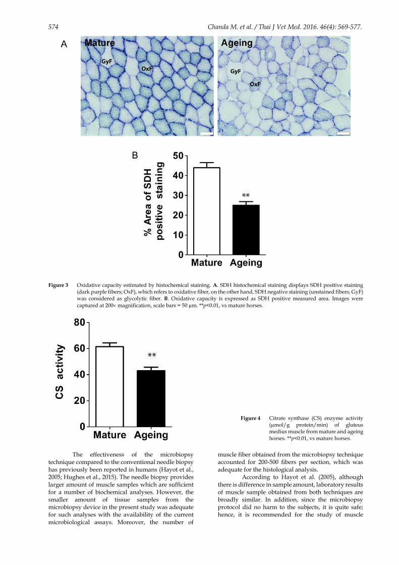
Histochemical and biochemical analyses: The microbiopsy procedure of horse gluteus medius muscle took approximately 5 sec per one single sampling. The wet weight of muscle sample from one single collection was approximately 26.6 ± 2.2 mg and contained 400-600 muscle fibers, which were sufficient for the analysis of histological features. With three consecutive biopsies, the amount of sample (65.4 ± 8.6 mg) was sufficient for the histochemical and biochemical analyses. By using plasma membrane dystrophin protein staining, muscle integrity of both groups of horses was found to be well preserved. However, the ageing horses showed high variation in muscle fiber size (Figs. 2A and B). The determination of cross-sectional area of muscle fibers (CSA) confirmed that the ageing horses had smaller CSA compared to the mature horses (mature, 3428.0 ± 36.6 µm2 vs. ageing, 3064.0 ± 49.3 µm2; p<0.01) (Fig. 2C). In addition, the ageing horses had more number of small fibers (fiber size <3000 µm2) (mature, 35.7 ± 3.6% fibers vs. ageing, 61.8 ± 3.7% fibers; p<0.01) (Fig. 2D). The oxidative capacity of skeletal muscle in the mature and ageing horses was also different. As shown in Fig. 3, the mature muscle had greater SDH positive staining area compared to the ageing muscle (mature, 44.0 ± 2.6% vs. ageing, 25.0 ± 1.8%; p<0.01). Moreover, the catalytic activity of CS enzyme in the mature horse muscle was significantly higher than that in the ageing horse muscle (mature, 61.4 ± 2.9 µmol/g/min vs. ageing, 43.0 ± 2.6 µmol/g/min; p<0.01) (Fig. 4).
Discussion
The present study demonstrates the effectiveness of semi-automated microbiopsy device for obtaining horse skeletal muscle samples. It not only provided satisfactory amount of muscle sample for analysis but also yielded well-preserved muscle quality showing intact anatomical fiber structures in the histological staining. More importantly, because of the small biopsy needle of this device, the biopsy procedure did not affect any routine activities or competitions as the horses were able to return to work immediately after biopsy maneuver and no complications were observed.
The quality of muscle tissue from semi-automated microbiopsy device was evaluated by means of histological and biochemical analyses and compared between mature and ageing horses. As it is known that muscle properties in mature and ageing horses are different, ageing is characterized by loss of

Chanda M. et al. / Thai J Vet Med. 2016. 46(4): 569-577. 573
muscle mass and functional capacity (Larsson, 1995). The atrophy of muscle has been reported to be attributed to the progressive loss of motor neuron during ageing (Doherty et al., 1993; Vandervoort, 2002; Aagaard et al., 2010). Our results are consistent with those of previous studies that showed increase in the proportion of glycolytic fiber with decrease in oxidative fiber. The enzyme activities showed noticeable changes as a function of advanced age
(Lehnhard et al., 2004; Kim et al., 2005; Doria et al., 2012). In our ageing horses, the size of muscle fiber reduced, as reported in human (Vandervoort, 2002; Deschenes, 2004; Aagaard et al., 2010; Nilwik et al., 2013), accompanied by the decrease in the proportion of oxidative fiber and enzyme activity (Fig. 3), proving that the muscles obtained by the semi-automated device provided similar results to previous reports elsewhere.
Figure 2 Skeletal muscle fiber size of mature vs ageing horses. A. Immunostaining of dystrophin illustrates the size of individual
muscle fiber in mature and ageing horses. B. Fiber size distribution analysis reveals a leftward shift of muscle fiber size in ageing horses. C. Mean fiber cross-sectional area (CSA) decreases accompanied by increase in number of muscle fibers size less than 3,000 µm2 in ageing horses (D). CSA was quantified from 1,108 fibers of mature horses (n=6) and 554 fibers
of ageing horses (n=3). Images were captured at 200 magnification, scale bars = 50 µm. **p<0.01, vs mature horses.
574 Chanda M. et al. / Thai J Vet Med. 2016. 46(4): 569-577.
Figure 3 Oxidative capacity estimated by histochemical staining. A. SDH histochemical staining displays SDH positive staining
(dark purple fibers; OxF), which refers to oxidative fiber, on the other hand, SDH negative staining (unstained fibers; GyF) was considered as glycolytic fiber. B. Oxidative capacity is expressed as SDH positive measured area. Images were
captured at 200 magnification, scale bars = 50 µm. **p<0.01, vs mature horses.
The effectiveness of the microbiopsy technique compared to the conventional needle biopsy has previously been reported in humans (Hayot et al., 2005; Hughes et al., 2015). The needle biopsy provides larger amount of muscle samples which are sufficient for a number of biochemical analyses. However, the smaller amount of tissue samples from the microbiopsy device in the present study was adequate for such analyses with the availability of the current microbiological assays. Moreover, the number of
muscle fiber obtained from the microbiopsy technique accounted for 200-500 fibers per section, which was adequate for the histological analysis.
According to Hayot et al. (2005), although there is difference in sample amount, laboratory results of muscle sample obtained from both techniques are broadly similar. In addition, since the microbiopsy protocol did no harm to the subjects, it is quite safe; hence, it is recommended for the study of muscle
Figure 4 Citrate synthase (CS) enzyme activity (µmol/g protein/min) of gluteus medius muscle from mature and ageing horses. **p<0.01, vs mature horses.

Chanda M. et al. / Thai J Vet Med. 2016. 46(4): 569-577. 575
physiology and pathology in horse. The microbiopsy technique may also provide a definitive diagnosis.
The present study indicates the advantages of the microbiopsy technique. The semi-automated microbiopsy device can be effectively used to obtain high quality muscle sample, sufficient muscle fiber per section as documented in human (Cote et al., 1992). Furthermore, this device causes least pain or discomfort during and after microbiopsy; horse can return to routine work immediately after biopsy procedure, which is the ultimate goal for muscle sampling in horse. No suture is required after microbiopsy and wound can completely heal within five days without any complications.
Conclusion
The semi-automated microbiopsy device is a potential tool for obtaining good quality and sufficient amount of horse muscle samples for histological and biochemical analyses. The advantages of using the semi-automated microbiopsy device for sampling horse skeletal muscle are safety, painlessness, and fast repair. Therefore, this microbiopsy device can be the alternative choice for muscle sampling, especially for studies performed in sport horses, in which the least discomfort and fastest recovery are critical.
Acknowledgements
This work was financially supported by Kasetsart Veterinary Development Funds. We would like to thank Mr. Harald Link, the President of Thailand Equestrian Federation and Thai Polo and Equestrian Club, and Ms. Nunthinee Tanner for the allocation of horses in this study. We also thank Dr. Santiago Bachmann and Dr. Teerapol Sathaporn for their practical helps in animal arrangement, restrain, and care throughout the experiment. Critical reading of the manuscript and valuable comments by Professor Dr. Chumpol Pholpramool and Dr.Witchuda Saengsawang are also appreciated.
References
Aagaard, P., Suetta, C., Caserotti, P., Magnusson, S.P., Kjær, M., 2010. Role of the nervous system in sarcopenia and muscle atrophy with aging: strength training as a countermeasure. Scand. J. Med. Sci. Sports 20(1): 49-64.
Aagaard, P., Suetta, C., Caserotti, P., Magnusson, S.P., Kjær, M., 2010. Role of the nervous system in sarcopenia and muscle atrophy with aging: strength training as a countermeasure. Scand. J. Med. Sci. Sports 20, 49-64.
Cote, A.M., Jimenez, L., Adelman, L.S., Munsat, T.L., 1992. Needle muscle biopsy with the automatic Biopty instrument. Neurology 42(11): 2212-2213.
Dalla Costa, E., Minero, M., Lebelt, D., Stucke, D., Canali, E., Leach, M.C., 2014. Development of the Horse Grimace Scale (HGS) as a pain assessment tool in horses undergoing routine castration. PLoS One 9(3): e92281.
Dengler, J., Linke, P., Gdynia, H.J., Wolf, S., Ludolph, A.C., Vajkoczy, P., Meyer, T., 2014. Differences in pain perception during open muscle biopsy and
Bergstroem needle muscle biopsy. J. Pain Res. 17(7): 645-650.
Deschenes, M.R., 2004. Effects of aging on muscle fibre type and size. Sports Med. 34(12): 809-824.
Doherty, T.J., Vandervoort, A.A., Taylor, A.W., Brown, W.F., 1993. Effects of motor unit losses on strength in older men and women. J. Appl. Physiol. 74(2): 868-874.
Doria, E., Buonocore, D., Focarelli, A., Marzatico, F., 2012. Relationship between human aging muscle and oxidative system pathway. Oxid. Med. Cell. Longev. 2012.
Ellefsen, S., Vikmoen, O., Zacharoff, E., Rauk, I., Slettaløkken, G., Hammarström, D., Strand, T., Whist, J., Hanestadhaugen, M., Vegge, G., 2014. Reliable determination of training‐induced alterations in muscle fiber composition in human skeletal muscle using quantitative polymerase chain reaction. Scand. J. Med. Sci. Sports 24(5): e332-e342.
Franck, T., Votion, D.M., Ceusters, J., de la Rebière de Pouyade, G., Mouithys-Mickalad, A., Niesten, A., Fraipont, A., Van Erck, E., Goachet, A., Robert, C., 2010. Specific immuno‐extraction followed by enzymatic detection (SIEFED) of myeloperoxidase and mitochondrial complex I in muscular microbiopsies: preliminary results in endurance horses. Equine Vet. J. 42(s38): 296-302.
Friedmann-Bette, B., Schwartz, F.R., Eckhardt, H., Billeter, R., Bonaterra, G., Kinscherf, R., 2012. Similar changes of gene expression in human skeletal muscle after resistance exercise and multiple fine needle biopsies. J. Appl. Physiol. 112(2): 289-295.
Hayot, M., Michaud, A., Koechlin, C., Caron, M.A., Leblanc, P., Prefaut, C., Maltais, F., 2005. Skeletal muscle microbiopsy: a validation study of a minimally invasive technique. Eur. Respir. J. 25(3): 431-440.
Hennessey, J.V., Chromiak, J.A., Della Ventura, S., Guertin, J., MacLean, D.B., 1997. Increase in percutaneous muscle biopsy yield with a suction-enhancement technique. J Appl Physiol (1985) 82(6): 1739-1742.
Hiatt, W.R., Regensteiner, J.G., Wolfel, E.E., Carry, M.R., Brass, E.P., 1996. Effect of exercise training on skeletal muscle histology and metabolism in peripheral arterial disease. J. Appl. Physiol. 81(2): 780-788.
Houben, R., Leleu, C., Fraipont, A., Serteyn, D., Votion, D.M., 2015. Determination of muscle mitochondrial respiratory capacity in Standardbred racehorses as an aid to predicting exertional rhabdomyolysis. Mitochondrion 24, 99-104.
Hughes, M.C., Ramos, S.V., Turnbull, P.C., Nejatbakhsh, A., Baechler, B.L., Tahmasebi, H., Laham, R., Gurd, B.J., Quadrilatero, J., Kane, D.A., 2015. Mitochondrial bioenergetics and fiber type assessments in microbiopsy vs. bergstrom percutaneous sampling of human skeletal muscle. Front. Physiol. 6:360.
Kim, J.S., Hinchcliff, K.W., Yamaguchi, M., Beard, L.A., Markert, C.D., Devor, S.T., 2005. Age-related changes in metabolic properties of equine skeletal

576 Chanda M. et al. / Thai J Vet Med. 2016. 46(4): 569-577.
muscle associated with muscle plasticity. Vet J 169(3): 397-403.
Klitgaard, H., Clausen, T., 1989. Increased total concentration of Na-K pumps in vastus lateralis muscle of old trained human subjects. J. Appl. Physiol. 67(6): 2491-2494.
Larsson, L., 1995. Motor units: remodeling in aged animals. J. Gerontol. A Biol. Sci. Med. Sci. 50 Spec No, 91-95.
Lehnhard, R.A., McKeever, K.H., Kearns, C.F., Beekley, M.D., 2004. Myosin heavy chain profiles and body composition are different in old versus young Standardbred mares. Vet J 167(1): 59-66.
Lindholm, A., Piehl, K., 1974. Fibre composition, enzyme activity and concentrations of metabolites and electrolytes in muscles of standardbred horses. Acta Vet Scand 15(1): 287-309.
Mann, P.B., Jiang, W., Zhu, Z., Wolfe, P., McTiernan, A., Thompson, H.J., 2010. Wheel running, skeletal muscle aerobic capacity and 1-methyl-1-nitrosourea induced mammary carcinogenesis in the rat. Carcinogenesis 31(7): 1279-1283.
Nilwik, R., Snijders, T., Leenders, M., Groen, B.B., van Kranenburg, J., Verdijk, L.B., van Loon, L.J., 2013. The decline in skeletal muscle mass with aging is mainly attributed to a reduction in type II muscle fiber size. Exp. Gerontol. 48(5): 492-498.
Pietrangelo, T., D'Amelio, L., Doria, C., Mancinelli, R., Fulle, S., Fano, G., 2011. Tiny percutaneous needle biopsy: An efficient method for studying cellular and molecular aspects of skeletal muscle in humans. Int. J. Mol. Med. 27, 361-367.
Rinnankoski-Tuikka, R., Silvennoinen, M., Torvinen, S., Hulmi, J.J., Lehti, M., Kivela, R., Reunanen, H., Kainulainen, H., 2012. Effects of high-fat diet and physical activity on pyruvate dehydrogenase kinase-4 in mouse skeletal muscle. Nutr Metab (Lond) 9(1): 53.
Snow, D.H., Guy, P.S., 1976. Percutaneous needle muscle biopsy in the horse. Equine Vet. J. 8(4): 150-155.
Srere, P., 1969. Citrate synthase. Methods Enzymol. 13, 3-11.
Srikuea, R., Symons, T.B., Long, D.E., Lee, J.D., Shang, Y., Chomentowski, P.J., Yu, G., Crofford, L.J., Peterson, C.A., 2013. Association of fibromyalgia with altered skeletal muscle characteristics which may contribute to postexertional fatigue in postmenopausal women. Arthritis Rheum. 65(2): 519-528.
Staron, R.S., Hagerman, F.C., Hikida, R.S., Murray, T.F., Hostler, D.P., Crill, M.T., Ragg, K.E., Toma, K., 2000. Fiber type composition of the vastus lateralis muscle of young men and women. J. Histochem. Cytochem. 48(5): 623-629.
Stefaniuk, M., Ropka-Molik, K., Piórkowska, K., Bereta, A., Szpar, P., Czerwonka, Z., Podstawski, Z., 2015. Evaluation of minimally invasive muscle biopsy method for genetic analysis in horse. Annals of Animal Science 15(3): 621-627.
Vandervoort, A.A., 2002. Aging of the human neuromuscular system. Muscle Nerve 25(1): 17-25.
Votion, D.M., Fraipont, A., Goachet, A.G., Robert, C., van Erck, E., Amory, H., Ceusters, J., de la Rebiere de Pouyade, G., Franck, T., Mouithys-Mickalad, A., Niesten, A., Serteyn, D., 2010. Alterations in mitochondrial respiratory function in response to endurance training and endurance racing. Equine Vet. J. v.42(s38) Suppl., 268-274.
Votion, D.M., Gnaiger, E., Lemieux, H., Mouithys-Mickalad, A., Serteyn, D., 2012. Physical fitness and mitochondrial respiratory capacity in horse skeletal muscle. PLoS One 7(4): e34890.

Chanda M. et al. / Thai J Vet Med. 2016. 46(4): 569-577. 575
บทคดยอ
อปกรณไมโครไบออฟซแบบกงอตโนมต: ศกยภาพในการใชเกบตวอยางกลามเนอลายในมา
เมธา จนดา1,2 รชกฤต ศรเกอ1 ภาวณ ปยะจตรวฒน1*
ในการประเมนพยาธสภาพและความสมบรณของกลามเนอลายในมา การเกบตวอยางเนอเยอของกลามเนอมาศกษาเปนวธมาตรฐานทใช ในปจจบนการวจยกลามเนอลายในมา มความนยมน าอปกรณไมโครไบออฟซแบบกงอตโนมตมาใชเพอเกบตวอยางชนเนอ อยางไรกตามยงไมมรายงานการศกษาถงคณภาพของชนเนอตวอยางทเกบไดจากการใชอปกรณดงกลาว วตถประสงคของการศกษานเพอตรวจสอบศกยภาพของอปกรณไมโครไบออฟซแบบกงอตโนมตในการใชเกบตวอยางเนอเยอกลามเนอลายมา โดยไดประเมนคณภาพของชนเนอจากลกษณะทางเนอเยอวทยาและทางชวเคม ในการวจยใชมาโปโลวยหนมจ านวน 6 ตวและมาโปโลแกจ านวน 3 ตว เกบตวอยางจากกลามเนอกลเตยส มเดยสโดยใชอปกรณไมโครไบออฟซแบบกงอตโนมต คณสมบตของกลามเนอลายทท าการประเมนไดแก ขนาดพนทหนาตดเสนใยกลามเนอ ชนดของเสนใยและสมรรถนะการท างานของเอนไซมออกซเดชนของกลามเนอ โดยเปรยบเทยบคณสมบตดงกลาวของกลามเนอลายในมาแกกบมาวยหนม จากการใชอปกรณไมโครไบออฟซในการตดเกบชนกลามเนอตวอยาง โดยท าการเกบซ า 3 ครงตอการเกบหนงตวอยาง ไดตวอยางเนอเยอประมาณ 65.4± 8.6 มลลกรมซงเพยงพอส าหรบการวเคราะหลกษณะทางเนอเยอวทยาและทางชวเคมของกลามเนอลาย ในระหวางและหลงการเกบตวอยางชนเนอ มาไมมการแสดงอาการเจบปวด แผลทเกดไดหายสนทอยางสมบรณภายใน 5 วนโดยไมมภาวะแทรกซอนใดๆ ยงพบวามาแกมขนาดหนาตดของเสนใยกลามเนอเลกกวามาวยหนม และมจ านวนเสนใยกลามเนอทใชพลงงานแบบมออกซเจนลดลง รวมทงสมรรถนะการท างานของเอนไซมซเตรทซนเธสลดลงดวย เมอเปรยบเทยบกบมาวยหนม โดยสรป ตวอยางกลามเนอทไดรบจากการใชอปกรณไมโครไบออฟซแบบกงอตโนมตในการเกบ มคณภาพทด มความเหมาะสมในการน าไปใชวเคราะหลกษณะทางเนอเยอวทยาและทางชวเคม ดงนน อปกรณไมโครไบออฟซแบบกงอตโนมตนนบเปนเครองมอทมศกยภาพส าหรบใชในการเกบตวอยางกลามเนอลายในมา โดยจะไดเนอเยอทมคณภาพด ปรมาณมากเพยงพอ มามการบาดเจบนอยและแผลหายเรว ค าส าคญ: มา ไมโครไบออฟซ อปกรณกงอตโนมต เสนใยกลามเนอลาย 1ภาควชาสรรวทยา คณะวทยาศาสตร มหาวทยาลยมหดล กรงเทพมหานคร 10400 2ภาควชาเวชศาสตรคลนกสตวใหญและสตวปา คณะสตวแพทยศาสตร มหาวทยาลยเกษตรศาสตร วทยาเขตก าแพงแสน นครปฐม 73140 *ผรบผดชอบบทความ E-mail: [email protected]
577