selective screening, isolation and … · mohan et al. int j pharm pharm sci, vol 5, suppl 4,...
TRANSCRIPT
Research Article
SELECTIVE SCREENING, ISOLATION AND CHARACTERIZATION OF ANTIMICROBIAL AGENTS FROM MARINE ACTINOMYCETES
Y.S.Y.V. JAGAN MOHAN1*, B. SIRISHA1, R. HARITHA2, T. RAMANA3
1Department of Biotechnology, College of Science and Technology, Andhra University,Visakhapatnam 530003, 2Department of Biotechnology, Visakha Government Degree College (Women), Visakhapatnam530020, 3Dean of school of life sciences, GITAM University,
Visakhapatnam 530045, India. Email: [email protected]
Received: 14 Sep 2013, Revised and Accepted: 19 Oct 2013
ABSTRACT
Objectives: The present study was concerned with the isolation, screening and characterization of the actinomycetes from the sediments, which are collected from different locations of Bay of Bengal.
Materials: Selective enrichment and pretreatment strategies can enhance isolation and screening of novel marine actinomycetes. A total of 15 marine samples were collected from different locations of the Bay of Bengal starting from Visakhapatnam to Singarayakonda. The pre-heat treatment method and a combination of 3 enrichment media were found to be effective in selectively isolating marine actinomycetes. The top five potent isolates were subjected to detailed morphological, cultural, biochemical and physiological characterization.
Results: A total of 93 marine actinomycetes were isolated. The antimicrobial activity was studied with all the 93 isolates. The preliminary study of 93 isolates for antimicrobial activity by cross streak method indicated that 36 isolates have antagonistic properties. All these 36 isolates were subjected to submerged fermentation studies. It was observed that 16 isolates (17.2%) exhibited antibacterial activity, 9 isolates (9.6%) showed antifungal activity while 11 isolates (11.8%) showed both antibacterial and antifungal activities.
Conclusion: The present study was an attempt to use different methods to screen, select and isolate marine actinomycetes, with intrinsic antimicrobial activity against a variety of microbial pathogens, from the sediments of Bay of Bengal.
Keywords: Marine actinomycetes, Antimicrobial activity, Bioactive compounds, Characterization.
INTRODUCTION
The biodiversity of marine environment proved to be an important resource for isolation of potent microorganisms to produce biologically active secondary metabolites [1,2]. Actinomycetes occur in a wide range of environments in which they have the ability to grow on most naturally occurring substrates [3]. Actinomycetes are of prime interest, since they are known to produce chemically diverse compounds with a wide range of unique and biologically active metabolites [4], therapeutically useful compounds and enzymes having application in industry.
Microorganisms in marine environments attract a great deal of attention, due to their adaptability to extreme environments and production of novel natural compounds [5]. Marine environmental conditions show great variation and are extremely different from terrestrial ones; hence marine actinomycetes have different characteristics from those of terrestrial counterparts. This allows the organisms to produce different types of bioactive compounds with unique properties and applications [6]. For successful isolation of actinomycetes from marine environments different methods like choice of screening source, pretreatment, selective medium, culture
condition and selection of potential colonies on a primary isolation plate are important.
Marine environment in Bay of Bengal is believed to have rich microbial diversity and the vast pool of indigenous marine micro flora is not fully explored. Recent focuses on marine actinomycetes show that they are being extensively explored for the discovery of drugs and other bioactive metabolites [7]. Though reports are available on antibiotic production by marine actinomycetes the demand for new antibiotics continues to grow due to the rapid emergence of multidrug resistant pathogens.
MATERIALS AND METHODS
Sampling
A total number of 15 marine sediments were collected along the South East coast of the Bay of Bengal at various depths of 30 – 200mts using grab sampler. They were maintained at ambient temperature with sea water and brought to the laboratory in sterile polypropylene bags for isolation of marine actinomycetes. The sampling locations are given in Table-1.
Table 1: Locations of sampling stations
Sediment no. Sampling location Latitude Longitude Depth No of isolates AUBT 01 Visakhapatnam 17°50.814N 84°01.422E 265 AUBT 101-109 (9) AUBT 02 Visakhapatnam 17°50.556N 83°01.228E 52.93 AUBT 201-208 (8) AUBT 03 Visakhapatnam 17°51.264N 83°32.060E 29.64 AUBT 301-303 (3) AUBT 04 Kakinada 16°59.832N 82°58.065E 201.17 AUBT 401-408 (8) AUBT 05 Kakinada 16°59.507N 82°43.923E 108.05 AUBT 501-506 (6) AUBT 06 Kakinada 16°49.885N 82°25.665E 34.61 AUBT 601-609 (9) AUBT 07 Divipopint 15°59.813N 81°29.045E 191 AUBT 701-708 (8) AUBT 08 Divipopint 15°59.813N 81°24.737E 88.11 AUBT 801-805 (5) AUBT 09 Divipopint 15°59.943N 81°20.229E 31.28 AUBT 901-908 (8) AUBT 10 Singarayakonda 15°00.551N 80°24.985E 192.60 AUBT 1001-1003 (3) AUBT 11 Singarayakonda 15°00.199N 80°16.943E 56.19 AUBT 1101- 1106 (6) AUBT 12 Singarayakonda 15°00.296N 80°12.826E 34.40 AUBT 1201-1206 (6) AUBT 13 Chennai 13°08.149N 80°35.251E 195 AUBT 1301-1303 (3) AUBT 14 Chennai 13°08.490N 80°31.962E 99 AUBT 1401-1405 (5) AUBT 15 Chennai 13°08.768N 80°26.478E 53.6 AUBT 1501-1506 (6)
International Journal of Pharmacy and Pharmaceutical Sciences
ISSN- 0975-1491 Vol 5, Suppl 4, 2013
AAccaaddeemmiicc SScciieenncceess

Mohan et al. Int J Pharm Pharm Sci, Vol 5, Suppl 4, 443-449
444
Selective Isolation of actinomycetes
Three different pre-treatment methods were performed to selectively enhance isolation and growth of marine actinomycetes.
Serial dilution method
Isolation and enumeration of marine actinomycetes were performed by the serial dilution plate technique [8]. 1 g each of the marine sediment sample was taken in 250 ml Erlenmeyer flask containing 50 ml of sterile water. Flasks were shaken on rotary shaker for 30min for the detachment of spore chains. The particulate matter was allowed to settle down and the suspension was serially diluted up to 10 -6 times. 1ml each of these dilutions were added to 50ml of sterile molten starch casein agar medium thoroughly mixed and poured into Petri plates and incubated at 28°C for 3 days to 3 weeks. Different media like starch casein agar, actinomycetes isolation agar, glycerol asparagine agar, oat meal agar, glucose yeast extract malt extract agar are used for isolation technique.
Heat treatment
The samples were heated by incubating at 55 °C for 15 min in a water bath [1]. 10 fold serial dilutions of the sediment samples were made using sterile 50% sea water [9]. About 0.1ml of the serially diluted samples was spread over Starch casein agar medium [10] and Actinomycetes isolation agar medium. Both the media were supplemented with 5μg/ml rifampicin and 25μg/ml of Nystatin (Himedia, Mumbai) to minimize the other bacterial and fungal growth [11].
All the plates were incubated at 28°C for 21 days. The appearance and growth of marine actinomycetes colonies were recognized by their characteristic chalky to leathery appearance. All the morphologically different actinomycete colonies were sub-cultured on yeast extract malt extract agar medium.
Pre-enrichment method
One gram of sediment was transferred to conical flasks containing 100 ml of sterile sea water, starch casein broth and glucose asparagine broth prepared with natural sea water separately for the pre-enrichment of samples. The flasks were incubated at 30 ºC for 14 days in a shaker incubator. A loop-full of inoculum from the pre-enriched starch casein broth and glucose asparagine broth was streaked on starch casein agar (SCA) and glucose asparagine agar separately and the plates were incubated at 30 ºC for 7 days. Single discrete colonies were isolated and identified. All the morphologically different actinomycete colonies were sub cultured on yeast extract malt extract agar medium (ISP No. 2) [12] by streak plate technique. After growth appeared, the actinomycetes colonies were maintained in ISP No. 2 agar slants.
Primary screening for antimicrobial activity
The antimicrobial activity of the isolates were tested by Cross-Streak method employing nutrient agar medium for bacteria and potato dextrose agar medium for fungi and yeast. The media was sterilized by autoclaving at 121°C and 15lbs pressure for 15 min and the molten sterile media was cooled to 40-45°C, poured into Petri plates (4 inch diameter) and allowed to solidify. Each plate was streaked with one isolate at the center and incubated at 28°C for 7 days. After 7 days, test organisms were streaked perpendicular to the growth of the isolate; 24 hour old cultures of bacteria, 4 day old cultures of fungi and 2 day old cultures of yeast were used to test the organisms. All the test organisms employed in the present investigation were procured from Microbial Type Culture Collection (MTCC), Chandigarh, India.
The test organisms used for the determination of antimicrobial activity are Staphylococcus aureus (MTCC 3160), Bacillus subtilis (MTCC 441), Bacillus cereus (MTCC 430), Pseudomonas aeruginosa (MTCC 424), Escherichia coli (MTCC 443), Proteus vulgaris (MTCC 426), Saccharomyces cerevisiae (MTCC 170), Candida albicans (MTCC 227), Aspergillus niger (MTCC 961), and Aspergillus flavus (MTCC 3396).
Secondary screening for antimicrobial activity
Based on the results of primary screening, 11 putative Streptomyces isolates namely AUBT-103, AUBT-205, AUBT-302, AUBT-506, AUBT-702, AUBT-708, AUBT-801, AUBT-902, AUBT- 1001, AUBT-1202, and AUBT-1503 were selected for the fermentation and determination of antibiotic production.
Secondary screening for antibiotic production Agar well diffusion method
Secondary screening of promising isolates was done by submerged fermentation. Slant cultures of mature actinomycete strains were inoculated in the medium containing soya bean meal 20 g, glucose 20 g, NaCl 4 g, K2HPO4 0.05 g, MgSO4 0.50 g and CaCO3 5 g for 1000 ml and maintained at pH 7.2. The cultures were incubated in a rotary shaker (180rev/min) at 27°C for 7 days and the fermented broth was centrifuged at 10,000 rpm at 4 °C for 20 min. The supernatant was filtered using 0.45μm pore size membrane filter (Millipore) [13]. The clear supernatant samples were tested for their antimicrobial activity by agar well diffusion method. To determine the antibacterial spectrum, pathogenic bacteria cultured on nutrient broth at 37°C for 24 h; the cultures were swapped on nutrient agar media. The relative activities of metabolites are determined based on the diameter of zones of inhibition formed. Secondary screening of potent actinomycetes confirmed the results of primary screening.
Characterization of actinomycetes cultures
The top five potent actinomycetes isolates selected from screening were characterized by morphological, cultural, biochemical and physiological features. Morphological and cultural characteristics such as type of aerial hyphae, growth of vegetative hyphae, diffusible pigment and spore formation was observed. Biochemical tests including melanin pigmentation, H2S production, tyrosine reaction, starch, casein, gelatin hydrolysis, Milk coagulation & peptonization, Methyl red, Voges-Proskauer, citrate, Oxidase, Urease and Catalase tests were also performed by starch casein agar. Physiological characterization such as the effect of pH (5-9) and temperature (10oC-50oC) were also tested.
Utilization of carbon sources such as Glucose, Fructose, Mannitol, Rhamnose, Raffinose, Maltose, Lactose, Sucrose, Glycerol, Starch and nitrogen sources namely L-Arginine, L-Tyrosine, L-Asparagine, L-Leucine, L-Cysteine, L-Histidine, L-Valine, and L-Glycine were tested on starch casein agar medium (Table-6&7).
Sodium chloride tolerance: Sodium chloride tolerance level [14] of the isolates was evaluated on starch casein agar supplemented with graded doses of NaCl (1, 4, 7, 10 and 13%) maximum NaCl tolerance concentration in the medium allowing any growth was recorded (Table-8).
RESULTS AND DISCUSSION
Marine sediments from South East coast of Bay of Bengal were selected as a potential source of marine actinomycetes and possible bioactivity. Various pretreatment procedures and selective media were applied to assess the optimal conditions for the isolation of marine actinomycetes from sediments. Three different pre-treatment methods were employed for maximum isolation of actinomycetes.
Serial dilution technique allowed the growth of actinomycetes, bacterial and fungal colonies (Fig 1). But the next two pre treatment methods (Fig 2 & 3) inhibited growth of bacterial and fungal colonies. Hence it has been inferred that when the sediments were cultured without pretreatment, large number of bacterial and fungal colonies were grown, the dominance of other bacterial and fungal contamination was found to inhibit the colonization of actinomycetes. Whereas when the soil was pretreated, their numbers decreased on culture plates. Previously, this type of pre-treatment methods for isolation of actinomycetes has also been suggested by several researchers [5,9,15].
The pretreatment of wet-heating for 55°C for 15min. Starch casein agar and glucose asparagine agar media were the most effective for the isolation of actinomycetes. The bacterial and fungal contamination was diminish by pre-heat treatment and allowed

Mohan et al. Int J Pharm Pharm Sci, Vol 5, Suppl 4, 443-449
445
selective isolation of actinomycetes. When antibacterial and antifungal agents rifampicin 5μg/ml and nystatin 25μg/ml were supplemented into the isolation medium, the number of bacteria and fungi were further decreased.
The growth of marine actinomycetes colonies was recognized by their characteristic chalky to leathery appearance. All the morphologically different actinomycete colonies were sub-cultured on yeast extract malt extract agar slants (ISP No. 2).
Fig. 1: Actinomycetes isolation agar plate showing growth of actinomycete colonies
Fig. 2: Starch casein agar plate showing selective growth of actinomycete colonies.
Fig. 3: Glycerol Asparagine agar plate Showing selective growth of actinomycete colonies.
Fig. 4: Pie diagram showing percentage frequency of isolated actinomycete genera
Fig. 5: Pure culture of AUBT – 205 streptomycetes on GYM agar plate.
Fig. 6: Pure culture of AUBT – 113 Nocardia on Starch casein agar plate.
Mohan et al. Int J Pharm Pharm Sci, Vol 5, Suppl 4, 443-449
446
Among 15 sediments screened, 93 actinomycete colonies were isolated and 36 isolates exhibited antimicrobial activity. 11 isolates showed both antibacterial and antifungal activities (Table 2 & 3). All the 93 isolates were identified at generic level based on the colony morphology and microscopic morphology.
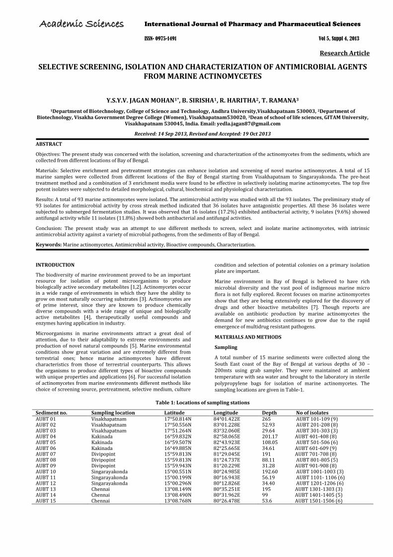
Their distribution pattern was shown in Fig 4. 69 % of the isolates belonged to white and grey colour series and morphologically similar to Streptomyces spp (Fig 5), 18% belonged to family Nocardia (Fig 6), 11 % to Micromonospora and 2% to Rhodococcus (Fig 7).
Fig. 7: Pure culture of AUBT - 1008 Rhodococcus on Starch casein agar plate.
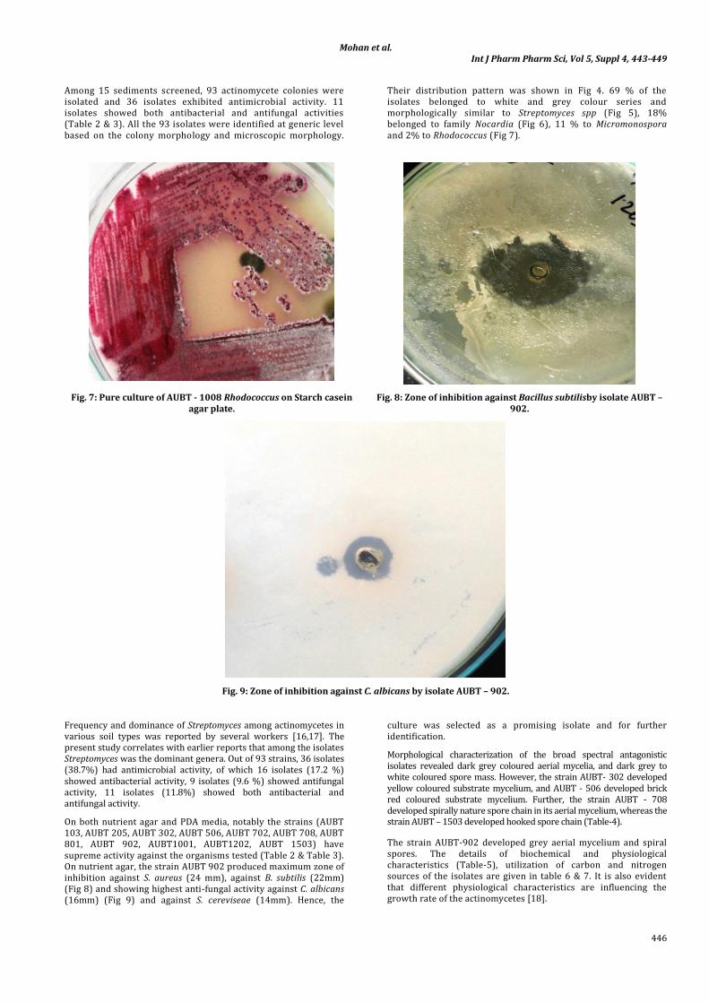
Fig. 8: Zone of inhibition against Bacillus subtilisby isolate AUBT – 902.
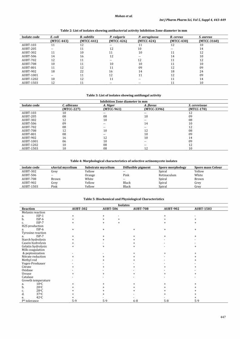
Fig. 9: Zone of inhibition against C. albicans by isolate AUBT – 902.
Frequency and dominance of Streptomyces among actinomycetes in various soil types was reported by several workers [16,17]. The present study correlates with earlier reports that among the isolates Streptomyces was the dominant genera. Out of 93 strains, 36 isolates (38.7%) had antimicrobial activity, of which 16 isolates (17.2 %) showed antibacterial activity, 9 isolates (9.6 %) showed antifungal activity, 11 isolates (11.8%) showed both antibacterial and antifungal activity.
On both nutrient agar and PDA media, notably the strains (AUBT 103, AUBT 205, AUBT 302, AUBT 506, AUBT 702, AUBT 708, AUBT 801, AUBT 902, AUBT1001, AUBT1202, AUBT 1503) have supreme activity against the organisms tested (Table 2 & Table 3). On nutrient agar, the strain AUBT 902 produced maximum zone of inhibition against S. aureus (24 mm), against B. subtilis (22mm) (Fig 8) and showing highest anti-fungal activity against C. albicans (16mm) (Fig 9) and against S. cereviseae (14mm). Hence, the
culture was selected as a promising isolate and for further identification.
Morphological characterization of the broad spectral antagonistic isolates revealed dark grey coloured aerial mycelia, and dark grey to white coloured spore mass. However, the strain AUBT- 302 developed yellow coloured substrate mycelium, and AUBT - 506 developed brick red coloured substrate mycelium. Further, the strain AUBT - 708 developed spirally nature spore chain in its aerial mycelium, whereas the strain AUBT – 1503 developed hooked spore chain (Table-4).
The strain AUBT-902 developed grey aerial mycelium and spiral spores. The details of biochemical and physiological characteristics (Table-5), utilization of carbon and nitrogen sources of the isolates are given in table 6 & 7. It is also evident that different physiological characteristics are influencing the growth rate of the actinomycetes [18].
Mohan et al. Int J Pharm Pharm Sci, Vol 5, Suppl 4, 443-449
447
Table 2: List of isolates showing antibacterial activity Inhibition Zone diameter in mm
Isolate code E. coli B. subtilis P. vulgaris P. aeruginosa B. cereus S. aureus (MTCC-443) (MTCC-441) (MTCC-426) (MTCC-424) (MTCC-430) (MTCC-3160) AUBT-103 11 12 -- 11 12 10 AUBT-205 -- 11 12 10 -- 14 AUBT-302 11 10 11 10 11 12 AUBT-506 14 16 12 -- 14 10 AUBT-702 12 11 -- 12 11 12 AUBT-708 10 11 10 10 11 10 AUBT-801 11 12 11 09 12 09 AUBT-902 18 22 16 14 18 24 AUBT-1001 -- 11 12 11 12 09 AUBT-1202 10 12 11 -- 11 14 AUBT-1503 12 11 -- -- 11 10
Table 3: List of isolates showing antifungal activity
Inhibition Zone diameter in mm Isolate code C. albicans A. Niger A. flavus S. cereviseae (MTCC-227) (MTCC-961) (MTCC-3396) (MTCC-170) AUBT-103 10 -- -- 12 AUBT-205 08 08 10 09 AUBT-302 12 10 -- 08 AUBT-506 09 -- 14 10 AUBT-702 08 -- -- 12 AUBT-708 12 10 12 08 AUBT-801 08 -- 10 09 AUBT-902 16 12 10 14 AUBT-1001 06 10 -- 09 AUBT-1202 10 08 -- 12 AUBT-1503 10 08 12 10
Table 4: Morphological characteristics of selective actinomycete isolates
isolate code sAerial mycelium Substrate mycelium Diffusible pigment Spore morphology Spore mass Colour AUBT-302 Grey Yellow -- Spiral Yellow AUBT-506 -- Orange Pink Retinaculum White AUBT-708 Brown White -- Spiral Brown AUBT-902 Grey Yellow Black Spiral Grey AUBT-1503 Pink Yellow Black Spiral Grey
Table 5: Biochemical and Physiological Characteristics
Isolates Reaction AUBT-302 AUBT-506 AUBT-708 AUBT-902 AUBT-1503 Melanin reaction a. ISP-1 + + - + - b. ISP-6 + + + + + c. ISP-7 + + + + + H2S production a. ISP-6 + + + + + Tyrosine reaction a. ISP-7 + + + + + Starch hydrolysis + + + - - Casein hydrolysis + - + - - Gelatin hydrolysis + + + - + Milk coagulation & peptonization - - + + + Nitrate reduction + + + - + Methyl red - + + - + Voges-Proskauer - - - - - Citrate + + + + + Oxidase - - - - - Urease + + + + + Catalase - - - - - Growth temperature a. 10oC + + + + + b. 20oC + + + + + c. 28oC + + + + + d. 37oC + + - + + e. 42oC + - - - + PH tolerance 5-9 5-9 6-8 5-8 5-9

Mohan et al. Int J Pharm Pharm Sci, Vol 5, Suppl 4, 443-449
448
Table 6: Utilization of Carbon source
Isolates Carbon source AUBT-302 AUBT-506 AUBT-708 AUBT-902 AUBT-1503 D-Glucose Good Good Good Good Good D-Fructose Good Good Good Good Good D-Mannitol Good Good Good Good Good Raffinose No growth No growth No growth No growth No growth Maltose Good No growth Moderate Good No growth Lactose Moderate No growth Moderate Good Moderate Sucrose Good Moderate Moderate Good Moderate Glycerol Good Moderate Moderate Good Good Starch Good Moderate Moderate Good Good
Table 7: Utilization of Nitrogen source
Isolates Nitrogen source KNO3 L-Arginine L-Tyrosine L-Asparagine L-Leucine L-Histidine L-Valine L-Glycine
AUBT-302 + + - + - + + - AUBT-506 + + - + - + + + AUBT-708 + + + + + + + + AUBT-902 + + + + + + + + AUBT-1503 + + + + + - - -
Table 8: Sodium chloride tolerance
Sodium chloride Tolerance Isolates 1% 4% 7% 10% 13% AUBT-302 Good Good Good Moderate No growth AUBT-506 Good Good Good No growth No growth AUBT-708 Good Good Good No growth No growth AUBT-902 Good Good Good Moderate No growth AUBT-1503 Good Good Good No growth No growth
In general, biochemical and physiological characteristics and antimicrobial susceptibility patterns of the actinomycetes vary from isolate to isolate depending on the growth conditions. The present investigation concluded that the physiological characteristics of actinomycetes varied depending on the available nutrients in the medium and the physical conditions. Thus, it was concluded on the basis of the present and previous studies that the nutrient compositions of the medium greatly influence the growth and morphology of organisms [19]. The cultural characteristics and spore morphology place the organism under the family Streptomycetaceae and genus Streptomyces. Further study is in progress to evaluate the potential of the organism for production of anti microbial compounds
CONCLUSION
The search for novel metabolites especially from actinomycetes requires screening large number of isolates (over thousands) in order to discover actinomycete population with novel compound of pharmaceutical interest. The present study was an attempt to use pretreatment methods to screen, select and isolate marine actinomycetes, with intrinsic antimicrobial activity against a variety of microbial pathogens, from the sediments of Bay of Bengal.
ACKNOWLEDGEMENT
We thank the Department of Biotechnology, Andhra University for providing the facilities used in the work.
REFERENCES
1. Bhaskaran R, Vijayakumar R and Mohan PM. Enrichment method for the isolation of bioactive actinomycetes from mangrove sediments of Andaman Islands, India. Malaysian Journal of Microbiology 2011; 7(1), pp. 26-32.
2. Ramesh S, Mathivanan N. Screening of marine actinomycetes isolated from the Bay of Bengal, India for antimicrobial activity
and industrial enzymes. World J. Microbiol. Biotechnol 2009; 25:2103-2011.
3. Goodfellow M and ST Williams. Ecology of actinomycetes. Annu. Rev. Microbiol 1983; 37: 189-216.
4. Bredholt H, Fjaervik E, Jhonsen G and Zotechev SB. Actinomycetes from sediments in the TrondheinFjrod, Norway: Diversity and biological activity. Journal of Marine Drugs 2008; 6: 12-24.
5. Solingen VP, Meijer D, Kleij WA, Branett C, Bolle R, Power SD, Jones BE. Cloning and expression of an endocellulase gene from a novel Streptomyces isolated from an East African soda lake. Extremophiles 2001; 5: 333-341.
6. Goshev I, Gousterova A, Vasileva Tonkova E, Nedkov P. Characterization of the enzyme complexed produced by two newly isolated thermophilic actinomycete strains during growth on collagen rich materials. Process Biochem 2005; 40:1627–1631.
7. Prabavathy VR, Mathivanan N, Murugesan K. Control of blast and sheath blight diseases of rice using antifungal metabolites produced by Streptomyces sp. PM5. Biol Control 2006; 39:313– 319.
8. Haritha R, Sivakumar K, Jagan Mohan YSYV and Ramana T. Amylolytic and Proteolytic Actinobacteria Isolated from Marine Sediments of Bay of Bengal. International Journal of Microbiological Research 2010; 1(2): 37-44.
9. Kim CM, Lec KH, Kwon OS, Shimazu A and Yoo ID. Selective isolation of Actinomycetes by physical pre-treatment of soil sample. Journal of Applied Environmental Microbial Biotechnology 1994; 22: 222-225.
10. Wellington EMH and Cross T. Taxonomy of antibiotic producing Actinomycetes and new approaches to their selective isolation. In: “Progress in industrial microbiology?” Bushell, M. E. (Eds.). Elsevier, Amsterdam 1983; pp: 36.
11. Sivakumar K, Haritha R, Jagan Mohan YSYV and Ramana T. Screening of marine actinobacteria for antimicrobial

Mohan et al. Int J Pharm Pharm Sci, Vol 5, Suppl 4, 443-449
449
compounds. Research journal of Microbiology 2011; 6(4):385- 393.
12. Shirling EB and Gottlieb D. Methods for characterization of Streptomyces species. International Journal of Systematic Bacteriology1966; 16: 312-340.
13. Ruan JS. The basis of taxonomy of actinomycetes. The Chinese Academic Press, Beijing 1977; pp: 139-146.
14. Tresner, HD, JA Hayes and EJ Backns. Differential tolerance of Streptomyces to sodium chloride as a taxonomic acid aid. Applied Microbiol 1968; 16: 1134-1136.
15. Jensen P, R Dwight and W Fenical. Distribution of actinomycetes in near-shore tropical marine sediments. Applied Environ. Microbiol 1991; 57: 1102-1108.
16. Kim CJ, Lee KH, Shimazu A, Kwon OS, Park DJ. Isolation of rare actinomycetes on various types of soil. J. Appl. Microbiol. Biotechnol 1995; 23:36-42.
17. Jensen PR and C Mafnas. Biogeography of the actinomycete genus Salinispora. Environ. Microbiol 2006; 8:1881–1888.
18. Shimizu M, Nakagawa Y, Sato Y, Furumai T, Igarashi Y, Onaka H, Yoshida R, Kunch H. Studies on endophytic actinomycetes (1) Streptomyces sp. Isolated from Rhododendron and its antimicrobial activity. J Gen Pl Pathol 2000; 66: 360-366.
19. Gesheva V, Gesheva R. Structure of the Streptomyces hygroscopicus 111-81 population and characteristics of its variants. Actinomycetes 1993; 4: 65-72.