selecting protein structure/s for docking-based …
TRANSCRIPT

Bhojwani and Joshi, IJPSR, 2019; Vol. 10(6): 2998-3011. E-ISSN: 0975-8232; P-ISSN: 2320-5148
International Journal of Pharmaceutical Sciences and Research 2998
IJPSR (2019), Volume 10, Issue 6 (Research Article)
Received on 09 October 2018; received in revised form, 19 December 2018; accepted, 30 December 2018; published 01 June 2019
SELECTING PROTEIN STRUCTURE/S FOR DOCKING-BASED VIRTUAL SCREENING: A
CASE STUDY ON TYPE II INHIBITORS OF VEGFR-2 KINASE
H. R. Bhojwani and U. J. Joshi *
Department of Pharmaceutical Chemistry, Prin. K. M. Kundnani College of Pharmacy, Cuffe Parade,
Mumbai - 400005, Maharashtra, India.
ABSTRACT: In this study, 36 crystal structures available with type I-V
inhibitors of VEGFR-2 kinase in the RCSB PDB were classified into
DFG-in/-out conformation using visual analysis and KLIFS database. The
focus was on Type II inhibitors as most kinase inhibitors belong to this
category. Therefore, the crystal structures with DFG-out confirmation
with a type II inhibitor were selected depending on the resolution and r-
free value. 11 selected crystal structures were subjected to self-docking
studies and interaction analysis, leading to the elimination of one crystal
structure viz. PDB id 3U6J. 10 crystal structures were subjected to cross-
docking analysis. No crystal structures were eliminated at this stage as
50% ligands were docked accurately at RMSD cut off ≤ 2Å. These
structures were further evaluated for screening performance by
calculation of five performance indicating terms. A rank order was
established by performance terms. The next stage of selection was the
calculation of enrichment factor and assessment of the number of
chemical classes retrieved after docking of the DUD set along with
actives. Considering the EF values and the rank order of performance
terms; 5 crystal structures were eliminated. Lastly, advanced enrichment
parameters such as ROC, AUC, RIE, the average number of outranked
decoys, and BEDROC were calculated for the remaining 5 structures.
After considering all the stages of evaluation, 4ASE was identified as the
most suitable crystal structure.
INTRODUCTION: Docking - based virtual
screening (DBVS) is a method of choice for
identification of chemically diverse hits when the 3
dimensions (3D) structures of the target are
available 1. The success of the docking-based
virtual screening is sensitive to the choice of the 3D
structure of the target 2.
QUICK RESPONSE CODE
DOI: 10.13040/IJPSR.0975-8232.10(6).2998-11
The article can be accessed online on www.ijpsr.com
DOI link: http://dx.doi.org/10.13040/IJPSR.0975-8232.10(6).2998-11
When a limited number of crystal structures were
available for a particular target, selection of a high-
resolution crystal structure was a method of choice
for the selection of crystal structure 3.
However, recent years have seen an explosion in
the availability of the crystal structures of many
“druggable” targets 4. It is a well-known fact that
the crystal structure of any target in a complex with
a bound ligand represents a confirmation of the
target, optimally adapted to accommodate that
particular ligand. The flexibility of the target
protein allows the target to adopt a different
conformation in the presence of a chemically
diverse ligand 5, 6
.
Keywords:
VEGFR-2 kinase,
Crystal Structures, DFG-in/out
confirmation, Type II inhibitors, Performance Indices, DUD Set
Validation, Enrichment Studies
Correspondence to Author:
Dr. Urmila Joshi
Head of Department,
Department of Pharmaceutical
Chemistry, Prin. K. M. Kundnani
College of Pharmacy, Cuffe Parade,
Mumbai - 400005, Maharashtra,
India.
E-mail: [email protected]

Bhojwani and Joshi, IJPSR, 2019; Vol. 10(6): 2998-3011. E-ISSN: 0975-8232; P-ISSN: 2320-5148
International Journal of Pharmaceutical Sciences and Research 2999
DBVS in such cases is more likely to identify more
hits belonging to the chemical class of co-
crystallized ligand. Docking is a computationally
intensive procedure; multiple runs of docking-
based virtual screening using multiple crystal
structures are difficult. Selection of appropriate
crystal structure for virtual screening assumes
importance in such a situation.
The vascular endothelial growth factor receptor
type 2 (VEGFR-2), a tyrosine kinase linked
receptor; represents one such case. It plays an
important role in normal physiological processes
such as cell proliferation, differentiation, migration,
and angiogenesis, which makes VEGFR-2 an
attractive target for cancer 7. VEGFR-2 exists in
two conformations, active and inactive depending
on the orientation of the DFG - motif of the
activation segment. In the „DFG-in‟ or active
conformation, the aspartate (Asp1046) residue
orients toward the ATP binding cleft, and the
phenylalanine (Phe1047) is buried in a hydrophobic
pocket adjacent to the ATP site. In the „DFG-out‟
or inactive conformation, the flipping of the DFG
motif causes the phenylalanine side chain to
occupy the ATP binding cleft, thus uncovering the
hydrophobic pocket 7-10
.
Different types of inhibitors occupy different sites
and bind to a different conformation of VEGFR-2.
Type I inhibitors bind to the DFG-in/active as well
as DFG-out/inactive conformation of the receptor,
whereas type II inhibitors bind to the inactive
conformation only, thereby occupying the
hydrophobic pocket in addition to ATP-binding
site. Presently, most of the inhibitors of kinases
belong to the type II category, and they impart
selectivity as well 11
. There are other types of
inhibitors, such as type I1/2, which bind to an
inactive conformation without occupying the
hydrophobic pocket that is characteristic of Type II
inhibitors 12
. Type III as well as IV inhibitors, do
not compete with ATP and bind solely to allosteric
pockets 13, 14
. Type V inhibitors bind to ATP-
binding site and neighboring region like the type II
inhibitor, however, they lock the kinase in the
DFG-in form as reported 14
.
There are several reports in the literature on the use
of docking or docking-based virtual screening for
the discovery of VEGFR-2 inhibitors 15-18
.
None of the studies, take the confirmation of the
DFG motif of the activation segment into
consideration. This conformational analysis is
important as the binding modes for different types
of inhibitors differ and could lead to inaccurate
results. The present study focuses on a systematic
approach for selection of crystal structure/s for
docking-based virtual screening of VEGFR-2
kinase inhibitors using Glide software with a
special emphasis on the conformational
complexities that are involved.
MATERIALS AND METHODS:
Classification and Selection of Crystal
Structures: 36 crystal structures (resolution 1.5 -
2.95Å) of VEGFR-2 kinase domain bound to
inhibitors have been deposited in the RCSB Protein
Data Bank (PDB) and listed in the KLIFS database 19
. A series of sequential filters were applied for
selecting the relevant crystal structures for the
study. The first filter was selection of crystal
structures with resolution < 2.5 Å and R-free value
<0.25 20, 21
. The second filter was the selection of
only those crystal structures present in DFG-out
conformation. These were identified on the basis of
visual analysis and literature report. They were in
complex with a Type II inhibitor as per literature.
The sequential filtering procedure gave 11 crystal
structures which were used for further study.
Detailed Analysis of Ligand Binding Mode using
KLIFS Database: In addition to the structural
information, the KLIFS database also reports the
ligand binding mode. Ligand binding mode
includes the details on the pockets and sub-pockets
occupied. Therefore, pocket and sub-pocket
occupancy were studied for the 11 selected crystal
structures using the KLIFS database 19
.
Receptor Preparation: 11 crystal structures (PDB
id 1YWN, 2OH4, 3BE2, 3EWH, 3U6J, 3VHE,
3VNT, 3VO3, 3WZE, 4ASD, and 4ASE) were
prepared using the protein preparation workflow in
Maestro 10.2 24
. Hydrogen atoms were added to the
protein structures consistent with a pH of 7.4.
Missing residues and loops were added. Since none
of the crystal structures had water-mediated
protein-ligand interactions, all the water molecules
were deleted. Hydrogen atoms were also added to
the co-crystallized ligand molecules followed by
the generation of energetically accessible ionization

Bhojwani and Joshi, IJPSR, 2019; Vol. 10(6): 2998-3011. E-ISSN: 0975-8232; P-ISSN: 2320-5148
International Journal of Pharmaceutical Sciences and Research 3000
and tautomeric states using Epik 3.2 25
. The protein
assignment program in Maestro was used to set
terminal rotamer states for Asn, Gln automatically,
and His as well as tautomeric and protonation states
of His to optimize the hydrogen-bonding network
in the complex. Besides, hydroxyl and thiol
torsions in Cys, Ser, and Tyr were optimized. The
optimized protein-ligand complexes were subjected
to a restrained minimization using the Imperf tool
available in Protein Preparation Wizard of Maestro.
An RMSD cutoff value of 0.3 Å was used.
Ligand Preparation: Two sets of VEGFR-2
ligands were used in this study: (1) Co-crystallized
ligands from the VEGFR-2 PDB complexes listed
above were used to access the ability of each
receptor to dock the ligands accurately in cross-
docking studies and (2) a collection of 42 VEGFR-
2 kinase inhibitors selected from the literature 26-36
with IC50 values ranging from 0.035 nM to 930 nM
belonging to diverse chemical classes were used for
the performance indices calculation and database
enrichment studies. The 42 VEGFR-2 kinase
inhibitors were chosen in a manner such that most
of the chemical classes of the type II inhibitors
could be included. The DUD decoy set of 2906
molecules with „„drug-like‟‟ characteristics and a
molecular weight less than or equal to 500 was
used for the database enrichment studies 37
. All
ligands were prepared using LigPrep 3.4 38
by
generating low energy ionization and tautomeric
states with a pH of 7.4. In the case of the co-
crystallized ligands and any analogs in set 2,
chiralities were retained from the input structure.
All molecules subjected to LigPrep 3.4 were energy
minimized using the OPLS 2005 force field.
Generation of Receptor Grid and Molecular
Docking: Energy grids for docking studies were
computed for each of the prepared protein-ligand
complexes using default settings in the Receptor
Grid Generation Tab in Glide 6.7 39, 40
. The
centroid of the co-crystallized ligand was used to
define the center of each grid box. Default values
were accepted for van der Waals scaling a partial
input charges were used. Extra precision docking
was used for docking accuracy, and standard
precision docking runs for performance indices and
enrichment studies, with default settings for all
other parameters and no constraints or similarity
scoring were applied 39, 41
.
Docking Accuracy and Interaction-Based
Analysis: To assess docking accuracy, all possible
self and cross-docking studies were carried out
with the extra precision (XP) scoring function. The
final docked conformation of the inhibitor was
aligned to the original conformation, and root
means square deviation (RMSD) was calculated.
After performing the self-docking studies, the
interaction-based analysis was carried out wherein
the hydrogen bonding and the other (π-π and π-
cation as well as hydrophobic) interactions
produced by the co-crystallized ligand after the
self-docking studies were compared with the
interactions reported in RCSB for that particular
ligand. The crystal structures which did not
reproduce hydrogen-bonding interactions were
eliminated during self-docking studies, and
remaining were subjected to cross-docking 42
.
Calculation of Screening Performance Index
and Enrichment Studies: Docking of the set 2
ligands (42 Type II VEGFR-2 kinase inhibitors
mentioned earlier in the Ligand Preparation
section) for the calculation of performance indices
was performed on each of the 9 prepared PDB
structures using the standard precision (SP) mode
of Glide with default parameters into the shortlisted
crystal structures 39
. Following this, the docking
performance was evaluated by calculating five
parameters referred to as term 1-5 using the
mathematical equations as reported in the literature 43
. These five terms include most favorable docking
energies, comparison of average docking energies
of all actives with the docking energy of the best
binder, number of actives giving favorable docking
energies, number of actives successfully docked
and the screening performance index (SPI).
Docking using the standard precision (SP) mode of
Glide 6.7 was performed on each of the shortlisted
PDB structures. Enrichment studies involved the
calculation of classical and advanced enrichment
parameters after the screening of the enriched
decoy set. Standard procedures reported in the
literature 44, 45
were used for the calculation of
enrichment factor (at 1, 2, 5, 10, and 20% of the
ranked dataset), receiver-operating characteristic
(ROC) curve, area under the curve, average number
of out-ranking decoys, robust initial enhancement
(RIE), and Boltzmann-enhanced receiver operating
curve (BEDROC) 44, 45
.

Bhojwani and Joshi, IJPSR, 2019; Vol. 10(6): 2998-3011. E-ISSN: 0975-8232; P-ISSN: 2320-5148
International Journal of Pharmaceutical Sciences and Research 3001
RESULTS AND DISCUSSION:
Classification and Selection of Crystal
Structures: The details of 36 crystal structures
have been given in Table 1 and 2. Three filters
were used for the selection of the crystal structures.
TABLE 1: A LIST OF CRYSTAL STRUCTURES OF VEGFR-2 KINASE USED IN THIS STUDY WITH THEIR
CORRESPONDING RESOLUTION, R-FREE VALUE, CHAIN, DFG-CONFORMATION AND THE TYPE OF
INHIBITOR (I, I1/2, III, AND V) HAS BEEN PROVIDED
S. no. PDB ID Resolution (Å) Chain R-value
Free
Type of
Inhibitor
DFG-conformation
as per KLIFS as per a visual
analysis
1 1Y6A 2.10 A 0.217 I Out-like Out
2 1Y6B 2.10 A 0.236 I Out-like Out
3 2P2H 1.95 A 0.229 I In In
4 2XIR 1.50 A 0.238 I1/2 Out Out
5 3B8R 2.70 A/B 0.259 I1/2 In In
6 3C7Q 2.10 A 0.279 I Out Out
7 3CJF 2.15 A 0.258 I Out Out
8 3CJG 2.25 A 0.253 I In In 9 3VHK 2.49 A 0.262 III Out Out
10 3VID 2.30 A 0.281 I Out Out
11 3WZD 1.57 A 0.212 V In In
12 4AG8 1.95 A 0.241 I1/2 Out Out
13 4AGC 2.00 A 0.251 I1/2 Out-Like Out
14 4AGD 2.81 A 0.307 I Out-Like Out
TABLE 2: A LIST OF CRYSTAL STRUCTURES OF VEGFR-2 KINASE USED IN THIS STUDY IS GIVEN WITH
THEIR CORRESPONDING RESOLUTION, R-FREE VALUE, CHAIN, DFG-CONFORMATION WITH TYPE II
INHIBITORS HAS BEEN PROVIDED
S. no. PDB ID Resolution (Å) Chain R-value
Free
Type of
Inhibitor
DFG-conformation
as per KLIFS as per the visual
analysis
1 1YWN 1.71 A 0.230 II Not Mentioned Out
2 2OH4 2.05 A 0.231 II Out Out
3 2P2I 2.40 A/B 0.266 II Out Out
4 2QU5 2.95 A 0.251 II Out Out
5 2QU6 2.10 A/B 0.272 II Out Out
6 2RL5 2.65 A 0.233 II Out Out
7 3B8Q 2.75 A/B 0.276 II Out Out
8 3BE2 1.75 A 0.226 II Out Out
9 3CP9 2.50 A/B 0.263 II Out Out
10 3CPB 2.70 A/B 0.286 II Out Out
11 3CPC 2.40 A/B 0.272 II Out Out 12 3DTW 2.90 A/B 0.279 II Out Out
13 3EFL 2.20 A/B 0.264 II Out Out
14 3EWH 1.60 A 0.234 II Out Out
15 3U6J 2.15 A 0.230 II Out Out
16 3VHE 1.55 A 0.209 II Out Out
17 3VNT 1.64 A 0.192 II Out Out
18 3VO3 1.52 A 0.182 II Out Out
19 3WZE 1.90 A 0.224 II Out Out
20 4ASD 2.03 A 0.230 II Out Out
21 4ASE 1.83 A 0.231 II Out Out
22 5EW3 2.50 A/B 0.254 II Out Out
The first filter used was the resolution of the crystal
structure for selecting good quality structures only.
The limit of filter value was set to < 2.5 Å which
resulted in the elimination of eight crystal
structures (2QU5, 2RL5, 3B8Q, 3B8R, 3CP9,
3CPB, 3DTW, and 4AGD). The next criterion used
was R-free value <0.25, which led to the
elimination of 13 crystal structures. The third
criterion used for selecting crystal structures was
the activation loop representing a DFG-out
Bhojwani and Joshi, IJPSR, 2019; Vol. 10(6): 2998-3011. E-ISSN: 0975-8232; P-ISSN: 2320-5148
International Journal of Pharmaceutical Sciences and Research 3002
confirmation with a Type II inhibitor. This
procedure resulted in the elimination of 4 crystal
structures (PDB ID: 1Y6A, 1Y6B, 2XIR, and
3WZD), as these crystal structures either do not
represent DFG-out confirmation or do not possess a
Type II inhibitor. 11 crystal structures with PDB
IDs 1YWN, 2OH4, 3BE2, 3EWH, 3U6J, 3VHE,
3VNT, 3VO3, 3WZE, 4ASD, and 4ASE were
selected for further study.
Ligand-Binding Mode Analysis: The ligand
binding modes of the 11 shortlisted crystal
structures were assessed using the KLIFS database,
as shown in Table 3.
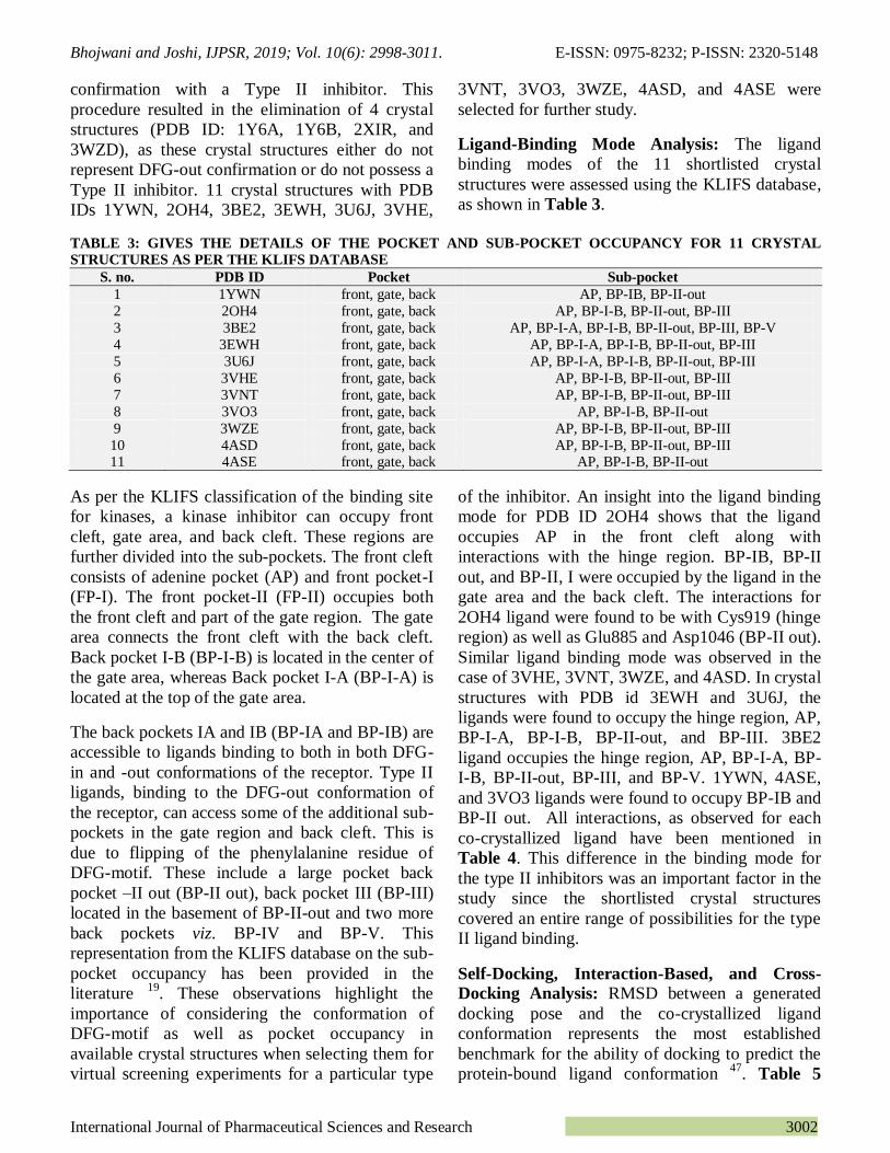
TABLE 3: GIVES THE DETAILS OF THE POCKET AND SUB-POCKET OCCUPANCY FOR 11 CRYSTAL
STRUCTURES AS PER THE KLIFS DATABASE
S. no. PDB ID Pocket Sub-pocket
1 1YWN front, gate, back AP, BP-IB, BP-II-out
2 2OH4 front, gate, back AP, BP-I-B, BP-II-out, BP-III
3 3BE2 front, gate, back AP, BP-I-A, BP-I-B, BP-II-out, BP-III, BP-V
4 3EWH front, gate, back AP, BP-I-A, BP-I-B, BP-II-out, BP-III
5 3U6J front, gate, back AP, BP-I-A, BP-I-B, BP-II-out, BP-III
6 3VHE front, gate, back AP, BP-I-B, BP-II-out, BP-III
7 3VNT front, gate, back AP, BP-I-B, BP-II-out, BP-III
8 3VO3 front, gate, back AP, BP-I-B, BP-II-out
9 3WZE front, gate, back AP, BP-I-B, BP-II-out, BP-III
10 4ASD front, gate, back AP, BP-I-B, BP-II-out, BP-III 11 4ASE front, gate, back AP, BP-I-B, BP-II-out
As per the KLIFS classification of the binding site
for kinases, a kinase inhibitor can occupy front
cleft, gate area, and back cleft. These regions are
further divided into the sub-pockets. The front cleft
consists of adenine pocket (AP) and front pocket-I
(FP-I). The front pocket-II (FP-II) occupies both
the front cleft and part of the gate region. The gate
area connects the front cleft with the back cleft.
Back pocket I-B (BP-I-B) is located in the center of
the gate area, whereas Back pocket I-A (BP-I-A) is
located at the top of the gate area.
The back pockets IA and IB (BP-IA and BP-IB) are
accessible to ligands binding to both in both DFG-
in and -out conformations of the receptor. Type II
ligands, binding to the DFG-out conformation of
the receptor, can access some of the additional sub-
pockets in the gate region and back cleft. This is
due to flipping of the phenylalanine residue of
DFG-motif. These include a large pocket back
pocket –II out (BP-II out), back pocket III (BP-III)
located in the basement of BP-II-out and two more
back pockets viz. BP-IV and BP-V. This
representation from the KLIFS database on the sub-
pocket occupancy has been provided in the
literature 19
. These observations highlight the
importance of considering the conformation of
DFG-motif as well as pocket occupancy in
available crystal structures when selecting them for
virtual screening experiments for a particular type
of the inhibitor. An insight into the ligand binding
mode for PDB ID 2OH4 shows that the ligand
occupies AP in the front cleft along with
interactions with the hinge region. BP-IB, BP-II
out, and BP-II, I were occupied by the ligand in the
gate area and the back cleft. The interactions for
2OH4 ligand were found to be with Cys919 (hinge
region) as well as Glu885 and Asp1046 (BP-II out).
Similar ligand binding mode was observed in the
case of 3VHE, 3VNT, 3WZE, and 4ASD. In crystal
structures with PDB id 3EWH and 3U6J, the
ligands were found to occupy the hinge region, AP,
BP-I-A, BP-I-B, BP-II-out, and BP-III. 3BE2
ligand occupies the hinge region, AP, BP-I-A, BP-
I-B, BP-II-out, BP-III, and BP-V. 1YWN, 4ASE,
and 3VO3 ligands were found to occupy BP-IB and
BP-II out. All interactions, as observed for each
co-crystallized ligand have been mentioned in
Table 4. This difference in the binding mode for
the type II inhibitors was an important factor in the
study since the shortlisted crystal structures
covered an entire range of possibilities for the type
II ligand binding.
Self-Docking, Interaction-Based, and Cross-
Docking Analysis: RMSD between a generated
docking pose and the co-crystallized ligand
conformation represents the most established
benchmark for the ability of docking to predict the
protein-bound ligand conformation 47
. Table 5

Bhojwani and Joshi, IJPSR, 2019; Vol. 10(6): 2998-3011. E-ISSN: 0975-8232; P-ISSN: 2320-5148
International Journal of Pharmaceutical Sciences and Research 3003
summarizes the results of self-docking of all the
cognate ligands into their native protein structures.
10 of 11 cognate ligands had an RMSD ≤ 1Å while
3BE2 had RMSD of 1.52. It is not only important
that the native ligands are docked accurately, but
also they must have a similar binding-pattern
reproduced as they possess in their native crystal
structure. For this purpose, an interaction-based
analysis was conducted wherein the hydrogen
bonds, hydrophobic interactions, π-π, and π-cation
interactions formed by the ligand with the receptor
in the crystal structure, were studied. This
assessment was based on the fact 42
that correctly
docked ligands will reproduce correct hydrogen-
bonding and the other interactions with the protein.
If the hydrogen-bonding interactions were
represented accurately, but the other interactions
differed moderately, the docking pose was
considered „nearly correct‟ and accepted. However,
if the hydrogen bonding interactions were not
reproduced, then that pose is incorrect. 11 crystal
structures were subjected to interaction-based
analysis. The result for interaction-based analysis
has been given in Table 5. 1YWN, 2OH4, 3BE2,
3VHE, 3VO3, and 4ASE; reproduced a correct
pose. 3EWH, 3WZE, 3VNT, and 4ASD could
reproduce a nearly correct pose. 3U6J did not
reproduce a correct or a nearly correct pose. It was
observed that the hydrogen bonding interactions
with Cys919 and Asp 1046 were not reproduced for
3U6J ligand post the self-docking study although it
had an acceptable RMSD value of 0.26. Taking this
into consideration, 3U6J was eliminated from the
study at this stage.
TABLE 4: INTERACTIONS AS OBSERVED IN THE 11 SELECTED CRYSTAL STRUCTURES DEPOSITED IN
THE PROTEIN DATA BANK
S.
no.
PDB ID Hydrogen Bonding Other interactions
Glu
917
Cys
919
Glu
885
Asp
1046
Hydrophobic Pi-Pi and Pi-cation
1 1YWN * * * * Val 916, Phe 918, Ile 890, Ile 1017, Leu 840
2 2OH4 * * * Leu1035, Phe1047, Val916, Ala866, Asp1046 3 3BE2 * * * Thr916, Phe918, Leu840, Val848, Ala866,
Lys868, Leu889, Asp1046, Phe1047
pi-pi (Phe1047)
4 3EWH * * Thr916, Leu840, Val848, Ala866,
Lys868, Leu889, Asp1046, Phe1047
pi-cation (Lys868)
pi-pi (Phe1047) 5 3U6J * * Ile892, Leu1019, Phe918, Leu840, Ala866,
Leu889, Asp1046, Phe1047
pi-pi (Phe1047)
6 3VHE * * * Phe918,Val848,Cys919, Leu840,
Ala866, Leu889, Asp1046, Phe1047, Cys1045
7 3VNT * * * Val916, Ala866 Leu889, Lys868 Cys1045,
Leu1035 Asp1046
pi-cation (Lys868)
8 3VO3 * * * Ala866, Val916, Lys868, Leu1035
9 3WZE * * * Val848, Val916, Leu1035, Ala866, Cys1045, Phe1047
10 4ASD * * * Val848, Phe918, Leu1035, Ala866, Cys1045, Lys868 Leu840, Phe1047
11 4ASE * * * Val848, Phe918, Leu1035, Ala866, Cys1045, Lys868 Leu840, Phe1047
* Indicates hydrogen bonding interaction was observed for a co-crystallized ligand with that particular amino acid residue.
TABLE 5: INDICATES THE RMSD VALUES FOR COGNATE LIGAND OF EACH CRYSTAL STRUCTURE
OBTAINED AFTER SELF-DOCKING STUDIES AND DETAILS OF THE INTERACTION-BASED ANALYSIS FOR
11 SELECTED CRYSTAL STRUCTURES
S. no. PDB ID Self-docking RMSD H-bonding Other interactions
1 1YWN 0.24 Reproduced Reproduced
2 2OH4 0.21 Reproduced Reproduced 3 3BE2 1.52 Reproduced Reproduced
4 3EWH 0.22 Reproduced Not Reproduced 5 3U6J 0.26 Not Reproduced Reproduced
6 3VHE 0.23 Reproduced Reproduced 7 3VNT 0.29 Reproduced Not Reproduced
8 3VO3 0.18 Reproduced Reproduced 9 3WZE 0.09 Reproduced Not Reproduced
10 4ASD 0.29 Reproduced Not Reproduced 11 4ASE 0.11 Reproduced Reproduced

Bhojwani and Joshi, IJPSR, 2019; Vol. 10(6): 2998-3011. E-ISSN: 0975-8232; P-ISSN: 2320-5148
International Journal of Pharmaceutical Sciences and Research 3004
The structures that have the best ability to dock
non-native ligands with a lower RMSD are
possibly are more successful in virtual screening 47
.
This was investigated by cross-docking studies for
type II inhibitors of the VEGFR-2 kinase. The
remaining 10 crystal structures were subjected to
cross-docking studies. Each of the non-native
ligands was docked into each of the protein
structure and RMSD calculated. The calculated
RMSD values for cross-docking studies are as
shown in Table 6. The average RMSD values were
found to be 0.52, 0.92, and 0.94 for 3EWH,
1YWN, and 4ASE; respectively. Other crystal
structures had average RMSD values higher than 1.
3VHE had the highest median RMSD value while
3EWH had the lowest. For shortlisting the crystal
structures at this stage, the cut-off value for RMSD
was set to value of ≤ 2Å. 3EWH could cross-dock
all the ligands accurately at cutoff ≤ 2Å.
Furthermore, all the crystal structures could dock at
least 5 out of 9 non-native ligands at cutoff ≤ 2Å.
Additionally, the performance was evaluated at
RMSD cutoff ≤ 3Å, 4Å, and 5Å in addition to
above mentioned one and the results have been
given in Table 6. 3VHE was the only crystal
structure to cross-dock at RMSD ≤ 5Å while
2OH4, 3VNT, 3VO3, and 3WZE cross-docked 1
ligand each at RMSD ≤ 4Å. Since RMSD ≤ 2Å is
most widely used cut off and as all the crystal
structures could cross-dock more than 50% (viz. 5
out of 9) ligands accurately at this cut off; none of
them was eliminated at this stage. Furthermore,
similar to self-docking, the interactions after the
cross-docking studies were analyzed.
A close look at the RMSD values for all the ligands
revealed that 3BE2 and 3EWH ligands had higher
values in most cross-docking screens. As
mentioned earlier, only 3BE2 and 3EWH ligands
occupied IA BP- and BP-IA and BP-V,
respectively, in addition to the hinge region, AP,
FP, BP-IB, BP-II out and BP-III occupied in most
other cases. It can be presumed that 3BE2 and
3EWH ligands result in induced fit effects such that
they occupy these additional pockets. Thus, the use
of other crystal structures can lead to binding of
these ligands in a pose that is different than their
native poses. Therefore, this could result in high
RMSD values for these two ligands in the cross-
docking screens. To further verify that the docked
pose for 3BE2 and 3EWH differs from the native
one, an inspection of the interactions made by these
ligands in the non-native crystal structures was
undertaken. It was observed that in most of the
cases, all the parent hydrogen bond interactions
characteristic for the type II binding were not
reproduced when docked into non-native structures.
Additionally, it was observed that except for 3BE2
ligand, either partial or complete hydrogen bond
interactions were reproduced in all crystal
structures.
TABLE 6: RMSD VALUES OF THE 9 CO-CRYSTALLIZED LIGANDS IN THE CROSS-DOCKING STUDIES
PERFORMED FOR 10 CRYSTAL STRUCTURES OF VEGFR-2 KINASE
PDB ID
Ligand 1YWN 2OH4 3EWH 3BE2 3VHE 3VNT 3VO3 3WZE 4ASD 4ASE
1YWN 2.19 0.47 0.57 2.30 1.74 ND 2.40 1.46 1.79
2OH4 0.34 0.37 1.22 1.92 0.69 0.50 0.52 0.51 0.75
3EWH 1.33 2.34 1.33 2.55 2.76 2.64 2.30 2.52 2.51
3BE2 0.53 3.59 1.08 4.15 3.85 3.67 3.83 2.63 1.94
3VHE 0.89 0.37 0.35 2.05 0.40 0.36 0.33 0.45 0.40
3VNT 2.40 0.76 0.93 0.76 2.68 0.74 0.69 0.57 0.40
3VO3 1.23 0.37 0.27 0.21 0.49 0.13 0.36 0.24 0.29
3WZE 0.26 0.27 0.21 1.43 0.79 0.19 0.81 0.18 0.12
4ASD 0.51 0.33 0.19 1.43 0.72 0.34 0.44 0.67 0.23
4ASE 0.80 0.85 0.83 1.32 0.94 0.83 0.89 0.24 0.83 Minimum RMSD 0.26 0.27 0.19 0.57 0.49 0.13 0.36 0.24 0.18 0.12
Maximum RMSD 2.40 3.59 1.08 1.43 4.15 3.85 3.67 3.83 2.63 2.51
Average
Cross-docking RMSD
0.92 1.23 0.52 1.15 1.84 1.21 1.26 1.26 1.04 0.94
Cross-docked at < 2Å 8 6 9 8 5 7 6 6 7 8
Cross-docked at <3Å 1 2 0 1 3 1 1 2 2 1
Cross-docked at < 4Å 0 1 0 0 0 1 1 1 0 0
Cross-docked at < 5Å 0 0 0 0 1 0 0 0 0 0
ND: Not Docked

Bhojwani and Joshi, IJPSR, 2019; Vol. 10(6): 2998-3011. E-ISSN: 0975-8232; P-ISSN: 2320-5148
International Journal of Pharmaceutical Sciences and Research 3005
Calculation of Screening Performance Index:
Screening performance index calculations are
based on the docking results of actives only and
were carried out for all the 10 crystal structures,
and results have been mentioned in Table 7.
Calculation of term-1 was done intending to
identifying the structure that binds ligands with the
most favorable docking energies 43
. These
structures are proposed to be more likely to select
other true actives and to reject false positives.
In simple terms, term 1 measured how favorable
the best docking energy within a receptor structure
was in comparison to the best docking energies to
all structures. The results indicated that 3WZE
docked an active with the most favorable docking
energy (-15.397 kcal/mol) and a term 1 value of 1
in comparison to the 9 other crystal structures. It
can also be seen that 1YWN had the least value for
term 1 and it could dock an active with most
favorable docking energy of -11.351 kcal/mol.
TABLE 7: TERM 1, 2, 3, AND 5 VALUES OBTAINED AFTER CALCULATION FOR 10 CRYSTAL STRUCTURES
PBD ID Lowest Docked
Energy (kcal/mol)
Average Docking
Energy (kcal/mol)
Term
1
Term
2
Term
3
Term
4
Term
5
Average
Rank
1YWN -11.350 -9.759 0.737 (10) 0.859 (2) 0.542 (1) NC 0.261 (10) 5.75
2OH4 -14.096 -10.721 0.915 (4) 0.760 (7) 0.254 (7) NC 0.619 (3) 5.25 3BE2 -12.339 -9.713 0.801 (9) 0.787 (5) 0.523 (2) NC 0.380 (7) 5.75
3EWH -12.447 -10.215 0.808 (8) 0.820 (3) 0.386 (3) NC 0.357 (9) 5.75 3VHE -12.557 -11.189 0.815 (7) 0.891 (1) 0.362 (4) NC 0.714 (1) 3.25
3VNT -13.565 -10.773 0.881 (5) 0.794 (4) 0.295 (5) NC 0.547 (4) 4.5 3VO3 -13.401 -9.995 0.870 (6) 0.745 (8) 0.245 (8) NC 0.380 (7) 7.25
3WZE -15.397 -10.639 1 (1) 0.691 (10) 0.185 (10) NC 0.404 (5) 6.5 4ASD -14.334 -10.322 0.930 (3) 0.720 (9) 0.217 (9) NC 0.404 (5) 6.5
4ASE -14.360 -11.275 0.932 (2) 0.785 (6) 0.276 (6) NC 0.642 (2) 4
Numbers in brackets indicate the rank obtained for that term value. NC: Not Calculated
Term-2 and Term-3 used the average docking
energies and the average deviation of the docking
energies from the most favorable one to evaluate
whether compounds other than the one with the
most favorable docking energy also possessed
favorable docking energies - the more compounds
with docking energies close to the most favorable
one the better 43
. Comparison of average docking
energies of all actives with the docking energy of
the best binder (Term-2) indicated that if the
average docking energy of all the actives to a
crystal structure is closer to the docking energy of
the best binder, more actives are bound with
favorable energies to this structure, thereby
suggesting that this structure could pick out actives
more readily 43
. It was observed that the term 2
value was the maximum for 3VHE for which the
lowest docking energy/most favorable energy and
the average docking energies of 42 actives were -
12.557 kcal/mol and -11.189 kcal/mol,
respectively. Likewise, the term 2 values for other
crystal structures were found to be as follows:
1YWN (0.859), 3EWH (0.82), 3VNT (0.794),
3BE2 (0.787), 4ASE (0.785), 2OH4 (0.76), 3VO3
(0.745), 4ASD (0.72), and 3WZE (0.691).
A value nearer to 1 indicates the lesser difference
between the lowest docking energy and the average
energy for the 42 active compounds. Term-3 is
qualitatively similar to Term-2 but quantitatively
different. It is based on the rationale that if more
actives give favorable docking energies as the best
binder, this structure is more likely to pick out
actives 43
. 3BE2 had the highest value for term 3,
while 3WZE had the least (0.185). These values
indicate that 3BE2 is likely to pick out more actives
and is followed by 3EWH, 3VHE, 3VNT, 4ASE,
2OH4, 3VO3, and 4ASD. Term-4 penalized
structures to which fewer compounds could be
docked successfully 43
. Term 4 was not calculated
as all 42 actives were successfully docked into 10
selected crystal structures, and therefore, none of
the crystal structures could be penalized.
The calculation of the number of actives giving
favorable docking energies (term-5) was based on
the assumption that if many actives can dock to a
structure with docking energies more favorable
than the overall average docking energies to all
structures, this structure might be more likely to
pick out many actives in virtual screening 43
. The
overall average docking energy considering 10
structures were found to be -10.460 kcal/mol.
During the calculations, we observed that 3VHE
could dock 30 out of 42 actives with favorable
energies greater than the overall average docking

Bhojwani and Joshi, IJPSR, 2019; Vol. 10(6): 2998-3011. E-ISSN: 0975-8232; P-ISSN: 2320-5148
International Journal of Pharmaceutical Sciences and Research 3006
energy (-10.460 kcal/mol). Likewise, the number of
actives (out of 42) that could be docked with better
energies by other crystal structures in comparison
to overall average docking energy were as follows:
4ASE (27), 2OH4 (26), 3VNT (23), 4ASD (17),
3WZE (18), 3VO3 (16), 3BE2 (16), 3EWH (15),
and 1YWN (11). Thus, the screening performance
index viz. term 5 was the maximum for 3VHE
(0.71), and the other crystal structures followed in
the same order.
Furthermore, the crystal structures were ranked for
their values obtained for each term so that it could
be understood which crystal structure had better
performance when considering all the performance
indices. The ranking order observed based on the
average ranks was 3VHE, 4ASE, 3VNT, 2OH4,
1YWN, 3BE2, 3EWH, 3WZE, 4ASD, and 3VO3,
as shown in Table 7.
The performance indices calculations indicated the
ability of the crystal structures to identify the
actives. However, in the virtual screening process,
the ability of a crystal structure to reject the false
positives is equally important. Given this, we
decided to subject each of these crystal structures to
docking-based virtual screening using a decoy set
enriched with the same 42 actives. In the next step,
10 crystal structures were evaluated for their
comparative ability to discriminate between active
and inactive during docking protocol. This was
done by use of a decoy set, which is a set of
compounds that are presumed to be inactive. The
largest publicly accessible database of decoys is the
Directory of Useful Decoys, which was used in the
present study along with 42 actives.
Enrichment Studies:
Calculation of Standard and Advanced
Enrichment Parameters: Enrichment studies
provide useful insights into the screening efficiency
of a crystal structure. They quantify the number of
active compounds found in the hit list, concerning
the fraction of inactive. The success of virtual
screening is correlated with its ability to rank the
active compounds at high positions of the hit list
since only the first fraction of a hit list will be
screened experimentally.
Enrichment Factor: The first enrichment
parameter calculated during the enrichment studies
was the enrichment factor at 1, 2, 5, 10, and 20% of
the ranked data set 44, 45
. The theoretical maximum
enrichment factors at 1, 2, 5, 10, and 20% that
could be obtained were 70.19, 49.97, 20.05, 9.99,
and 5.00, respectively. The values of enrichment
factors for 10 PDB structures are as shown in
Table 8.
The results of the calculation indicated that six
PDB structures viz. 1YWN, 2OH4, 3BE2, 3EWH,
3VHE, 3VNT, and 4ASE could produce EF1%
greater than 50. The remaining three crystal
structures produced EF1% values of 38.72 and
41.15. The values of enrichment factors at 2, 5, 10,
and 20% of the ranked dataset also showed similar
trends with 3VO3, 3WZE, and 4ASD remaining at
the bottom.
TABLE 8: ENRICHMENT FACTOR VALUES CALCULATED AT VARIOUS PERCENTAGES OF THE RANKED
DATASET FOR 10 SELECTED CRYSTAL STRUCTURES
PDB EF1% EF2% EF5% EF10% EF20% Number of Chemical Classes in
1% of the dataset
1YWN 65.35 32.12 14.32 7.37 3.92 8 2OH4 65.35 35.69 16.23 8.33 4.28 8 3BE2 55.67 27.36 13.85 7.61 4.40 7
3EWH 55.67 35.69 16.23 8.56 4.40 7 3VHE 62.93 35.69 14.80 7.61 4.04 8 3VNT 55.67 27.36 12.89 7.12 3.69 9
3VO3 38.72 19.03 7.64 5.23 2.74 6 3WZE 41.15 22.60 10.50 5.47 3.57 6 4ASD 41.15 21.41 10.50 5.47 3.09 8
4ASE 60.51 34.50 15.28 8.80 4.64 9
Since, chemical diversity is an important aspect in
identifying hits in virtual screening, the number of
chemical classes to which the actives belonged was
identified for 1% of the ranked data set. It was
seen that 3VNT and 4ASE identified actives
belonging to 9 chemical classes. 1YWN, 2OH4,
3VHE, and 4ASD could identify actives belonging
to 8 chemical classes while 3EWH and 3BE2
identified 7 chemical classes. The active
compounds belonging to 6 chemical classes were

Bhojwani and Joshi, IJPSR, 2019; Vol. 10(6): 2998-3011. E-ISSN: 0975-8232; P-ISSN: 2320-5148
International Journal of Pharmaceutical Sciences and Research 3007
retrieved by 3VO3 and 3WZE. Therefore, by
results of lower ranking in both screening
performance and values of enrichment factors;
3BE2, 3EWH, 3VO3, 3WZE, and 4ASD were
eliminated from the study.
ROC Curves: ROC curves allow a visual
comparison of the ability of the crystal structures to
discriminate the actives and decoys reflected in the
form of sensitivity and specificity pairs. Fig. 1
represents the ROC curve for 1YWN, 2OH4,
3VHE, 3VNT, and 4ASE. Usually, the ROC curve
representing ideal distributions will be the one
where there is no overlap between the scores of
active molecules and decoys. The ideal ROC curve
continues as a horizontal straight line to the upper-
right corner where all actives and all decoys are
retrieved, which corresponds to sensitivity = 1 and
specificity = 0.
In contrast to that, the ROC curve for a set of
actives and decoys with randomly distributed
scores tends towards the Se = 1-Sp line
asymptotically with an increasing number of
actives and decoys. This represents a random
performance and is reflected as a diagonal. The
ROC curves for all 5 crystal structures in this study
indicated that they performed better than random
screening. ROC curve for 4ASE depicts that the
curve starts from the origin and closely follows the
y-axis till the point where Se = 0.8 and after which
it begins to drift upwards and right with subsequent
retrieval of actives and decoys.
FIG. 1: REPRESENTS ROC CURVES OBTAINED FOR 5 SELECTED CRYSTAL STRUCTURES WHERE (A)
1YWN, (B) 2OH4, (C) 3VHE, (D) 3VNT, AND (E) 4ASE
A
B C
D E

Bhojwani and Joshi, IJPSR, 2019; Vol. 10(6): 2998-3011. E-ISSN: 0975-8232; P-ISSN: 2320-5148
International Journal of Pharmaceutical Sciences and Research 3008
Following this, when the Se > 0.9 at a particular
point, only decoys were retrieved, and hence the
curve moved only rightwards. The remaining
actives were retrieved after this. When the last
activity was identified, the Se = 1 and 1-Sp was
slightly lesser than 0.8. Likewise, the ROC curves
were analyzed for the other 4 crystal structures.
However, it was difficult to differentiate between
the crystal structures using ROC curves only.
The area under the ROC Curve: In the ROC
context, the area under the ROC curve (AUC)
measures the performance numerically and can
provide insights for quantitative comparison. A
general guide for classifying the accuracy of
screening is as follows: 0.9≤ AUC ≤1 is excellent;
0.80 ≤ AUC < 0.9 is good; 0.70 ≤ AUC < 0.8 is
fair; 0.50 ≤ AUC < 0.7 is poor; and AUC < 0.5 is a
failure 44, 45
. AUC values have been mentioned in
Table 9.
TABLE 9: GIVES THE ADVANCED ENRICHMENT PARAMETER VALUES FOR 5 SELECTED CRYSTAL STRUCTURES
Enrichment Parameters 1YWN 2OH4 3VHE 3VNT 4ASE
AUC 0.87
(good)
0.94
(excellent)
0.87
(good)
0.85
(good)
0.95
(excellent)
Ave. Number of outranking decoys 375 179 366 444 158
RIE 12.79 13.49 13.05 10.95 14.77
BEDROC(alpha=20.0, alpha*Ra=0.2849) 0.735 0.775 0.750 0.629 0.849
An Average Number of Outranking Decoys: The
rank of each action is adjusted by the number of
outranking actives. The number of outranking
decoys is then defined as the adjusted rank of that
active minus one. The number of outranking
decoys is calculated for each docked active and
averaged. The average number of outranking
decoys was calculated in the same manner for the 5
crystal structures. It was seen that this value was
the least for 4ASE. 4ASE was followed by 2OH4,
3VHE, 1YWN, and 3VNT, as shown in Table 9.
From the results, it was evident that 4ASE docked
fewer decoys higher than the actives when
compared with other 4 crystal structures. However,
the classical enrichment parameters such as
enrichment factors, ROC and AUC suffer from the
problem of “early recognition”.
In other words, these parameters do not distinguish
high ranked active molecules from actives ranked
at the end of a rank-ordered list.
In other words, two crystal structures that differ in
the ability to rank the highest scored active
molecules at the beginning of such an ordered list,
but show the same enrichment for active molecules,
would be assessed to perform equal and therefore,
it was decided to compare them based on advanced
enrichment parameters. Therefore, it was important
to compare these 5 crystal structures from
advanced enrichment descriptors such as Robust
Initial Enhancement (RIE) and Boltzmann-
Enhanced Discrimination of ROC (BEDROC).
Robust Initial Enhancement: RIE is an advanced
enrichment metric which quantitatively indicates
the ability of a ranking method to achieve a
distribution of actives better than a method
performing randomly. The RIE values for selected
crystal structures which have been given in Table
9. An RIE value of greater than 1 indicates
performance better than random. In our study, all
four crystal structures produced RIE greater than
1.Since, the aim was to identify the best crystal
structure to carry out virtual screening for type II
VEGFR-2 inhibitors, and a crystal structure that
performs exceptionally well will be of importance.
4ASE had the highest RIE of 14.77, followed by
2OH4, 3VHE, 1YWN, and 3VNT with 13.49,
13.05, 12.79, and 10.95, respectively.
The Boltzmann-Enhanced Discrimination of
ROC (BEDROC): This metric is related to α-
values. The α-value is a value that contributes to
θ% of the total score at z% of the rank. The α-value
of 20 indicates that 80% of the maximum
contribution comes from the first 8% of the list,
thereby ensuring the measurement of early
recognition. If we keep the maximum contribution
value to be constant at 80%, then the α-value of 20
will indicate this contribution coming from 8% of
the list, and for comparing any two structures an
important criterion is αRa<< 1 47
.
Table 9 gives the BEDROC values for crystal
structures under consideration. Taking into
consideration, the criterion for αRa, BEDROC
value was considered at α = 20 where αRa =

Bhojwani and Joshi, IJPSR, 2019; Vol. 10(6): 2998-3011. E-ISSN: 0975-8232; P-ISSN: 2320-5148
International Journal of Pharmaceutical Sciences and Research 3009
0.2849. At this stated criterion, it was found that
BEDROC value was the maximum for 4ASE
followed by 2OH4, 3VHE, 1YWN, and 3VNT for
8% of the ranked data set. On the basis of results
obtained for self- and cross-docking studies,
interaction analysis, performance indices as well as
classical and advanced enrichment metrics, it could
be inferred that 4ASE performed consistently and
was the most appropriate crystal structure for
virtual screening of type II VEGFR-2 inhibitors.
Comparative Analysis with Previous Studies:
1Y6A, 2P2H, 2QU5, 2RL5, 3C7Q, 3CJG, and
3CJF that were used in the study conducted by
Planes as and co-workers 17
have not been included
in our study after applying the selection criteria.
PDB 1YWN was selected by them as the working
structure after taking into account the
crystallographic resolution and the analysis of the
docking results obtained for each PDB, to use for
VEGFR-2 docking-based virtual screening.
Likewise, 1YWN was identified as one of the top
three crystal structures by Zhang and co-workers 18
.
In the present study, 1YWN performed fairly but
was not identified as the best one taking the
conformational state and type II inhibitors into
consideration. The conformational analysis, along
with the type of inhibitor, is an important
consideration in the case of kinases, as mentioned
earlier. However, the lack of conformational
analysis (DFG-in/out) is fairly evident in the results
given by Zhang and co-workers 18
. Amongst the 31
crystal structures included in their study, 2P2H,
3CJG, and 3B8R are present in DFG-in
conformation. The count is given for ligands cross-
docked at RMSD ≤ 2Å for 2P2H, 3CJG, and 3B8R
are 5, 4, and 8; respectively.
Additionally, there are structures present in DFG-
out form but in complex with other types of
inhibitors. The number of ligands cross-docked at
RMSD≤ 2Å in each of these cases is as follows:
1Y6A (4), 1Y6B (4), 3C7Q (2), 3CJF (4), 3VHK
(6), 3VID (4), 4AGC (5) and 4AGD (1). This
indicates the incapability of these cavities to
accommodate chemically diverse type II ligands.
These results highlight the importance of
considering the conformations in the beginning, not
only to have the correct data being taken ahead but
also to avoid intensive computational procedures. It
is essential to mention that the conformational
consideration, along with the type of inhibitors,
will significantly change statistical RMSD results
for all the structures in the study reported by Zhang
and co-workers. In comparison to Zhang‟s study,
3B8Q was eliminated at the primary stage due to its
resolution while 3EWH was eliminated at a later
stage owing to low performance in comparison to
other structures as evident from enrichment factor
and number of chemical classes retrieved. Lastly,
in the present study, an evenhanded chance was
given to all the structures by considering them at
each stage and eventually eliminating the poor
performers after the enrichment factor and
chemical class considerations.
CONCLUSION: A methodical study of arriving at
the most appropriate crystal structure of VEGFR-2
which can be used in docking-based virtual
screening to identify Type II inhibitors was
undertaken. The methods serially employed
classification of crystal structures, ligand binding
mode analysis, self-docking, interaction-based
analysis, cross-docking, docking of known actives
followed by calculation of screening performance
index, docking of enriched decoy set and
calculation of enrichment parameters; both
classical and advanced. Taking the results of all the
studies into consideration, it is proposed that 4ASE
is the most promiscuous structure that can be used
for the docking-based virtual screening studies of
type II inhibitors. Lastly, this work is a more
accurate representation of a systematic approach
that can be applied for selection of crystal structure
for virtual screening from limitless data available in
a protein data bank for the difficult targets (along
with a particular type of inhibitor which is
particularly, applicable to kinases).
ACKNOWLEDGEMENT: The authors
acknowledge the Department of Biotechnology
(Project File No. BT /PR14373 /MED/30/
530/2010) for sanction of grant
CONFLICT OF INTEREST: Both authors have
none to declare.
REFERENCES:
1. Tuccinardi T: Docking-based virtual screening: recent developments. Comb Chem High Throughput Screening 2009; 12(3): 303-14.
2. Erickson JA, Jalaie M, Robertson DH, Lewis RA and Vieth M: Lessons in molecular recognition: the effects of

Bhojwani and Joshi, IJPSR, 2019; Vol. 10(6): 2998-3011. E-ISSN: 0975-8232; P-ISSN: 2320-5148
International Journal of Pharmaceutical Sciences and Research 3010
ligand and protein flexibility on molecular docking accuracy. J Med Chem 2004; 47(1): 45-55.
3. Anderson AC. The process of structure-based drug design: Chem Biol 2003; 10(9): 787-97.
4. Andricopulo AD, Salum LB and Abraham DJ: Structure-based drug design strategies in medicinal chemistry. Curr Top Med Chem 2009; 9(9): 771-90.
5. Rueda M, Bottegoni G and Abagyan R: Recipes for the selection of experimental protein conformations for virtual screening. J Chem Inf Model 2009; 50(1): 186-93.
6. Teague SJ: Implications of protein flexibility for drug
discovery. Nat Rev Drug Discovery 2003; 2(7): 527-41. 7. Neufeld G, Cohen T, Gengrinovitch S and Poltorak Z:
Vascular endothelial growth factor (VEGF) and its receptors. FASEB J 1999; 13(1): 9-22.
8. Roskoski Jr R: VEGF receptor protein–tyrosine kinases: Structure and regulation. Biochem Biophys Res Comm 2008; 375(3): 287-91.
9. Holmes K, Roberts OL, Thomas AM and Cross MJ:
Vascular endothelial growth factor receptor-2: structure, function, intracellular signaling and therapeutic inhibition. Cell Signal 2007; 19(10): 2003-12.
10. Treiber DK and Shah NP: the ins and outs of kinase DFG motifs. Chem Biol 2013; 20(6): 745-46.
11. Muller S, Chaikuad A, Gray NS and Knapp S: The ins and outs of selective kinase inhibitor development. Nature Chemical Biology 2015; 11(11): 818-21.
12. Huang L, Huang Z, Bai Z, Xie R, Sun L and Lin K: Development and strategies of VEGFR-2/KDR inhibitors. Future Med Chem 2012; 4(14): 1839-52.
13. Wu P, Nielsen TE and Clausen MH: Small-molecule kinase inhibitors: an analysis of FDA-approved drugs. Drug Discovery Today 2016; 21(1): 5-10.
14. Okamoto K, Ikemori-Kawada M, Jestel A, Von Koonig K, Funahashi Y, Matsushima T, Tsuruoka A, Inoue A and
Matsui J: Distinct binding mode of multikinase inhibitor lenvatinib revealed by biochemical characterization. ACS Medicinal Chemistry Letters 2014; 6(1): 89-94.
15. Ai G, Tian C, Deng D, Fida G, Chen H, Ma Y, Ding L and Gu Y: A combination of 2D similarity search, pharmacophore, and molecular docking techniques for the identification of vascular endothelial growth factor rece-ptor-2 inhibitors. Anti-cancer Drugs 2015; 26(4): 399-09.
16. Kar RK, Suryadevara P, Sahoo BR, Sahoo GC, Dikhit MR
and Das P: Exploring novel KDR inhibitors based on pharmaco-informatics methodology. SAR QSAR Environ Res 2013; 24(3): 215-34.
17. Planesas JM, Claramunt RM, Teixidó J, Borrell JI and Perez-Nueno VI: Improving VEGFR-2 docking-based screening by pharmacophore post filtering and similarity search postprocessing. J Chem Inf Model 2011; 51(4): 777-87.
18. Zhang Y, Yang S, Jiao Y, Liu H, Yuan H, Lu S, Ran T, Yao S, Ke Z, Xu J and Xiong X: An integrated virtual screening approach for VEGFR-2 inhibitors. J Chem Inf Model 2013; 53(12): 3163-77.
19. Vijayan RS, He P, Modi V, Duong-Ly KC, Ma H, Peterson JR, Dunbrack Jr RL and Levy RM: Conformational analysis of the DFG-out kinase motif and biochemical profiling of structurally validated type II
inhibitors. J Med Chem 2014; 58(1): 466-79. 20. Van Linden OP, Kooistra AJ, Leurs R, De Esch IJ and De
Graaf C: KLIFS: a knowledge-based structural database to navigate kinase-ligand interaction space. J Med Chem 2013; 57(2): 249-77.
21. Wlodawer A, Minor W, Dauter Z and Jaskolski M: Protein crystallography for non‐crystallographers, or how to get
the best (but not more) from published macromolecular structures. The FEBS Journal 2008; 275(1): 1-21.
22. Kleywegt GJ: Validation of protein crystal structures. Acta Crystallogr Sect D: Biol Crystallogr 2000; 249-65.
23. Lintnerova L, Garcia-Caballero M, Gregan F, Melichercik M, Quesada AR, Dobias J, Lac J, Salisova M and Bohac A: A development of chimeric VEGFR2 TK inhibitor based on two ligand conformers from PDB: 1Y6A complex–Medicinal chemistry consequences of a TKs analysis. Eur J Med Chem 2014; 72: 146-59.
24. Maestro, version 10.2, Schrodinger, LLC, New York, NY,
2015. 25. Epik, version 3.2, Schrödinger, LLC, New York, NY,
2015. 26. Dai Y, Guo Y, Frey RR, Ji Z, Curtin ML, Ahmed AA,
Albert DH, Arnold L, Arries SS, Barlozzari T and Bauch JL: Thienopyrimidine ureas as novel and potent multitargeted receptor tyrosine kinase inhibitors. J Med Chem 2005; 48(19): 6066-83.
27. Dai Y, Hartandi K, Ji Z, Ahmed AA, Albert DH, Bauch JL, Bouska JJ, Bousquet PF, Cunha GA, Glaser KB and Harris CM: , Discovery of N-(4-(3-Amino-1 H-indazol-4-yl) phenyl)-N „-(2-fluoro-5-methyl phenyl) urea (ABT-869), a 3-Aminoindazole-Based Orally Active Multitargeted Receptor Tyrosine Kinase Inhibitor. J Med Chem 2007; 50: 1584-97.
28. Hasegawa M, Nishigaki N, Washio Y, Kano K, Harris PA,
Sato H, Mori I, West RI, Shibahara M, Toyoda H and Wang L: Discovery of novel benzimidazoles as potent inhibitors of TIE-2 and VEGFR-2 tyrosine kinase receptors. J Med Chem 2007; 50(18): 4453-70.
29. Frey RR, Curtin ML, Albert DH, Glaser KB, Pease LJ, Soni NB, Bouska JJ, Reuter D, Stewart KD, Marcotte P and Bukofzer G: 7-Aminopyrazolo 1, 5-a pyrimidines as potent multitargeted receptor tyrosine kinase inhibitors. J
Med Chem 2008; 51(13): 3777-87. 30. Kubo K, Shimizu T, Ohyama SI, Murooka H, Iwai A,
Nakamura K, Hasegawa K, Kobayashi Y, Takahashi N, Takahashi K and Kato S: Novel potent orally active selective VEGFR-2 tyrosine kinase inhibitors: synthesis, structure-activity relationships, and antitumor activities of n-phenyl-n „-{4-(4-quinolyloxy) phenyl} ureas. J Med Chem 2005; 48(5): 1359-66.
31. Wang C, Gao H, Dong J, Zhang Y, Su P, Shi Y and Zhang
J: Biphenyl derivatives incorporating urea unit as novel VEGFR-2 inhibitors: Design, synthesis and biological evaluation. Bioorg Med Chem 2014; 22(1): 277-84.
32. Curtin ML, Frey RR, Heyman HR, Sarris KA, Steinman DH, Holmes JH, Bousquet PF, Cunha GA, Moskey MD, Ahmed AA and Pease LJ: Isoindolinone ureas: a novel class of KDR kinase inhibitors. Bioorg Med Chem Lett 2004; 14(17): 4505-09.
33. Matsumoto S, Miyamoto N, Hirayama T, Oki H, Okada K, Tawada M, Iwata H, Nakamura K, Yamasaki S, Miki H and Hori A: Structure-based design, synthesis, and evaluation of imidazo 1, 2-b pyridazine and imidazo 1, 2-a pyridine derivatives as novel dual c-Met and VEGFR2 kinase inhibitors. Bioor Med Chem 2013; 21(24): 7686-98.
34. Liu L, Siegmund A, Xi N, Kaplan-Lefko P, Rex K, Chen A, Lin J, Moriguchi J, Berry L, Huang L and Teffera Y:
Discovery of a potent, selective, and orally bioavailable c-Met inhibitor: 1-(2-Hydroxy-2-methyl propyl)-N-(5-(7-methoxyquinolin-4-yloxy) pyridine-2-yl)-5-methyl-3-oxo-2-phenyl-2, 3-dihydro-1 H-pyrazole-4-carboxamide (AMG 458). J Med Chem 2008; 51(13): 3688-91.
35. Liu L, Norman MH, Lee M, Xi N, Siegmund A, Boezio AA, Booker S, Choquette D, D‟Angelo ND, Germain J

Bhojwani and Joshi, IJPSR, 2019; Vol. 10(6): 2998-3011. E-ISSN: 0975-8232; P-ISSN: 2320-5148
International Journal of Pharmaceutical Sciences and Research 3011
and Yang K: Structure-based design of novel class II c-Met inhibitors: 2. SAR and kinase selectivity profiles of
the pyrazolone series. J Med Che 2012; 55(5): 1868-97. 36. Musumeci F, Radi M, Brullo C and Schenone S: Vascular
endothelial growth factor (VEGF) receptors: drugs and new inhibitors. J Med Chem 2012; 55(24): 10797-22.
37. Lagarde N, Zagury JF and Montes M: Benchmarking data sets for the evaluation of virtual ligand screening methods: review and perspectives. Journal of chemical information and modeling. 2015; 55(7): 1297-07.
38. LigPrep, version 3.4, Schrodinger, LLC, New York, NY,
2015. 39. Glide, version 6.7, Schrödinger, LLC, New York, NY,
2015. 40. Joshi AK, Gadhwal MA and Joshi UJ: Identification of
potential novel EGFR inhibitors using a combination of pharmacophore and docking methods. International Journal of Pharmacy and Pharmaceutical Sciences. 2015; 7(6): 77-91.
41. Bhojwani HR and Joshi UJ: Pharmacophore and Docking Guided Virtual Screening Study for Discovery of Type I Inhibitors of VEGFR-2 Kinase. Current Computer-Aided Drug Design 2017; 13(3): 186-07.
42. Kroemer RT, Vulpetti A, McDonald JJ, Rohrer DC, Trosset JY, Giordanetto F, Cotesta S, McMartin C, Kihlen
M and Stouten PF: Assessment of docking poses: interactions-based accuracy classification (IBAC) versus crystal structure deviations. J Chem Inf Comput Sci 2004; 44(3): 871-81.
43. Huang Z and Wong CF: Inexpensive method for selecting receptor structures for virtual screening. J Chem Inf Model 2015; 56(1): 21-34.
44. Kirchmair J, Markt P, Distinto S, Wolber G and Langer T: Evaluation of the performance of 3D virtual screening
protocols: RMSD comparisons, enrichment assessments, and decoy selection-What can we learn from earlier mistakes? J Comp Aided Mol Des 2008; 22(3-4): 213-28.
45. Braga RC and Andrade CH: Assessing the performance of 3D pharmacophore models in virtual screening: how good are they? Curr Top Med Chem 2013; 13(9): 1127-38.
46. Ramezani M and Shamsara J: A cross-docking study on matrix metalloproteinase family. Anti-Inflammatory Anti-
Allergy Agents Med Chem 2015; 14(3): 164-71. 47. Truchon JF and Bayly CI: Evaluating virtual screening
methods: good and bad metrics for the “early recognition” problem. J Chem Inf Model 2007; 47(2): 488-08.
All © 2013 are reserved by International Journal of Pharmaceutical Sciences and Research. This Journal licensed under a Creative Commons Attribution-NonCommercial-ShareAlike 3.0 Unported License.
This article can be downloaded to Android OS based mobile. Scan QR Code using Code/Bar Scanner from your mobile. (Scanners are available on Google
Play store)
How to cite this article: Bhojwani HR and Joshi UJ: Selecting protein structure/s for docking-based virtual screening: a case study on type II inhibitors of VEGFR-2 kinase. Int J Pharm Sci & Res 2019; 10(6): 2998-11. doi: 10.13040/IJPSR.0975-8232.10(6).2998-11.