see page 3 for storage instructions. - edvotek of restriction sites on plasmid dna see page 3 for...
TRANSCRIPT

The Biotechnology Education Company ®
EDVOTEK, Inc. • 1-800-EDVOTEK • www.edvotek.com
EVT 100202AM
EDVO-Kit
105Mapping of Restriction Sites on Plasmid DNA
See Page 3 for storage instructions.
ExPERIMENT OBjECTIVE:
The objective of this experiment module is to develop an under-standing of the principles of DNA mapping using various restric-
tion enzymes to generate DNA fragments.
�The Biotechnology Education Company® • 1-800-EDVOTEK • www.edvotek.com
Mapping of Restriction Sites on Plasmid DNA
EVT 100202AM
All components are intended for educational research only. They are not to be used for diag-nostic or drug purposes, nor administered to or consumed by humans or animals.
THIS EXPERIMENT DOES NOT CONTAIN HUMAN DNA. None of the experiment components are derived from human sources.
EDVOTEK, The Biotechnology Education Company, and InstaStain are registered trademarks of EDVOTEK, Inc.. Ready-to-Load, UltraSpec-Agarose and FlashBlue are trademarks of EDVOTEK, Inc.
Page
Experiment Components 3
Experiment Requirements 3
Background Information 4
Experiment Procedures
Experiment Overview and General Instructions 7
Agarose Gel Electrophoresis 9
Size Determination of DNA Restriction Fragments 10
Mapping of DNA Restriction Sites 12
Study Questions 13
Instructor's Guidelines
Notes to the Instructor and Pre-Lab Preparations 15
Experiment Results and Analysis 21
Study Questions and Answers 22
Appendices 23
Material Safety Data Sheets 34
Table of Contents
�
105Experiment
Mapping of Restriction Sites on Plasmid DNA
EVT 100202AM
EDVOTEK - The Biotechnology Education Company® 1-800-EDVOTEK • www.edvotek.com
FAx: (�01) �40-058� • email: [email protected]
READy-TO-LOAD™ DNA SAMPLES FOR ELECTROPhORESIS
A Standard DNA Fragments B Plasmid cut with Enzyme 1 C Plasmid cut with Enzyme 2 D Plasmid cut with Enzyme 1 and Enzyme 2
REAgENTS & SuPPLIES
• UltraSpec-Agarose™ powder • Concentrated electrophoresis buffer • FlashBlue™ DNA Stain • InstaStain® Blue cards • Practice Gel Loading Solution • 1 ml pipet • Microtipped Transfer Pipets
Note: If you ordered Experiment #105-Q, the experiment components in-clude InstaStain® Ethidium bromide instead of FlashBlue™ and InstaStain® Blue DNA stains.
DNA samples are stable at room temperature. However, if the experiment will not be conducted within one month of receipt, it is recommended that the DNA samples be stored in the refrigerator.
DNA samples do not require heating prior to gel loading.
Experiment Components
Requirements
• Horizontal gel electrophoresis apparatus • D.C. power supply • Automatic micropipets with tips • Balance • Microwave, hot plate or burner • Pipet pump • 250 ml flasks or beakers • Hot gloves • Safety goggles and disposable laboratory gloves • Small plastic trays or large weigh boats (for gel destaining) • DNA visualization system (white light) • Distilled or deionized water
4
Duplication of this document, in conjunction with use of accompanying reagents, is permitted for classroom/laboratory use only. This document, or any part, may not be reproduced or distributed for any other purpose without the written consent of EDVOTEK, Inc.
Copyright © 1989,1992,1994,1997,1998, 2000, 2004, 2007, 2009, EDVOTEK, Inc., all rights reserved. EVT 100202AM
The Biotechnology Education Company® • 1-800-EDVOTEK • www.edvotek.com
Mapping of Restriction Sites on Plasmid DNA105 Experiment
Background Information
The Human Genome and other genome projects are extremely significant accomplish-ments with important applications to biology and medicine. The explosion of this new information is leading to dramatic changes in the way we are able to improve life. Part of the challenge in dealing with the enormous amounts of data is to determine what genes are responsible for different functions. Scientists must determine the location of genes through DNA mapping, and then begin the arduous task of determining what the individual genes do.
Mapping the positions of restriction enzyme cleavage sites on a DNA molecule is an important prerequisite to DNA sequencing, which provides the primary nucleotide sequence information in DNA. Mapping involves the de-termination of the relative distances between restriction enzyme cleavage sites. An illustrative analogy would be somewhat similar to the following: If DNA mapping were compared to identifying the streets on a city map, then DNA sequencing would be analogous to identifying the specific houses on the streets.
DNA mapping is performed by determining the size of the DNA fragments generated by single or combina-tions of restriction enzyme digestions, and subsequent construction of a DNA map. For example:
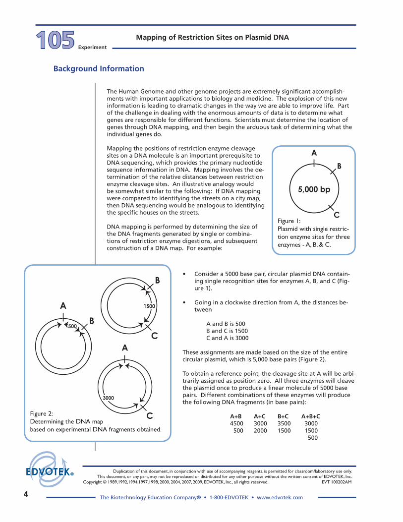
Figure 2:Determining the DNA map based on experimental DNA fragments obtained.
B
A
500
A
C
3000
B
C
1500
Figure 1:Plasmid with single restric-tion enzyme sites for three enzymes - A, B, & C.
B
A
C
5,000 bp
• Consider a 5000 base pair, circular plasmid DNA contain-ing single recognition sites for enzymes A, B, and C (Fig-ure 1).
• Going in a clockwise direction from A, the distances be-tween
A and B is 500 B and C is 1500 C and A is 3000
These assignments are made based on the size of the entire circular plasmid, which is 5,000 base pairs (Figure 2).
To obtain a reference point, the cleavage site at A will be arbi-trarily assigned as position zero. All three enzymes will cleave the plasmid once to produce a linear molecule of 5000 base pairs. Different combinations of these enzymes will produce the following DNA fragments (in base pairs):
A+B A+C B+C A+B+C 4500 3000 3500 3000 500 2000 1500 1500 500

5
Duplication of this document, in conjunction with use of accompanying reagents, is permitted for classroom/laboratory use only. This document, or any part, may not be reproduced or distributed for any other purpose without the written consent of EDVOTEK, Inc. Copyright © 1989,1992,1994,1997,1998, 2000, 2004, 2007, 2009, EDVOTEK, Inc., all rights reserved. EVT 100202AM
The Biotechnology Education Company® • 1-800-EDVOTEK • www.edvotek.com
105Experiment
Mapping of Restriction Sites on Plasmid DNA
Background Information
This data shows that the cleavage site at B is closest to A since cleavage A+B generated the smallest fragment (500) out of all the pairs of enzymes. The shortest distance be-tween A and C is 2000 base pairs since the smallest fragment in the A + C pair is 2000. Similarly, the shortest distance between B and C is 1500 base pairs. It remains to be deter-mined if B is in between A and C (Figure 1) or alternatively, B is between C and A (going in a clockwise direction from A around the plasmid, Figure 3).
If C was in between A and B, the 500 base pair fragment would have been cleaved into two smaller fragments. However, when all three enzymes are used, the 500 base pair fragment remains. In addition, only the 2000 base pair fragment found in the A + C pair is cleaved into 1500 and 500 base pair fragments when all three enzymes are used, verify-ing the location of B. This kind of logic enables the construction of a map, as previously shown, from DNA fragment sizes.
Note that the data from this experiment cannot tell us the absolute orientation of the cleavage sites since it can lead to an alternative map as shown in Figure 2. However, the relative positions are still the same (B is in between A and C). The assignment (Figure 1 or Figure 3) can be made upon further analysis.
Unknown DNA fragment sizes are determined by comparing the relative mobilities of DNA fragments of known size as standards. DNA fragments, from plasmid digests, and standard DNA fragments (also known as markers) are electrophoretically separated in parallel on the same agarose gel. After electrophoretic separation, DNA fragments are stained for visualization, and migration distances of known and unknown fragments are measured.
Figure 3:Construction of a plasmid map using three enzymes.
B
A
C
5,000 bp
500 bp
1500
bp 20
00 bp

�
Duplication of this document, in conjunction with use of accompanying reagents, is permitted for classroom/laboratory use only. This document, or any part, may not be reproduced or distributed for any other purpose without the written consent of EDVOTEK, Inc.
Copyright © 1989,1992,1994,1997,1998, 2000, 2004, 2007, 2009, EDVOTEK, Inc., all rights reserved. EVT 100202AM
The Biotechnology Education Company® • 1-800-EDVOTEK • www.edvotek.com
Mapping of Restriction Sites on Plasmid DNA105 Experiment
Background Information
Standard fragments are used to make a standard curve by plotting their size on the y-axis versus the migration distance on the x-axis. The size of the fragments on the y-axis are expressed as the log of the number of base pairs they contain or the log of their molecu-lar weight. Most of the plotted data obtained from the markers will yield a straight line. The migration distance of the unknown DNA fragment(s) are located on the X-axis and their size is estimated from the standard curve.
After determining the size of the DNA fragments generated by single and combinations of restriction enzymes, a DNA map is constructed as previously described.
In this experiment, you will determine the relative locations of three restriction enzyme cleavage sites on a circular plasmid DNA. The plasmid has been cleaved with three re-striction enzymes. Enzyme 1 cleaves the plasmid once at site A. Assume that the Enzyme 1 site is at position 0. Enzyme 2 and 3 also cut the plasmid once at sites B and C. The objective is to calculate the distances in base pairs between the points of cleavage and to determine whether the Enzyme 1 site is in between the Enzyme 2 sites.
8
10
765
4
3
2
1
9
8765
4
3
2
1
9
8765
4
3
2
1
9
1 cm 2 cm 3 cm 4 cm 5 cm
Migration Distance
Log
base
pai
rs
10,000 base pairs
1,000 base pairs
1 2 5
3 4C
entimeters
23130
43616557
725
9416
2027
570
30002322
basepairs
Standard DNAFragments
Quick Reference:
A standard curve will be made on semi-log graph paper. The following are the Standard DNA fragment sizes, which are expressed in base pairs.
23130 9416 6557 4361 3000 2322 2027 725 570

�
Duplication of this document, in conjunction with use of accompanying reagents, is permitted for classroom/laboratory use only. This document, or any part, may not be reproduced or distributed for any other purpose without the written consent of EDVOTEK, Inc. Copyright © 1989,1992,1994,1997,1998, 2000, 2004, 2007, 2009, EDVOTEK, Inc., all rights reserved. EVT 100202AM
The Biotechnology Education Company® • 1-800-EDVOTEK • www.edvotek.com
105Experiment
Mapping of Restriction Sites on Plasmid DNAExp
erimen
t Proced
ure
Experiment Overview and general Instructions
ExPERIMENT OBjECTIVE:
The objective of this experiment module is to develop an understanding of the principles of DNA mapping using various restriction enzymes to generate DNA fragments.
LABORATORy SAFETy
1. Gloves and goggles should be worn routinely as good laboratory practice.
2. Exercise extreme caution when working with equipment that is used in conjunction with the heating and/or melting of reagents.
3. DO NOT MOUTH PIPET REAGENTS - USE PIPET PUMPS.
4. Exercise caution when using any electrical equipment in the laboratory.
5. Always wash hands thoroughly with soap and water after handling reagents or biological materials in the laboratory.
LABORATORy NOTEBOOK RECORDINgS:
Address and record the following in your laboratory notebook or on a separate worksheet.
Before starting the Experiment:
• Write a hypothesis that reflects the experiment. • Predict experimental outcomes.
During the Experiment: • Record (draw) your observations, or photograph the results.
Following the Experiment: • Formulate an explanation from the results. • Determine what could be changed in the experiment if the experiment were repeated. • Write a hypothesis that would reflect this change.

8
Duplication of this document, in conjunction with use of accompanying reagents, is permitted for classroom/laboratory use only. This document, or any part, may not be reproduced or distributed for any other purpose without the written consent of EDVOTEK, Inc.
Copyright © 1989,1992,1994,1997,1998, 2000, 2004, 2007, 2009, EDVOTEK, Inc., all rights reserved. EVT 100202AM
The Biotechnology Education Company® • 1-800-EDVOTEK • www.edvotek.com
Mapping of Restriction Sites on Plasmid DNA105 Experiment
Exp
erim
ent
Pro
ced
ure
After electrophoresis, transfer gel for staining
Analysis on white
light source
FlashBlue™DNA stain
Attach safety cover,connect leads to power
source and conduct electrophoresis
Load eachsample in
consecutive wells
Remove end blocks & comb, then submerge
gel under buffer in electrophoresis
chamber
Prepare agarose gel in
casting tray
6
5
4
3
2
1
Gel pattern will vary depending upon experiment.
( - )
( + )
1 2 3 4 5 6
Experiment Overview: Flow Chart
�
Duplication of this document, in conjunction with use of accompanying reagents, is permitted for classroom/laboratory use only. This document, or any part, may not be reproduced or distributed for any other purpose without the written consent of EDVOTEK, Inc. Copyright © 1989,1992,1994,1997,1998, 2000, 2004, 2007, 2009, EDVOTEK, Inc., all rights reserved. EVT 100202AM
The Biotechnology Education Company® • 1-800-EDVOTEK • www.edvotek.com
105Experiment
Mapping of Restriction Sites on Plasmid DNAExp
erimen
t Proced
ure
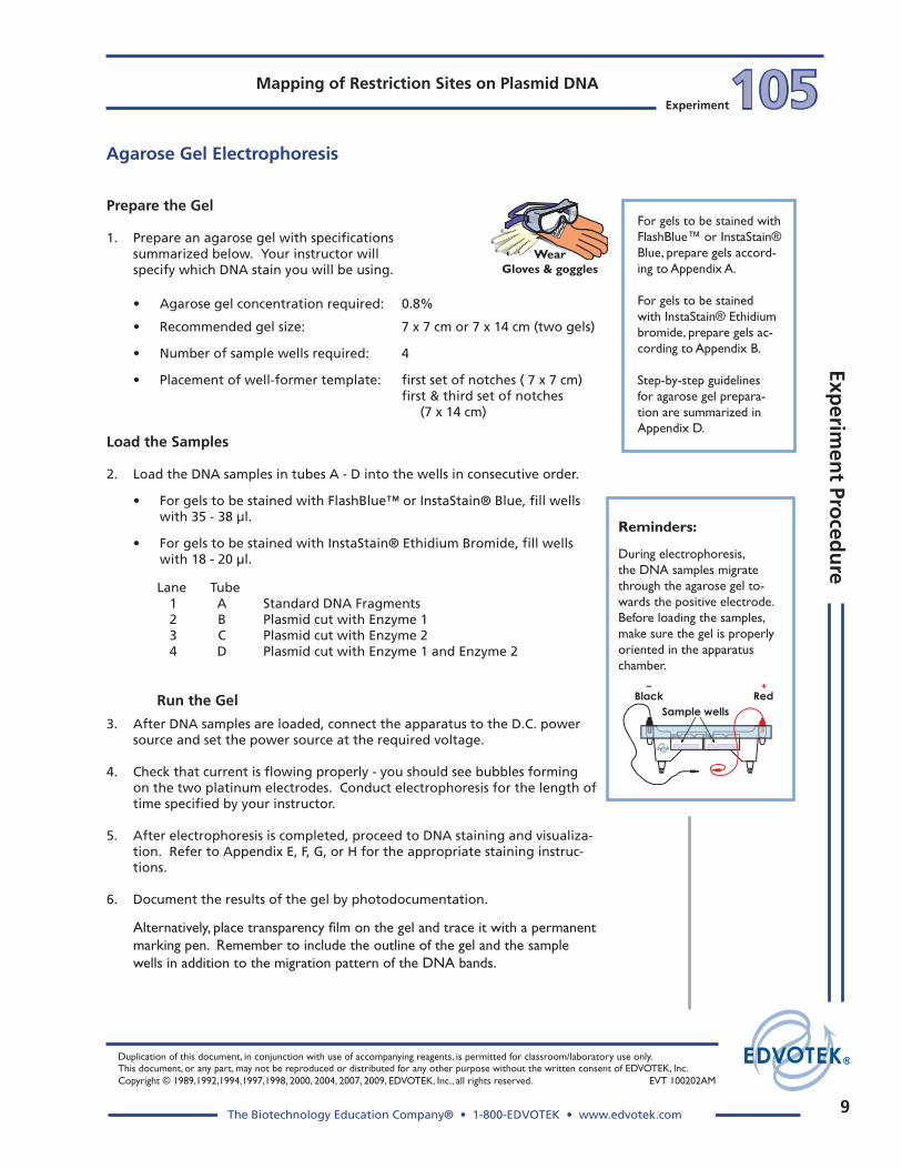
Prepare the gel
1. Prepare an agarose gel with specifications summarized below. Your instructor will specify which DNA stain you will be using.
• Agarose gel concentration required: 0.8%
• Recommended gel size: 7 x 7 cm or 7 x 14 cm (two gels)
• Number of sample wells required: 4
• Placement of well-former template: first set of notches ( 7 x 7 cm) first & third set of notches (7 x 14 cm)
Reminders:
During electrophoresis, the DNA samples migrate through the agarose gel to-wards the positive electrode. Before loading the samples, make sure the gel is properly oriented in the apparatus chamber.
+Black Red
Sample wells
–
Agarose gel Electrophoresis
Lane Tube 1 A Standard DNA Fragments 2 B Plasmid cut with Enzyme 1 3 C Plasmid cut with Enzyme 2 4 D Plasmid cut with Enzyme 1 and Enzyme 2
Run the gel
3. After DNA samples are loaded, connect the apparatus to the D.C. power source and set the power source at the required voltage.
4. Check that current is flowing properly - you should see bubbles forming on the two platinum electrodes. Conduct electrophoresis for the length of time specified by your instructor.
5. After electrophoresis is completed, proceed to DNA staining and visualiza-tion. Refer to Appendix E, F, G, or H for the appropriate staining instruc-tions.
6. Document the results of the gel by photodocumentation.
Alternatively, place transparency film on the gel and trace it with a permanent marking pen. Remember to include the outline of the gel and the sample wells in addition to the migration pattern of the DNA bands.
For gels to be stained with FlashBlue™ or InstaStain® Blue, prepare gels accord-ing to Appendix A.
For gels to be stained with InstaStain® Ethidium bromide, prepare gels ac-cording to Appendix B.
Step-by-step guidelines for agarose gel prepara-tion are summarized in Appendix D.
Wear Gloves & goggles
Load the Samples
2. Load the DNA samples in tubes A - D into the wells in consecutive order.
• For gels to be stained with FlashBlue™ or InstaStain® Blue, fill wells with 35 - 38 µl.
• For gels to be stained with InstaStain® Ethidium Bromide, fill wells with 18 - 20 µl.

10
Duplication of this document, in conjunction with use of accompanying reagents, is permitted for classroom/laboratory use only. This document, or any part, may not be reproduced or distributed for any other purpose without the written consent of EDVOTEK, Inc.
Copyright © 1989,1992,1994,1997,1998, 2000, 2004, 2007, 2009, EDVOTEK, Inc., all rights reserved. EVT 100202AM
The Biotechnology Education Company® • 1-800-EDVOTEK • www.edvotek.com
Mapping of Restriction Sites on Plasmid DNA105 Experiment
Exp
erim
ent
Pro
ced
ure
This exercise focuses on the first step for mapping DNA restriction sites, which is to determine the size of "unknown" DNA fragments generated after electrophoresis. The assignment of sizes for DNA fragments separated by agarose gel electrophoresis can have ± 10% margin of error. The sizes of the "unknowns" will be extrapolated by their migra-tion distances relative to the Standard DNA Fragments (Sample A), for which the frag-ment sizes are known.
1. Measure and record the distance traveled in the agarose gel by each Standard DNA fragment (except the largest 23,130 bp fragment, which will not fit in a straight line in step 4).
In each case, measure from the lower edge of the sample well to the lower end of each band. Record the distance traveled in centimeters (to the nearest millime-ter).
2. Label the semi-log graph paper:
A. Label the non-logarithmic horizontal x-axis "Migra-tion Distance" in centimeters at equal intervals.
B. Label the logarithmic vertical y-axis "Log base pairs". Choose your scales so that the data points are well spread out. Assume the first cycle on the y-axis represents 100-1,000 base pairs and the second cycle represents 1,000-10,000 base pairs.
3. For each Standard DNA fragment, plot the measured migration distance on the x-axis versus its size in base pairs, on the y-axis.
4. Draw the best average straight line through all the points. The line should have approximately equal num-bers of points scattered on each side of the line. Some points may be right on the line (see Figure 1 for an example).
5. Measure the migration distance of each of the "un-known" fragments from samples B, C, and D.
Figure 1
6. Using the graph of the Standard DNA fragments, determine the sizes in base pairs of each "unknown" fragment.
• Find the migration distance of the unknown fragment on the x-axis. Draw a vertical line from that point until the standard graph line is inter-sected.
• From the point of intersection, draw a second line horizontally to the y-axis and determine the approximate size of the fragment in base pairs (refer to Figure 1 for an example).
Quick Reference:
Standard DNA fragment sizes - length is expressed in base pairs.
23130 9416 6557 4361 3000 2322 2027 725 570


1�
Duplication of this document, in conjunction with use of accompanying reagents, is permitted for classroom/laboratory use only. This document, or any part, may not be reproduced or distributed for any other purpose without the written consent of EDVOTEK, Inc.
Copyright © 1989,1992,1994,1997,1998, 2000, 2004, 2007, 2009, EDVOTEK, Inc., all rights reserved. EVT 100202AM
The Biotechnology Education Company® • 1-800-EDVOTEK • www.edvotek.com
Mapping of Restriction Sites on Plasmid DNA105 Experiment
Exp
erim
ent
Pro
ced
ure
The size of the plasmid used in this experiment is 4300 bp.
1. Draw a circle representing a 4300 bp plasmid on a transparent sheet of acetate.
2. Mark the positions of Enzyme #2 (Lane 3) sites corresponding to the sizes of fragments obtained upon digestion of the plasmid on the gel.
3. Draw a second circle representing a 4300 bp plasmid on a transparent sheet of acetate.
4. Mark the position of the Enzyme #1 (Lane 2) site at the top (12:00 o'clock).
5. To draw a composite map of both enzymes, overlay the Enzyme #2 map on top of the Enzyme #1 map.
6. Keeping the Enzyme #1 site at the 12:00 o'clock position, rotate the Enzyme #2 map until the relative distances between the sites approximate the relative sizes of the frag-ments of Enzyme #1 and #2 combined.
7. Specify, in base pairs, the distances between all the sites.
4,300 bp
Enzyme 1
4,300 bp
Enzyme 23,650
650overlay
Mapping of DNA Restriction Sites

1�
Duplication of this document, in conjunction with use of accompanying reagents, is permitted for classroom/laboratory use only. This document, or any part, may not be reproduced or distributed for any other purpose without the written consent of EDVOTEK, Inc. Copyright © 1989,1992,1994,1997,1998, 2000, 2004, 2007, 2009, EDVOTEK, Inc., all rights reserved. EVT 100202AM
The Biotechnology Education Company® • 1-800-EDVOTEK • www.edvotek.com
105Experiment
Mapping of Restriction Sites on Plasmid DNA
1. Describe DNA mapping and list some important uses for this technology.
�. When plotting the sizes of DNA fragments, which axis is used to plot the migration distances of the known and unknown fragments?
Which axis is used to plot the sizes of the known and unknown fragments?
�. A plasmid DNA was cut with several restriction enzymes and the following fragment sizes were determined by comparing the unknown fragments to a standard DNA marker:
Enzyme 1 �000 Enzyme � �000 Enzyme � 1800 & 1�00 Enzymes 1 & � 1450 & 1550 Enzymes � & � 1800, �50, & 550 Enzymes 1 & � 1�00, 1000, & 800
Draw a restriction map based on the data.
Study Questions

14The Biotechnology Education Company® • 1-800-EDVOTEK • www.edvotek.com
Mapping of Restriction Sites on Plasmid DNA
EVT 100202AM

15
105Experiment
Mapping of Restriction Sites on Plasmid DNA
EVT 100202AM
EDVOTEK - The Biotechnology Education Company® 1-800-EDVOTEK • www.edvotek.com
FAx: (�01) �40-058� • email: [email protected]
Instructor’s guide
Class size, length of laboratory sessions, and availability of equipment are factors which must be considered in planning and implementing this experi-ment with your students. These guidelines can be adapted to fit your spe-cific set of circumstances. If you do not find the answers to your questions in this section, a variety of resources are continuously being added to the EDVOTEK web site. Technical Service is available from 9:00 am to 6:00 pm, Eastern time zone. Call for help from our knowledgeable technical staff at 1-800-EDVOTEK (1-800-338-6835).
EDuCATIONAL RESOuRCES, NATIONAL CONTENT AND SKILL STANDARDS
By performing this experiment, students will learn to load samples and run agarose gel electrophoresis. Experiment analysis will provide students the means to transform an abstract concept into a concrete explanation.
Notes to the Instructor & Pre-Lab Preparations
Visit our web site for information about EDVOTEK's complete line of experiments for biotechnology
and biology education.
EDVOTEK Ready-to-Load Electrophoresis Ex-periments are easy to perform and are designed for maximum success in the classroom setting. However, even the most experienced students and teachers occasionally encounter experimen-tal problems or difficulties. EDVOTEK web site resources provide suggestions and valuable hints for conducting electrophoresis, as well as answers to frequently asked electrophoresis questions.
Laboratory Extensions and Supplemental Activities
Laboratory extensions are easy to perform using EDVOTEK experiment kits. For example, a DNA sizing determination activity can be performed on any electrophoresis gel result if DNA markers are run in parallel with other DNA samples. For DNA Sizing instructions, and other laboratory ex-tension suggestions, please refer to the EDVOTEK website.
Visit the EDVOTEK web site often for continuously updated information.
Mon - Fri 9 am - 6 pm ET
(1-800-338-6835)
EDVO-TECH SERVICE
1-800-EDVOTEK
Mon - Fri9:00 am to 6:00 pm ET
FAX: (301) 340-0582Web: www.edvotek.comemail: [email protected]
Please have the following information ready:
• Experiment number and title• Kit lot number on box or tube• Literature version number (in lower right corner)• Approximate purchase date
Technical ServiceDepartment
OrderOnline

1�
Duplication of this document, in conjunction with use of accompanying reagents, is permitted for classroom/laboratory use only. This document, or any part, may not be reproduced or distributed for any other purpose without the written consent of EDVOTEK, Inc.
Copyright © 1989,1992,1994,1997,1998, 2000, 2004, 2007, 2009, EDVOTEK, Inc., all rights reserved. EVT 100202AM
The Biotechnology Education Company® • 1-800-EDVOTEK • www.edvotek.com
105Experiment
Inst
ruct
or’
s g
uid
eInstructor’s Guide Mapping of Restriction Sites on Plasmid DNA
APPROxIMATE TIME REQuIREMENTS
1. Gel preparation: Whether you choose to prepare the gel(s) in advance or have the students prepare
their own, allow approximately 30 minutes for this procedure. Generally, 20 minutes of this time is required for gel solidification.
2. Micropipeting and Gel Loading: If your students are unfamiliar with using micropipets and sample loading tech-
niques, a micropipeting or practice gel loading activity is suggested prior to conduct-ing the experiment. Two suggested activities are:
• EDVOTEK Expt. # S-44, Micropipetting Basics, focuses exclusively on using micro-pipets. Students learn pipeting techniques by preparing and delivering various dye mixtures to a special Pipet Card™.
• Practice Gel Loading: EDVOTEK Series 100 electrophoresis experiments contain a tube of practice gel loading solution for this purpose. It is highly recommended that a separate agarose gel be cast for practice sample delivery. This activity can require anywhere from 10 minutes to an entire laboratory session, depending upon the skill level of your students.
Notes to the Instructor & Pre-Lab Preparations
3. Conducting Electrophoresis: The approximate time for electrophoresis will vary from
approximately 15 minutes to 2 hours. Different models of electrophoresis units will separate DNA at different rates depending upon its design configuration. Generally, the higher the voltage applied the faster the samples migrate. However, maximum voltage should not exceed the indicated recommendations. The Table C example at left shows Time and Voltage recommendations. Refer to Table C in Appendi-ces A or B for specific experiment guidelines.
PREPARINg AgAROSE gELS FOR ELECTROPhORESIS
There are several options for preparing agarose gels for the electrophoresis experiments:
1. Individual Gel Casting: Each student lab group can be responsible for casting their own individual gel prior to conducting the experiment.
2. Batch Gel Preparation: A batch of agarose gel can be prepared for sharing by the class. To save time, a larger quantity of UltraSpec-Agarose can be prepared for shar-ing by the class. See instructions for "Batch Gel Preparation".
3. Preparing Gels in Advance: Gels may be prepared ahead and stored for later use. Solidified gels can be stored under buffer in the refrigerator for up to 2 weeks.
Do not store gels at -20°C. Freezing will destroy the gels.
Time and VoltageRecommendations
Minimum / Maximum
Volts
150
125
70
50
15 / 20 min
20 / 30 min
35 / 45 min
50 / 80 min
Table
CEDVOTEK Electrophoresis Model
M6+ M12 & M36
Minimum / Maximum
25 / 35 min
35 / 45 min
60 / 90 min
95 / 130 min

1�
Duplication of this document, in conjunction with use of accompanying reagents, is permitted for classroom/laboratory use only. This document, or any part, may not be reproduced or distributed for any other purpose without the written consent of EDVOTEK, Inc. Copyright © 1989,1992,1994,1997,1998, 2000, 2004, 2007, 2009, EDVOTEK, Inc., all rights reserved. EVT 100202AM
The Biotechnology Education Company® • 1-800-EDVOTEK • www.edvotek.com
105Experiment
Mapping of Restriction Sites on Plasmid DNAIn
structo
r’s gu
ide
Instructor’s Guide
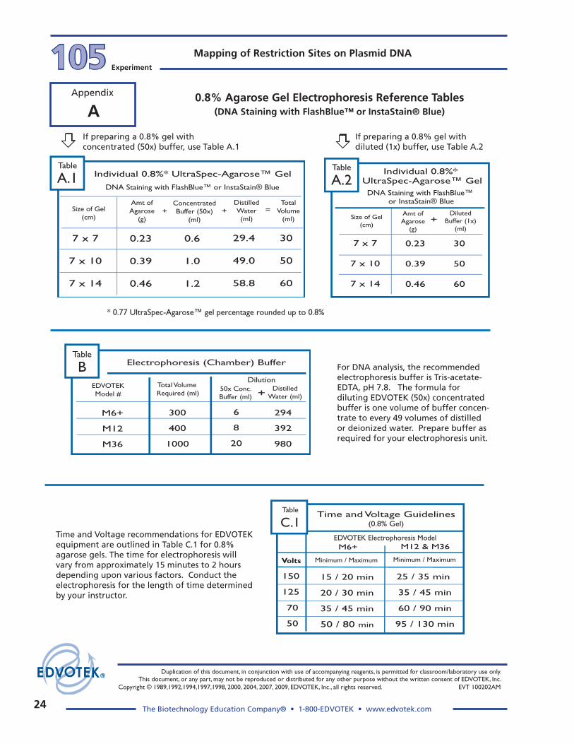
* 0.77 UltraSpec-Agarose™ gel percentage rounded up to 0.8%
Amt ofAgarose
(g)
ConcentratedBuffer (50x)
(ml)
Size of Gel(cm)
DistilledWater(ml)
TotalVolume
(ml)
7 x 7
7 x 10
7 x 14
0.23
0.39
0.46
0.6
1.0
1.2
29.4
49.0
58.8
+ =+
Individual 0.8%* UltraSpec-Agarose™ Gel
DNA Staining with FlashBlue™ or InstaStain® Blue
Table
A.1
30
50
60
Table
A.2
Amt ofAgarose
(g)
DilutedBuffer (1x)
(ml)
Size of Gel(cm)
7 x 7
7 x 10
7 x 14
0.23
0.39
0.46
30
50
60
+
Individual 0.8%* UltraSpec-Agarose™ Gel
DNA Staining with FlashBlue™ or InstaStain® Blue
If preparing a 0.8% gel with concentrated (50x) buffer, use Table A.1
If preparing a 0.8% gel with diluted (1x) buffer, use Table A.2
uSINg AgAROSE gELS ThAT hAVE BEEN PREPARED IN ADVANCE
If gels have been removed from their trays for storage, they should be "anchored" back to the tray with a few drops of hot, molten agarose before placing the gels into the apparatus for electrophoresis. This will prevent the gel from sliding around in the tray and/or floating around in the electrophoresis chamber.
AgAROSE gEL CONCENTRATION AND VOLuME
Gel concentration is one of many factors which affect the mobility of molecules during electrophoresis. Higher percentage gels are sturdier and easier to handle. However, the mobility of molecules and staining will take longer because of the tighter matrix of the gel. Gel volume varies depending on the size of the casting tray, as well as the type of stain to be used for DNA staining after electrophoresis. Gels which will be stained with InstaStain® Ethidium Bromide require less sample amount (volume) than gels that will be stained with FlashBlue™ or InstaStain® Blue.
This experiment requires a 0.8% gel. It is a common agarose gel concentration for sepa-rating dyes or DNA fragments in EDVOTEK experiments.
• Specifications for preparing a 0.8% gel to be stained with FlashBlue™ or In-staStain® Blue can be found in Appendix A.
• Specifications for preparing a 0.8% gel to be stained with InstaStain® Ethidium bromide can be found in Appendix B.
Tables A-1 and A-2 below are examples of tables from Appendix A. The first (left) table shows reagent volumes using concentrated (50x) buffer. The second (right) table shows reagent volumes using diluted (1x) buffer.
Notes to the Instructor & Pre-Lab Preparations

18
Duplication of this document, in conjunction with use of accompanying reagents, is permitted for classroom/laboratory use only. This document, or any part, may not be reproduced or distributed for any other purpose without the written consent of EDVOTEK, Inc.
Copyright © 1989,1992,1994,1997,1998, 2000, 2004, 2007, 2009, EDVOTEK, Inc., all rights reserved. EVT 100202AM
The Biotechnology Education Company® • 1-800-EDVOTEK • www.edvotek.com
105Experiment
Inst
ruct
or’
s g
uid
eInstructor’s Guide Mapping of Restriction Sites on Plasmid DNA
gEL STAININg AND DESTAININg AFTER ELECTROPhORESIS DNA stains FlashBlue™ and InstaStain® Blue are included in EDVOTEK standard Series 100 experiments. For Series 100-Q experiments, InstaStain® Ethidium Bromide (InstaStain® EtBr) is included. InstaStain® is a proprietary staining method which saves time and reduces liquid waste. EDVOTEK also offers Protein InstaStain® for staining Protein polyacrylamide gels, which can be purchased separately.
Instructions for DNA staining options are provided in the Appendices section.
Option 1: FlashBlue™ liquid - Appendix E.
This simple and rapid liquid staining and destaining procedure yields excellent visibil-ity of DNA bands in less than 25 minutes (5 minutes staining, 20 minutes destaining).
Option �: InstaStain® Blue cards, One-step Staining and Destaining- Appendix F.
Agarose gels can be stained and destained in one easy step.
Option �: InstaStain® Blue cards - Appendix g.
Using InstaStain® Blue cards, staining is completed in approximately 5-10 minutes. DNA bands will become visible after destaining for approximately 20 minutes. Results will become sharper with additional destaining. For the best photographic results, allow the gel to destain for several hours to overnight. This will allow the stained gel to "equilibrate" in the destaining solution, resulting in dark blue DNA bands contrasting against a uniformly light blue background.
Option 4: InstaStain® Ethidium Bromide - Appendix h
Staining with ethidium bromide is very sensitive and can detect as little as 5 to 10 nanograms of DNA with the use of a U.V. transilluminator. Ethidium Bromide is a dye that is commonly used by scientific researchers. It is a listed mutagen and forms a tight complex with DNA by intercalating between the bases within the double helix. The complex strongly fluoresces when exposed to ultraviolet light.
CAUTION: Ethidium Bromide is a listed mutagen. Disposal of the InstaStain® EtBr cards, which contain microgram amounts of ethidium bromide, is minimal compared to the large volume of liquid waste generated by traditional ethidium bromide stain-ing procedures. Disposal of InstaStain® cards and gels should follow institutional guidelines for chemical waste.
Notes to the Instructor & Pre-Lab Preparations

1�
Duplication of this document, in conjunction with use of accompanying reagents, is permitted for classroom/laboratory use only. This document, or any part, may not be reproduced or distributed for any other purpose without the written consent of EDVOTEK, Inc. Copyright © 1989,1992,1994,1997,1998, 2000, 2004, 2007, 2009, EDVOTEK, Inc., all rights reserved. EVT 100202AM
The Biotechnology Education Company® • 1-800-EDVOTEK • www.edvotek.com
105Experiment
Mapping of Restriction Sites on Plasmid DNAIn
structo
r’s gu
ide
Instructor’s Guide
READy-TO-LOAD DNA SAMPLES FOR ELECTROPhORESIS
No heating required before gel loading. EDVOTEK offers the widest selection of electrophoresis experiments which minimize expensive equipment requirements and save valuable time for integrating important biotechnology concepts in the teaching laboratory. Series 100 experiments feature DNA samples which are predigested with restriction enzymes and are stable at room temperature. DNA samples are ready for immediate delivery onto agarose gels for electrophoretic separation and do not require pre-heating in a waterbath.
Electrophoresis samples and reagents in EDVOTEK experiments are packaged in various formats. The samples in Series 100 and S-series electrophoresis experiments will be packaged in one of the following ways:
1) Pre-aliquoted Quickstrip™ connected tubes OR 2) Individual 1.5 ml (or 0.5 ml) microtest tubes
SAMPLES FORMAT: PRE-ALIQuOTED QuICKSTRIP™ CONNECTED TuBES
Convenient QuickStrip™ connected tubes contain pre-aliquoted ready-to-load samples. The samples are packaged in a microtiter block of tubes covered with a protective overlay. Separate the microtiter block of tubes into strips for a complete set of samples for one gel.
1. Use sharp scissors to separate the block of samples into individual strips as shown in the diagram at right.
Each row of samples (strip) constitutes a complete set of samples for each gel. The number of samples per set will vary depending on the experiment. Some tubes may be empty.
2. Cut carefully between the rows of samples. Do not cut or puncture the protective overlay directly covering the sample tubes.
FEDCBA
Carefully cut betweeneach set of tubes
EDV
OTE
K®
•
DO
NO
T BE
ND
A
B
C
D
E
F
G
H
CU
T H
ERE
A
B
C
D
E
F
G
H
CU
T H
ERE
A
B
C
D
E
F
G
H
CU
T H
ERE
CU
T H
ERE
A
B
C
D
E
F
G
HC
UT
HER
E
A
B
C
D
E
F
G
H
A
B
C
D
E
F
G
H
3. Each gel will require one strip of samples.
4. Remind students to tap the tubes before gel loading to ensure that all of the sample is at the bottom of the tube.
Notes to the Instructor & Pre-Lab Preparations

�0
Duplication of this document, in conjunction with use of accompanying reagents, is permitted for classroom/laboratory use only. This document, or any part, may not be reproduced or distributed for any other purpose without the written consent of EDVOTEK, Inc.
Copyright © 1989,1992,1994,1997,1998, 2000, 2004, 2007, 2009, EDVOTEK, Inc., all rights reserved. EVT 100202AM
The Biotechnology Education Company® • 1-800-EDVOTEK • www.edvotek.com
105Experiment
Inst
ruct
or’
s g
uid
eInstructor’s Guide Mapping of Restriction Sites on Plasmid DNA
Notes to the Instructor & Pre-Lab Preparations
SAMPLES FORMAT: INDIVIDuAL 1.5 ML MICROTEST TuBES
It is recommended that samples packaged in 1.5 ml individual microtest tubes be aliquot-ed for each gel. DNA Samples packaged in this format are available in three standard quantities:
Standard experiment kit 240 µl Bulk B-Series 480 µl Bulk C Series 960 µl
1. Check all sample volumes for possible evaporation. Samples will become more con-centrated if evaporation has occurred.
2. If needed, tap or centrifuge the sample tubes. Then add distilled water to slightly above the following level:
1.3 cm level for Standard experiment kit 1.9 cm level for the B-Series 2.8 cm level for the C-Series
3. Mix well by inverting and tapping the tubes several times.
4. After determining that the samples are at their proper total volumes, aliquot each sample into appropriately labeled 0.5 ml or 1.5 ml microtest tubes.
• For gels to be stained with Flash-Blue™ or InstaStain® Blue:
35-38 µl of each sample
• For gels to be stained with In-staStain® Ethidium bromide:
18-20 µl of each sample
4.5
cm
B Ser
ies
4
80 µ
lC S
erie
s
9
60 µ
l
Expt.
Kit
2
40 µ
l
1.5 cm tubeApproximate
VolumeMeasurements
1.3
cm
1.9
cm 2.8
cm
Custom bulk quantities are also available by request.
5. If students have difficulty retrieving the entire aliquoted volume of sample because some of it clings to the side walls of the tubes, remind students to make sure all of the sample is at the bottom of the tube before gel loading. They should centrifuge the samples tubes, or tap the tubes on the tabletop.

�1
Duplication of this document, in conjunction with use of accompanying reagents, is permitted for classroom/laboratory use only. This document, or any part, may not be reproduced or distributed for any other purpose without the written consent of EDVOTEK, Inc. Copyright © 1989,1992,1994,1997,1998, 2000, 2004, 2007, 2009, EDVOTEK, Inc., all rights reserved. EVT 100202AM
The Biotechnology Education Company® • 1-800-EDVOTEK • www.edvotek.com
105Experiment
Mapping of Restriction Sites on Plasmid DNAIn
structo
r’s gu
ide
Instructor’s Guide
Experiment Results and Analysis
In the idealized schematic, the relative positions of DNA fragments are shown but are not depicted to scale.
Lane Tube
1 A Standard DNA Fragments (expressed in approximate base pairs) 23130 9416 6557 4361 3000 2322 2027 725 570 The3000bpand725bpfragmentshavebeenaddedtothe HindIIIfragmentstofacilitatemeasurements.
2 B Enzyme 1 4,300 bp ± 420 3 C Enzyme 2 3,650 bp ± 365 650 bp ± 65 4 D Enzyme 1 & 2 2,810 bp ± 365 840 bp ± 84 650 bp ± 65
Note: This technique has a ± 10 - 15% margin of error.
Referring to Figure B, going in a clockwise direction, the approximate distance, in base pairs between:
Enzyme 1 and nearest Enzyme 2: 840
Enzyme 2 and Enzyme 2: 650
Enzyme 1 and farthest Enzyme 2: 1490 Enzyme 2
Enzyme 1
Figure A
orEnzyme 2
Figure B
Enzyme 1
Enzyme 2
Enzyme 2
( - )
( + )
1 2 3 4 5 6

��
Duplication of this document, in conjunction with use of accompanying reagents, is permitted for classroom/laboratory use only. This document, or any part, may not be reproduced or distributed for any other purpose without the written consent of EDVOTEK, Inc.
Copyright © 1989,1992,1994,1997,1998, 2000, 2004, 2007, 2009, EDVOTEK, Inc., all rights reserved. EVT 100202AM
The Biotechnology Education Company® • 1-800-EDVOTEK • www.edvotek.com
105Experiment
Inst
ruct
or’
s g
uid
eInstructor’s Guide Mapping of Restriction Sites on Plasmid DNA
Study Questions and Answers
1. Describe DNA mapping and list some important uses for this technol-ogy.
DNA mapping involves the determination of the positions of the restric-tion enzyme cleavage sites in a DNA molecule. The map is constructed by comparing the patterns of single and combinations of restriction enzyme digestions and determination of the DNA bands. Some impor-tant uses include mapping of genes and human genetic linkage studies in The Human Genome Project.
�. When plotting the sizes of DNA fragments, which axis is used to plot the migration distances of the known and unknown fragments? Which axis is used to plot the sizes of the known and unknown fragments?
The migration distances of the DNA fragments are plotted on the x-axis and the sizes of the DNA fragments are plotted on the y-axis.
�. A plasmid DNA was cut with several restriction enzymes and the fol-lowing fragment sizes were determined by comparing the unknown fragments to a standard DNA marker:
Enzyme 1 �000 Enzyme � �000 Enzyme � 1800 & 1�00 Enzymes 1 & � 1450 & 1550 Enzymes � & � 1800, �50, & 550 Enzymes 1 & � 1�00, 1000, & 800
Draw a restriction map based on the data.
800
1
3
23
1000
550
650

��
105Experiment
Mapping of Restriction Sites on Plasmid DNA
EVT 100202AM
EDVOTEK - The Biotechnology Education Company® 1-800-EDVOTEK • www.edvotek.com
FAx: (�01) �40-058� • email: [email protected]
A 0.8 % Agarose Gel Electrophoresis Reference Tables For DNA Staining with FlashBlue™ or InstaStain® Blue
B 0.8% Agarose Gel Electrophoresis Reference Tables For DNA Staining with InstaStain® Ethidium Bromide
C Quantity Preparations for Agarose Gel Electrophoresis
D Agarose Gel Preparation Step by Step Guidelines
E Staining and Visualization of DNA FlashBlue™ liquid
F Staining and Visualization of DNA InstaStain® Blue One-step Staining and destaining
G Staining and Visualization of DNA InstaStain® Blue Cards
H Staining and Visualization of DNA InstaStain® Ethidium Bromide Cards
Appendices
�4
Duplication of this document, in conjunction with use of accompanying reagents, is permitted for classroom/laboratory use only. This document, or any part, may not be reproduced or distributed for any other purpose without the written consent of EDVOTEK, Inc.
Copyright © 1989,1992,1994,1997,1998, 2000, 2004, 2007, 2009, EDVOTEK, Inc., all rights reserved. EVT 100202AM
The Biotechnology Education Company® • 1-800-EDVOTEK • www.edvotek.com
Mapping of Restriction Sites on Plasmid DNA105 Experiment
Appendix
* 0.77 UltraSpec-Agarose™ gel percentage rounded up to 0.8%
0.8% Agarose gel Electrophoresis Reference Tables(DNA Staining with FlashBlue™ or InstaStain® Blue)
Time and Voltage recommendations for EDVOTEK equipment are outlined in Table C.1 for 0.8% agarose gels. The time for electrophoresis will vary from approximately 15 minutes to 2 hours depending upon various factors. Conduct the electrophoresis for the length of time determined by your instructor.
A
Amt ofAgarose
(g)
ConcentratedBuffer (50x)
(ml)
Size of Gel(cm)
DistilledWater(ml)
TotalVolume
(ml)
7 x 7
7 x 10
7 x 14
0.23
0.39
0.46
0.6
1.0
1.2
29.4
49.0
58.8
+ =+
Individual 0.8%* UltraSpec-Agarose™ Gel
DNA Staining with FlashBlue™ or InstaStain® Blue
Table
A.1
30
50
60
Table
A.2
Amt ofAgarose
(g)
DilutedBuffer (1x)
(ml)
Size of Gel(cm)
7 x 7
7 x 10
7 x 14
0.23
0.39
0.46
30
50
60
+
Individual 0.8%* UltraSpec-Agarose™ Gel
DNA Staining with FlashBlue™ or InstaStain® Blue
For DNA analysis, the recommended electrophoresis buffer is Tris-acetate-EDTA, pH 7.8. The formula for diluting EDVOTEK (50x) concentrated buffer is one volume of buffer concen-trate to every 49 volumes of distilled or deionized water. Prepare buffer as required for your electrophoresis unit.
50x Conc.Buffer (ml)
DistilledWater (ml)+
EDVOTEKModel #
Total Volume Required (ml)
Electrophoresis (Chamber) Buffer
M6+
M12
M36
300
400
1000
Dilution
Table
B
6
8
20
294
392
980
Time and Voltage Guidelines(0.8% Gel)
Minimum / MaximumVolts
150
125
70
50
15 / 20 min
20 / 30 min
35 / 45 min
50 / 80 min
Table
C.1EDVOTEK Electrophoresis ModelM6+ M12 & M36
Minimum / Maximum
25 / 35 min
35 / 45 min
60 / 90 min
95 / 130 min
If preparing a 0.8% gel with concentrated (50x) buffer, use Table A.1
If preparing a 0.8% gel with diluted (1x) buffer, use Table A.2

�5
Duplication of this document, in conjunction with use of accompanying reagents, is permitted for classroom/laboratory use only. This document, or any part, may not be reproduced or distributed for any other purpose without the written consent of EDVOTEK, Inc. Copyright © 1989,1992,1994,1997,1998, 2000, 2004, 2007, 2009, EDVOTEK, Inc., all rights reserved. EVT 100202AM
The Biotechnology Education Company® • 1-800-EDVOTEK • www.edvotek.com
105Experiment
Mapping of Restriction Sites on Plasmid DNA
Appendix
If preparing a 0.8% gel with concentrated (50x) buffer, use Table A.3
0.8% Agarose gel Electrophoresis Reference Tables(DNA Staining with InstaStain® Ethidium Bromide)
If preparing a 0.8% gel with diluted (1x) buffer, use Table A.4
* 0.77 UltraSpec-Agarose™ gel percentage rounded up to 0.8%
B
Amt ofAgarose
(g)
ConcentratedBuffer (50x)
(ml)
Size of Gel(cm)
DistilledWater(ml)
TotalVolume
(ml)
7 x 7
7 x 10
7 x 14
0.15
0.23
0.31
0.4
0.6
0.8
19.6
29.4
39.2
+ =+
Individual 0.8%* UltraSpec-Agarose™ Gel
DNA Staining with InstaStain® Ethidium Bromide
Table
A.3
20
30
40
Table
A.4
Amt ofAgarose
(g)
DilutedBuffer (1x)
(ml)
Size of Gel(cm)
7 x 7
7 x 10
7 x 14
0.15
0.23
0.31
20
30
40
+
Individual 0.8%* UltraSpec-Agarose™ Gel
DNA Staining with InstaStain® Ethidium Bromide
Time and Voltage recommendations for EDVOTEK equipment are outlined in Table C.1 for 0.8% agarose gels. The time for electrophoresis will vary from approximately 15 minutes to 2 hours depending upon various factors. Conduct the electrophoresis for the length of time determined by your instructor.
For DNA analysis, the recommended electrophoresis buffer is Tris-acetate-EDTA, pH 7.8. The formula for diluting EDVOTEK (50x) concentrated buffer is one volume of buffer concen-trate to every 49 volumes of distilled or deionized water. Prepare buffer as required for your electrophoresis unit.
50x Conc.Buffer (ml)
DistilledWater (ml)+
EDVOTEKModel #
Total Volume Required (ml)
Electrophoresis (Chamber) Buffer
M6+
M12
M36
300
400
1000
Dilution
Table
B
6
8
20
294
392
980
Time and Voltage Guidelines(0.8% Gel)
Minimum / MaximumVolts
150
125
70
50
15 / 20 min
20 / 30 min
35 / 45 min
50 / 80 min
Table
C.1EDVOTEK Electrophoresis ModelM6+ M12 & M36
Minimum / Maximum
25 / 35 min
35 / 45 min
60 / 90 min
95 / 130 min
��
Duplication of this document, in conjunction with use of accompanying reagents, is permitted for classroom/laboratory use only. This document, or any part, may not be reproduced or distributed for any other purpose without the written consent of EDVOTEK, Inc.
Copyright © 1989,1992,1994,1997,1998, 2000, 2004, 2007, 2009, EDVOTEK, Inc., all rights reserved. EVT 100202AM
The Biotechnology Education Company® • 1-800-EDVOTEK • www.edvotek.com
Mapping of Restriction Sites on Plasmid DNA105 Experiment
Appendix
C
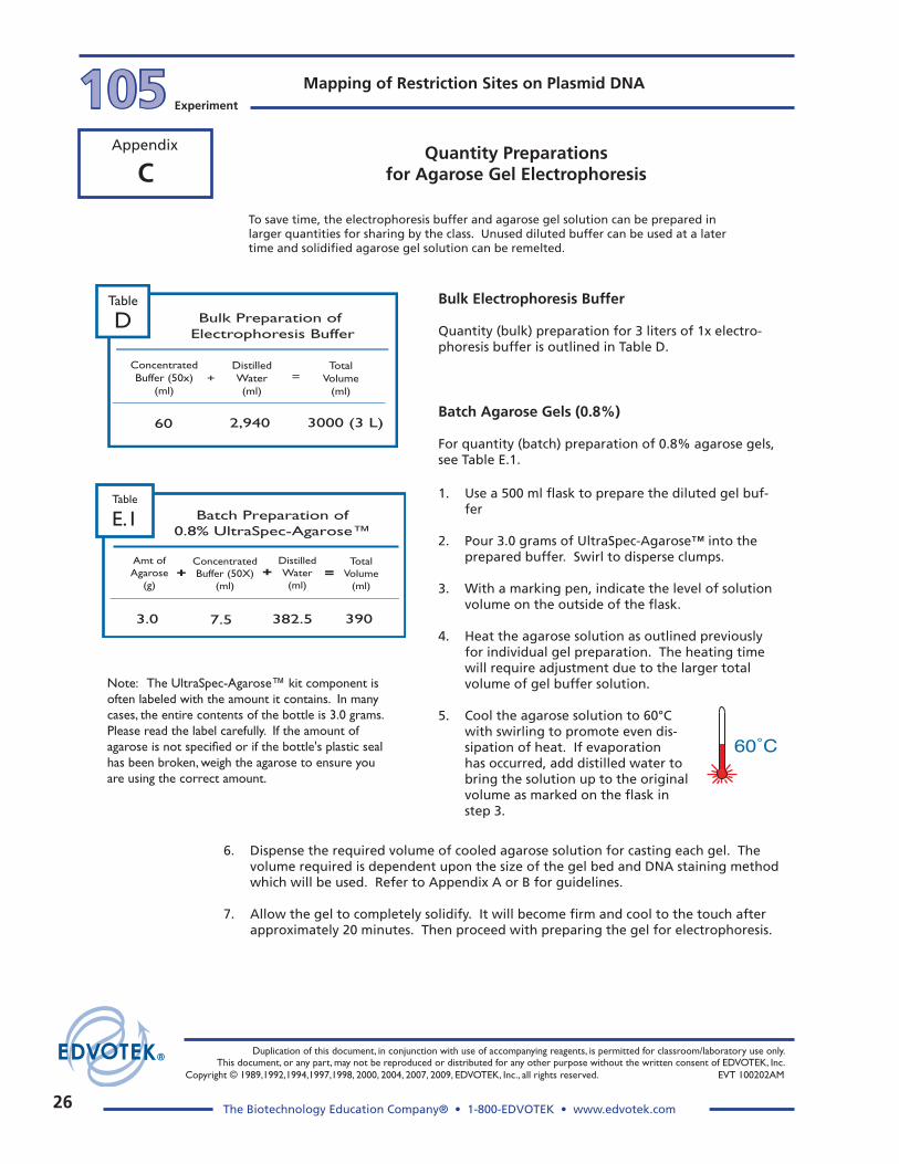
To save time, the electrophoresis buffer and agarose gel solution can be prepared in larger quantities for sharing by the class. Unused diluted buffer can be used at a later time and solidified agarose gel solution can be remelted.
Bulk Electrophoresis Buffer
Quantity (bulk) preparation for 3 liters of 1x electro-phoresis buffer is outlined in Table D.
Batch Agarose gels (0.8%)
For quantity (batch) preparation of 0.8% agarose gels, see Table E.1.
1. Use a 500 ml flask to prepare the diluted gel buf-fer
2. Pour 3.0 grams of UltraSpec-Agarose™ into the prepared buffer. Swirl to disperse clumps.
3. With a marking pen, indicate the level of solution volume on the outside of the flask.
4. Heat the agarose solution as outlined previously for individual gel preparation. The heating time will require adjustment due to the larger total volume of gel buffer solution.
5. Cool the agarose solution to 60°C with swirling to promote even dis-sipation of heat. If evaporation has occurred, add distilled water to bring the solution up to the original volume as marked on the flask in step 3.
60˚C
Note: The UltraSpec-Agarose™ kit component is often labeled with the amount it contains. In many cases, the entire contents of the bottle is 3.0 grams. Please read the label carefully. If the amount of agarose is not specified or if the bottle's plastic seal has been broken, weigh the agarose to ensure you are using the correct amount.
ConcentratedBuffer (50x)
(ml)
DistilledWater(ml)
TotalVolume
(ml)
60 2,940 3000 (3 L)
=+
Bulk Preparation of Electrophoresis Buffer
Table
D
3.0 7.5 382.5 390
Batch Preparation of
0.8% UltraSpec-Agarose™
Amt ofAgarose
(g)
ConcentratedBuffer (50X)
(ml)+
DistilledWater(ml)
TotalVolume
(ml)=+
Table
E.1
6. Dispense the required volume of cooled agarose solution for casting each gel. The volume required is dependent upon the size of the gel bed and DNA staining method which will be used. Refer to Appendix A or B for guidelines.
7. Allow the gel to completely solidify. It will become firm and cool to the touch after approximately 20 minutes. Then proceed with preparing the gel for electrophoresis.
Quantity Preparations for Agarose gel Electrophoresis

��
Duplication of this document, in conjunction with use of accompanying reagents, is permitted for classroom/laboratory use only. This document, or any part, may not be reproduced or distributed for any other purpose without the written consent of EDVOTEK, Inc. Copyright © 1989,1992,1994,1997,1998, 2000, 2004, 2007, 2009, EDVOTEK, Inc., all rights reserved. EVT 100202AM
The Biotechnology Education Company® • 1-800-EDVOTEK • www.edvotek.com
105Experiment
Mapping of Restriction Sites on Plasmid DNA
Appendix
EDVOTEK electrophoresis units include 7 x 7 cm or 7 x 14 cm gel casting trays.
A. Using Rubber dams:
• Place a rubber dam on each end of the bed. Make sure the rubber dam fits firmly in contact with the sides and bottom of the bed.
B. Taping with labeling or masking tape:
• Extend 3/4 inch wide tape over the sides and bottom edge of the bed. • Fold the extended tape edges back onto the sides and bottom. Press contact
points firmly to form a good seal.
If gel trays and rubber end caps are new, they may be initially somewhat difficult to assemble. Here is a helpful hint:
2. Place a well-former template (comb) in the first set of notches at the end of the bed. Make sure the comb sits firmly and evenly across the bed.
Preparing the gel bed
1. Close off the open ends of a clean and dry gel bed (casting tray) by using rubber dams or tape.
DAgarose gel Preparation - Step by Step guidelines
Place one of the black end caps with the wide “u” shaped slot fac-ing up on the lab bench.
Push one of the corners of the gel tray into one of the ends of the black cap. Press down on the tray at an angle, working from one end to the other until the end of the tray completely fits into the black cap. Repeat the process with the other end of the gel tray and the other black end cap.
Casting Agarose gels
3. Use a 250 ml flask or beaker to prepare the gel solution.
4. Refer to the appropriate Reference Table (i.e. 0.8%, 1.0% or 2.0%) for agarose gel preparation. Add the specified amount of agarose powder and buffer. Swirl the mixture to disperse clumps of agarose powder.
5. With a lab marking pen, indicate the level of the solution volume on the outside of the flask.
At high altitudes, use a microwave oven to reach boiling temperatures.
6. Heat the mixture to dissolve the agarose powder.
A. Microwave method:
• Cover the flask with plastic wrap to minimize evaporation.
• Heat the mixture on High for 1 minute. • Swirl the mixture and heat on High in bursts of 25 seconds
until all the agarose is completely dissolved.
B. Hot plate method:
• Cover the flask with aluminum foil to minimize evaporation. • Heat the mixture to boiling over a burner with occasional
swirling. Boil until all the agarose is completely dissolved.
Continue heating until the final solution appears clear (like water) with-out any undissolved particles. Check the solution carefully. If you see "crystal" particles, the agarose is not completely dissolved.

�8
Duplication of this document, in conjunction with use of accompanying reagents, is permitted for classroom/laboratory use only. This document, or any part, may not be reproduced or distributed for any other purpose without the written consent of EDVOTEK, Inc.
Copyright © 1989,1992,1994,1997,1998, 2000, 2004, 2007, 2009, EDVOTEK, Inc., all rights reserved. EVT 100202AM
The Biotechnology Education Company® • 1-800-EDVOTEK • www.edvotek.com
Mapping of Restriction Sites on Plasmid DNA105 Experiment
Appendix
7. Cool the agarose solution to 60°C with careful swirling to promote even dissipation of heat. If detectable evaporation has occurred, add distilled water to bring the solution up to the original volume marked in step 5.
After the gel is cooled to �0°C:
• If you are using rubber dams, go to step 9. • If you are using tape, continue with step 8.
DO NOT POUR BOILING HOT AGAROSE INTO THE GEL BED.
Hot agarose solution may irreversibly warp the bed.
60˚C
+Black Red
Sample wells
–
During electrophoresis, the DNA samples migrate through the agarose gel towards the positive electrode.
Agarose gel Preparation Step by Step guidelines, continued D
8. Seal the interface of the gel bed and tape to prevent aga-rose solution from leaking.
• Use a transfer pipet to deposit a small amount of the cooled agarose to both inside ends of the bed.
• Wait approximately 1 minute for the agarose to solidify.
9. Place the bed on a level surface and pour the cooled agarose solution into the bed.
10. Allow the gel to completely solidify. It will become firm and cool to the touch after approximately 20 minutes.
Preparing the gel for electrophoresis
11. After the gel is completely solidified, carefully and slowly remove the rubber dams or tape from the gel bed. Be especially careful not to damage or tear the gel wells when removing the rubber dams. A thin plastic knife, spatula or pipet tip can be inserted between the gel and the dams to break possible surface tension.
12. Remove the comb by slowly pulling straight up. Do this carefully and evenly to prevent tearing the sample wells.
13. Place the gel (on its bed) into the electrophoresis chamber, properly oriented, centered and level on the platform.
14. Fill the electrophoresis apparatus chamber with the appropriate amount of diluted (1x) electrophoresis buffer (refer to Table B on the Appendix page provided by your instructor).
15. Make sure that the gel is completely submerged under buffer before proceeding to loading the samples and conducting electrophoresis.
��
Duplication of this document, in conjunction with use of accompanying reagents, is permitted for classroom/laboratory use only. This document, or any part, may not be reproduced or distributed for any other purpose without the written consent of EDVOTEK, Inc. Copyright © 1989,1992,1994,1997,1998, 2000, 2004, 2007, 2009, EDVOTEK, Inc., all rights reserved. EVT 100202AM
The Biotechnology Education Company® • 1-800-EDVOTEK • www.edvotek.com
105Experiment
Mapping of Restriction Sites on Plasmid DNA
Appendix
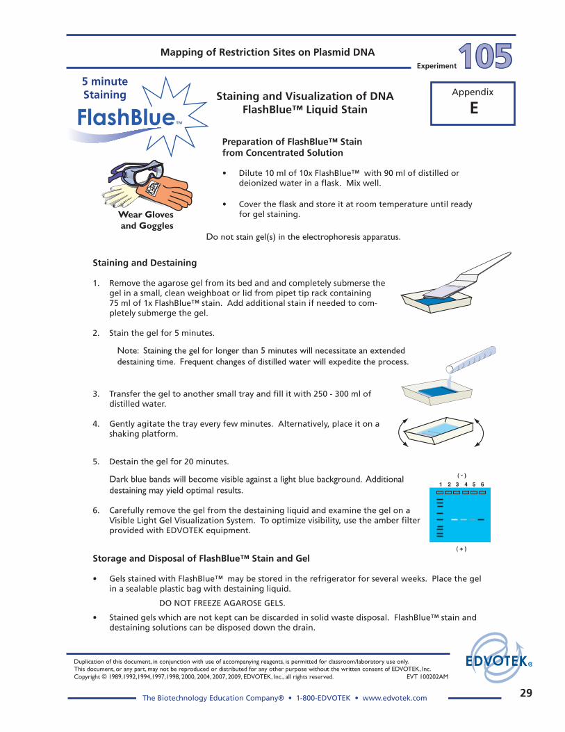
EStaining and Visualization of DNA
FlashBlue™ Liquid Stain
Preparation of FlashBlue™ Stain from Concentrated Solution
• Dilute 10 ml of 10x FlashBlue™ with 90 ml of distilled or deionized water in a flask. Mix well.
• Cover the flask and store it at room temperature until ready for gel staining.
Do not stain gel(s) in the electrophoresis apparatus.
Staining and Destaining
1. Remove the agarose gel from its bed and and completely submerse the gel in a small, clean weighboat or lid from pipet tip rack containing
75 ml of 1x FlashBlue™ stain. Add additional stain if needed to com-pletely submerge the gel.
2. Stain the gel for 5 minutes.
Note: Staining the gel for longer than 5 minutes will necessitate an extended destaining time. Frequent changes of distilled water will expedite the process.
Wear Gloves and Goggles
5. Destain the gel for 20 minutes.
Dark blue bands will become visible against a light blue background. Additional destaining may yield optimal results.
6. Carefully remove the gel from the destaining liquid and examine the gel on a Visible Light Gel Visualization System. To optimize visibility, use the amber filter provided with EDVOTEK equipment.
( - )
( + )
1 2 3 4 5 6
3. Transfer the gel to another small tray and fill it with 250 - 300 ml of distilled water.
4. Gently agitate the tray every few minutes. Alternatively, place it on a shaking platform.
Storage and Disposal of FlashBlue™ Stain and gel
• Gels stained with FlashBlue™ may be stored in the refrigerator for several weeks. Place the gel in a sealable plastic bag with destaining liquid.
DO NOT FREEZE AGAROSE GELS.
• Stained gels which are not kept can be discarded in solid waste disposal. FlashBlue™ stain and destaining solutions can be disposed down the drain.
5 minuteStaining

�0
Duplication of this document, in conjunction with use of accompanying reagents, is permitted for classroom/laboratory use only. This document, or any part, may not be reproduced or distributed for any other purpose without the written consent of EDVOTEK, Inc.
Copyright © 1989,1992,1994,1997,1998, 2000, 2004, 2007, 2009, EDVOTEK, Inc., all rights reserved. EVT 100202AM
The Biotechnology Education Company® • 1-800-EDVOTEK • www.edvotek.com
Mapping of Restriction Sites on Plasmid DNA105 Experiment
Appendix
FOne-Step Staining and Destaining
with InstaStain® Blue
1. Remove the 7 x 7 cm agarose gel from its bed and com-pletely submerse the gel in a small, clean tray containing
75 ml of distilled or deionized water, or used electrophore-sis buffer. The agarose gel should be completely covered with liquid.
Examples of small trays include large weigh boats, or small plastic food containers
Do not stain gel(s) in the electrophoresis apparatus.
InstaStain™
One Step Stain and Destain
2. Wearing gloves, gently float a 7 x 7 cm card of InstaStain® Blue with the stain side (blue) facing the liquid.
Note: If staining a 7 x 14 cm gel, use two InstaStain® Blue cards.
3. Let the gel soak undisturbed in the liquid for approximately 3 hours. The gel can be left in the liquid overnight (cover with plastic wrap to prevent evaporation).
4. After staining and destaining, the gel is ready for visualization and photography.
Storage and Disposal of InstaStain® Blue Cards and gels
• Stained gels may be stored in the refrigerator for several weeks. Place the gel in a sealable plastic bag with destaining liquid.
DO NOT FREEZE AGAROSE GELS!
• Used InstaStain® cards and destained gels can be discarded in solid waste disposal.
• Destaining solutions can be disposed down the drain.
Wear Gloves and Goggles
Agarose gels can be stained and destained in one easy step with InstaStain™ Blue cards. This one-step method can be completed in approximately 3 hours, or can be left overnight.
�1
Duplication of this document, in conjunction with use of accompanying reagents, is permitted for classroom/laboratory use only. This document, or any part, may not be reproduced or distributed for any other purpose without the written consent of EDVOTEK, Inc. Copyright © 1989,1992,1994,1997,1998, 2000, 2004, 2007, 2009, EDVOTEK, Inc., all rights reserved. EVT 100202AM
The Biotechnology Education Company® • 1-800-EDVOTEK • www.edvotek.com
105Experiment
Mapping of Restriction Sites on Plasmid DNA
Appendix
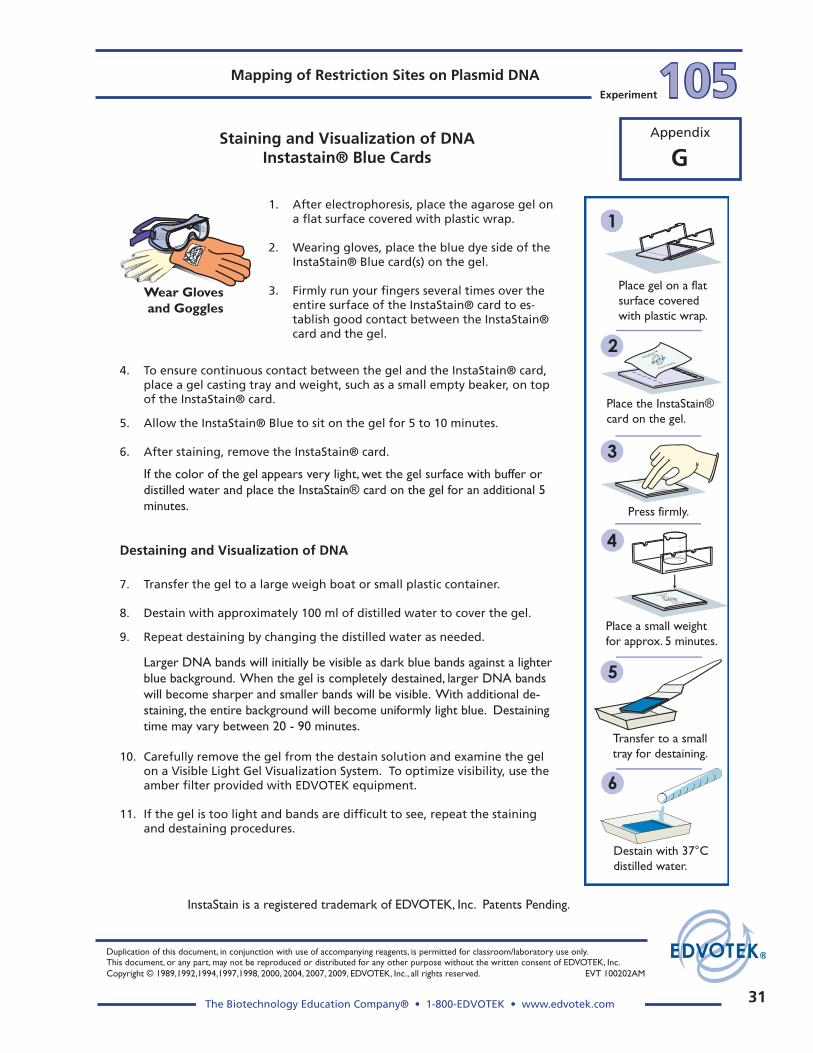
1. After electrophoresis, place the agarose gel on a flat surface covered with plastic wrap.
2. Wearing gloves, place the blue dye side of the InstaStain® Blue card(s) on the gel.
3. Firmly run your fingers several times over the entire surface of the InstaStain® card to es-tablish good contact between the InstaStain® card and the gel.
InstaStain™
Patents Pending
DNA InstaStain™
Patents Pending
Patents Pending
InstaStain™
-----
1
2
3
4
5
6
Place gel on a flatsurface covered with plastic wrap.
Place the InstaStain®card on the gel.
Place a small weightfor approx. 5 minutes.
Transfer to a smalltray for destaining.
Destain with 37°Cdistilled water.
Press firmly.
InstaStain is a registered trademark of EDVOTEK, Inc. Patents Pending.
Staining and Visualization of DNAInstastain® Blue Cards
4. To ensure continuous contact between the gel and the InstaStain® card, place a gel casting tray and weight, such as a small empty beaker, on top of the InstaStain® card.
5. Allow the InstaStain® Blue to sit on the gel for 5 to 10 minutes.
6. After staining, remove the InstaStain® card.
If the color of the gel appears very light, wet the gel surface with buffer or distilled water and place the InstaStain® card on the gel for an additional 5 minutes.
Destaining and Visualization of DNA
7. Transfer the gel to a large weigh boat or small plastic container.
8. Destain with approximately 100 ml of distilled water to cover the gel.
9. Repeat destaining by changing the distilled water as needed.
Larger DNA bands will initially be visible as dark blue bands against a lighter blue background. When the gel is completely destained, larger DNA bands will become sharper and smaller bands will be visible. With additional de-staining, the entire background will become uniformly light blue. Destaining time may vary between 20 - 90 minutes.
10. Carefully remove the gel from the destain solution and examine the gel on a Visible Light Gel Visualization System. To optimize visibility, use the amber filter provided with EDVOTEK equipment.
11. If the gel is too light and bands are difficult to see, repeat the staining and destaining procedures.
Wear Gloves and Goggles
g

��
Duplication of this document, in conjunction with use of accompanying reagents, is permitted for classroom/laboratory use only. This document, or any part, may not be reproduced or distributed for any other purpose without the written consent of EDVOTEK, Inc.
Copyright © 1989,1992,1994,1997,1998, 2000, 2004, 2007, 2009, EDVOTEK, Inc., all rights reserved. EVT 100202AM
The Biotechnology Education Company® • 1-800-EDVOTEK • www.edvotek.com
Mapping of Restriction Sites on Plasmid DNA105 Experiment
Appendix Staining and Visualization of DNAInstastain® Blue Cards
continued
Destaining Notes for InstaStain® Blue
• Use of warmed distilled water at 37°C will accelerate destaining. Destaining will take longer with room temperature water.
• DO NOT EXCEED 37°C ! Warmer temperatures will soften the gel and may cause it to break.
• The volume of distilled water for destaining depends upon the size of the tray. Use the small-est tray available that will accommodate the gel. The gel should be completely submerged during destaining.
• Do not exceed 3 changes of water for destaining. Excessive destaining will cause the bands to be very light.
Storage and Disposal of InstaStain® Blue Cards and gels
• Stained gels may be stored in the refrigerator for several weeks. Place the gel in a sealable plastic bag with destaining liquid.
DO NOT FREEZE AGAROSE GELS!
• Used InstaStain® cards and destained gels can be discarded in solid waste disposal.
• Destaining solutions can be disposed down the drain.
g
��
Duplication of this document, in conjunction with use of accompanying reagents, is permitted for classroom/laboratory use only. This document, or any part, may not be reproduced or distributed for any other purpose without the written consent of EDVOTEK, Inc. Copyright © 1989,1992,1994,1997,1998, 2000, 2004, 2007, 2009, EDVOTEK, Inc., all rights reserved. EVT 100202AM
The Biotechnology Education Company® • 1-800-EDVOTEK • www.edvotek.com
105Experiment
Mapping of Restriction Sites on Plasmid DNA
Appendix
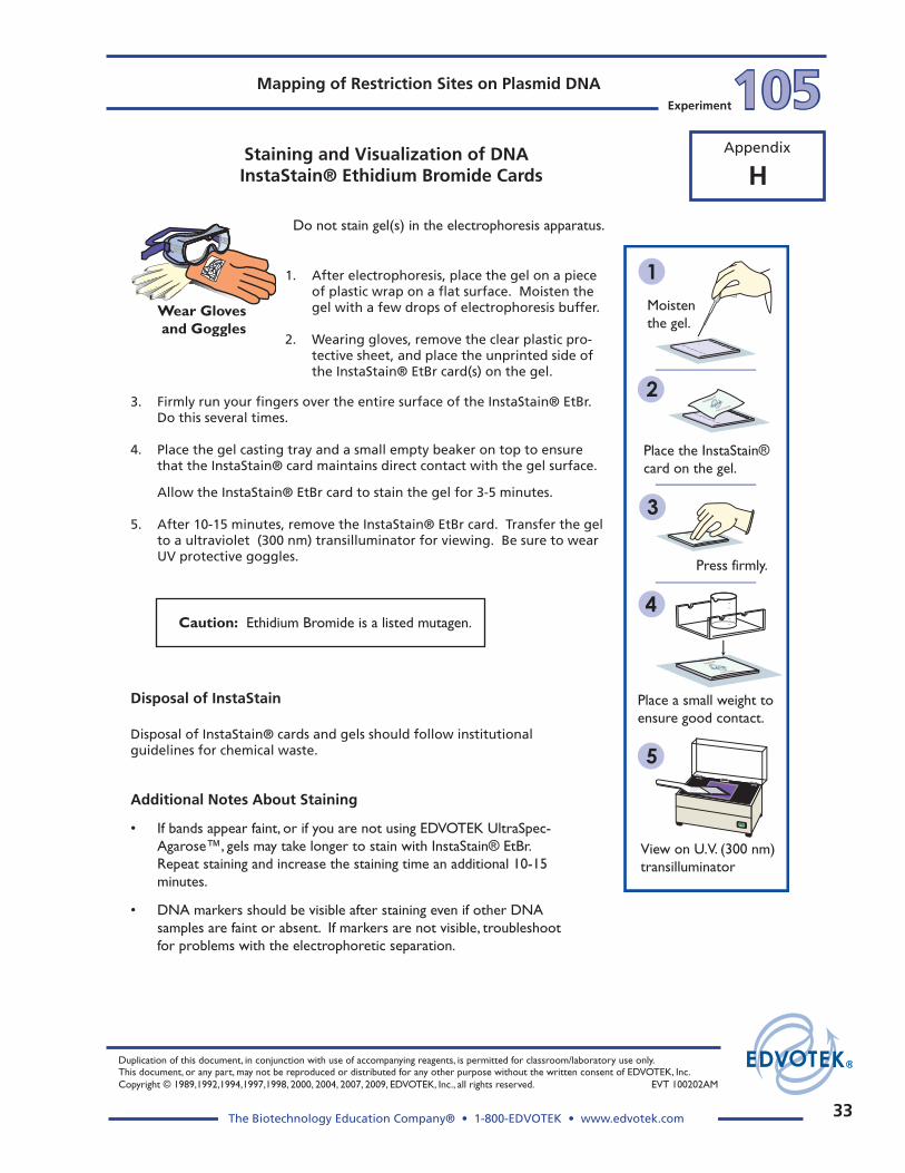
Do not stain gel(s) in the electrophoresis apparatus.
1. After electrophoresis, place the gel on a piece of plastic wrap on a flat surface. Moisten the gel with a few drops of electrophoresis buffer.
2. Wearing gloves, remove the clear plastic pro-tective sheet, and place the unprinted side of the InstaStain® EtBr card(s) on the gel.
Disposal of InstaStain
Disposal of InstaStain® cards and gels should follow institutional guidelines for chemical waste.
Additional Notes About Staining
• If bands appear faint, or if you are not using EDVOTEK UltraSpec-Agarose™, gels may take longer to stain with InstaStain® EtBr. Repeat staining and increase the staining time an additional 10-15 minutes.
• DNA markers should be visible after staining even if other DNA samples are faint or absent. If markers are not visible, troubleshoot for problems with the electrophoretic separation.
Caution: Ethidium Bromide is a listed mutagen.
Staining and Visualization of DNA InstaStain® Ethidium Bromide Cards
3. Firmly run your fingers over the entire surface of the InstaStain® EtBr. Do this several times.
4. Place the gel casting tray and a small empty beaker on top to ensure that the InstaStain® card maintains direct contact with the gel surface.
Allow the InstaStain® EtBr card to stain the gel for 3-5 minutes.
5. After 10-15 minutes, remove the InstaStain® EtBr card. Transfer the gel to a ultraviolet (300 nm) transilluminator for viewing. Be sure to wear UV protective goggles.
Wear Gloves and Goggles
h
1
2
3
4
5
Press firmly.
Moistenthe gel.
Place the InstaStain®card on the gel.
Place a small weight toensure good contact.
View on U.V. (300 nm) transilluminator
InstaStain™
Patents Pending
DNA InstaStain™
Patents Pending
Patents Pending
InstaStain™
-----

Material Safety Data SheetsFull-size (8.5 x 11”) pdf copy of MSDS is available at www. edvotek.com or by request.105
Experiment
�4
Mat
eria
l Saf
ety
Dat
a Sh
eet
May
be
use
d t
o c
om
ply
wit
h O
SHA
's H
azar
d C
om
mu
nic
atio
nSt
and
ard
. 29
CFR
191
0.12
00 S
tan
dar
d m
ust
be
con
sult
ed f
or
spec
ific
req
uir
emen
ts.
IDEN
TITY
(A
s U
sed
on
Lab
el a
nd
Lis
t)N
ote
: B
lan
k sp
aces
are
no
t p
erm
itte
d.
If a
ny
item
is n
ot
app
licab
le, o
r n
o in
form
atio
n is
ava
ilab
le, t
he
spac
e m
ust
b
e m
arke
d t
o in
dic
ate
that
.
Sect
ion
IM
anu
fact
ure
r's
Nam
e
Sect
ion
II -
Haz
ard
ou
s In
gre
die
nts
/Id
enti
fy In
form
atio
n
Emer
gen
cy T
elep
ho
ne
Nu
mb
er
Tele
ph
on
e N
um
ber
fo
r in
form
atio
n
Dat
e Pr
epar
ed
Sig
nat
ure
of
Prep
arer
(o
pti
on
al)
Ad
dre
ss (
Nu
mb
er, S
tree
t, C
ity,
Sta
te,
Zip
Co
de)
EDV
OTE
K, I
nc.
1467
6 R
oth
geb
Dri
veR
ock
ville
, MD
208
50
Haz
ard
ou
s C
om
po
nen
ts [
Spec
ific
C
hem
ical
Iden
tity
; C
om
mo
n N
ame(
s)]
O
SHA
PEL
AC
GIH
TLV
Oth
er L
imit
s R
eco
mm
end
ed%
(O
pti
on
al)
(301
) 25
1-59
90
(301
) 25
1-59
90
Bo
ilin
g P
oin
t
Sect
ion
III -
Ph
ysic
al/C
hem
ical
Ch
arac
teri
stic
s
Un
usu
al F
ire
and
Exp
losi
on
Haz
ard
s
Spec
ial F
ire
Fig
hti
ng
Pro
ced
ure
s
Vap
or
Pres
sure
(m
m H
g.)
Vap
or
Den
sity
(A
IR =
1)
Solu
bili
ty in
Wat
er
Ap
pea
ran
ce a
nd
Od
or
Sect
ion
IV -
Ph
ysic
al/C
hem
ical
Ch
arac
teri
stic
sFl
ash
Po
int
(Met
ho
d U
sed
)
Exti
ng
uis
hin
g M
edia
Flam
mab
le L
imit
sU
ELLE
L
Mel
tin
g P
oin
t
Evap
ora
tio
n R
ate
(Bu
tyl A
ceta
te =
1)
Spec
ific
Gra
vity
(H
0 =
1)
2
Ag
aro
se
10/0
5/06
This
pro
du
ct c
on
tain
s n
o h
azar
do
us
mat
eria
ls a
s d
efin
ed b
y th
e O
SHA
Haz
ard
Co
mm
un
icat
ion
Stan
dar
d.
CA
S #9
012-
36-6
For
1% s
olu
tio
n 1
94 F
N
o d
ata
N
o d
ata
No
dat
a
No
dat
a
No
dat
a
Inso
lub
le -
co
ld
W
hit
e p
ow
der
, no
od
or
N.D
. = N
o d
ata
No
dat
a
N
.D.
N.D
.
Wat
er s
pra
y, d
ry c
hem
ical
, car
bo
n d
ioxi
de,
hal
on
or
stan
dar
d f
oam
Poss
ible
fir
e h
azar
d w
hen
exp
ose
d t
o h
eat
or
flam
e
No
ne
ED
VO
TE
K®
Stab
ility
Sect
ion
V -
Rea
ctiv
ity
Dat
aU
nst
able
Sect
ion
VI -
Hea
lth
Haz
ard
Dat
a
Inco
mp
atib
ility
Co
nd
itio
ns
to A
void
Ro
ute
(s)
of
Entr
y:In
hal
atio
n?
Ing
esti
on
?Sk
in?
Oth
er
Stab
le
Haz
ard
ou
s Po
lym
eriz
atio
nM
ay O
ccu
rC
on
dit
ion
s to
Avo
id
Will
No
t O
ccu
r
Hea
lth
Haz
ard
s (A
cute
an
d C
hro
nic
)
Car
cin
og
enic
ity:
NTP
?O
SHA
Reg
ula
tio
n?
IAR
C M
on
og
rap
hs?
Sig
ns
and
Sym
pto
ms
of
Exp
osu
re
Med
ical
Co
nd
itio
ns
Gen
eral
ly A
gg
rava
ted
by
Exp
osu
re
Emer
gen
cy F
irst
Aid
Pro
ced
ure
s
Sect
ion
VII
- Pr
ecau
tio
ns
for
Safe
Han
dlin
g a
nd
Use
Step
s to
be
Take
n in
cas
e M
ater
ial i
s R
elea
sed
fo
r Sp
illed
Was
te D
isp
osa
l Met
ho
d
Prec
auti
on
s to
be
Take
n in
Han
dlin
g a
nd
Sto
rin
g
Oth
er P
reca
uti
on
s
Sect
ion
VIII
- C
on
tro
l Mea
sure
s
Ven
tila
tio
nLo
cal E
xhau
stSp
ecia
l
Mec
han
ical
G
en. d
iluti
on
ven
tila
tio
n
Res
pir
ato
ry P
rote
ctio
n (
Spec
ify
Typ
e)
Pro
tect
ive
Glo
ves
Oth
er P
rote
ctiv
e C
loth
ing
or
Equ
ipm
ent
Wo
rk/H
ygie
nic
Pra
ctic
es
Eye
Pro
tect
ion
Haz
ard
ou
s D
eco
mp
osi
tio
n o
r B
ypro
du
cts
Yes
Sp
lash
pro
of
go
gg
les
Imp
ervi
ou
s cl
oth
ing
to
pre
ven
t sk
in c
on
tact
No
neX
N
on
e
No
dat
a av
aila
ble
X
No
ne
Yes
Y
es
Yes
Inh
alat
ion
: N
o d
ata
avai
lab
le
In
ges
tio
n:
Larg
e am
ou
nts
may
cau
se d
iarr
hea
No
dat
a av
aila
ble
No
dat
a av
aila
ble
Trea
t sy
mp
tom
atic
ally
an
d s
up
po
rtiv
ely
Swee
p u
p a
nd
pla
ce in
su
itab
le c
on
tain
er f
or
dis
po
sal
No
rmal
so
lid w
aste
dis
po
sal
No
ne
No
ne
Ch
emic
al c
artr
idg
e re
spir
ato
r w
ith
fu
ll fa
cep
iece
.
ED
VO
TE
K®
Mat
eria
l Saf
ety
Dat
a Sh
eet
May
be
use
d t
o c
om
ply
wit
h O
SHA
's H
azar
d C
om
mu
nic
atio
nSt
and
ard
. 29
CFR
191
0.12
00 S
tan
dar
d m
ust
be
con
sult
ed f
or
spec
ific
req
uir
emen
ts.
IDEN
TITY
(A
s U
sed
on
Lab
el a
nd
Lis
t)N
ote
: B
lan
k sp
aces
are
no
t p
erm
itte
d.
If a
ny
item
is n
ot
app
licab
le, o
r n
o in
form
atio
n is
ava
ilab
le, t
he
spac
e m
ust
b
e m
arke
d t
o in
dic
ate
that
.
Sect
ion
IM
anu
fact
ure
r's
Nam
e
Sect
ion
II -
Haz
ard
ou
s In
gre
die
nts
/Id
enti
fy In
form
atio
n
Emer
gen
cy T
elep
ho
ne
Nu
mb
er
Tele
ph
on
e N
um
ber
fo
r in
form
atio
n
Dat
e Pr
epar
ed
Sig
nat
ure
of
Prep
arer
(o
pti
on
al)
Ad
dre
ss (
Nu
mb
er, S
tree
t, C
ity,
Sta
te,
Zip
Co
de)
EDV
OTE
K, I
nc.
1467
6 R
oth
geb
Dri
veR
ock
ville
, MD
208
50
Haz
ard
ou
s C
om
po
nen
ts [
Spec
ific
C
hem
ical
Iden
tity
; C
om
mo
n N
ame(
s)]
O
SHA
PEL
AC
GIH
TLV
Oth
er L
imit
s R
eco
mm
end
ed%
(O
pti
on
al)
(301
) 25
1-59
90
(301
) 25
1-59
90
Bo
ilin
g P
oin
t
Sect
ion
III -
Ph
ysic
al/C
hem
ical
Ch
arac
teri
stic
s
Un
usu
al F
ire
and
Exp
losi
on
Haz
ard
s
Spec
ial F
ire
Fig
hti
ng
Pro
ced
ure
s
Vap
or
Pres
sure
(m
m H
g.)
Vap
or
Den
sity
(A
IR =
1)
Solu
bili
ty in
Wat
er
Ap
pea
ran
ce a
nd
Od
or
Sect
ion
IV -
Ph
ysic
al/C
hem
ical
Ch
arac
teri
stic
sFl
ash
Po
int
(Met
ho
d U
sed
)
Exti
ng
uis
hin
g M
edia
Flam
mab
le L
imit
sU
ELLE
L
Mel
tin
g P
oin
t
Evap
ora
tio
n R
ate
(Bu
tyl A
ceta
te =
1)
Spec
ific
Gra
vity
(H
0 =
1)
2
50x
Elec
tro
ph
ore
sis
Bu
ffer
This
pro
du
ct c
on
tain
s n
o h
azar
do
us
mat
eria
ls a
s d
efin
ed b
y th
e O
SHA
Haz
ard
Co
mm
un
icat
ion
Sta
nd
ard
.
No
dat
a
No
dat
a
No
dat
a
No
dat
a
No
dat
a
No
dat
a
Ap
pre
ciab
le, (
gre
ater
th
an 1
0%)
Cle
ar, l
iqu
id, s
ligh
t vi
neg
ar o
do
r
No
dat
a
N.D
. = N
o d
ata N.D
.
N.D
.
Use
ext
ing
uis
hin
g m
edia
ap
pro
pri
ate
for
surr
ou
nd
ing
fir
e.
Wea
r p
rote
ctiv
e eq
uip
men
t an
d S
CB
A w
ith
fu
ll fa
cep
iece
op
erat
ed in
po
siti
ve p
ress
ure
mo
de.
No
ne
iden
tifi
ed
10/0
5/06
Stab
ility
Sect
ion
V -
Rea
ctiv
ity
Dat
aU
nst
able
Sect
ion
VI -
Hea
lth
Haz
ard
Dat
a
Inco
mp
atib
ility
Co
nd
itio
ns
to A
void
Ro
ute
(s)
of
Entr
y:In
hal
atio
n?
Ing
esti
on
?Sk
in?
Oth
er
Stab
le
Haz
ard
ou
s Po
lym
eriz
atio
nM
ay O
ccu
rC
on
dit
ion
s to
Avo
id
Will
No
t O
ccu
r
Hea
lth
Haz
ard
s (A
cute
an
d C
hro
nic
)
Car
cin
og
enic
ity:
NTP
?O
SHA
Reg
ula
tio
n?
IAR
C M
on
og
rap
hs?
Sig
ns
and
Sym
pto
ms
of
Exp
osu
re
Med
ical
Co
nd
itio
ns
Gen
eral
ly A
gg
rava
ted
by
Exp
osu
re
Emer
gen
cy F
irst
Aid
Pro
ced
ure
s
Sect
ion
VII
- Pr
ecau
tio
ns
for
Safe
Han
dlin
g a
nd
Use
Step
s to
be
Take
n in
cas
e M
ater
ial i
s R
elea
sed
fo
r Sp
illed
Was
te D
isp
osa
l Met
ho
d
Prec
auti
on
s to
be
Take
n in
Han
dlin
g a
nd
Sto
rin
g
Oth
er P
reca
uti
on
s
Sect
ion
VIII
- C
on
tro
l Mea
sure
s
Ven
tila
tio
nLo
cal E
xhau
stSp
ecia
l
Mec
han
ical
(G
ener
al)
Res
pir
ato
ry P
rote
ctio
n (
Spec
ify
Typ
e)
Pro
tect
ive
Glo
ves
Oth
er P
rote
ctiv
e C
loth
ing
or
Equ
ipm
ent
Wo
rk/H
ygie
nic
Pra
ctic
es
Eye
Pro
tect
ion
Haz
ard
ou
s D
eco
mp
osi
tio
n o
r B
ypro
du
cts
X
N
on
e
Stro
ng
oxi
diz
ing
ag
ents
Car
bo
n m
on
oxi
de,
Car
bo
n d
ioxi
de
X
N
on
e
Yes
Y
es
Y
es
No
ne
No
ne
iden
tifi
ed
Irri
tati
on
to
up
per
res
pir
ato
ry t
ract
, ski
n, e
yes
No
ne
Ing
esti
on
: If
co
nsc
iou
s, g
ive
larg
e am
ou
nts
of
wat
er
Eyes
: Fl
ush
wit
h w
ater
In
hal
atio
n:
Mo
ve t
o f
resh
air
Sk
in:
Was
h w
ith
so
ap a
nd
wat
er
Wea
r su
itab
le p
rote
ctiv
e cl
oth
ing
. M
op
up
sp
ill
and
rin
se w
ith
wat
er, o
r co
llect
in a
bso
rpti
ve m
ater
ial a
nd
dis
po
se o
f th
e ab
sorp
tive
mat
eria
l.
Dis
po
se in
acc
ord
ance
wit
h a
ll ap
plic
able
fed
eral
, sta
te, a
nd
loca
l en
viro
men
tal r
egu
lati
on
s.
Avo
id e
ye a
nd
ski
n c
on
tact
.
No
ne
Yes
N
on
e
Yes
N
on
e
Yes
_Saf
ety
go
gg
les
No
ne
No
ne
Stab
ility
Sect
ion
V -
Rea
ctiv
ity
Dat
aU
nst
able
Sect
ion
VI -
Hea
lth
Haz
ard
Dat
a
Inco
mp
atib
ility
Co
nd
itio
ns
to A
void
Ro
ute
(s)
of
Entr
y:In
hal
atio
n?
Ing
esti
on
?Sk
in?
Oth
er
Stab
le
Haz
ard
ou
s Po
lym
eriz
atio
nM
ay O
ccu
rC
on
dit
ion
s to
Avo
id
Will
No
t O
ccu
r
Hea
lth
Haz
ard
s (A
cute
an
d C
hro
nic
)
Car
cin
og
enic
ity:
NTP
?O
SHA
Reg
ula
tio
n?
IAR
C M
on
og
rap
hs?
Sig
ns
and
Sym
pto
ms
of
Exp
osu
re
Med
ical
Co
nd
itio
ns
Gen
eral
ly A
gg
rava
ted
by
Exp
osu
re
Emer
gen
cy F
irst
Aid
Pro
ced
ure
s
Sect
ion
VII
- Pr
ecau
tio
ns
for
Safe
Han
dlin
g a
nd
Use
Step
s to
be
Take
n in
cas
e M
ater
ial i
s R
elea
sed
fo
r Sp
illed
Was
te D
isp
osa
l Met
ho
d
Prec
auti
on
s to
be
Take
n in
Han
dlin
g a
nd
Sto
rin
g
Oth
er P
reca
uti
on
s
Sect
ion
VIII
- C
on
tro
l Mea
sure
s
Ven
tila
tio
nLo
cal E
xhau
stSp
ecia
l
Mec
han
ical
(G
ener
al)
Res
pir
ato
ry P
rote
ctio
n (
Spec
ify
Typ
e)
Pro
tect
ive
Glo
ves
Oth
er P
rote
ctiv
e C
loth
ing
or
Equ
ipm
ent
Wo
rk/H
ygie
nic
Pra
ctic
es
Eye
Pro
tect
ion
Haz
ard
ou
s D
eco
mp
osi
tio
n o
r B
ypro
du
cts
X
N
on
e
No
ne
Sulf
ur
oxi
des
, an
d b
rom
ides
X
N
on
e
Yes
Y
es
Yes
Acu
te e
ye c
on
tact
: M
ay c
ause
irri
tati
on
. N
o d
ata
avai
lab
le f
or
oth
er r
ou
tes.
No
dat
a av
aila
ble
May
cau
se s
kin
or
eye
irri
tati
on
No
ne
rep
ort
ed
Trea
t sy
mp
tom
atic
ally
an
d s
up
po
rtiv
ely.
Rin
se c
on
tact
ed a
rea
wit
h c
op
iou
s am
ou
nts
of
wat
er.
Wea
r ey
e an
d s
kin
pro
tect
ion
an
d m
op
sp
ill a
rea.
Rin
se w
ith
wat
er.
Ob
serv
e al
l fed
eral
, sta
te, a
nd
loca
l reg
ula
tio
ns.
Avo
id e
ye a
nd
ski
n c
on
tact
.
No
ne
Yes
No
ne
Yes
No
ne
Yes
Spla
sh p
roo
f g
og
gle
s
No
ne
req
uir
ed
Avo
id e
ye a
nd
ski
n c
on
tact
Mat
eria
l Saf
ety
Dat
a Sh
eet
May
be
use
d t
o c
om
ply
wit
h O
SHA
's H
azar
d C
om
mu
nic
atio
nSt
and
ard
. 29
CFR
191
0.12
00 S
tan
dar
d m
ust
be
con
sult
ed f
or
spec
ific
req
uir
emen
ts.
IDEN
TITY
(A
s U
sed
on
Lab
el a
nd
Lis
t)N
ote
: B
lan
k sp
aces
are
no
t p
erm
itte
d.
If a
ny
item
is n
ot
app
licab
le, o
r n
o in
form
atio
n is
ava
ilab
le, t
he
spac
e m
ust
b
e m
arke
d t
o in
dic
ate
that
.
Sect
ion
IM
anu
fact
ure
r's
Nam
e
Sect
ion
II -
Haz
ard
ou
s In
gre
die
nts
/Id
enti
fy In
form
atio
n
Emer
gen
cy T
elep
ho
ne
Nu
mb
er
Tele
ph
on
e N
um
ber
fo
r in
form
atio
n
Dat
e Pr
epar
ed
Sig
nat
ure
of
Prep
arer
(o
pti
on
al)
Ad
dre
ss (
Nu
mb
er, S
tree
t, C
ity,
Sta
te,
Zip
Co
de)
EDV
OTE
K, I
nc.
1467
6 R
oth
geb
Dri
veR
ock
ville
, MD
208
50
Haz
ard
ou
s C
om
po
nen
ts [
Spec
ific
C
hem
ical
Iden
tity
; C
om
mo
n N
ame(
s)]
O
SHA
PEL
AC
GIH
TLV
Oth
er L
imit
s R
eco
mm
end
ed%
(O
pti
on
al)
(301
) 25
1-59
90
(301
) 25
1-59
90
Bo
ilin
g P
oin
t
Sect
ion
III -
Ph
ysic
al/C
hem
ical
Ch
arac
teri
stic
s
Un
usu
al F
ire
and
Exp
losi
on
Haz
ard
s
Spec
ial F
ire
Fig
hti
ng
Pro
ced
ure
s
Vap
or
Pres
sure
(m
m H
g.)
Vap
or
Den
sity
(A
IR =
1)
Solu
bili
ty in
Wat
er
Ap
pea
ran
ce a
nd
Od
or
Sect
ion
IV -
Ph
ysic
al/C
hem
ical
Ch
arac
teri
stic
sFl
ash
Po
int
(Met
ho
d U
sed
)
Exti
ng
uis
hin
g M
edia
Flam
mab
le L
imit
sU
ELLE
L
Mel
tin
g P
oin
t
Evap
ora
tio
n R
ate
(Bu
tyl A
ceta
te =
1)
Spec
ific
Gra
vity
(H
0 =
1)
2
Prac
tice
Gel
Lo
adin
g S
olu
tio
n
10/0
5/06
This
pro
du
ct c
on
tain
s n
o h
azar
do
us
mat
eria
ls a
s d
efin
ed b
y th
e O
SHA
Haz
ard
Co
mm
un
icat
ion
Stan
dar
d.
No
dat
a
No
dat
a
No
dat
a
No
dat
a
No
dat
a
No
dat
a
Solu
ble
B
lue
liqu
id, n
o o
do
r
No
dat
aN
o d
ata
No
dat
a
Dry
ch
emic
al, c
arb
on
dio
xid
e, w
ater
sp
ray
or
foam
Use
age
nts s
uita
ble
for t
ype
of su
rrou
ndin
g fir
e. K
eep
upw
ind,
avo
idb
reat
hin
g h
azar
do
us
sulf
ur
oxi
des
an
d b
rom
ides
. W
ear
SCB
A.
Un
kno
wn
ED
VO
TE
K®

�5
105Experiment
Material Safety Data SheetsFull-size (8.5 x 11”) pdf copy of MSDS is available at www. edvotek.com or by request.
Stab
ility
Sect
ion
V -
Rea
ctiv
ity
Dat
aU
nst
able
Sect
ion
VI -
Hea
lth
Haz
ard
Dat
a
Inco
mp
atib
ility
Co
nd
itio
ns
to A
void
Ro
ute
(s)
of
Entr
y:In
hal
atio
n?
Ing
esti
on
?Sk
in?
Oth
er
Stab
le
Haz
ard
ou
s Po
lym
eriz
atio
nM
ay O
ccu
rC
on
dit
ion
s to
Avo
id
Will
No
t O
ccu
r
Hea
lth
Haz
ard
s (A
cute
an
d C
hro
nic
)
Car
cin
og
enic
ity:
NTP
?O
SHA
Reg
ula
tio
n?
IAR
C M
on
og
rap
hs?
Sig
ns
and
Sym
pto
ms
of
Exp
osu
re
Med
ical
Co
nd
itio
ns
Gen
eral
ly A
gg
rava
ted
by
Exp
osu
re
Emer
gen
cy F
irst
Aid
Pro
ced
ure
s
Sect
ion
VII
- Pr
ecau
tio
ns
for
Safe
Han
dlin
g a
nd
Use
Step
s to
be
Take
n in
cas
e M
ater
ial i
s R
elea
sed
fo
r Sp
illed
Was
te D
isp
osa
l Met
ho
d
Prec
auti
on
s to
be
Take
n in
Han
dlin
g a
nd
Sto
rin
g
Oth
er P
reca
uti
on
s
Sect
ion
VIII
- C
on
tro
l Mea
sure
s
Ven
tila
tio
nLo
cal E
xhau
stSp
ecia
l
Mec
han
ical
(G
ener
al)
Res
pir
ato
ry P
rote
ctio
n (
Spec
ify
Typ
e)
Pro
tect
ive
Glo
ves
Oth
er P
rote
ctiv
e C
loth
ing
or
Equ
ipm
ent
Wo
rk/H
ygie
nic
Pra
ctic
es
Eye
Pro
tect
ion
Haz
ard
ou
s D
eco
mp
osi
tio
n o
r B
ypro
du
cts
X
N
on
e
Stro
ng
oxi
diz
ing
ag
ents
Car
bo
n m
on
oxi
de,
Car
bo
n d
ioxi
de,
nit
rog
en o
xid
es, h
ydro
gen
bro
mid
e g
as
X
N
on
e
Yes
Y
es
Yes
No
dat
a av
aila
ble
Irri
tati
on
to
mu
cou
s m
emb
ran
es a
nd
up
per
res
pir
ato
ry t
ract
No
dat
a
Trea
t sy
mp
tom
atic
ally
an
d s
up
po
rtiv
ely
Wea
r SC
BA
, ru
bb
er b
oo
ts, r
ub
ber
glo
ves
Mix
mat
eria
l wit
h c
om
bu
stib
le s
olv
ent
and
bu
rn in
a c
hem
ical
inci
ner
ato
r eq
uip
ped
aft
erb
urn
er a
nd
scr
ub
ber
Use
in c
hem
ical
fu
me
ho
od
wit
h p
rop
er p
rote
ctiv
e la
b g
ear.
Mu
tag
en
Yes
Ch
em. f
um
e h
oo
d
No
N
on
e
Ru
bb
er
C
hem
. saf
ety
go
gg
les
R
ub
ber
bo
ots
Use
in c
hem
ical
fu
me
ho
od
wit
h p
rop
er p
rote
ctiv
e la
b g
ear.
Acu
te: M
ater
ial i
rrit
atin
g t
o m
uco
us
mem
bra
nes
, up
per
res
pir
ato
ry t
ract
, eye
s, s
kin
Ch
ron
ic:
May
alt
er g
enet
ic m
ater
ial
SCB
A
Mat
eria
l Saf
ety
Dat
a Sh
eet
May
be
use
d t
o c
om
ply
wit
h O
SHA
's H
azar
d C
om
mu
nic
atio
nSt
and
ard
. 29
CFR
191
0.12
00 S
tan
dar
d m
ust
be
con
sult
ed f
or
spec
ific
req
uir
emen
ts.
IDEN
TITY
(A
s U
sed
on
Lab
el a
nd
Lis
t)N
ote
: B
lan
k sp
aces
are
no
t p
erm
itte
d.
If a
ny
item
is n
ot
app
licab
le, o
r n
o in
form
atio
n is
ava
ilab
le, t
he
spac
e m
ust
b
e m
arke
d t
o in
dic
ate
that
.
Sect
ion
IM
anu
fact
ure
r's
Nam
e
Sect
ion
II -
Haz
ard
ou
s In
gre
die
nts
/Id
enti
fy In
form
atio
n
Emer
gen
cy T
elep
ho
ne
Nu
mb
er
Tele
ph
on
e N
um
ber
fo
r in
form
atio
n
Dat
e Pr
epar
ed
Sig
nat
ure
of
Prep
arer
(o
pti
on
al)
Inst
aSta
in, I
nc.
P.O
. Bo
x 12
32W
est
Bet
hes
da,
MD
208
27
Haz
ard
ou
s C
om
po
nen
ts [
Spec
ific
C
hem
ical
Iden
tity
; C
om
mo
n N
ame(
s)]
O
SHA
PEL
AC
GIH
TLV
Oth
er L
imit
s R
eco
mm
end
ed%
(O
pti
on
al)
(301
) 25
1-59
90
(301
) 25
1-59
90
Bo
ilin
g P
oin
t
Sect
ion
III -
Ph
ysic
al/C
hem
ical
Ch
arac
teri
stic
s
Un
usu
al F
ire
and
Exp
losi
on
Haz
ard
s
Spec
ial F
ire
Fig
hti
ng
Pro
ced
ure
s
Vap
or
Pres
sure
(m
m H
g.)
Vap
or
Den
sity
(A
IR =
1)
Solu
bili
ty in
Wat
er
Ap
pea
ran
ce a
nd
Od
or
Sect
ion
IV -
Ph
ysic
al/C
hem
ical
Ch
arac
teri
stic
sFl
ash
Po
int
(Met
ho
d U
sed
)
Exti
ng
uis
hin
g M
edia
Flam
mab
le L
imit
sU
ELLE
L
Mel
tin
g P
oin
t
Evap
ora
tio
n R
ate
(Bu
tyl A
ceta
te =
1)
Spec
ific
Gra
vity
(H
0 =
1)
2
Inst
aSta
in®
Eth
idiu
m B
rom
ide
Eth
idiu
m B
rom
ide
D
ata
no
t av
aila
ble
No
dat
a
No
dat
a
No
dat
a
No
dat
a
No
dat
a
No
dat
a
Solu
ble
Ch
emic
al b
ou
nd
to
pap
er, n
o o
do
r
No
dat
a
N.D
. = N
o d
ata N.D
. N
.D.
Wat
er s
pra
y, c
arb
on
dio
xid
e, d
ry c
hem
ical
po
wd
er, a
lco
ho
l or
po
lym
er f
oam
Wea
r p
rote
ctiv
e cl
oth
ing
an
d S
CB
A t
o p
reve
nt
con
tact
wit
h s
kin
& e
yes
Emit
s to
xic
fum
es
10/0
5/06
CA
S# 1
39-3
3-3
(2,7
-Dia
min
o-1
0-Et
hyl
-9-P
hen
ylp
hen
anth
rid
iniu
m B
rom
ide)
ED
VO
TE
K®
Mat
eria
l Saf
ety
Dat
a Sh
eet
May
be
use
d t
o c
om
ply
wit
h O
SHA
's H
azar
d C
om
mu
nic
atio
nSt
and
ard
. 29
CFR
191
0.12
00 S
tan
dar
d m
ust
be
con
sult
ed f
or
spec
ific
req
uir
emen
ts.
IDEN
TITY
(A
s U
sed
on
Lab
el a
nd
Lis
t)N
ote
: B
lan
k sp
aces
are
no
t p
erm
itte
d.
If a
ny
item
is n
ot
app
licab
le, o
r n
o in
form
atio
n is
ava
ilab
le, t
he
spac
e m
ust
b
e m
arke
d t
o in
dic
ate
that
.
Sect
ion
IM
anu
fact
ure
r's
Nam
e
Sect
ion
II -
Haz
ard
ou
s In
gre
die
nts
/Iden
tify
Info
rmat
ion
Emer
gen
cy T
elep
ho
ne
Nu
mb
er
Tele
ph
on
e N
um
ber
fo
r in
form
atio
n
Dat
e Pr
epar
ed
Sig
nat
ure
of
Prep
arer
(o
pti
on
al)
Ad
dre
ss (
Nu
mb
er, S
tree
t, C
ity,
Sta
te,
Zip
Co
de)
EDV
OTE
K, I
nc.
1467
6 R
oth
geb
Dri
veR
ock
ville
, MD
208
50
Haz
ard
ou
s C
om
po
nen
ts [
Spec
ific
C
hem
ical
Iden
tity
; C
om
mo
n N
ame(
s)]
O
SHA
PEL
AC
GIH
TLV
Oth
er L
imit
s R
eco
mm
end
ed%
(O
pti
on
al)
(301
) 25
1-59
90
(301
) 25
1-59
90
Bo
ilin
g P
oin
t
Sect
ion
III -
Ph
ysic
al/C
hem
ical
Ch
arac
teri
stic
s
Un
usu
al F
ire
and
Exp
losi
on
Haz
ard
s
Spec
ial F
ire
Fig
hti
ng
Pro
ced
ure
s
Vap
or
Pres
sure
(m
m H
g.)
Vap
or
Den
sity
(A
IR =
1)
Solu
bili
ty in
Wat
er
Ap
pea
ran
ce a
nd
Od
or
Sect
ion
IV -
Ph
ysic
al/C
hem
ical
Ch
arac
teri
stic
sFl
ash
Po
int
(Met
ho
d U
sed
)
Exti
ng
uis
hin
g M
edia
Flam
mab
le L
imit
sU
ELLE
L
Mel
tin
g P
oin
t
Evap
ora
tio
n R
ate
(Bu
tyl A
ceta
te =
1)
Spec
ific
Gra
vity
(H
0 =
1)
2
ED
VO
TE
K®
Inst
aSta
in®
Blu
e, F
lash
Blu
e™
03-2
6-09
Met
hyl
ene
Blu
e
3.7
Bis
(D
imet
hyl
amin
o)
Phen
oth
iazi
n 5
IUM
C
hlo
rid
e
No
dat
a av
aila
ble
CA
S #
61-7
3-4
No
dat
a
No
dat
a
No
dat
a
No
dat
a
No
dat
a
No
dat
a
Solu
ble
- c
old
Ch
emic
al b
ou
nd
to
pap
er, n
o o
do
r
No
dat
a av
aila
ble
No
dat
a
N
o d
ata
Wat
er s
pra
y, c
arb
on
dio
xid
e, d
ry c
hem
ical
po
wd
er, a
lco
ho
l or
po
lym
er f
oam
Self
co
nta
ined
bre
ath
ing
ap
par
atu
s an
d p
rote
ctiv
e cl
oth
ing
to
pre
ven
t co
nta
ct
wit
h s
kin
an
d e
yes
Emit
s to
xid
fu
mes
un
der
fir
e co
nd
itio
ns
Stab
ility
Sect
ion
V -
Rea
ctiv
ity
Dat
aU
nst
able
Sect
ion
VI -
Hea
lth
Haz
ard
Dat
a
Inco
mp
atib
ility
Co
nd
itio
ns
to A
void
Ro
ute
(s)
of
Entr
y:In
hal
atio
n?
Ing
esti
on
?Sk
in?
Oth
er
Stab
le
Haz
ard
ou
s Po
lym
eriz
atio
nM
ay O
ccu
rC
on
dit
ion
s to
Avo
id
Will
No
t O
ccu
r
Hea
lth
Haz
ard
s (A
cute
an
d C
hro
nic
)
Car
cin
og
enic
ity:
NTP
?O
SHA
Reg
ula
tio
n?
IAR
C M
on
og
rap
hs?
Sig
ns
and
Sym
pto
ms
of
Exp
osu
re
Med
ical
Co
nd
itio
ns
Gen
eral
ly A
gg
rava
ted
by
Exp
osu
re
Emer
gen
cy F
irst
Aid
Pro
ced
ure
s
Sect
ion
VII
- Pr
ecau
tio
ns
for
Safe
Han
dlin
g a
nd
Use
Step
s to
be
Take
n in
cas
e M
ater
ial i
s R
elea
sed
fo
r Sp
illed
Was
te D
isp
osa
l Met
ho
d
Prec
auti
on
s to
be
Take
n in
Han
dlin
g a
nd
Sto
rin
g
Oth
er P
reca
uti
on
s
Sect
ion
VIII
- C
on
tro
l Mea
sure
s
Ven
tila
tio
nLo
cal E
xhau
stSp
ecia
l
Mec
han
ical
(G
ener
al)
Res
pir
ato
ry P
rote
ctio
n (
Spec
ify
Typ
e)
Pro
tect
ive
Glo
ves
Oth
er P
rote
ctiv
e C
loth
ing
or
Equ
ipm
ent
Wo
rk/H
ygie
nic
Pra
ctic
es
Eye
Pro
tect
ion
Haz
ard
ou
s D
eco
mp
osi
tio
n o
r B
ypro
du
cts
X
No
ne
Stro
ng
oxi
diz
ing
ag
ents
Toxi
c fu
mes
of
Car
bo
n m
on
oxi
de,
Car
bo
n d
ioxi
de,
n
itro
gen
oxi
des
, su
lfu
r o
xid
es, h
ydro
gen
, ch
lori
de
gas
X
N
on
e
Yes
Y
es
Yes
Skin
: M
ay c
ause
ski
n ir
rita
tio
n
Eyes
: M
ay c
ause
eye
irri
tati
on
In
hal
atio
n:
Cya
no
sis
Mee
ts c
rite
ria
for
pro
po
sed
OSH
A m
edic
al r
eco
rds
rule
PER
EAC
47.
3042
0.82
No
dat
a av
aila
ble
No
dat
a av
aila
ble
Trea
t sy
mp
tom
atic
ally
Ven
tila
te a
rea
and
was
h s
pill
sit
e
Mix
mat
eria
l wit
h a
co
mb
ust
ible
so
lven
t an
d b
urn
in c
hem
ical
inci
ner
ato
r eq
uip
ped
wit
h a
fter
bu
rner
an
d s
cru
bb
er.
Ch
eck
loca
l an
d s
tate
reg
ula
tio
ns.
Kee
p t
igh
tly
clo
sed
. St
ore
in c
oo
l, d
ry p
lace
No
ne
MIO
SH/O
SHA
ap
pro
ved
, SC
BA
Req
uir
ed
Ru
bb
erC
hem
. saf
ety
go
gg
les
Ru
bb
er b
oo
ts

��The Biotechnology Education Company® • 1-800-EDVOTEK • www.edvotek.com
Mapping of Restriction Sites on Plasmid DNA
EVT 100202AM
EDVOTEK Series 100 Electrophoresis Experiments:
Cat. # Title
101 Principles and Practice of Agarose Gel Electrophoresis
102 Restriction Enzyme Cleavage Patterns of DNA
105 Mapping of Restriction Sites on Plasmid DNA
104 Size Determination of DNA Restriction Fragments
105 Mapping of Restriction Sites on Plasmid DNA
105 DNA Fingerprinting - Identification of DNA by Restriction Fragmentation Patterns
112 Analysis of Eco RI Cleavage Patterns of Lambda DNA
114 DNA Paternity Testing Simulation
115 Cancer Gene Detection
116 Sickle Cell Gene Detection (DNA-based)
117 Detection of Mad Cow Disease
118 Cholesterol Diagnostiics
124 DNA-based Screening for Smallpox
130 DNA Fingerprinting - Amplification of DNA for Fingerprinting
Visit our web site for information about the above experiments and other products
in EDVOTEK’s comprehensive offerings for biotechnology and biology education.
Order Online