section 6 biological effects of radiofrequency exposure
TRANSCRIPT
RF Toolkit–BCCDC/NCCEH Section 6A 83
Section 6
Biological Effects of Radiofrequency Exposure
Section 6A Cell Culture Studies
Table of Contents
Summary ................................................................................................................................................ 84
6A.1 Introduction ................................................................................................................................ 85
6A.2 Purpose ........................................................................................................................................ 85
6A.3 Methods ....................................................................................................................................... 86
6A.4 Cancer-Related Effects .......................................................................................................... 87
6A.4.1 DNA damage and RF fields (Table 1) .......................................................... 87
6A.4.2 Cell transformation and proliferation and RF fields (Table 2) ...................... 91
6A.4.3 Apoptosis and RF fields (Table 3) ............................................................... 96
6A.4.4 Reactive oxygen species and RF exposure (Table 4) ................................... 99
6A.4.5 Ornithine decarboxylase activity and RF fields (Table 5) ........................... 102
6A.5 Gene Expression and RF Fields ....................................................................................... 105
6A.5.1 Heat shock gene and protein changes and RF fields (Table 6) .................. 105
6A.5.2 Other gene and protein expression changes and RF fields (Table 7) ......... 109
6A.6 Other Specific Intracellular Effects ................................................................................ 112
6A.6.1 Changes in protein and RF fields (Table 8) ............................................... 112
6A.6.2 Calcium efflux and RF fields (Table 9) ...................................................... 115
6A.6.3 Cell permeability and RF fields (Table 10) ................................................ 116
6A.5 Discussion................................................................................................................................ 117
6A.5.1 Research gaps ......................................................................................... 118
6A.6 References ............................................................................................................................... 119
RF Toolkit–BCCDC/NCCEH Section 6A 84
Summary
• Use of cell culture models to investigate effects of environmental exposures can lead to elucidation of biologic mechanisms to explain adverse effects which help direct animal and human health research. Many cell culture studies have recently (2005–2012) been published to assess whether radiofrequency (RF) field exposure has adverse biological effects on a variety of cells.
• Studies of DNA damage and RF field exposure at non-thermal levels using indicators such as chromosomal aberrations and micronucleus have shown mixed results, with a few positive studies and many negative ones. There is no convincing evidence from cell culture studies that RF field exposure damages DNA.
• There is no evidence from recent cell culture studies that exposure to RF fields alone can induce transformation. Results of studies concerning the effect of RF fields on cell proliferation when RF fields are applied alone is mixed, with a few positive studies showing decreased proliferation with exposure to RF but many negative studies as well. More research is needed on the effect of RF fields in conjunction with known carcinogenic exposures such as ionizing radiation.
• Evidence of the presence of reactive oxygen species (ROS) or of apoptosis (cell death) in cell cultures due to exposure to RF fields is contradictory, with some studies showing evidence of generation of ROS or of apoptosis and others none. Recent studies on each of these putative study outcomes have been well conducted and no particular aspect of the study protocols characterize positive versus negative studies.
• Recent well conducted studies of the effect of RF fields in induction of ornithine decarboxylase (affecting tumour growth) have been predominantly negative, even under conditions of cell stress or stimulation.
• The question of whether non-thermal RF fields induce changes in expression of heat shock or other genes or proteins is open, as the results of studies are quite contradictory. However, as in most other aspects of cell culture research, there are no specific frequencies or characteristics (pulsed or continuous wave) of RF exposure which distinguish positive from negative studies.
• A variety of physiologic processes in neurologic and other cells have been tested under exposure to RF fields, with no weight of evidence to indicate that such RF exposure adversely affects any process.
• There is little evidence from recent studies that RF fields adversely affect calcium channelling in cultured cells.
RF Toolkit–BCCDC/NCCEH Section 6A 85
• There have been only a few studies recently assessing the effect of RF fields on cell cultures designed to mimic the blood-brain barrier, and these are mostly negative. Most of the work in this area has been recently conducted using animal models.
• Overall, in spite of the many well-conducted cell culture experiments examining a number of putative effects from RF fields, there is no convincing evidence that exposure to such fields has adverse biological effects. In many areas of research, the results are inconsistent and contradictory. The lack of features distinguishing positive studies from negative ones has prevented the development of any credible biologic mechanism by which such fields might adversely affect cells in culture.
6A.1 Introduction
Over the past 25 years, many studies have been conducted to determine whether RF field exposure can have adverse effects on human health, but in spite of the effort, there is still much uncertainty. Studies of the putative relationship between RF exposure and chronic diseases such as brain cancer carried out in humans are observational in nature rather than experimental. Most observational studies are retrospective in nature and consequently provide incomplete information on RF exposure and a lack of control of confounding variables, which complicates the process of determining cause and effect. In addition, a significant period of time elapses between exposure and subsequent disease, making causal relationships more difficult to establish.
Experimental studies under laboratory conditions allow manipulation of exposure and measurement of effect. Human-derived cell and tumour lines are plentiful, outcome measures can be achieved quickly, and biological processes known to be involved in chronic disease can be studied under controlled conditions. If such studies show that RF fields initiate or promote biological processes known to be involved in chronic disease, and these results are independently replicated by other researchers, then this can lead to development of testable biological mechanisms to better understand and predict effects of RF. Although cellular studies cannot determine the interactions between cells seen in living systems, biologic mechanisms suggested by cell line studies can then be rapidly tested in experimental animal models. Thus, studies in cell lines can play a key role in advancing knowledge about the possible relationship between RF exposure and human disease.
6A.2 Purpose The purpose of this review is to summarize the recent literature (2005–2011) on the effects of RF fields on cell cultures which are most relevant to possible adverse human health effects. A few a priori limitations were established. First of all, it is well known that RF fields at high power can cause thermal effects, including stimulation of heat-shock proteins, alterations in DNA, and in extreme cases, cell destruction. However, the fields to which humans are exposed in day-to-day use of RF devices do not cause

RF Toolkit–BCCDC/NCCEH Section 6A 86
any notable heating, and thus studies involving changes due to thermal effects were not included. Near field intensity of RF fields within cell cultures or tissue is described by the metric specific absorption rate (SAR) measured in “watts per kilogram” of tissue. SAR values under realistic day-to-day conditions of use from RF sources rarely exceed levels of about 2 W/kg in humans, and consequently studies which examine the effects in vitro and animal model studies that generate SAR levels around or below these levels will be emphasized whenever possible.
Further, as the major human concern to date with RF fields concerns use of cellular or mobile phones, the review will concentrate largely on studies of the frequencies between 800 MHz and 2450 MHz, as these are the commonly used frequencies in North American, Asian and Nordic telephony at the present time. Although use of the latest generation of RF devices using the Long Term Evolution (LTE) standard and marketed as 4G is rapidly expanding, little information on its effect on biologic systems is available at this time.
6A.3 Methods
A search of the online databases PubMed (MEDLINE), and EBSCO Academic Search was conducted using search terms “radiofrequency field,” “radiofrequency radiation,” “RF radiation,” “microwave,” “cellular phone,” “mobile phone,” and these key words were combined with terms for carcinogenesis, genotoxicity, DNA damage, chromosome(al) aberration, micronucleus formation, apoptosis, gene expression, ornithine decarboxylase, cell permeability, protein expression, gene expression, cell proliferation, and cell transformation. The search was restricted to peer-reviewed articles published in English since 2005 to 2011. After eliminating duplicate references picked up by multiple searches, there were 126 studies found for more detailed review. A separate search using the term “WiFi” linked to cancer, and various other terms including “health” produced only one genuine in vitro investigation. Review articles were separated out so bibliographies could be searched; and recent national reviews of RF fields and health such as the Latin American Experts Committee on High Frequency Electromagnetic Fields and Human Health report1 and the UK Health Protection Agency’s recent report2 were also examined for papers missed by other means.
Although this review will concentrate mainly on more recent studies (2005–2011), summary paragraphs at the end of each group of potential adverse biological effects will consider all available evidence and not just included studies published since 2005. The reason for the emphasis on more recent work is that these investigations are more likely to be characterized by good RF dosimetry and better experimental protocols offering good control of the potential confounding effect of thermal changes. Sometimes, earlier investigations will be referenced to provide context for study of a particular adverse effect.
Within each category of in vitro biological effects on cells, a representative group of studies were chosen for tabular presentation and discussion. These studies are, for the

RF Toolkit–BCCDC/NCCEH Section 6A 87
most part, characterized by good experimental methods, accurate RF dosimetry, use of RF frequencies that humans are exposed to on a day-to-day basis (such as GSM and CDMA mobile phone frequencies), and SAR values of around 2 W/kg.
6A.4 Cancer-Related Effects
To facilitate conduct of in vitro studies, blood lymphocytes, buccal, skin or other cells can be obtained from human volunteers or animals. In addition, cancer or other cells may be extracted from humans or animals, immortalized using a virus or other means, and cultured, forming cell lines. Such cell lines remain genetically constant over time and can be used for years to produce “test cells” for many studies. Thus, investigators seeking to repeat an experiment done by another scientist can use the same cell line as used in a previous study with reasonable assurance that the test cells are genetically very similar to the original.
A sham group refers to cells which are grown in exactly the same conditions and undergo all the manipulations that the RF-exposed cells go through except for the RF exposure itself. This helps ensure that other conditions of the experiment do not cause cellular changes which might then falsely be attributed to RF field exposure. Including a positive control group can also be a valuable addition to an experiment as it provides a standard against which changes in the experimental cells can be compared.
6A.4.1 DNA damage and RF fields (Table 1)
One of the principal concerns with RF fields is whether they have the ability to cause cancer alone, or to promote cancer in the presence of other known carcinogens. Since damaged DNA is characteristic of cancer cells, indications of damage due to RF field exposure are important. DNA damage is manifested in a number of ways in cells, including chromosomal aberrations, micronucleus formation, and DNA strand breaks. Chromosomal aberrations occur when a cell divides, and this process does not take place properly. Micronucleus formation occurs when a daughter cell inherits an incomplete complement of chromosomes plus a small micronucleus carrying the whole or partial chromosome missing from the actual nucleus. Chromosomal aberrations and micronucleus formation are characteristics of genetic instability and are associated with diseases such as cancers.
Vijayalaxmi (2006)3 at the University of Texas Health Sciences Centre extracted lymphocytes (while blood cells) from blood samples collected from non-smoking male donors and exposed the samples to pulsed 2450 MHz or 820 MHz RF fields or sham for two hours at SAR levels of 2.3 W/kg or 20.7 W/kg. Another group of lymphocytes was exposed to an acute gamma radiation exposure of 1.5 G, known to cause DNA damage, and was maintained as a positive control group. Cultured lymphocytes were then examined to determine the extent of cytogenetic damage incurred with the RF exposure. No differences were seen in percentage mitotic index, chromosomal exchange aberrations, or excess fragments in the RF-exposed cells by comparison with

RF Toolkit–BCCDC/NCCEH Section 6A 88
sham- exposed cells. As expected, the positive control cells showed elevated damage levels compared to both sham- and RF-exposed cells. The investigator concluded that the results showed no indications that RF field exposure increased DNA damage by comparison with sham exposure.
Stronati et al. (2006)4 exposed lymphocytes from 14 healthy donors to 935 MHz basic GSM signal (SAR 1.0 and 2.0 W/kg) or sham for 24 hours either alone or combined with one- minute exposure to 1.0 Gy of 250 kVp x-rays given either immediately before or after RF exposure. Results showed no elevation in DNA strand breakage, chromosomal aberrations, sister chromatid exchanges, or micronucleus formation in the RF-field- exposed cells by comparison with sham-exposed cells. In addition, RF exposure did not enhance DNA damaging effects in the x-ray exposed cells.
A further study in fibroblasts by Speit et al. (2007)5 used V79 hamster fibroblasts exposed to 1800 MHz continuous wave RF fields or sham exposure on an intermittent schedule (5 minutes on, 10 off) for 1 to 24 hours. The RF exposure was performed in a temperature-controlled wave guide chamber (SAR of 2.0 W/kg). Positive and negative control cultures were also included in the protocol. Evaluation after exposure using the Comet assay showed no increase in DNA damage in the RF-exposed cells compared to the sham-exposed and control groups. The Comet assay is a test in which RF-exposed cells are lysed in an agarose gel and exposed to pulsed electrophoresis. The lysed cell material, when observed using fluorescent microscopy, appears like a comet, and DNA damage is assessed by the size of the comet “tail.” In addition, the study did not detect any increased micronucleus formation, another indication of DNA damage, in the RF- field-exposed cells.
Mazor and his colleagues (2008)6 exposed lymphocytes from 10 volunteers to a continuous wave RF field at 800 MHz or sham in a wave-guide resonator at SARs of 2.9 and 4.1 W/kg for 72 hours. The study was conducted over a range of temperatures from 33.5 to 40.0ºC to evaluate the contribution of thermal effects to any changes observed. Assessment of the lymphocytes after exposure at 37ºC showed increased aneuploidy in chromosomes 1 and 10 at the higher SAR, and in chromosomes 11 and 17 at the lower SAR level, indicating damaged DNA in the RF-exposed cells. Aneuploidy is an abnormal number of chromosomes and occurs when chromosomes do not separate properly at cell division. Elevated levels of aneuploidy were also seen at other temperatures, leading investigators to conclude that elevated damage levels in RF-exposed cells might be independent of temperature.
In a further study, conducted by Manti et al. (2008),7 lymphocytes were exposed to x-rays (4 Gy) known to cause DNA damage, and subsequently to 1950 MHz UMTS signal at 0.5 or 2.0 W/kg SAR or to sham exposure for a period of 24 hours. Analysis revealed a small but statistically significant increase in the amount of DNA damage per cell in cells exposed to x-rays and 1950 MHz signal at a SAR of 2.0 W/kg compared to those exposed to x-rays and sham RF exposure. The authors suggested that this might be

RF Toolkit–BCCDC/NCCEH Section 6A 89
evidence of an inhibiting effect exerted by RF fields on cells’ DNA repair mechanism following damage by x-rays.
The study of Zeni et al. (2008)8 evaluated peripheral lymphocytes (circulating white blood cells) from healthy volunteers to exposure for 24–68 hours to intermittent 1950 MHz RF fields (six minutes RF on; two hours off; SAR 2.2 W/kg) or sham in a transverse electromagnetic cell (TEM). The protocol included temperature control measures as well as negative and positive control (mitomycin-C; methylmethanesulphonate exposure) cells. Results of comet and micronucleus assays showed no effects on DNA structure and no increase in micronucleus formation or changes in cell cycle kinetics attributable to RF field exposure.
Schwarz and colleagues (2008)9 exposed human-cultured fibroblasts to 1950 MHz UMTS signal (SAR below 2 W/kg) for 8, 12, or 24 hours in a commercial incubation chamber with good control of temperature. Results showed increased micronucleus formation and enhanced comet tail factor response in cells exposed for 24 hours at SAR 0.5 W/kg, indicating DNA damage.
Kim et al. (2008)10 exposed L5178Y mouse leukemia/lymphoma cells to 835 MHz CDMA signal in a TEM cell at 4.0 W/kg or sham for 24 or 48 hours. At Comet assay, no increase in chromosomal aberrations were seen in the exposed cells in comparison with the sham exposed cells; however, in conjunction with the clastogenic agents cyclophosphamide or 4-nitroquinoline 1-oxide, which are known to produce chromosomal damage, RF exposure appeared to potentiate the damage brought about by these agents. The relevance of this study to human health issues is questionable as the SAR level is much higher than is seen in day-to-day use of RF devices.
With Sannino et al. (2009),11 human dermal fibroblasts were exposed to 900 MHz pulsed GSM signal for 24 hours (SAR 1.0 W/kg) alone and in conjunction with the potent mutagen 3-Chloro-4-(dichloromethy)-5-Hydroxy-2(5h)furanone (MX). Comet assay results revealed no genotoxic or cytotoxic damage from RF field exposure alone or enhanced DNA damage due to the addition of RF exposure to MX.
In a further similar study, Hansteen et al. (2009)12 collected blood from six healthy donors, separated and cultured their lymphocytes, and exposed the cultured lymphocytes to 2300 MHz pulsed or continuous wave signal or sham in an anechoic chamber, alone or in conjunction with mitomycin C, a known clastogen. A clastogen is a compound known to cause chromosomal breaks. Field intensity was given to be 10 W/m2 although no SAR levels are noted. Results showed no differences in either damaged DNA in RF- exposed cells alone, compared with sham-exposed, and in addition no enhanced damage or slower DNA repair in those exposed to mitomycin C and RF fields, in comparison with sham and mitomycin C.
Campisi et al. (2010)13 exposed rat astroglial cells to 900 MHz continuous and pulsed GSM signal for 5, 10, or 20 minutes at a SAR of 0.25 W/kg and showed increased DNA
RF Toolkit–BCCDC/NCCEH Section 6A 90
damage as indicated by Comet assay results in RF cells compared to sham-exposed and control cells. In addition, the RF-exposed cells showed increased production of reactive oxygen species (ROS) by comparison with control cells.
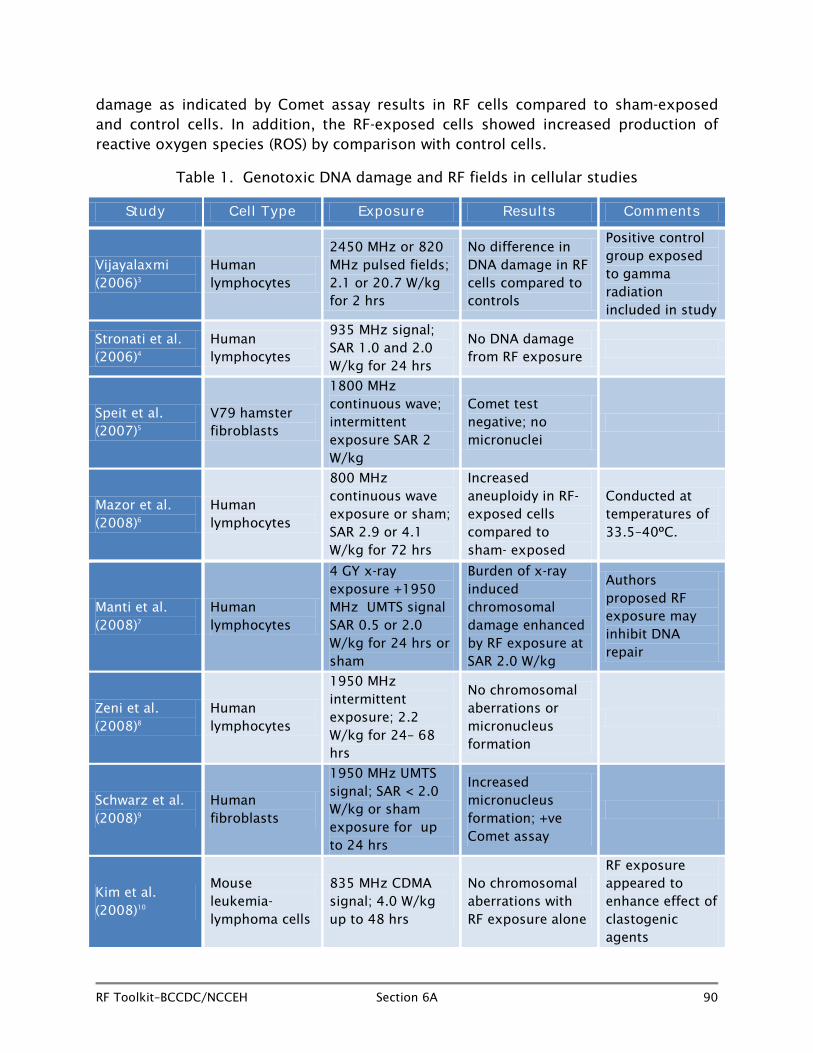
Table 1. Genotoxic DNA damage and RF fields in cellular studies
Study Cell Type Exposure Results Comments
Vijayalaxmi (2006)3
Human lymphocytes
2450 MHz or 820 MHz pulsed fields; 2.1 or 20.7 W/kg for 2 hrs
No difference in DNA damage in RF cells compared to controls
Positive control group exposed to gamma radiation included in study
Stronati et al. (2006)4
Human lymphocytes
935 MHz signal; SAR 1.0 and 2.0 W/kg for 24 hrs
No DNA damage from RF exposure
Speit et al. (2007)5
V79 hamster fibroblasts
1800 MHz continuous wave; intermittent exposure SAR 2 W/kg
Comet test negative; no micronuclei
Mazor et al. (2008)6
Human lymphocytes
800 MHz continuous wave exposure or sham; SAR 2.9 or 4.1 W/kg for 72 hrs
Increased aneuploidy in RF-exposed cells compared to sham- exposed
Conducted at temperatures of 33.5–40ºC.
Manti et al. (2008)7
Human lymphocytes
4 GY x-ray exposure +1950 MHz UMTS signal SAR 0.5 or 2.0 W/kg for 24 hrs or sham
Burden of x-ray induced chromosomal damage enhanced by RF exposure at SAR 2.0 W/kg
Authors proposed RF exposure may inhibit DNA repair
Zeni et al. (2008)8
Human lymphocytes
1950 MHz intermittent exposure; 2.2 W/kg for 24– 68 hrs
No chromosomal aberrations or micronucleus formation
Schwarz et al. (2008)9
Human fibroblasts
1950 MHz UMTS signal; SAR < 2.0 W/kg or sham exposure for up to 24 hrs
Increased micronucleus formation; +ve Comet assay
Kim et al. (2008)10
Mouse leukemia-lymphoma cells
835 MHz CDMA signal; 4.0 W/kg up to 48 hrs
No chromosomal aberrations with RF exposure alone
RF exposure appeared to enhance effect of clastogenic agents

RF Toolkit–BCCDC/NCCEH Section 6A 91
Study Cell Type Exposure Results Comments
Sannino et al. (2009)11
Human dermal fibroblasts
900 MHz pulsed GSM signal SAR 1.0 W/kg for 24 hrs and MX mutagen
Comet assay showed no enhancement of MX-induced DNA damage by RF exposure
Hansteen et al. (2009)12
Human lymphocytes
2300 MHz pulsed signal at 10 W/m2 for 53 hrs or sham, with and without Mitomycin C
No chromosomal differences in RF cells compared to controls either with or without Mitomycin C
No SAR levels found in paper
Campisi et al. (2010)13
Rat astroglial cells
900 MHz continuous and pulsed for 5, 10 or 20 min; SAR 0.25 W/kg
Comet test showed increased DNA damage in RF- exposed vs. sham and control cells
Production of ROS in RF exposed cells
Summary
For direct indicators of DNA damage such as chromosomal aberrations and micronucleus formation, the evidence for an effect of RF fields alone among cell cultures is not strong, largely because studies show such inconsistent results. For instance, among fibroblast cell culture studies, the investigations of Schwarz et al.9 showed DNA damage but that of Speit et al.5 did not. A comprehensive review of data by an expert group under the aegis of the International Agency for Research on Cancer concluded that for most end points in cell culture studies including DNA damage, studies of low intensity (non thermal) RF exposure provided only weak evidence of any effect.14 Adding to the difficulties of making sense of the contradictory results seen is the fact that most recent studies use first-rate cell culturing techniques, well-validated measures of DNA damage, excellent temperature control to rule out thermal effects, and well-described RF exposure protocols.
6A.4.2 Cell transformation and proliferation and RF fields (Table 2)
Cell transformation is an important step in the process of carcinogenesis, involving escape of a clone of cells from contact inhibition, by which cells surrounding the clone restrict its ability to proliferate. Cell proliferation in normal healthy cells is restricted to a rate commensurate with the function of those cells within the cellular matrix they are growing in. Although the process of carcinogenesis results in an increased rate of proliferation in cells, in normal routinely growing cultures proliferation can be an indication of cell stress.

RF Toolkit–BCCDC/NCCEH Section 6A 92
There have only been two recent studies involving the effect of RF field exposure on cell transformation since 2005, and both have been negative. Wang et al. (2005)15 exposed mouse C3H10T1/2 fibroblasts to continuous wave 2450 MHz electromagnetic fields at specific absorption rates of 5 to 200 W/kg for two hours in conjunction with methylcholanthrene, a known initiating chemical, or to methylcholanthrene alone. The transformation frequency of cells was slightly increased with the addition of 2450 MHz exposure, but only at SAR levels in excess of 100W/kg—almost 100 times as high as seen in normal human exposure to RF fields.
A Japanese study (2008)16 exposed BALB/3T3 mouse cells to 2142 MHz W-CDMA signal at SAR of 80 and 800 mW/kg for six weeks alone and in addition to 3-methylcholanthrene, and also on RF-exposed cells initiated with MCA and co-exposed to TPA. Results showed no significant increase or decrease in transformation frequency and no promotion effect resulting from RF exposure. Both these results confirmed negative cell transformational findings from an earlier 2001 investigation.17
Studies of the ability of RF fields to affect cell proliferation rates have been more frequent, with more than 30 conducted since 2006, although fewer than half used human cells.
The study of Miyakoshi et al. (2005)18 exposed MO54 human glioma cells to 1950 MHz continuous wave RF exposure at SARs of 1, 2, and 10 W/kg or sham in a temperature controlled incubation chamber for 10, 30, 60, or 120 minutes. Results indicated that RF exposure had not altered proliferation rates of the cells in comparison with sham- exposed cells.
Italian study investigators (2007)19 exposed SH-SY5Y cells from a human neuroblastoma cell line to pulsed 900 MHz fields at a SAR level of 1 W/kg or sham for periods of 5, 15, or 30 minutes, or 6 or 24 hours in an isothermal incubator. Cells RF exposed for 24 hours showed a transient increase in Egr-1 gene (a key transcriptional factor gene) expression and impaired cell cycling, with G
2M accumulation, indicating a
halt in cell cycling and a slowing in cell proliferation as well as onset of apoptosis, as indicated by down regulation of the Bcl-2 gene.
Proliferation studies have also been carried out using other cell types including fibroblasts. Pavicic and Trosic (2008)20 exposed V79 Chinese hamster fibroblasts to 864 MHz continuous wave RF signal at SAR of 0.08 W/kg, or 935 MHz RF field at 0.12 W/kg in a transverse electromagnetic field cell (TEM cell) for one, two or three hours, along with positive and negative controls, and showed decreased proliferation in the cells exposed to RF fields for two or three hours. No effect however, was seen on cell viability or colony forming ability due to RF exposure. This group of investigators showed similar results in another study21 also conducted in 2008.
Investigations using similar scientific protocols, but conducted in other labs using fibroblasts, did not show the same effects. Hoyto et al. (2008)22 exposed L929
RF Toolkit–BCCDC/NCCEH Section 6A 93
fibroblasts to 872 MHz continuous wave or pulsed GSM signal at a SAR of 5 W/kg for 1 or 24 hours with or without menedione (to induce production of reactive oxygen species) or tert-butylhydroperoxide (to induce lipid peroxidation, the oxidative destruction of fats) along with completely unexposed control cells. At analysis, the L929 cells exposed to pulsed but not to continuous wave RF fields, and menedione showed some increase in caspase-3 activity. Caspase-3 is a protein that plays a role in induction of apoptosis, the process of programmed cell destruction. However, in L929 cells exposed exclusively to any form of RF exposure alone, no effects at all including levels of caspase-3 activity or of cell proliferation were seen compared to control cells. In the same experiment, SH-SY5Y cells, (a human neuroblastoma cell line) were also exposed to the same RF fields as well as menedione or tert-butylperoxide. In this cell line, no changes in either cell proliferation or in caspase-3 induction were seen with application of RF fields alone or in conjunction with either of the oxidants.
A further study by the same investigator exposed L929 fibroblasts to pulsed 872 MHz RF fields (SAR 5 W/kg) in a waveguide chamber.23 However, during the experiment, the investigators also added a change of cell culture medium (known to increase proliferation) to the protocol to see if the RF exposure might further increase the expected rise in proliferation expected from the culture medium change. After exposure of 1 hour or 24 hours to RF fields, measurement of proliferative activity was assessed at 24 and 48 hours, and no significant differences were seen between cells exposed to RF fields, as well as a medium change by comparison with cells exposed to the medium change only.
Lee et al. (2008)24 exposed NIH3T3 mouse fibroblasts to 849 MHz signal at SAR levels of 2 or 10 W/kg or sham for either one hour or one hour per day for three days in an exposure chamber maintained isothermally using a circulating water jacket. After RF or sham exposure, cells were transferred to an incubator, and cell proliferation rates were measured 24 and 48 hours later. No significant difference was detected in proliferation rate between the RF-exposed and sham-exposed cells.
Cao and colleagues (2009)25 exposed SHG44 human glioma cells to 900 MHz or sham in an EMCO chamber two hours a day for three days. On day four, the cells were exposed or sham-exposed to 5 Gy gamma radiation at a dose rate of 1 Gy/minute. At the conclusion of the study, pre-exposure with 900 MHz fields prior to gamma radiation exposure appeared to enhance the decrease in cell proliferation induced in cells treated with gamma radiation, although in the groups of cells treated with RF alone, little difference was seen compared with control cells unexposed to either gamma radiation or RF fields. Cells exposed to RF and gamma rays also showed increased reactive oxygen species (ROS) compared with those exposed to gamma radiation alone, but the expression of hsp70 (heat shock protein) remained unaltered.
A Japanese study (2010)26 exposed two types of cells of human neurologic origin (A-172 glioblastoma; H4 neuroglioma) to continuous wave 2142 MHz W-CDMA signal at

RF Toolkit–BCCDC/NCCEH Section 6A 94
SARs of 80, 250 and 800 mW/kg or sham in anechoic chambers for up 24, 48, 72, or 96 hours and found no change in cell proliferation due to RF exposure.
Table 2. Cell proliferation and RF field exposure in cellular studies
Cell Transformation
Study Cell Type Exposure Results Comments
Wang et al. (2005)15
C3H10TI1/2 mouse cells
Methylcholanthrene alone or with 2450 MHz continuous wave signal; SAR levels 5 W/kg to 200 W/kg or sham for 2 hrs
Transformation with addition of RF field exposure increased slightly only at SAR levels of > 100 W/kg
SAR levels much higher than experienced by humans
Hirose et al. (2008)16
BALB/3T3 mouse cells
2142 MHz W-CDMA signal; SAR .08 or .8 W/kg or sham alone or with methylcholanthrene or alone and with TPA for 6 wks
RF fields up to 0.8 W/kg does not induce or co-promote cell transformation
Cell Proliferation
Study Cell Type Exposure Results Comments
Miyakoshi et al. (2005)18
MO54 human glioma cells
1950 MHz IMT-2000 signal SAR 1–10 W/kg for 1–2 hrs
No change in cell proliferation compared to non-RF- exposed cells
Buttigione et al. (2007)19
SH-SY5Y neuroblastoma cells
900 MHz pulsed field; SAR 1.0 W/kg or sham for 24 hrs
Impaired cell cycle with decreased proliferation
Apoptotic cells seen after 24 hrs
Pavicic and Trosic (2008a)20
V79 fibroblasts
864 MHz continuous wave; SAR .08 W/kg or 935 MHz continuous wave signal; SAR 0.12 W/kg for 1, 2, or 3 hrs or sham
Decreased cell proliferation rate in cells after 2–3 hrs RF exposure
Colony forming ability and cell viability not affected by RF exposure

RF Toolkit–BCCDC/NCCEH Section 6A 95
Cell Proliferation
Study Cell Type Exposure Results Comments
Pavicic and Trosic (2008b)21
V79 fibroblasts
864 MHz or 935 MHz continuous wave SAR 0.08 at 864 and 0.12 W/kg at 935 for 1, 2, or 3 hr controls
Decrease in proliferation 72 hrs post RF exposure vs. control cells
Hoyto et al. (2008a)22
L929 fibroblasts
872 MHz continuous or pulsed GSM signal; SAR 5 W/kg , with or without menedione or tert-butylhydroperoxide for 1 or 24 hrs or sham
No change in cell proliferation in RF-exposed compared with control cells
Hoyto et al. (2008b)23
Murine L929 fibroblasts
872 MHz continuous or pulsed signal; SAR 5W/kg or sham for 1 or 24 hrs
No change in cell proliferation 48 hrs after exposure
Slight increase in ODC activity but thought to be chance finding
Lee et al. (2008)24
NIH3T3 mouse fibroblasts
849 MHz CDMA signal; SAR 2 or 10 W/kg or sham for 1 hr only or 1 hr on each of 3 days
No alteration in cell proliferation 24 or 48 hr after RF exposure vs. control cells
Cao et al. (2009)25
SH 44 human glioma cells
900 MHz at 2, 4 or 6 W/cm, 2 hrs/day for 3 days; with or without γ radiation on day 4
Exposure of cells to 900 MHz prior to γ radiation enhanced decrease in proliferation vs. no RF
No SAR given. RF alone had no effect on proliferation vs. sham exposure alone
Sekijima et al. (2010)26
H4 neuroglioma cells and A172 glioblastoma cells
2142 MHz continuous wave W-CDMA SAR 80, 250, 800 mW/kg or sham up to 96 hrs
No change in cell proliferation in RF-exposed vs. unexposed cells
No change in gene expression in exposed vs. unexposed cells

RF Toolkit–BCCDC/NCCEH Section 6A 96
Summary
There is no convincing evidence that radiofrequency fields alone can induce transformation in cell culture studies. There is a lack of consistent results concerning cell proliferation in cells of human neurologic origin in these studies that characterizes the state of knowledge in cells of all types in this area. Positive results are usually not replicated. The finding of Cao et al.25 suggesting that pre-exposure to RF fields prior to exposure to gamma radiation, potentiates the cell cycling effects of ionizing radiation however, does merit follow-up studies. Studies of the ability of RF fields to alter proliferation in other types of cells such as keratinocytes, melanoma cell lines and in prokaryotic yeast, and bacterial cells have produced conflicting results, in the same fashion as seen in cells of neural origin or fibroblasts. Although the recent studies are in general of good quality with excellent cell culturing protocols well-established end point assays and good RF dosimetry, it is difficult to draw conclusions as to whether RF fields affect proliferation in any kind of animal or human cell. The results do not support the emergence of any plausible biologic mechanism which might explain altered proliferation due to RF fields.
6A.4.3 Apoptosis and RF fields (Table 3)
Apoptosis, or programmed cell death is a natural process in which cells which have undergone damage which cannot be repaired, particularly DNA damage, are eliminated by being engulfed by phagocytes rather than undergoing necrosis which would spread cell contents and initiate inflammation throughout the body. In cells which are becoming malignant due to irreparable genetic damage, apoptosis is considered positive; however, the presence of significant apoptosis in normal cell lines is generally indicative of cellular damage.
German investigators, Lantow et al. (2006),27 exposed human cultured monocytes (Mono Mac 6 cells) to 1800 MHz GSM-DTX fields with a SAR of 2 W/kg or sham in a CO
2
incubator alone or in conjunction with gliotoxin or phorbol-12-myrystate-13 acetate (PMA) for 12 hours. Gliotoxin is known to increase apoptosis, and PMA is a chemical which increases necrosis. The incubator assisted with temperature control and provided a chamber to ensure accurate RF dosimetry. After 72 hours, examination of the cells exposed to RF fields alone showed no difference in indicators of apoptosis by comparison with the sham exposed cells. In addition, RF exposure did not increase apoptosis levels in gliotoxin treated cells by comparison with sham-exposed cells treated with gliotoxin. RF exposure alone or in conjunction with PMA also did not increase necrosis levels by comparison with sham and sham +PMA treated cells.
Joubert and colleagues in France (2006)28 exposed human neuroblastoma SH-SY5Y cells to 900 MHz continuous wave (SAR 2 W/kg) or pulsed (0.25 W/kg) RF exposure or sham at either 37 or 39°C for 24 hours, and after assessing an increase in apoptosis using three methods, showed no significant alteration in RF-exposed cells by comparison with sham-exposed.

RF Toolkit–BCCDC/NCCEH Section 6A 97
Another study by the same team (2007)29 exposed cultured rat neuronal cells to 900 MHz GSM signal at SAR levels of .25 W/kg or sham for 24 hours in an incubator. Assessment of apoptosis was carried out immediately after RF exposure and at 24 hours post exposure using three different methods including evaluation of caspase-3. None of the three test methods gave an indication of increased apoptosis in RF-exposed cells compared to sham-exposed cultures. A positive control using the same rat cells exposed to staurosporine for three hours at 37°C was also included in this study.
Zhao et al. (2007)30 evaluated whether expression of genes related to apoptosis were dysregulated in cultured mouse neuron cells and astrocytes by exposure to 1900 MHz GSM mobile phone signal for two hours. An actual mobile phone was placed over the cultured cells for exposure, so SAR levels were not available. Gene array analysis showed up-regulation of caspase-2 and caspase-6 in neurons in both the “on” and “stand-by” phone modes but only in the “on” mode in astrocytes. An actual SAR value was not noted in the publication, and illustrations in the paper showed exposure of cells in culture dishes using an open flip-top mobile phone placed over the dishes. It should be noted that this type of exposure using an actual mobile phone that does not yield a homogeneous RF field and may interfere with temperature control.
In a study using continuous wave rather than pulsed RF fields at 900 MHz (SAR 2 W/kg) Joubert and her French team (2008)31 again evaluated whether exposure for 24 hours would induce apoptosis in rat neurons by comparison with sham exposure. Although no increase in caspase-3 activity (an indicator of apoptosis) was seen with RF exposure, a significant increase was seen in another measure of apoptosis; namely apoptosis inducing factor (AIF), a flavoprotein which initiates a non-caspase-related apoptotic cascade by causing DNA fragmentation.
Moquet et al. (2008)32 studied the effect of exposure to 935 MHz GSM basic, GSM talk or continuous wave unmodulated signal (compared to GSM pulsed signals) or sham for 24 hours on murine N2a neuroblastoma cells. A set of positive controls (exposed to 4 Gy x-rays) was included in the protocol. Three different assays (Annexin V, caspase activation, in situ end-labelling) were used to evaluate indications of apoptosis, but no differences were seen between any type of RF exposure and sham-exposed cells.
Palumbo and colleagues (2008)33 investigated the induction of apoptosis in quiescent and proliferating human peripheral lymphocytes (white blood cells) after exposure to 900 MHz GSM RF radiation or sham. The exposure was carried out at an average specific absorption rate of 1.35 W/kg in a dual wire patch cell exposure system where the temperature of cell cultures was accurately controlled. After one hour exposure to the RF field, a slight but statistically significant increase in caspase-3 activity, measured six hours post-exposure was observed in proliferating human PBLs (22%). In contrast, no effect was detected in quiescent human PBLs.

RF Toolkit–BCCDC/NCCEH Section 6A 98
Other cell lines such as leukemia, human fibroblasts, and mouse stem cells also showed mixed results for indications of apoptosis due to RF field exposure.
The study of Hoyto et al. (2008)22 noted above exposed SH-SY5Y neuroblastoma cells and mouse L929 fibroblasts to a continuous wave of pulsed 872 MHz fields for 1 or 24 hours, either alone or in conjunction with menedione, or tert-butylhydroperoxide. Results showed an increase in caspase-3 activity in the L929 cells but no increase in the SH-SY5Y cells by comparison with similarly treated sham groups.
A further study by the same investigators (2008)23 exposed murine L929 fibroblasts to 872 MHz pulsed or continuous wave RF fields at a SAR of 5 W/kg or sham for 1 or 24 hours and found no increase in caspase-3 activity in either short-term or long-term RF exposed cells compared to their respective sham groups.
Table 3. Apoptosis and exposure to RF fields in cellular studies
Study Cell type Exposure Results Comments
Lantow et al. (2006)27
Human Mono Mac 6 cells
1800 MHz GSM-DTX signal; SAR 2 W/kg for 12 hrs or sham; alone or with gliotoxin +PMA
No increased apoptosis (or necrosis) in monocytes exposed to RF fields alone or with gliotoxin +PMA
Joubert et al. (2006)28
SH-SY5Y human neuroblastoma cells
900 MHz GSM pulsed or CW signal; SAR .25 or 2 W/kg or sham for 24 hrs at 37 and 39ºC
No increased indications of apoptosis in RF-exposed cells
Joubert et al. (2007)29
Cultured rat neuronal cells
Pulsed 900 MHz GSM signal; average SAR 0.25 W/kg for 24 hrs or sham
No indications of increased apoptosis in RF-exposed cells compared to sham- exposed
Zhao et al. (2007)30
Cultured mouse neurons and astrocytes
1900 MHz GSM signal for 2 hrs from a phone in “stand-by” or “on” modes
Up regulation of caspase-2 and 6 genes in RF exposed cells
SAR not available as actual mobile phone placed over culture dishes was used for RF exposure

RF Toolkit–BCCDC/NCCEH Section 6A 99
Study Cell type Exposure Results Comments
Joubert et al. (2008)31
Cultured rat neuronal cells
900 MHz CW signal; SAR 2 W/kg; 24 hrs at 37 or 39ºC or sham
Indications of apoptosis through AIF pathway at 37 and 39ºC in RF-exposed cells compared to sham
Moquet et al. (2008)32
Murine neuroblastoma cells
935 MHz in GSM basic, talk or CW signal; SAR 2 W/kg, for 24 hrs or sham
No indication of increased apoptosis in RF-exposed cells
Palumbo et al. (2008)33
Human peripheral lymphocytes
900 MHz GSM signal; SAR 1.35 W/kg for 1 hr or sham
Increased caspase-3 in proliferating but not quiescent PBLs
Hoyto et al. (2008a)22
L929 fibroblasts and SH-SY5Y neuroblastoma cells
872 MHZ CW or pulsed GSM signal; SAR 5 W/kg , with or without menedione or tert-butylhydroperoxide for 1 or 24 hrs or sham
Increased caspase-3 in L929 cells with menedione + RF exposure
No increase in caspase-3 seen in SH-SY5Y cells
Hoyto et al. (2008b)23
Murine L929 fibroblasts
872 MHz continuous or pulsed signal; SAR 5 W/kg or sham for 1 or 24 hr
No differences in caspase-3 in RF-exposed vs. sham-exposed cells
Slight increase in ODC activity but thought to be chance finding
Summary
Studies of apoptosis in human cell lines, cultured monocytes, and fibroblasts provided conflicting evidence of apoptotic activity resulting from pulsed or continuous wave RF exposure. Very similar protocols, even with the same investigative teams, appear to provide conflicting results. With few exceptions, recent studies are well-conducted and do not provide evidence of a single factor or constellation of factors which are associated with whether study results will be positive or negative. The current state of knowledge does not provide any consistent support for the theory that RF fields increase apoptotic activity in any given cell type.
6A.4.4 Reactive oxygen species and RF exposure (Table 4)
Reactive oxygen species (ROS) form naturally in normal cell physiological processes involving oxygen; however, when cells are under stress due to adverse environmental conditions (for example, heat or ionizing radiation), more may be formed than can be scavenged by antioxidants. While low levels of ROS have a role in physiologic processes such as apoptosis, high levels can cause damage to cell structures, and because ROS contain free radicals, they can damage DNA.
RF Toolkit–BCCDC/NCCEH Section 6A 100
European investigators (2007)34 exposed L929 murine fibroblasts to either 900 MHz continuous wave or 900 MHz GSM pulsed signal for 10 or 30 minutes at SAR rates of 0.3 and 1.0 W/kg or sham with or without co-exposure to sub-toxic levels of 3-chloro-4-(dichloromethyl)-5-hydroxy-2(5H)-furanone (MX), a mutagen and carcinogen produced in chlorination of water. When MX was used, RF exposure followed within 10 or 30 minutes afterward. Formation of reactive oxygen species (ROS) was monitored and ROS harvested until one hour after RF exposure. Results indicate that ROS production in cells exposed to RF fields alone was not significantly different from sham cells. In addition, by comparison with MX and sham-exposed cells, RF field exposure did not enhance formation of reactive oxygen species known to take place in the presence of MX.
Cao et al. (2009)25 in a study mentioned earlier, exposed SHG44 human glioma cells to gamma radiation (5 Gy over five minutes) with or without 900 MHz RF field exposure of two hours per day for six days. No increase in oxidative stress levels as indicated by increased levels of superoxide dismutase (SOD) or malondialdehyde (MDA) were seen with RF exposure alone by comparison with control cells. However, enhanced formation of reactive oxygen species (elevated SOD and MDA) were seen when RF field exposure preceded gamma radiation exposure by comparison with levels seen with ionizing radiation alone.
Brescia et al. (2009)35 exposed immortalized human lymphoblastoid T-cells (Jurkat cells) to 1950 MHz UMTS (3 G) signal or sham at SAR levels of 0.5 or 2 W/kg for time periods between 5 and 60 minutes (short-term exposure) or 24 hours (long-term exposure). Concurrent studies were carried out with cells exposed to both ferrous sulphate (known to induce ROS) and RF fields, to see if RF exposure enhanced the reactive oxygen species levels induced by FeSO
4. No change in cell viability consistent
with increased ROS production was seen for cells exposed to RF fields alone compared to sham-exposed cells, and no enhanced ROS effect was seen in the iron-exposed cells.
Chinese investigators, Xu et al. (2010),36 exposed cultured cortical neurons to 1800 MHz pulsed fields at SAR 2 W/kg or sham, for a period of 24 hours to determine whether exposure caused an increase in reactive oxygen species which might damage mitochondrial DNA in cells. Another group of cells were exposed to hydrogen peroxide to provide a positive control for reactive oxygen species production, and a further group was exposed to melatonin four hours prior to administration of RF exposure. Analysis 24 hours post-exposure showed increased indications of ROS formation, including increased levels of 8-hydroxyguanine, decrease in the copy number of mitochondrial DNA and decreased levels of mitochondrial RNA transcripts. Interestingly, cells exposed to melatonin, a potent antioxidant, prior to RF exposure showed no increase in ROS.

RF Toolkit–BCCDC/NCCEH Section 6A 101
Campisi et al. (2010)13 exposed cultured astroglial cells isolated from newborn rats to 900 MHz carrier wave or amplitude modulated RF fields for 5, 10, or 20 minutes at 10 V/m. A significant increase in ROS levels and DNA fragmentation was seen in cells exposed to amplitude-modulated fields for 20 minutes but none for shorter periods. No effect was seen with continuous wave exposure for any of the three time periods. The investigators hypothesized that the positive effect of increased ROS levels for modulated RF exposure might be due to hyperstimulation of glutamine receptors in the brain. The authors also noted that the observed increase in ROS levels might be modified in vivo by neural repair mechanisms.
Table 4. Reactive oxygen species and RF field exposure in cellular studies
Study Cell Type Exposure Results Comments
Zeni et al. (2007)34
L929 mouse fibroblasts
900 MHz continuous and pulsed GSM signal; SAR 0.3 or 1.0 W/kg for 10 or 30 min + MX (mutagen)
No ROS increase with RF alone; no increase with RF exposure over MX level and sham
Cao et al. (2009)25
SHG44 human glioma cells
900 MHz GSM signal at power density of 2,4, or 6 mW/cm2 for 2 hrs/day for 6 days with or without 5 Gy γ radiation
RF exposure increases ROS over that seen with γ radiation alone. No increase in ROS with RF exposure alone
Brescia et al. (2009)35
Jurkat cells
1950 MHz UMTS signal (SAR 0.5, 2.0 W/kg) or sham for 5–60 min or 24 hrs, with or without FeSO
4
No increase in ROS from RF alone. No enhancement of ROS in FeSO
4 treated
cells
Xu et al. (2010)36
Cortical neurons
1800 MHz pulsed signal; SAR 2 W/kg; or sham for 24 hrs, with and without prior melatonin exposure
Increased production of ROS in exposed cells. No increase when RF preceded by melatonin
Campisi et al. (2010)13
Rat astroglial cells
900 MHz amplitude modulated or CW fields; power density .26 W/m or sham for 5, 10, or 20 min
Increase in ROS levels and DNA fragmentation
No SAR given

RF Toolkit–BCCDC/NCCEH Section 6A 102
Summary
Recent studies of RF exposure and production of reactive oxygen species show both positive and negative results. There is no consistent evidence from cellular studies that a specific type of cell is more or less susceptible to increased ROS formation under conditions of RF field exposure alone. Some but not all studies have indicated that RF exposure might enhance production in conjunction with administration of agents known to increase ROS in cells. More research is needed in this area.
6A.4.5 Ornithine decarboxylase activity and RF fields (Table 5)
Ornithine decarboxylase (ODC) is a key enzyme which is activated in polyamine biosynthesis. Polyamines are essential for cell growth and proliferation, and cancers have higher levels of polyamines than normal tissue. Activation of ODC is thought to be associated with tumour promotion and progression. This has increased interest in whether exposure of cells to RF fields results in activation of ODC.
An American study, Penafiel et al.,37 conducted in 1997 exposed mouse L929 cells to analogue and digital 835 MHz signals. The RF signals in the study were produced using analogue and digital mobile telephone, and the authors noted that uniformity of electrical fields over the cells in growth flasks may not have been uniform. The analogue fields produced a 90% transient increase in ODC levels that peaked at eight hours after RF exposure and disappeared by 24 hours post exposure, and a TDMA pulsed digital signal produced a 40% increase. Continuous wave exposure produced no change in ODC levels. Results of this study must be treated with caution due to potential problems with RF dosimetry.
More recent studies of the effect of RF field exposure using more modern exposure methods and research protocols are available.
Hoyto et al. (2006)38 evaluated the effects of RF fields and changes in temperature on ODC activity in L929 fibroblasts in an attempt to confirm the results of the Penafiel study.37 After exposure to pulsed or continuous wave 900 MHz GSM signal in an aluminum RF resonator at SAR levels of 0.2 or 0.4 W/kg for 2, 8, or 24 hours, the RF- exposed cells showed no increase in ODC activity by comparison with sham-exposed cells. The investigators noted in the course of the study that an increase in temperature of less than 1ºC did produce an increased level of ODC activity. This study did not confirm the results of Penafiel et al.37 but did suggest that ODC was very sensitive to changes in temperature in the cell culture.
In a similar study carried out in 2007 with a more extensive variety of cell lines, Hoyto et al.39 exposed L929 fibroblasts, rat C6 glioblastoma cells, human SH-SH5Y neuroblastoma cells, and rat primary astrocytes to 872 MHz pulsed or continuous wave RF fields at SAR levels of 1.5, 2.5, or 6.0 W/kg or sham exposure for 2, 8, or 24 hours. L929 cells, rat C6 glioblastoma cells and SH-SH5Y cell types showed no elevation in

RF Toolkit–BCCDC/NCCEH Section 6A 103
ODC activity with RF exposure for 2, 8, or 24 hours by comparison with sham-exposed cells. However, rat primary astrocytes showed significantly decreased levels of ODC with exposure levels of 1.5 or 6.0 W/kg using pulsed or continuous wave exposure. The authors noted that since the activity levels of primary astrocytes were likely to be closer in response to living tissue, and as these cells showed decreased ODC activity, the results did not support the theory that RF field exposure increased ODC levels.
Hoyto and her colleagues23 conducted a further study searching for possible alterations in ODC levels in cells exposed to RF fields. The authors hypothesized that stressing cells by serum deprivation, or stimulating cells by the addition of fresh culture medium, might change their ODC response to RF fields. As in previous studies, L929 fibroblasts were exposed to 872 MHz pulsed or continuous wave RF exposure or sham in a waveguide exposure chamber at a SAR of 5 W/kg for 1 or 24 hours, with and without the addition of fresh culture medium and with or without serum deprivation. ODC levels assessed at 1 and 24 hours showed slight increases in levels after RF exposure in cultures either stressed from serum deprivation or stimulated with fresh medium, by comparison with sham-exposed cultures similarly treated. However, only one of the 15 slightly increased levels was statistically significant, and the authors concluded that the one significant increase was a chance result due to multiple testing. They concluded that stressed and stimulated cells were not more sensitive to RF field-induced ODC effects than cells in a normal metabolic state.
A French study, Billaudel et al. (2009)40 exposed L929 fibroblasts to 835 MHz pulsed Digital Advanced Mobile Phone System (DAMPS) signal, 900 MHz or 1800 MHz pulsed GSM or sham for eight hours with a SAR level of 2.5 or 6.0 W/kg in an attempt to replicate the findings of Penafiel et al.37 The different RF exposures were carried out in appropriate vessels with fans to control temperature at the high SAR levels under which the experiments were conducted. The investigators found no alterations in ODC activity in RF-exposed cells at any of the test frequencies by comparison with sham-exposed cells and concluded that the results did not support the earlier findings of the American study.37

RF Toolkit–BCCDC/NCCEH Section 6A 104
Table 5. Ornithine decarboxylase activity and RF field exposure in cellular studies
Study Cell Culture Exposure Results Comments
Hoyto et al. (2006)38
Murine L929 fibroblasts
915 MHz pulsed or CW signal; SAR 0.2 or 0.4 W/kg or sham for 2, 8, or 24 hrs
No increase in ODC with pulsed or continuous wave RF exposure
Increase in temperature of 0.8°C produced increase in ODC activity
Hoyto et al. (2007)39
Murine L929 fibroblasts; rat C6 glioblastoma cells; human SH-SH5Y glioblastoma cells; rat primary astrocytes
872 MHz GSM pulsed or CW signal; SAR 1.5, 2.5, or 6.0 W/kg for 2, 8, or 24 hrs or sham
No increase in ODC levels with RF in any cells except rat primary astrocytes where ODC levels decreased with pulsed or CW RF exposure
Hoyto et al. (2008b)23
L929 fibroblasts
872 MHz pulsed or CW signal; SAR 5W/kg for 1 or 24 hrs or sham ± stimulation with fresh culture medium± serum deprivation
Cells responded to medium change and to serum deprivation as expected. No significant increase in ODC activity in stressed or stimulated cells with RF exposure
Billaudel et al. (2009)40
L929 cells
835 MHz pulsed DAMPS signal; or 900 MHz or 1800 MHz pulsed signal SAR 2.5 W/kg for 8 hrs
No increased ODC activity for any of the RF- exposed cell cultures
Summary
Results from recent well conducted studies appear to indicate that no increase in ODC activity results from either pulsed or continuous wave RF field exposure. Further, even under conditions of cell stress or stimulation, very little or no increase in ODC levels are seen with RF field exposure.
RF Toolkit–BCCDC/NCCEH Section 6A 105
6A.5 Gene Expression and RF Fields
Gene expression is the process by which the information genes carry is used to make RNA and protein products. Most genes produce copies of themselves called RNA transcripts; proteins are made using these transcripts as instructions. A gene can be up-regulated or down-regulated at the DNA level (by causing the gene to produce more (or less) RNA transcripts) or at the RNA level (by stabilizing the transcript so that it can make more (or less) protein molecules). Some genes are expressed quite uniformly with little variation over time, routinely producing proteins to maintain the normal functions of the cell, while expression of other genes can be induced or repressed by signals that depend on external stimuli from agents either alone or in combination with other factors. Several studies recently have been conducted evaluating the effect of RF fields on a number of genes. These are described in two categories, namely studies of expression of heat shock genes and proteins, and studies of other types of genes and protein expression changes.
6A.5.1 Heat shock gene and protein changes and RF fields (Table 6)
One of the most commonly used indicators of cellular stress in RF health research is the alteration in expression of heat shock genes or proteins. Heat shock proteins are involved in the folding and unfolding of other proteins and have been highly conserved throughout evolution. They act as intra-cellular chaperones, moving other proteins around and preventing polypeptide chains from aggregating into non-functional structures. Heat shock protein levels increase in conditions of environmental stress such as excess heat, inflammation, and exposure to toxins. Their up-regulation is considered part of a generalized stress response on the part of a cell, and this is why they have been used extensively in RF research. Indications of increased or reduced synthesis of proteins can also be useful as measures of cell stress under adverse environmental conditions, and a number of studies have focussed on heat shock proteins. Many early studies showing heat shock protein changes with RF exposure have had inadequate control of RF heating,41 but more recent studies have been better designed.
Czyz et al. (2004)42 exposed p53 deficient and wild type embryonic stem cells to 1710 MHz pulsed RF fields at SAR levels of 0.4 to 2.0 W/kg or sham intermittently (5 minutes on and 30 minutes off) for between 6 and 72 hours in hanging drops and in suspension. Results showed an up-regulation of heat shock protein Hsp70 in the p53 deficient differentiating cells but not in wild type cells.
Miyakoshi and other Japanese investigators18 exposed MO54 human glioma cells to 1950 MHz continuous wave RF exposure at SARs of 1, 2, and 10 W/kg or sham in a temperature controlled incubation chamber for 10, 30, 60, or 120 minutes. No altered expression levels were seen for Hsp27 or Hsp70 heat shock proteins in RF-exposed cells by comparison to sham-exposed cells.

RF Toolkit–BCCDC/NCCEH Section 6A 106
Wang and colleagues (2006)43 studied the effect of exposure of A172 human glioblastoma cells on expression levels of heat shock genes Hsp70 and Hsp27. Cells were subjected to 2450 MHz RF fields at SAR levels of 5 to 200 W/kg or sham for one to three hours in an incubator. As exposure at high SAR levels is likely to cause temperature increases in culture medium, appropriate heat control cell groups (38–44°C) were incorporated into the protocol. Results showed no changes in expression levels of Hsp70 or Hsp27 at 5 W/kg, a level much higher than seen in day-to-day human use of RF devices. However, it may induce a transient increase in Hsp27 phosphorylation in the A127 cells at SAR levels greater than 100 W/kg, although such high levels have no relevance to normal human exposure.
Sanchez et al. (2006)44 in France evaluated the effect of 900 MHz pulsed signal at a SAR of 2 W/kg for 48 hours on the expression of Hsp70, Hsp27, and Hsc70 in human isolated keratinocytes and in human reconstructed epidermis (hRE). No change was seen in any of the gene expression parameters in isolated keratinocytes following RF exposure, but at three weeks and again at five weeks, slight but significant increases in Hsp70 expression was seen in the hRE, although there were no changes in hRE thickness or in proliferation, suggesting the gene expression change has no functional effect. The authors interpreted the results as indicating that exposure to 900 MHz RF fields was unlikely to have adverse effects at the human skin level.
Chauhan et al. (2006)45 in Canada exposed human lymphoblastoma cells to 1900 MHz pulsed RF fields at SAR levels of 1 and 10 W/kgor sham for periods of five minutes on exposure, 10 minutes off for six hours. Evaluation of levels of Hsp70 expression and Hsp27 expression were assessed and no significant differences were seen between RF-exposed cells and sham-exposed cells.
In a further experiment, the Canadian group (2006)46 exposed several different cell lines (HL-60 and Mono Mac 6) to 1900 MHz pulsed RF fields at SAR levels of 1 and 10 W/kg at 37°C—essentially the same protocol as used in their earlier 2006 study. Again, evaluation of levels of Hsp70 and Hsp27 expression showed no alterations in RF field-exposed cells of either type compared to analogous sham-exposed cells.
Vanderwaal et al. (2006)47 exposed cultured HeLa, S3, and E.A. Hy296 cells to 847 MHz TDMA pulsed signal at SAR levels of 5 W/kg for 1, 2, or 24 hours, or to 900 MHz pulsed GSM signal at a SAR level of 3.7 W/kg for 1, 2, or 5 hours. Sham exposures were paired with each RF exposure, and a positive heat control arm (30 minutes at 45°C or two hours at 41°C) was also included. No increase in Hsp27 phosphorylation was seen in cells in either of the RF-exposed arms of the study by comparison with sham exposure. Both positive control arms saw an increase in Hsp27 phosphorylation, as expected.
French investigators, Sanchez et al. (2007),48 exposed human skin cells (keratinocytes and fibroblasts) to 1800 MHz pulsed RF signal at an average SAR of 2 W/kg for 48 hours. A positive control (exposure to UVR in a single dose plus one hour at 45°C) was

RF Toolkit–BCCDC/NCCEH Section 6A 107
included in the protocol. Results showed no changes in Hsp70, Hsc70, or Hsp27 proteins in either keratinocytes or fibroblasts exposed to 1800 MHz RF fields for 24 hours compared to unexposed cells.
Chauhan et al. (2007)49 in Canada, again exposed human glioblastoma cultured cells (U78MG) and a human monocyte cell line (MM6) to 1900 MHz pulsed RF signal at SARs of 0.1–10 W/kg intermittently (5 minutes on, 10 off) for a longer period (24 hours) instead of the six hours of the earlier studies. Gene expression was evaluated immediately after RF exposure and again 18 hours post-exposure, and no changes were seen in Hsp gene expression in the RF-exposed U78 MG or the MM6 cells. Positive control cells (43°C for one hour) did show Hsp expression changes.
Franzellitti and his Italian colleagues (2008)50 exposed human trophoblast cells to 1800 MHz continuous wave or pulsed GSM signal at a SAR of 2.0 W/kg for 4–24 hours intermittently (5 minutes on, 10 off) in a temperature controlled incubator and found Hsp70C transcript enhanced (but no protein) after 24 hours of pulsed signal compared to unexposed cells. Positive control cells (one hour at 43°C) were also included in the experiment.
Valbonesi and colleagues (2008)51 used HTR-8/SV neo cells exposed to pulsed 1817 MHz signal for one hour to determine whether Hsp70 or Hsc70 mediated stress response was elicited by comparison with control cells. No evidence was seen in RF-exposed cells for change in Hsp70 or Hsc70 gene or protein expression.
Table 6. Heat shock gene and protein expression changes and RF field exposure
Study Cell Type Exposure Results Comments
Czyz et al. (2004)42
P53 deficient and wild-type embryonic stem cells
1710 MHz pulsed RF Avg SAR 0.4–2.0 W/kg, 5 min on, 30 off for periods of 6–72 hrs
Up-regulation of Hsp70 in p53 deficient stem cells but not in wild type
Miyakoshi et al. (2005)18
MO54 human glioma cells
1950 MHz IMT-2000 signal SAR 1–10 W/kg for 1–2 hrs
No change in expression of Hsp27 or Hsp70 proteins
Wang et al. (2006)43
A 172 human glioblastoma cells
2450 MHz SAR 5–200 W/kg for 1–3 hrs or sham
No effect on Hsp70 or Hsp27 gene expression at 5 W/kg. Increase in phosphorylation of Hsp27 but only at 100 W/kg
Increase in protein phosphorylation may not be relevant due to high SAR levels

RF Toolkit–BCCDC/NCCEH Section 6A 108
Study Cell Type Exposure Results Comments
Sanchez et al. (2006)44
Human cultured keratinocytes and human reconstructed epidermis (hRE)
900 MHz GSM; SAR 2 W/kg or sham for 48 hrs
No change in Hsp70, Hsp27 or Hsc70 in RF exposed keratinocytes. Increase in Hsp70 in hRE after 3–5 wks
Increase in Hsp 70 in hRE did not result in changes in thickness or proliferation
Chauhan et al. (2006a)45
Human lymphoblastoma cells
1900 MHz pulsed RF fields; SAR 1 or 10 W/kg or sham 5 min on, 10 min off for 6 hrs
No evidence of increased expression of Hsp70 or Hsp27 in RF-exposed cells compared to sham
Positive controls did show increased expression as expected
Chauhan et al. (2006b)46
HL-60 and Mono Mac 6 human derived cells
1900 MHz pulsed RF fields; SAR 1 or 10 W/kg or sham 5 min on, 10 min off for 6 hrs
No evidence of increased expression of Hsp70 or Hsp27 in RF-exposed cells compared to sham
Vanderwaal et al. (2006)47
HeLa, S3 and EA Hy 296 cells
847 MHz TDMA signal; SAR 5 W/Kg or sham for 1, 2, or 24 hrs or 900 MHz pulsed GSM; SAR 3.7 W/kg for 1, 2, or 5 hrs
No increase in Hsp27 phosphorylation with exposure to either RF exposure for any cell line
Positive control (heat) showed increased phosphorylation in cell lines
Sanchez et al. (2007)48
Human keratinocytes and fibroblasts
1800 MHz pulsed signal; SAR 2 W/kg or sham continuous for 48 hrs
No effect of 48- hr RF fields on Hsp70, Hsc70 or Hsp27
Heat shock positive control; single dose of UVR + 45ºC for 1 hr
Chauhan et al. (2007)49
U87MG human glioblastoma cells and monocytes
1900 MHz pulsed signal; SAR 0.1–10 W/kg; 5 min on and 10 min off for 6 or 24 hrs
No alterations in Hsp gene expression after 24 hrs exposure to RF fields
Positive heat shock control included
Franzellitti et al. (2008)50
Human trophoblasts
1800 MHz GSM continuous wave or pulsed signal; SAR 2 W/kg 5 min on, 30 off for 4–24 hrs or sham
Increased Hsp70C transcript in pulsed RF- exposed cells
Heat shock control cells (1 hr at 43ºC used as positive control)
Valbonesi et al. (2008)51
HTR-8/SV neo human trophoblasts
1817 MHz pulsed signal; SAR 2 W/kg or sham for 1 hr
No evidence that exposure to RF induced Hsp70 stress response

RF Toolkit–BCCDC/NCCEH Section 6A 109
6A.5.2 Other gene and protein expression changes and RF fields (Table 7)
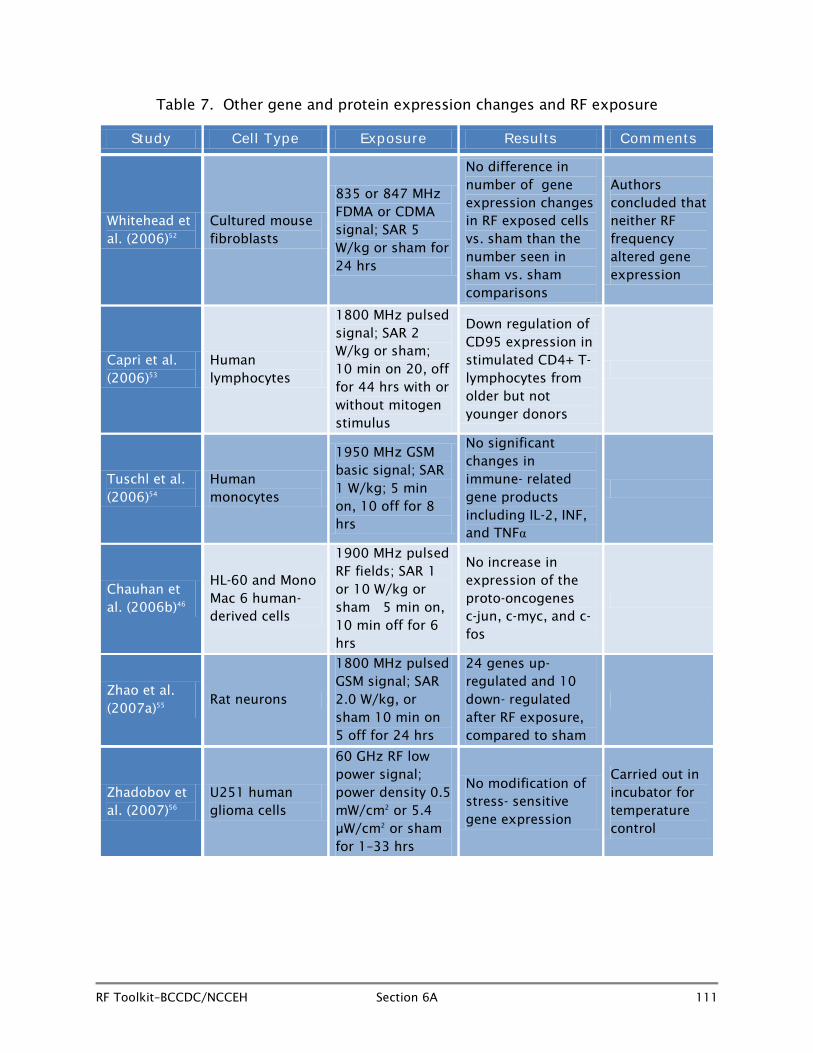
US investigators, Whitehead et al. (2006)52 exposed cultured mouse C3H 10T 1/2 cells to 835 MHz Frequency Division Multiple Access (FDMA) or 847 MHz Code Division Multiple Access (CDMA) RF fields at a SAR of 5 W/kg or sham for 24 hours, using an Affymetrix U74AV2 gene chip (which employs 12,448 probes over 9198 genes) to search for oncogenes (genes involved in initiating cancer) or stress genes which were over or under expressed. Three separate flasks of cells were exposed to each of the two radiofrequencies, along with matched sham flasks. A positive control group of cells exposed to 0.68 Gy of x-rays included in the protocol demonstrated the expected gene expression changes by comparison with sham-exposed cells. However, the expression changes found in RF field-exposed cells versus the sham-exposed cells did not exceed the number seen in multiple comparisons of sham versus sham-exposed cells. The authors considered that the changes seen in RF exposed cells were false positives and concluded that there was no evidence that either 835 MHz FDMA or 847 MHz CDMA RF exposure altered gene expression.
Capri et al. (2006)53 analysed levels of CD95 (a molecule which is important in starting and terminating the immunologic response) in CD4+ and in CD8+ T-cells in vitro in peripheral blood mononuclear cells taken from young (age 26 ± 5 years) and older (age 88 ± 2 years) donors and exposed or sham-exposed to 1800 MHz pulsed RF fields (SAR 2 W/kg) intermittently with or without stimulation by mitogens. Mitogens are agents which stimulate cell division. After RF exposure, a small but significant down-regulation of CD95 expression in mitogen-stimulated CD4+ T-lymphocytes was seen among older, but not younger donors. The fact that the down-regulation was seen only in older volunteers suggests that the RF-related effect, if real and eventually replicated in other studies, affects the relatively weaker immune systems seen in older individuals rather than the more robust systems seen in the young.
Tuschl et al. (2006)54 evaluated human monocytes from donors for effects of exposure to 1950 MHz GSM basic signal or sham for eight hours, alternating five minutes on and 10 minutes off at a SAR of 1 W/kg. The study evaluated intracellular production of IL-2 and activity of immune relevant genes. No significant changes were seen in expression of products of immune relevant genes in RF-exposed cells after eight hours by comparison with sham-exposed cells.
The Canadian group noted earlier, Chauhan et al.,46 exposed several different cell lines (HL-60 and Mono Mac 6) to 1900 MHz pulsed RF fields at SAR levels of 1 and 10 W/kg or sham at 37ºC to assess heat shock protein related genes. However, the investigators took advantage of the opportunity to measure changes in a number of proto-oncogenes (c-jun, c-myc, and c-fos) as well. Proto-oncogenes are normal genes which, through mutation or increased expression, can become oncogenes and initiate the process of carcinogenesis. No significant changes were seen in the expression of c-jun, c-myc or c-fos in either type of cells exposed to the pulsed RF fields by comparison

RF Toolkit–BCCDC/NCCEH Section 6A 110
with sham-exposed cells. The findings mimic those noted in an earlier study by Czyz et al.42 described in the section on heat shock gene and protein expression. In that study of p53 gene deficient and wild type embryonic stem cells exposed to 1710 MHz pulsed RF signal, exposure produced no change in levels of c-jun, or c-myc in wild type cells and only very modest and transient changes in the p53 deficient cells.
Zhao et al. (2007)55 evaluated gene expression profiles in rat neurons exposed to 1800 MHz pulsed GSM signal 10 minutes on and 5 minutes off for 24 hours at an average SAR of 2.0 W/kg or sham in a test chamber at 37°C. Among 1,200 candidate genes evaluated using an Affymetrix U34 gene chip, 24 were up-regulated and an additional 10 were down-regulated after 24-hour intermittent exposure at an average SAR of 2.0 W/kg. The genes were associated with multiple cellular functions including signal transduction pathway and metabolism. Some caution is needed in interpreting these results because, although statistically significant p-values were found for the 34 genes, none of the up-regulated change values exceeded two-fold, and many are as little as 1.15, suggesting the possibility of false positive findings due to chance in so many markers.
Zhadobov and colleagues in France (2007)56 exposed U25 human glioma cultured cells to 60 GHz low power fields at power densities of 0.5 m W/cm2 or 5.4 µW/cm2
for
periods of 1 to 33 hours in an incubator to achieve adequate temperature control. The 60 GHz range has a number of upcoming applications including use in indoor high-data rate communications over wireless 4G local area networks (LAN). No changes in expression of any stress-sensitive genes were seen compared to sham-exposed cells.
Gerner et al. (2010)57 exposed human Jurkat cells, human diploid fibroblasts, and quiescent mononuclear cells to 1800 MHz pulsed signal at a SAR of 2 W/kg or sham for eight hours and found increases in protein synthesis in both Jurkat cells and fibroblasts exposed to RF fields, by comparison with sham-exposed cells, but no difference in the exposed quiescent mononuclear cells. The authors interpreted the results as indicating an increased protein in the cells turnover due to interference in hydrogen bonds by RF fields.
Japanese scientists, Hirose et al. (2010),58 studied the effect of 1950 MHz modulated IMT-2000 W-CDMA signal at SARs of 0.2, 0.8 and 2.0 W/kg or sham exposure for two hours on rat microglial cells. Results were assessed at 24 and 72 hours after exposure, and no significant differences were seen between RF-exposed cells and sham-exposed cells for expression of immune related cytokines including tumour necrosis factor-α, interleukin 1-β, or IL6. Cytokines are regulatory proteins that play a central role in the immune system by modulating functions in the system, including lymphocyte activation, immune cell proliferation, differentiation, and survival.
RF Toolkit–BCCDC/NCCEH Section 6A 111
Table 7. Other gene and protein expression changes and RF exposure
Study Cell Type Exposure Results Comments
Whitehead et al. (2006)52
Cultured mouse fibroblasts
835 or 847 MHz FDMA or CDMA signal; SAR 5 W/kg or sham for 24 hrs
No difference in number of gene expression changes in RF exposed cells vs. sham than the number seen in sham vs. sham comparisons
Authors concluded that neither RF frequency altered gene expression
Capri et al. (2006)53
Human lymphocytes
1800 MHz pulsed signal; SAR 2 W/kg or sham; 10 min on 20, off for 44 hrs with or without mitogen stimulus
Down regulation of CD95 expression in stimulated CD4+ T-lymphocytes from older but not younger donors
Tuschl et al. (2006)54
Human monocytes
1950 MHz GSM basic signal; SAR 1 W/kg; 5 min on, 10 off for 8 hrs
No significant changes in immune- related gene products including IL-2, INF, and TNFα
Chauhan et al. (2006b)46
HL-60 and Mono Mac 6 human- derived cells
1900 MHz pulsed RF fields; SAR 1 or 10 W/kg or sham 5 min on, 10 min off for 6 hrs
No increase in expression of the proto-oncogenes c-jun, c-myc, and c-fos
Zhao et al. (2007a)55
Rat neurons
1800 MHz pulsed GSM signal; SAR 2.0 W/kg, or sham 10 min on 5 off for 24 hrs
24 genes up- regulated and 10 down- regulated after RF exposure, compared to sham
Zhadobov et al. (2007)56
U251 human glioma cells
60 GHz RF low power signal; power density 0.5 mW/cm2 or 5.4 µW/cm2 or sham for 1–33 hrs
No modification of stress- sensitive gene expression
Carried out in incubator for temperature control

RF Toolkit–BCCDC/NCCEH Section 6A 112
Study Cell Type Exposure Results Comments
Gerner et al. (2010)57
Human Jurkat cells; human diploidfibroblasts; human quiescent mononuclear cells
1800 MHz pulsed GSM signal; SAR 2.0 W/kg or sham for 8 hrs
Increased protein synthesis in Jurkat cells and human fibroblasts in RF-exposed vs. sham-exposed cells
No change due to RF exposure in quiescent mononuclear cells
Hirose et al. (2010)58
Rat microglial cells
1950 MHz modulated IMT-2000 W-CDMA signal; SAR 0.2, 0.8, 2.0 W/kg or sham for 2 hrs
No differences between RF- and sham- exposed cells in cell activation or expression of immune function cytokines
Summary
Overall, although some recent studies have shown alterations in heat shock-related gene expression or protein expression, a similar number or more have shown negative results. The same situation prevails in studies of RF fields and other non-heat shock-related gene and protein expression studies. As in other areas of investigation concerning potentially adverse effects of RF fields on physiological processes in cell cultures, most recent studies are well-conducted, and there are no specific features which appear to distinguish positive studies from those finding no association. Although this area of research will undoubtedly continue, there is no compelling evidence at present that RF fields of the type and strength to which humans are exposed are responsible for gene or protein expression changes.
6A.6 Other Specific Intracellular Effects
6A.6.1 Changes in protein and RF fields (Table 8)
In addition to gene expression changes, which could result in over- or under- production of proteins, other studies have been conducted to determine whether exposure to RF fields can alter physiologic processes within different types of cells.
Belyaev et al. (2005)59 conducted a study to evaluate whether there were differences in response to RF fields in lymphocytes taken from electro-sensitive individuals by comparison with those from non-sensitive subjects. The research group exposed human lymphocytes from seven healthy individuals and seven electro-sensitive persons to 915 MHz GSM signal at SAR of 37 mW/kg or sham for a period of two hours, in a TEM cell. The study was conducted to determine whether RF exposure altered chromatin conformation in electro-sensitive individuals by comparison with non-

RF Toolkit–BCCDC/NCCEH Section 6A 113
sensitive subjects. Chromatin conformation capture examines protein-DNA combination in chromosome structures within the cell nucleus which have a variety of functions including helping facilitate gene expression. A positive control group was also part of the protocol and cells in this group were exposed to 41°C for two hours. Evaluation at 24 and 48 hours after exposure showed changes in the conformation of chromatin in lymphocytes from both radio-sensitive and non-sensitive RF-exposed subjects compared with sham-exposed cells from each group. No significant differences were seen between healthy and electrically sensitive participants. The authors reported that the changes seen in the RF-exposed cells were similar to the stress response seen in the positive control heat shock cells.
A German study, Sukhotina et al. (2006)60 attempted to confirm results of an earlier investigation61 conducted back in 2002 suggesting that melatonin synthesis is suppressed by exposure to RF fields. Isolated hamster pineal glands were exposed to 1800 MHz continuous wave or pulsed GSM signal at SAR levels of 8, 80, 800, and 2700 mW/kg for periods of seven hours, and perfusate samples were collected every hour. At SAR rates characteristic of the use of mobile phones (8, 80, and 800 mW/kg) melatonin release was enhanced by both continuous and pulsed exposure by comparison with control glands. At 2.7 W/kg pulsed 1800 MHz exposure appeared to suppress melatonin levels, but as the exposure increased temperature by 1.2°C, the suppression was actually due to thermal effects. The authors concluded that the study did not support the theory that exposure to RF fields at levels produced by use of mobile phones suppresses melatonin.
Friedman et al. (2007)62 studied the effect of RF fields on mitogen activated protein kinase (MAPK) cascades, which are important in cell survival and apoptosis. The investigators subjected human cultured epithelial (HeLa) cells and Rat 1 cells to 875 MHz at intensities of 0.005–0.3 mW/cm2 for periods of 0, 5, 10, 20, or 30 minutes. Results showed a temporary increase in phosphorylation of extracellular signal-regulated kinase (ERK), one of the MAPK pathways, at five minutes, which decreased to basal levels within 30 minutes. The authors suggested that the activation of ERK is mediated by reactive oxygen species (ROS) produced by the RF fields, and the ERK activates the MAPK cascade in both types of cells tested. The authors suggest the finding might indicate that interference with intracellular signalling by RF fields can inappropriately activate ERK functioning with adverse effects on apoptosis. However, it should be noted that studies of RF fields and apoptosis have been predominantly negative.
Bormusov et al. (2008)63 in Israel evaluated the possibility that RF field exposure might damage eye tissue. The investigators exposed bovine lenses to 1100 MHz RF fields at 2.22 mW intensity for 90 cycles of 50 minutes each, followed by a 10-minute pause. The lenses were then cultured for 15 days. Control lenses were simply cultured for 10–15 days. A further group was exposed to heat (39.5°C) three times for two hours each time with 24 hours between exposures and cultured for 11 days. Results showed
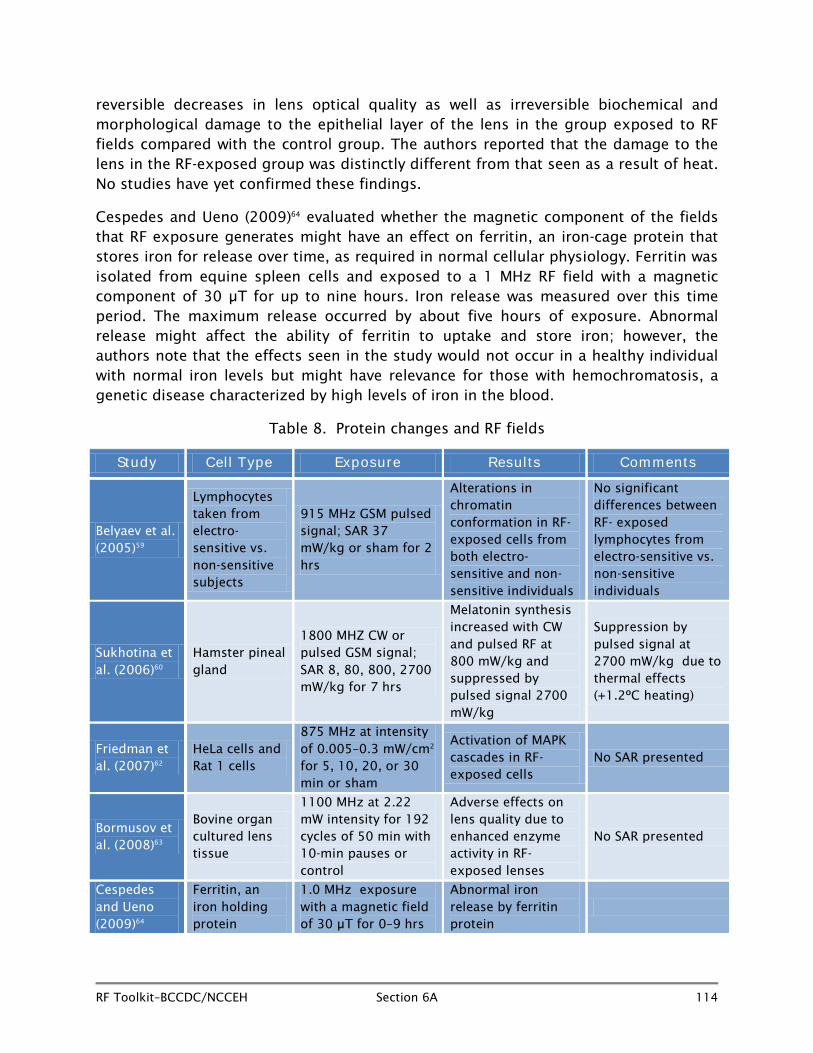
RF Toolkit–BCCDC/NCCEH Section 6A 114
reversible decreases in lens optical quality as well as irreversible biochemical and morphological damage to the epithelial layer of the lens in the group exposed to RF fields compared with the control group. The authors reported that the damage to the lens in the RF-exposed group was distinctly different from that seen as a result of heat. No studies have yet confirmed these findings.
Cespedes and Ueno (2009)64 evaluated whether the magnetic component of the fields that RF exposure generates might have an effect on ferritin, an iron-cage protein that stores iron for release over time, as required in normal cellular physiology. Ferritin was isolated from equine spleen cells and exposed to a 1 MHz RF field with a magnetic component of 30 µT for up to nine hours. Iron release was measured over this time period. The maximum release occurred by about five hours of exposure. Abnormal release might affect the ability of ferritin to uptake and store iron; however, the authors note that the effects seen in the study would not occur in a healthy individual with normal iron levels but might have relevance for those with hemochromatosis, a genetic disease characterized by high levels of iron in the blood.
Table 8. Protein changes and RF fields
Study Cell Type Exposure Results Comments
Belyaev et al. (2005)59
Lymphocytes taken from electro-sensitive vs. non-sensitive subjects
915 MHz GSM pulsed signal; SAR 37 mW/kg or sham for 2 hrs
Alterations in chromatin conformation in RF-exposed cells from both electro-sensitive and non-sensitive individuals
No significant differences between RF- exposed lymphocytes from electro-sensitive vs. non-sensitive individuals
Sukhotina et al. (2006)60
Hamster pineal gland
1800 MHZ CW or pulsed GSM signal; SAR 8, 80, 800, 2700 mW/kg for 7 hrs
Melatonin synthesis increased with CW and pulsed RF at 800 mW/kg and suppressed by pulsed signal 2700 mW/kg
Suppression by pulsed signal at 2700 mW/kg due to thermal effects (+1.2ºC heating)
Friedman et al. (2007)62
HeLa cells and Rat 1 cells
875 MHz at intensity of 0.005–0.3 mW/cm2 for 5, 10, 20, or 30 min or sham
Activation of MAPK cascades in RF-exposed cells
No SAR presented
Bormusov et al. (2008)63
Bovine organ cultured lens tissue
1100 MHz at 2.22 mW intensity for 192 cycles of 50 min with 10-min pauses or control
Adverse effects on lens quality due to enhanced enzyme activity in RF-exposed lenses
No SAR presented
Cespedes and Ueno (2009)64
Ferritin, an iron holding protein
1.0 MHz exposure with a magnetic field of 30 µT for 0–9 hrs
Abnormal iron release by ferritin protein

RF Toolkit–BCCDC/NCCEH Section 6A 115
Summary
It is difficult to draw conclusions from the evidence from studies in this area at present. Although all of these representative studies indicate that RF fields may affect physiologic processes, the studies have a variety of endpoints, and essentially none of the different findings have been independently replicated. As well, none provide the basis for a convincing biologic mechanism for the action of RF fields. If replicated, the in vitro changes seen in culture would need to be tested in animal models to see whether they persist in the face of interactions that take place in living organisms.
The most frequently used cells for examining the effect of RF fields on single cell motility are sperm cells as their characteristics are well-known, and they are easy to obtain. A complete review of issues surrounding the effects of RF fields and male fertility, including analysis of sperm cells is presented in Section 10.
6A.6.2 Calcium efflux and RF fields (Table 9)
Cells control internal calcium levels tightly, and it is known that a number of cell-signalling pathways include temporary changes in intracellular calcium. Early studies had indicated that pulsed RF fields might allow calcium efflux from brain tissue,65-67 although the evidence appearing in more recent studies is quite contradictory. Since 2006, only three new studies have appeared.
Platano et al. (2007)67 exposed neurons from Sprague-Dawley rats to one, two, or three sessions of 90-second exposure to 900 MHz continuous wave or pulsed RF fields at SAR 2 W/kg for each type of RF exposure to evaluate whether voltage-gated calcium channels (VGCC) were affected in their ability to control calcium levels in cells. VGCCs are an important transport system for moving sodium and calcium ions in and out of cells. In conducting the experiment, the investigators used Ba2+ ions in order to avoid Ca2+ inactivation of the currents induced with RF fields. Results showed no alterations in voltage-gates calcium channel brought about by either the continuous wave or pulsed 900 MHz exposure.
Rao et al. (2008)68 in the US, exposed neuronal cells from a mouse embryonic cell line to 700 –1100 MHz signal at 100 MHz intervals at 0.5 to 5 W/kg for one hour. The study found that at 800 MHz (SAR .5 W/kg), the number of Ca2+ spikes per hour was significantly greater than the number on the control cells. The authors reported that the increase was RF frequency dependent but not SAR dependent.
A recent study, O’Connor et al. (2010)69 exposed human endothelial cells, PC-12, neuroblastoma cells and primary hippocampal neurons to a pulsed 900 MHz GSM signal at SAR levels from .012–2 W/kg, similar to the levels incurred using a GSM mobile phone, for a period of 30 minutes. Data from the pulsed field experiment were compared to analogous result using a continuous wave signal or sham. Neither the pulsed nor the continuous wave exposure had any effect on Ca2+ signalling even at the

RF Toolkit–BCCDC/NCCEH Section 6A 116
highest SAR levels. The evidence for any effect of RF fields on calcium channelling remains uncertain.
Table 9. Calcium efflux and RF fields
Study Cell Type Exposure Result Comments
Platano et al. (2007)67
Sprague-Dawley rat-cultured neurons
900 MHz pulsed or continuous wave exposure for 1, 2 , or 3 sessions of 90 sec
No changes seen in voltage-gated calcium channels from pulsed or CW exposure
Rao et al. (2008)68
Mouse embryonic neurons
700–1100 MHz signal; SAR .5–5 W/kg for 60 min + control cells
Cells exposed to 800 MHz fields had significantly more Ca2+ spikes per hour than control cells
O’Connor et al. (2010)69
Human endothelial cells, PC-12 neuroblastoma cells, and primary hippocampal neurons
900 MHz GSm pulsed or CW signal; SAR .12–2 W/kg or sham for 30 min
No change in Ca2+ signaling in any cell type with pulsed or CW exposure compared to sham exposed
Summary
There is little evidence from recent studies that RF fields adversely affect calcium channelling in cell cultures.
6A.6.3 Cell permeability and RF fields (Table 10)
Although most work on possible effects of RF fields on blood-brain barrier permeability are carried out in animal models (reviewed in Section 6B), there have been several studies since 2005 looking at permeability after RF exposure in endothelial cells.
Franke et al. (2005)70 exposed an endothelial cell/astrocyte co-culture model to pulsed 1800 MHz RF fields at SARs of 0.03 or 0.46 W/kg or sham over five days in an attempt to confirm findings from an earlier study71 conducted in 2000 which showed increased permeability with RF exposure. The co-culture cell model used in the present paper featured significantly higher physiologic tightness than that used in the 2000 study and more closely mimicked blood-brain barrier characteristics in living animals. Results showed that the outcome measure, sucrose permeation across the cell layers, was not affected by exposure to the 1800 MHz RF exposure.
A further study by the same group72 used brain capillary endothelial cells isolated from pigs and cultured on a collagen-coated Transwell cell culture insert to mimic the blood-brain barrier to test for disruption by RF fields. The cultured multi-cell membranes were exposed to 1966 MHz UMTS signal or sham exposure for between 24 and 72
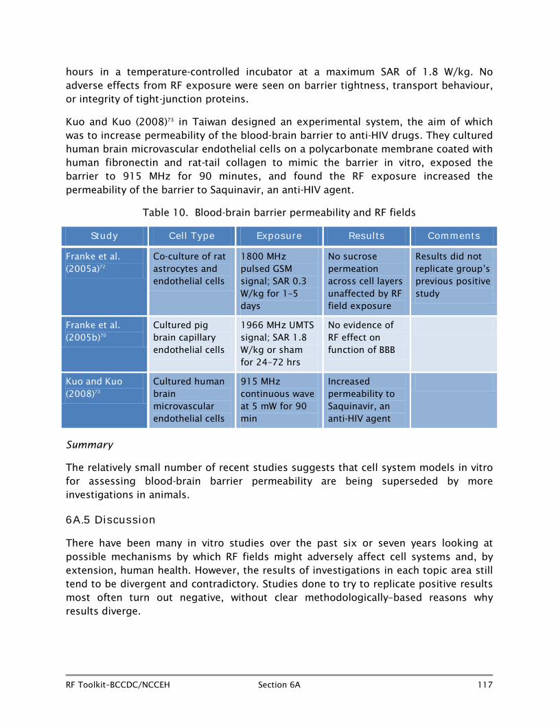
RF Toolkit–BCCDC/NCCEH Section 6A 117
hours in a temperature-controlled incubator at a maximum SAR of 1.8 W/kg. No adverse effects from RF exposure were seen on barrier tightness, transport behaviour, or integrity of tight-junction proteins.
Kuo and Kuo (2008)73 in Taiwan designed an experimental system, the aim of which was to increase permeability of the blood-brain barrier to anti-HIV drugs. They cultured human brain microvascular endothelial cells on a polycarbonate membrane coated with human fibronectin and rat-tail collagen to mimic the barrier in vitro, exposed the barrier to 915 MHz for 90 minutes, and found the RF exposure increased the permeability of the barrier to Saquinavir, an anti-HIV agent.
Table 10. Blood-brain barrier permeability and RF fields
Study Cell Type Exposure Results Comments
Franke et al. (2005a)72
Co-culture of rat astrocytes and endothelial cells
1800 MHz pulsed GSM signal; SAR 0.3 W/kg for 1–5 days
No sucrose permeation across cell layers unaffected by RF field exposure
Results did not replicate group’s previous positive study
Franke et al. (2005b)70
Cultured pig brain capillary endothelial cells
1966 MHz UMTS signal; SAR 1.8 W/kg or sham for 24–72 hrs
No evidence of RF effect on function of BBB
Kuo and Kuo (2008)73
Cultured human brain microvascular endothelial cells
915 MHz continuous wave at 5 mW for 90 min
Increased permeability to Saquinavir, an anti-HIV agent
Summary
The relatively small number of recent studies suggests that cell system models in vitro for assessing blood-brain barrier permeability are being superseded by more investigations in animals.
6A.5 Discussion
There have been many in vitro studies over the past six or seven years looking at possible mechanisms by which RF fields might adversely affect cell systems and, by extension, human health. However, the results of investigations in each topic area still tend to be divergent and contradictory. Studies done to try to replicate positive results most often turn out negative, without clear methodologically–based reasons why results diverge.
RF Toolkit–BCCDC/NCCEH Section 6A 118
There is no consistent evidence that RF fields produce chromosomal aberrations or micronucleus formation or that they generate the type of DNA damage characteristic of carcinogenic mechanisms. Recent evidence does not appear to support the notion that RF fields cause mutations or cell transformation. Studies of the effect of RF fields on cell proliferation have, in the main, been negative although in some cases results have indicated reduced cell proliferation. Studies looking at the production of ROS have been contradictory, and this field will require still more research. Early evidence that RF fields might stimulate ODC activity has not been confirmed by results from recent studies. Several apoptosis studies have shown positive results, but an equal or greater number have shown no effect. In vitro studies designed to explore whether RF fields facilitate leakage through the blood-brain barrier have not shown any consistent evidence of any effect due to RF fields. Most research on blood-brain barrier permeability is currently is being carried out in animal models.
Finally, there is no agreement as to which types of cells might be most sensitive to adverse effects of RF fields and no agreement on which RF frequencies and characteristics are most likely to elicit a biological effect. Because of this, no plausible mechanism has emerged to explain how RF fields might produce adverse biologic effects.
At the present time, there is no convincing body of evidence from in vitro investigations that exposure to RF fields at levels expected in day-to-day use of mobile phones and other RF emitting devices have the ability to initiate adverse biologic processes characteristic of human disease.
6A.5.1 Research gaps
More research is needed to:
• Encourage some degree of standardization among research protocols investigating any given putative adverse effect to allow direct comparisons with other studies to confirm or refute positive findings.
• Explore the joint effects of RF fields in conjunction with known cellular-stressing agents.
• Evaluate cellular response to RF in cells obtained from younger versus older donors.
• Develop cellular models that are more closely related to human biological processes.
RF Toolkit–BCCDC/NCCEH Section 6A 119
6A.6 References
1. Latin American Experts Committee on High Frequency Electromagnetic Fields and Human Health. Non-ionizing electromagnetic radiation in the radiofrequency spectrum and its effects on human health. Campinas/SP, Brasil: Instituto Edumed - Edumed Institute for Education in Medicine and Health Independent Research Group on the Impacts of Mobile Technologies on Health; 2010 Jun. Available from: http://www.wireless-health.org.br/downloads/LatinAmericanScienceReviewReport.pdf.
2. Health Protection Agency. Health effects from radiofrequency electromagnetic fields. London, UK: HPA; 2012 Apr. Available from: http://www.hpa.org.uk/webw/HPAweb&HPAwebStandard/HPAweb_C/1317133826368.
3. Vijayalaxmi. Cytogenetic studies in human blood lymphocytes exposed in vitro to 2.45 GHz or 8.2 GHz radiofrequency radiation. Radiat Res. 2006 Sep;166(3):532-8.
4. Stronati L, Testa A, Moquet J, Edwards A, Cordelli E, Villani P, et al. 935 MHz cellular phone radiation. An in vitro study of genotoxicity in human lymphocytes. Int J Radiat Biol. 2006 May;82(5):339-46.
5. Speit G, Schütz P, Hoffmann H. Genotoxic effects of exposure to radiofrequency electromagnetic fields (RF-EMF) in cultured mammalian cells are not independently reproducible. Mutat Res. 2007 Jan;626(1-2):42-7.
6. Mazor R, Korenstein-Ilan A, Barbul A, Eshet Y, Shahadi A, Jerby E, et al. Increased levels of numerical chromosome aberrations after in vitro exposure of human peripheral blood lymphocytes to radiofrequency electromagnetic fields for 72 hours. Radiat Res. 2008 Jan;169(1):28-37.
7. Manti L, Braselmann H, Calabrese ML, Massa R, Pugliese M, Scampoli P, et al. Effects of modulated microwave radiation at cellular telephone frequency (1.95 GHz) on X-ray-induced chromosome aberrations in human lymphocytes in vitro. Radiat Res. 2008 May;169(5):575-83.
8. Zeni O, Schiavoni A, Perrotta A, Forigo D, Deplano M, Scarfi MR. Evaluation of genotoxic effects in human leukocytes after in vitro exposure to 1950 MHz UMTS radiofrequency field. Bioelectromagnetics. 2008 Apr;29(3):177-84.
9. Schwarz C, Kratochvil E, Pilger A, Kuster N, Adlkofer F, Rudiger HW. Radiofrequency electromagnetic fields (UMTS, 1,950 MHz) induce genotoxic effects in vitro in human fibroblasts but not in lymphocytes. Int Arch Occup Environ Health. 2008 May;81(6):755-67.
RF Toolkit–BCCDC/NCCEH Section 6A 120
10. Kim JY, Hong SY, Lee YM, Yu SA, Koh WS, Hong JR, et al. In vitro assessment of clastogenicity of mobile-phone radiation (835 MHz) using the alkaline comet assay and chromosomal aberration test. Environ Toxicol. 2008 Jun;23(3):319-27.
11. Sannino A, Di Costanzo G, Brescia F, Sarti M, Zeni O, Juutilainen J, et al. Human fibroblasts and 900 MHz radiofrequency radiation: evaluation of DNA damage after exposure and co-exposure to 3-chloro-4-(dichloromethyl)-5-hydroxy-2(5h)-furanone (MX). Radiat Res. 2009 Jun;171(6):743-51.
12. Hansteen IL, Clausen KO, Haugan V, Svendsen M, Svendsen MV, Eriksen JG, et al. Cytogenetic effects of exposure to 2.3 GHz radiofrequency radiation on human lymphocytes in vitro. Anticancer Res. 2009 Nov;29(11):4323-30.
13. Campisi A, Gulino M, Acquaviva R, Bellia P, Raciti G, Grasso R, et al. Reactive oxygen species levels and DNA fragmentation on astrocytes in primary culture after acute exposure to low intensity microwave electromagnetic field. Neurosci Lett. 2010 Mar 31;473(1):52-5.
14. Baan R, Grosse Y, Lauby-Secretan B, El Ghissassi F, Bouvard V, Benbrahim-Tallaa L, et al. Carcinogenicity of radiofrequency electromagnetic fields. Lancet Oncol. 2011;12(7):624-6.
15. Wang J, Sakurai T, Koyama S, Komatubara Y, Suzuki Y, Taki M, et al. Effects of 2450 MHz electromagnetic fields with a wide range of SARs on methylcholanthrene-induced transformation in C3H10T1/2 cells. J Radiat Res (Tokyo). 2005 Sep;46(3):351-61.
16. Hirose H, Suhara T, Kaji N, Sakuma N, Sekijima M, Nojima T, et al. Mobile phone base station radiation does not affect neoplastic transformation in BALB/3T3 cells. Bioelectromagnetics. 2008 Jan;29(1):55-64.
17. Roti Roti JL, Malyapa RS, Bisht KS, Ahern EW, Moros EG, Pickard WF, et al. Neoplastic transformation in C3H 10T(1/2) cells after exposure to 835.62 MHz FDMA and 847.74 MHz CDMA radiations. Radiat Res. 2001 Jan;155(1 Pt 2):239-47.
18. Miyakoshi J, Takemasa K, Takashima Y, Ding GR, Hirose H, Koyama S. Effects of exposure to a 1950 MHz radio frequency field on expression of Hsp70 and Hsp27 in human glioma cells. Bioelectromagnetics. 2005 May;26(4):251-7.
19. Buttiglione M, Roca L, Montemurno E, Vitiello F, Capozzi V, Cibelli G. Radiofrequency radiation (900 MHz) induces Egr-1 gene expression and affects cell-cycle control in human neuroblastoma cells. J Cell Physiol. 2007;213(3):759-67.
20. Pavicic I, Trosic I. Impact of 864 MHz or 935 MHz radiofrequency microwave radiation on the basic growth parameters of V79 cell line. Acta Biol Hung. 2008 Mar;59(1):67-76.
RF Toolkit–BCCDC/NCCEH Section 6A 121
21. Pavicic I, Trosic I. In vitro testing of cellular response to ultra high frequency electromagnetic field radiation. Toxicol In Vitro. 2008;22(5):1344-8.
22. Hoyto A, Luukkonen J, Juutilainen J, Naarala J. Proliferation, oxidative stress and cell death in cells exposed to 872 MHz radiofrequency radiation and oxidants. Radiat Res. 2008 Aug;170(2):235-43.
23. Hoyto A, Sokura M, Juutilainen J, Naarala J. Radiofrequency radiation does not significantly affect ornithine decarboxylase activity, proliferation, or caspase-3 activity of fibroblasts in different physiological conditions. Int J Radiat Biol. 2008 Sep;84(9):727-33.
24. Lee JJ, Kwak HJ, Lee YM, Lee JW, Park MJ, Ko YG, et al. Acute radio frequency irradiation does not affect cell cycle, cellular migration, and invasion. Bioelectromagnetics. 2008 Dec;29(8):615-25.
25. Cao Y, Zhang W, Lu MX, Xu Q, Meng QQ, Nie JH, et al. 900-MHz microwave radiation enhances gamma-ray adverse effects on SHG44 cells. J Toxicol Environ Health A. 2009;72(11-12):11-2.
26. Sekijima M, Takeda H, Yasunaga K, Sakuma N, Hirose H, Nojima T, et al. 2-GHz band CW and W-CDMA modulated radiofrequency fields have no significant effect on cell proliferation and gene expression profile in human cells. J Radiat Res (Tokyo). 2010;51(3):277-84.
27. Lantow M, Viergutz T, Weiss DG, Simkó M. Comparative study of cell cycle kinetics and induction of apoptosis or necrosis after exposure of human Mono Mac 6 cells to radiofrequency radiation. Radiat Res. 2006;166(3).
28. Joubert V, Leveque P, Rametti A, Collin A, Bourthoumieu S, Yardin C. Microwave exposure of neuronal cells in vitro: study of apoptosis. Int J Radiat Biol. 2006 Apr;82(4):267-75.
29. Joubert V, Leveque P, Cueille M, Bourthoumieu S, Yardin C. No apoptosis is induced in rat cortical neurons exposed to GSM phone fields. Bioelectromagnetics. 2007 Feb;28(2):115-21.
30. Zhao TY, Zou SP, Knapp PE. Exposure to cell phone radiation up-regulates apoptosis genes in primary cultures of neurons and astrocytes. Neurosci Lett. 2007 Jan 22;412(1):34-8.
31. Joubert V, Bourthoumieu S, Leveque P, Yardin C. Apoptosis is induced by radiofrequency fields through the caspase-independent mitochondrial pathway in cortical neurons. Radiat Res. 2008 Jan;169(1):38-45.
32. Moquet J, Ainsbury E, Bouffler S, Lloyd D. Exposure to low level GSM 935 MHZ radiofrequency fields does not induce apoptosis in proliferating or differentiated murine neuroblastoma cells. Radiat Prot Dosimetry. 2008;131(3):287-96.

RF Toolkit–BCCDC/NCCEH Section 6A 122
33. Palumbo R, Brescia F, Capasso D, Sannino A, Sarti M, Capri M, et al. Exposure to 900 MHz radiofrequency radiation induces caspase 3 activation in proliferating human lymphocytes. Radiat Res. 2008 Sep;170(3):327-34.
34. Zeni O, Di Pietro R, d'Ambrosio G, Massa R, Capri M, Naarala J, et al. Formation of reactive oxygen species in L929 cells after exposure to 900 MHz RF radiation with and without co-exposure to 3-chloro-4-(dichloromethyl)-5-hydroxy-2(5H)-furanone. Radiat Res. 2007 Mar;167(3):306-11.
35. Brescia F, Sarti M, Massa R, Calabrese ML, Sannino A, Scarfi MR. Reactive oxygen species formation is not enhanced by exposure to UMTS 1950 MHz radiation and co-exposure to ferrous ions in Jurkat cells. Bioelectromagnetics. 2009 Oct;30(7):525-35.
36. Xu S, Zhou Z, Zhang L, Yu Z, Zhang W, Wang Y, et al. Exposure to 1800 MHz radiofrequency radiation induces oxidative damage to mitochondrial DNA in primary cultured neurons. Brain Res. 2010 Jan 22;1311:189-96.
37. Penafiel LM, Litovitz T, Krause D, Desta A, Mullins JM. Role of modulation on the effect of microwaves on ornithine decarboxylase activity in L929 cells. Bioelectromagnetics. 1997;18(2):132-41.
38. Hoyto A, Sihvonen AP, Alhonen L, Juutilainen J, Naarala J. Modest increase in temperature affects ODC activity in L929 cells: Low-level radiofrequency radiation does not. Radiat Environ Biophys. 2006 Sep;45(3):231-5.
39. Hoyto A, Juutilainen J, Naarala J. Ornithine decarboxylase activity is affected in primary astrocytes but not in secondary cell lines exposed to 872 MHz RF radiation. Int J Radiat Biol. 2007 Jun;83(6):367-74.
40. Billaudel B, Taxile M, Ruffie G, Veyret B, Lagroye I. Effects of exposure to DAMPS and GSM signals on ornithine decarboxylase (ODC) activity: I. L-929 mouse fibroblasts. Int J Radiat Biol. 2009 Jun;85(6):510-8.
41. Dawe AS, Bodhicharla RK, Graham NS, May ST, Reader T, Loader B, et al. Low-intensity microwave irradiation does not substantially alter gene expression in late larval and adult Caenorhabditis elegans. Bioelectromagnetics. 2009 Dec;30(8):602-12.
42. Czyz J, Guan K, Zeng Q, Nikolova T, Meister A, Schonborn F, et al. High frequency electromagnetic fields (GSM signals) affect gene expression levels in tumor suppressor p53-deficient embryonic stem cells. Bioelectromagnetics. 2004 May;25(4):296-307.
43. Wang J, Koyama S, Komatsubara Y, Suzuki Y, Taki M, Miyakoshi J. Effects of a 2450 MHz high-frequency electromagnetic field with a wide range of SARs on the induction of heat-shock proteins in A172 cells. Bioelectromagnetics. 2006 Sep;27(6):479-86.
RF Toolkit–BCCDC/NCCEH Section 6A 123
44. Sanchez S, Milochau A, Ruffie G, Poulletier de Gannes F, Lagroye I, Haro E, et al. Human skin cell stress response to GSM-900 mobile phone signals. In vitro study on isolated primary cells and reconstructed epidermis. Febs J. 2006 Dec;273(24):5491-507.
45. Chauhan V, Mariampillai A, Gajda GB, Thansandote A, McNamee JP. Analysis of proto-oncogene and heat-shock protein gene expression in human derived cell-lines exposed in vitro to an intermittent 1.9 GHz pulse-modulated radiofrequency field. Int J Radiat Biol. 2006 May;82(5):347-54.
46. Chauhan V, Mariampillai A, Bellier PV, Qutob SS, Gajda GB, Lemay E, et al. Gene expression analysis of a human lymphoblastoma cell line exposed in vitro to an intermittent 1.9 GHz pulse-modulated radiofrequency field. Radiat Res. 2006 Apr;165(4):424-9.
47. Vanderwaal RP, Cha B, Moros EG, Roti Roti JL. HSP27 phosphorylation increases after 45 degrees C or 41 degrees C heat shocks but not after non-thermal TDMA or GSM exposures. Int J Hyperthermia. 2006 Sep;22(6):507-19.
48. Sanchez S, Haro E, Ruffie G, Veyret B, Lagroye I. In vitro study of the stress response of human skin cells to GSM-1800 mobile phone signals compared to UVB radiation and heat shock. Radiat Res. 2007 May;167(5):572-80.
49. Chauhan V, Qutob SS, Lui S, Mariampillai A, Bellier PV, Yauk CL, et al. Analysis of gene expression in two human-derived cell lines exposed in vitro to a 1.9 GHz pulse-modulated radiofrequency field. Proteomics. 2007 Nov;7(21):3896-905.
50. Franzellitti S, Valbonesi P, Contin A, Biondi C, Fabbri E. HSP70 expression in human trophoblast cells exposed to different 1.8 Ghz mobile phone signals. Radiat Res. 2008 Oct;170(4):488-97.
51. Valbonesi P, Franzellitti S, Piano A, Contin A, Biondi C, Fabbri E. Evaluation of HSP70 expression and DNA damage in cells of a human trophoblast cell line exposed to 1.8 GHz amplitude-modulated radiofrequency fields. Radiat Res. 2008 Mar;169(3):270-9.
52. Whitehead TD, Moros EG, Brownstein BH, Roti Roti JL. The number of genes changing expression after chronic exposure to code division multiple access or frequency DMA radiofrequency radiation does not exceed the false-positive rate. Proteomics. 2006 Sep;6(17):4739-44.
53. Capri M, Salvioli S, Altilia S, Sevini F, Remondini D, Mesirca P, et al. Age-dependent effects of in vitro radiofrequency exposure (mobile phone) on CD95+ T helper human lymphocytes. Ann N Y Acad Sci. 2006 May;1067:493-9.
54. Tuschl H, Novak W, Molla-Djafari H. In vitro effects of GSM modulated radiofrequency fields on human immune cells. Bioelectromagnetics. 2006;27(3):188-96.
RF Toolkit–BCCDC/NCCEH Section 6A 124
55. Zhao R, Zhang S, Xu Z, Ju L, Lu D, Yao G. Studying gene expression profile of rat neuron exposed to 1800MHz radiofrequency electromagnetic fields with cDNA microassay. Toxicology. 2007 Jun 25;235(3):167-75.
56. Zhadobov M, Sauleau R, Le Coq L, Debure L, Thouroude D, Michel D, et al. Low-power millimeter wave radiations do not alter stress-sensitive gene expression of chaperone proteins. Bioelectromagnetics. 2007 Apr;28(3):188-96.
57. Gerner C, Haudek V, Schandl U, Bayer E, Gundacker N, Hutter HP, et al. Increased protein synthesis by cells exposed to a 1,800-MHz radio-frequency mobile phone electromagnetic field, detected by proteome profiling. Int Arch Occup Environ Health. 2010 Aug;83(6):691-702.
58. Hirose H, Sasaki A, Ishii N, Sekijima M, Iyama T, Nojima T, et al. 1950 MHz IMT-2000 field does not activate microglial cells in vitro. Bioelectromagnetics. 2010 Feb;31(2):104-12.
59. Belyaev IY, Hillert L, Protopopova M, Tamm C, Malmgren LO, Persson BR, et al. 915 MHz microwaves and 50 Hz magnetic field affect chromatin conformation and 53BP1 foci in human lymphocytes from hypersensitive and healthy persons. Bioelectromagnetics. 2005 Apr;26(3):173-84.
60. Sukhotina I, Streckert JR, Bitz AK, Hansen VW, Lerchl A. 1800 MHz electromagnetic field effects on melatonin release from isolated pineal glands. J Pineal Res. 2006 Jan;40(1):86-91.
61. Burch JB, Reif JS, Noonan CW, Ichinose T, Bachand AM, Koleber TL, et al. Melatonin metabolite excretion among cellular telephone users. Int J Radiat Biol. 2002 Nov;78(11):1029-36.
62. Friedman J, Kraus S, Hauptman Y, Schiff Y, Seger R. Mechanism of short-term ERK activation by electromagnetic fields at mobile phone frequencies. Biochem J. 2007 Aug 1;405(3):559-68.
63. Bormusov E, U PA, Sharon N, Schachter L, Lahav A, Dovrat A. Non-thermal electromagnetic radiation damage to lens epithelium. Open Opthalmol J. 2008;2:102-6.
64. Cespedes O, Ueno S. Effects of radio frequency magnetic fields on iron release from cage proteins. Bioelectromagnetics. 2009 Jul;30(5):336-42.
65. Lin-Liu S, Adey WR. Low frequency amplitude modulated microwave fields change calcium efflux rates from synaptosomes. Bioelectromagnetics. 1982;3(3):309-22.
66. Dutta SK, Ghosh B, Blackman CF. Radiofrequency radiation-induced calcium ion efflux enhancement from human and other neuroblastoma cells in culture. Bioelectromagnetics. 1989;10(2):197-202.

RF Toolkit–BCCDC/NCCEH Section 6A 125
67. Platano D, Mesirca P, Paffi A, Pellegrino M, Liberti M, Apollonio F, et al. Acute exposure to low-level CW and GSM-modulated 900 MHz radiofrequency does not affect Ba 2+ currents through voltage-gated calcium channels in rat cortical neurons. Bioelectromagnetics. 2007 Dec;28(8):599-607.
68. Rao VS, Titushkin IA, Moros EG, Pickard WF, Thatte HS, Cho MR. Nonthermal effects of radiofrequency-field exposure on calcium dynamics in stem cell-derived neuronal cells: elucidation of calcium pathways. Radiat Res. 2008 Mar;169(3):319-29.
69. O'Connor RP, Madison SD, Leveque P, Roderick HL, Bootman MD. Exposure to GSM RF fields does not affect calcium homeostasis in human endothelial cells, rat pheocromocytoma cells or rat hippocampal neurons. PLoS ONE. 2010(7):1-16.
70. Franke H, Streckert J, Bitz A, Goeke J, Hansen V, Ringelstein EB, et al. Effects of Universal Mobile Telecommunications System (UMTS) electromagnetic fields on the blood-brain barrier in vitro. Radiat Res. 2005 Sep;164(3):258-69.
71. Schirmacher A, Winters S, Fischer S, Goeke J, Galla HJ, Kullnick U, et al. Electromagnetic fields (1.8 GHz) increase the permeability to sucrose of the blood-brain barrier in vitro. Bioelectromagnetics. 2000 Jul;21(5):338-45.
72. Franke H, Ringelstein EB, Stogbauer F. Electromagnetic fields (GSM 1800) do not alter blood-brain barrier permeability to sucrose in models in vitro with high barrier tightness. Bioelectromagnetics. 2005 Oct;26(7):529-35.
73. Kuo YC, Kuo CY. Electromagnetic interference in the permeability of saquinavir across the blood-brain barrier using nanoparticulate carriers. Int J Pharm. 2008 Mar 3;351(1-2):271-81.

RF Toolkit–BCCDC/NCCEH Section 6A 126