secreted proteases control autolysin-mediated biofilm growth of
TRANSCRIPT
Esp cleaves autolysin to regulate staphylococcal biofilms
1
Secreted proteases control autolysin-mediated biofilm growth of Staphylococcus aureus
Chen Chen1, Vengadesan Krishnan
2, Kevin Macon
3, Kartik Manne
3, Sthanam V.L. Narayana
3, and
Olaf Schneewind1
1From the Department of Microbiology, University of Chicago, 920 East 58
th Street, Chicago, IL 60637,
USA 2UNESCO Regional Centre for Biotechnology, Biotech Science Cluster, 180 Udyog Vihar Phase 1,
Gurgaon-122016, Haryana, India 3Center for Biophysical Sciences and Engineering, School of Optometry, University of Alabama at
Birmingham, Birmingham, AL 35294, USA
*Running title: Esp cleaves autolysin to regulate staphylococcal biofilms
To whom correspondence should be addressed: Olaf Schneewind, Department of Microbiology,
University of Chicago, 920 East 58th Street, Chicago, IL 60637, USA, Tel.: (773) 834-9060; Fax: (773)
834-8150; E-mail: [email protected]
Keywords: Staphylococcus aureus, Staphylococcus epidermidis, Esp, V8, autolysin, biofilm
Background: Esp, a secreted protease of
Staphylococcus epidermidis, blocks biofilm
formation of Staphylococcus aureus and its ability
to colonize human nares.
Results: Esp cleaves autolysin, thereby preventing
the release of staphylococcal DNA as biofilm
matrix.
Conclusion: Secreted proteases control S. aureus
biofilm development and host colonization.
Significance: Methods that promote autolysin
degradation may also prevent S. aureus
colonization of humans.
ABSTRACT
Staphylococcus epidermidis, a commensal of
humans, secretes Esp protease to prevent
Staphylococcus aureus biofilm formation and
colonization. Blocking S. aureus colonization
may reduce the incidence of invasive infectious
diseases, however the mechanism whereby Esp
disrupts biofilms is unknown. We show here
that Esp cleaves autolysin (Atl)-derived murein
hydrolases and prevents staphylococcal release
of DNA, which serves as extracellular matrix in
biofilms. The three-dimensional structure of
Esp was revealed by X-ray crystallography and
shown to be highly similar to that of S. aureus
V8 (SspA). Both atl and sspA are necessary for
biofilm formation and purified SspA cleaves
Atl-derived murein hydrolases. Thus, S. aureus
biofilms are formed via the controlled secretion
and proteolysis of autolysin, and this
developmental program appears to be
perturbed by the Esp protease of S. epidermidis.
Staphylococcus aureus is both a commensal
and an invasive pathogen that causes skin and soft
tissue infections (SSTI), sepsis and endocarditis
(1). The primary niche for S. aureus colonization
are the human nares (2). About 20% of the human
population is colonized persistently while 30%
represent intermittent carriers and 50% are non-
carriers (3). Nosocomial S. aureus bacteremia is
three times more frequent in carriers than in non-
carriers (3,4). Colonization with highly virulent,
multi-drug resistant strains (MRSA, methicillin-
resistant S. aureus) is associated with invasive
disease and treatment failure (5). S. aureus is
currently the most frequent cause of infectious
disease morbidity and mortality in the United
States (6). Thus, strategies are needed to prevent S.
aureus nasal colonization without selecting for
antibiotic-resistance and with the ultimate goal of
reducing the incidence of staphylococcal
infections.
Colonization of human nares is thought to
involve the establishment of S. aureus biofilms
(7). Work from many laboratories suggests that S.
aureus biofilm growth occurs as a developmental
program, whereby bacteria initially adhere to host
epithelial surfaces and subsequently release some
of their DNA as extracellular matrix to replicate as
biofilm communities (8,9). Biofilm growth is also
associated with the shedding of staphylococci,
http://www.jbc.org/cgi/doi/10.1074/jbc.M113.502039The latest version is at JBC Papers in Press. Published on August 22, 2013 as Manuscript M113.502039
Copyright 2013 by The American Society for Biochemistry and Molecular Biology, Inc.
by guest on April 10, 2019
http://ww
w.jbc.org/
Dow
nloaded from

Esp cleaves autolysin to regulate staphylococcal biofilms
2
where released bacteria promote invasive disease
or disseminate within host tissues (9). Several
secreted products have been reported to function
as adhesins for S. aureus biofilm formation,
including fibronectin binding proteins (FnbA and
FnbB) (10,11), the extracellular adhesion protein
(Eap)(12,13), and the extracellular matrix protein
(Emp) (13). S. aureus biofilms use bacterial DNA
as an extracellular matrix (14,15), which is
released via Atl, a multi-functional murein
hydrolase (16,17). In addition to atl, the release of
DNA by S. aureus grown in biofilms is also
dependent on the cidABC and lrgAB operons,
which appear to function as holins/antiholins by
either initiating or preventing staphylococcal entry
into a programmed-cell death pathway (17). The
expression of the cid and lrg operons is controlled
in response to environmental signals via the LysR
type regulator CidR and the two-component
regulator LytRS, respectively (8,18).
The 1256 residue autolysin precursor is
secreted via its N-terminal signal peptide.
Following signal peptide removal, pro-Atl is
cleaved at two sites, residues 302 and 874, thereby
generating the mature amidase [N-
acetylmuramoyl-L-alanine amidase (AM, residues
303-874) and N-acetylglucosaminidase domains
(GL, residues 875-1276)(19). Each of the two
enzymes is endowed with repeat domains (R1-R2-
R3), that are tethered either to the C-terminal end
of AM (R1-R2, residues 534-874) or the N-
terminal end of GL (R3, residues 875-1016) (20).
Each repeat domain folds into two half-open barrel
subunits that bind polyglycerol-phosphate
lipoteichoic acid at accessible sites in the bacterial
envelope (21). Surface access is limited to
peptidoglycan in the vicinity of the cell division
septum (22,23), as these sites are not occluded by
polyribitol-phosphate wall teichoic acids (24).
Deletion mutations in the atl gene abolish S.
aureus biofilm formation and atl mutants form
large clusters of cells with incompletely separated
cell wall envelopes (25).
Studying nasal colonization in human
volunteers, Iwase and colleagues observed a
negative correlation between the colonization of
Staphylococcus epidermidis strains expressing esp
and S. aureus (7). Co-culturing of S. epidermidis
strains expressing esp inhibited S. aureus biofilm
formation (7). Although Esp does not affect the
viability of S. aureus, the purified protease
prevents biofilm formation and promotes
disassembly of pre-established biofilms (7). Esp
was found to degrade 75 different proteins in S.
aureus biofilms (26). Nevertheless, previous work
left unresolved by what mechanism Esp may
interfere with S. aureus biofilms (26).
EXPERIMENTAL PROCEDURES
Bacterial strains and reagents- S. aureus
Newman (27) and its variant with a bursa aurealis
insertion in atl (28) have been previously
described. The atl mutational lesion was
transduced with bacteriophage φ85 into wild-type
S. aureus Newman. Staphylococci were grown in
tryptic soy broth (TSB) or on tryptic soy agar
(TSA) plates. Erythromycin (10 μg/ml) was used
to select for the bursa aurealis insertional variant.
Escherichia coli strains were grown in Luria broth
or on Luria agar supplemented with ampicillin
(100 μg/ml). Chemicals were purchased from
Sigma unless indicated otherwise.
Esp expression and purification- Pro-Esp (Met1
to Gln282
) with an N-terminal His-tag was cloned
into pET28b, expressed in E. coli BL21 (DE3)
cells and purified using nickel-affinity
chromatography (Ni-NTA Superflow Agarose
resin, Qiagen) (29). Mature Esp was purified by
cleaving pro-Esp with thermolysin followed by
gel-filtration chromatography (Superdex 75 10/30
column, GE Healthcare) with 20 mM Tris-HCl
(pH 7.2), 150 mM NaCl. Briefly, purified pro-Esp
was incubated with thermolysin at 37° C for 4
hours and cleavage was quenched by the addition
of 5 mM EDTA. The purity and proteolytic
activity of Esp were confirmed by SDS-PAGE and
azocasein assay, respectively (29). Esp was
concentrated to 22 mg/ml using an Amicon
ultrafiltration system.
Esp crystallization and structure
determination- Concentrated, mature Esp was
crystallized using the hanging-drop vapor
diffusion method (29). A droplet consisting of 1 µl
protein [22 mg ml-1
in 20 mM Tris-HCl (pH 7.2),
150 mM NaCl] and 1 µl reservoir solution (0.25 M
potassium-acetate, 22% PEG 3350) was
equilibrated against 1 ml reservoir solution at
22°C. Native diffraction data were collected to 1.8
Å resolution on a R-AXIS IV imaging-plate
detector mounted on an in-house RIGAKU®
rotating-anode X-ray generator operating at 100
mA and 50 kV, and using 20% (v/v) ethylene
by guest on April 10, 2019
http://ww
w.jbc.org/
Dow
nloaded from
Esp cleaves autolysin to regulate staphylococcal biofilms
3
glycol as a cryoprotectant. Diffraction data were
processed with D*TREK (30). The native Esp
crystals belonged to the monoclinic space group
P21 with one molecule in the asymmetric unit.
Data collection and processing statistics are
reported in Table 1.
The crystal structure of Esp was solved by
molecular replacement, with the help of PHASER
(31), implemented in the CCP4 suite (32), using
the crystal structure of V8 protease (PDB entry
1QY6) (33) as a search model. Model building and
refinement were completed with the help of
COOT (34), and REFMAC (35). The final model
consisted of 216 residues (Val67
to Gln282
) and the
refinement converged with Rwork/Rfree values
17.3/19.9% (Table 1). The final model is of good
quality, reflected by an excellent Ramachandran
plot, with all residues displaying backbone angles
in the allowed regions. The quality of the final
model was examined using COOT and
PROCHECK (36) and deposited into the Protein
Data Bank (ID: 4JCN).
Cleavage of GST-Atl hybrids by Esp and V8-
GST-AMΔR1R2, GST-GLΔR3 and GST-GL were
purified as described previously (20). GST-AM
was purified via a modified protocol (37). E. coli
BL21 (DE3) harboring pGST-AM was grown in 2
L of Luria broth at 37°C to OD600 0.5, expression
induced with 1 mM of IPTG and culture incubated
for additional 3 hours at 30°C. Cells were
harvested by centrifugation, suspended in STE
lysis buffer [10 mM Tris-HCl (pH 8.0), 150 mM
NaCl, 1 mM EDTA, 100 µg/ml lysozyme] and
incubated on ice for 15 min followed by the
addition of 5 mM DTT and 1.5 mM PMSF.
Sarkosyl was added to a final concentration of 2%
(w/v). Bacteria were disrupted in a French
pressure cell at 6,000 psi followed by
centrifugation for 10 min at 13,000 ×g. The
supernatant was transferred to a new tube, Triton
X-100 added to a final concentration of 2%,
samples incubated at room temperature for 30 min
and loaded onto 1 ml glutathione sepharose 4B
column (GE healthcare, Life Sciences Piscataway,
NJ, USA) pre-equilibrated with STE. The column
was washed with 100 ml STE buffer. GST-AM
was eluted with 10 ml 20 mM glutathione, 10%
glycerol, 10 mM Tris-HCl (pH 8.0), 120 mM
NaCl. Purified GST-Atl hybrids (5 µg GST-AM,
GST-AMΔR1R2, GST-GL or GST-GLΔR3) were
incubated with 400 nM Esp or V8 for 20 min at
37°C. Samples were subjected to SDS-PAGE and
proteins stained with Coomassie-Brilliant Blue.
Biofilm Assays- Static biofilm assays were
performed using a previously described protocol
(38). Single S. aureus colonies were used to
inoculate 2 ml of TSB with 0.2% glucose and
incubated overnight at 37°C with shaking (250
rpm). Cultures were diluted to OD600 0.05 and 200
µl aliquots were added to Costar 3596 96-well
polystyrene plates (Corning, Lowell, MA)
pretreated with 100 µl of 1 µg/ml human
fibronectin (BD, Franklin Lakes, NJ) in PBS
overnight at 4°C. Plates were incubated at 37°C
with 5% CO2 for 24 hours and washed with 200 µl
PBS twice. Washed samples were treated with 100
µl ethanol for 2 minutes and then stained with 100
µl 0.41% crystal violet in 12% ethanol for 2
minutes. Excess stain was removed by three
washes with 200 µl PBS. The remaining crystal
violet staining was solubilized with 100 µl 95%
ethanol for 10 min and absorbance of 595 nm light
measured. Average absorbance values of media
only-wells were subtracted from wells that had
been inoculated with S. aureus.
For biofilm restoration experiments, 2.5 µg of
purified GST-Atl hybrids were incubated with S.
aureus Newman on fibronectin-coated microtiter
plates at 37°C with 5% CO2 for 24 hours. For
biofilm disassembly experiments, S. aureus and S.
epidermidis biofilms were formed for 24 hours.
Purified Esp or V8 (2.5 µg) were added and
samples incubated for 24 hours at 37°C. Biofilms
were quantified as described above and analyzed
with the Student’s t-test using GraphPad Prism
version 5.0 for Windows (GraphPad Software, La
Jolla, CA.)
Biofilm substrates of Esp- S. aureus biofilms
were formed during growth in iron-depleted
CRPMI (RPMI 1640) medium. Culture medium
was depleted of iron by batch incubation with 6%
(wt/vol) Chelex 100 and then supplemented with
10% RPMI 1640 to provide trace amounts of
divalent cations for growth (13). S. aureus
overnight cultures in TSB were diluted to OD600
0.05 and 50 µl were added to FALCON 150 mm
culture dishes coated with 1 µg/ml human
fibronectin. Plates were incubated at 37°C with
5% CO2 for 24 hours and washed three times with
35 ml PBS each. Biofilm was removed with a cell
scraper, suspended in 1 ml PBS and incubated
with 400 nM Esp or left untreated for 16 hours at
by guest on April 10, 2019
http://ww
w.jbc.org/
Dow
nloaded from

Esp cleaves autolysin to regulate staphylococcal biofilms
4
37°C with rotation. Biofilm samples were
subsequently boiled in sample buffer, proteins
separated by 10-20% gradient SDS-PAGE and
visualized with Coomassie Blue staining. Protein
bands were excised and identified with liquid-
chromatography tandem mass spectrometry at the
Taplin Biological Mass Spectrometry Facility
(Harvard Medical School).
Extracellular DNA in staphylococcal biofilms-
S. aureus biofilms were formed in Costar 12-well
polystyrene plates with 12 mm glass coverslips
that had been pretreated with 1 µg/ml human
fibronectin. Wells were washed three times with
PBS and stained with 1 µM SYTO 9/propidium
iodide (PI) at room temperature for 20 minutes.
Wells were washed with PBS and samples fixed
with 4% paraformaldehdye. Coverslips were
mounted on glass slides and viewed via light
microscopy. Microscopy and image acquisition
were performed with the Olympus "live cell" DSU
Spinning Disk inverted confocal microscopy
(Integrated Microscopy Core Facility, The
University of Chicago). Images were obtained
using a 40× objective. Fluorescence intensities
from 15 random fields were quantified using
Image J software.
Peptidoglycan cleavage assay- S. aureus
peptidoglycan was purified as described
previously (39). Briefly, staphylococci were
grown in 2 L TSB to A600 0.6, centrifuged, bacteria
washed in water, suspended in 4% SDS, and
boiled for 30 min. Detergent was removed by
washing staphylococci extensively in water.
Staphylococci were subjected to bead beating,
glass beads were removed, and cell debris was
sedimented by centrifugation. The extract was
incubated with 100 µg/ml amylase for 2 hours,
followed by first the addition of 10 µg/ml DNase
and 50 µg/ml RNase for 2 hours, and then by
incubation with 100 µg/ml trypsin for 16 hours at
37°C. Peptidoglycan extracts were centrifuged,
washed with water, suspended in 1% SDS and
boiled for 15 min to heat-inactivate all enzymes.
Peptidoglycan was extensively washed with water
to remove all traces of SDS, followed by washing
with 8 m LiCl, 100 mm EDTA, and acetone. Cell
walls were then washed with water and
lyophilized. Hydrofluoric acid was added and
incubated for 48 hours at 4°C to remove teichoic
acid. Peptidoglycan was neutralized, and
sedimented murein sacculi were treated with
alkaline phosphatase for 16 hours at 37°C. Purified
peptidoglycan was boiled for 5 min, washed with
water, and stored at 4 °C. Peptidoglycan was
incubated with 5 µg purified GST-AM, GST-
AMΔR1R2, GST-GL or GST-GLΔR3 in 0.1 M
phosphate buffer (pH 7.0) for 16 hours at 37 °C.
Peptidoglycan cleavage was determined by
measuring the OD600 before and after incubation.
Staphylococcal cell cluster analysis- Overnight
cultures of S. aureus were diluted 1:100 in 100 µl
TSB and added to 96-well plates at 37°C with
shaking. Culture growth was monitored by reading
OD600 in 30 minute intervals. Overnight cultures
were diluted 1:100 in 1 ml of TSB and incubated
at 37°C for 2 hours with shaking with or without
25 µg GST-Atl hybrids. Cultures were
subsequently centrifuged at 7,000 ×g for 1 min
and fixed with 4% paraformaldehyde prior to
washing and suspension in 1 ml PBS.
Staphylococci were then analyzed at the
University of Chicago flow cytometry facility
using BD LSRII-Blue flow cytometer to measure
the cluster size of S. aureus cells.
Activity measurements of staphylococcal
proteases- Conditioned extracellular media,
obtained as the supernatant following
centrifugation of overnight cultures of S. aureus,
were concentrated fifteen-fold using the Amicon
ultrafiltration system. Concentrated culture media,
20 µl aliquots, were incubated with 480 µl reaction
mixture containing 1% azocasein, 100 mM Tris-
HCl (pH 8.0) at 37°C overnight. Following
incubation, 25 µl 100% TCA was added to quench
each reaction; following centrifugation at 15,000
×g for 10 min, soluble material was recovered
with the supernatant and absorbance at 440 nm
was measured to determine protease activity.
RESULTS
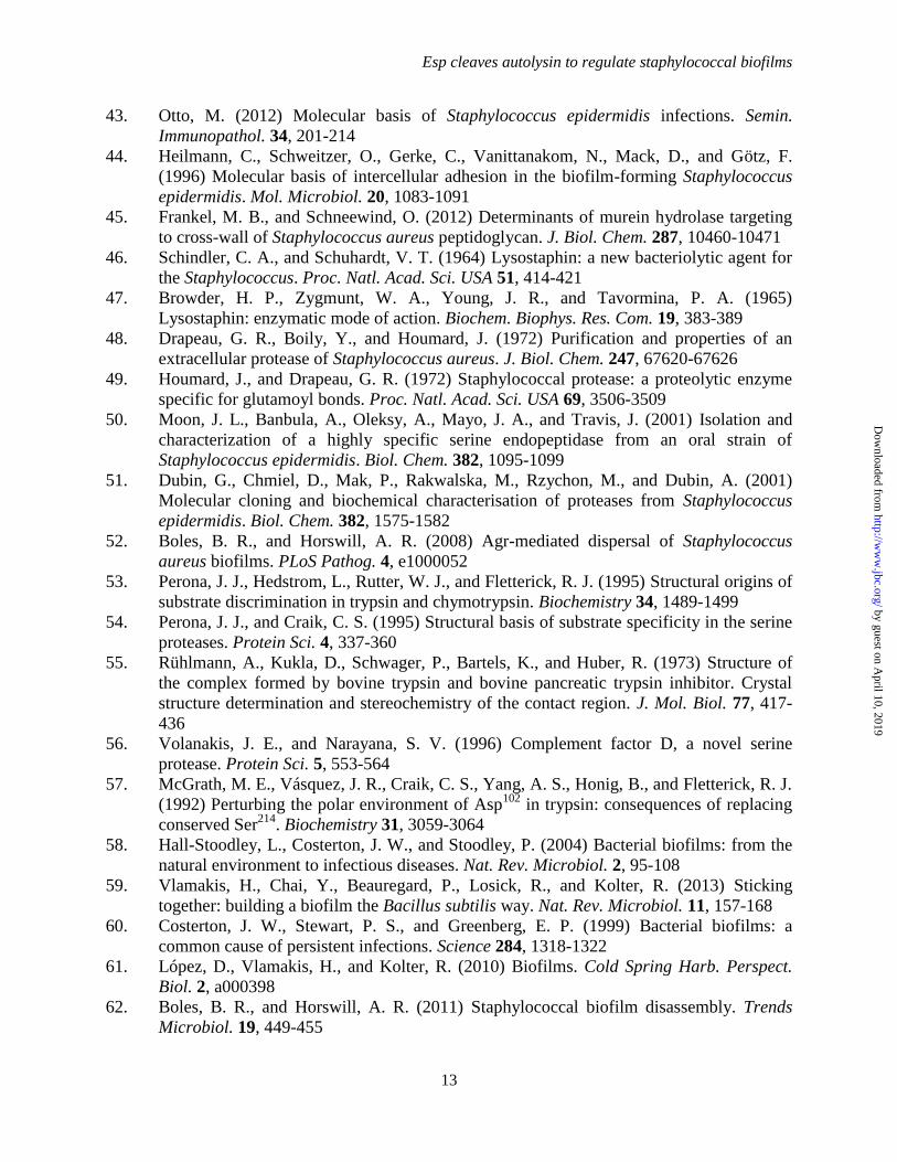
Esp cleaves Atl in staphylococcal biofilms-
Following signal peptide cleavage, the pro-form of
Esp (pro-Esp) is cleaved in the extracellular
medium of S. epidermidis cultures to generate
mature Esp protease, which mediates the
disassembly of S. aureus biofilms (7). We
expressed six-histidyl tagged pro-Esp in E. coli
and purified recombinant protein by affinity
chromatography (Fig. 1A). Thermolysin cleavage
and gel filtration chromatography were used to
obtain purified Esp (Fig. 1B). The variant EspS235A
harbors an alanyl substitution at the active site
by guest on April 10, 2019
http://ww
w.jbc.org/
Dow
nloaded from
Esp cleaves autolysin to regulate staphylococcal biofilms
5
serine residue of Esp (Fig. 1A). When examined
for protease activity with azocasein substrate (40),
Esp cleaved significantly more substrate than pro-
Esp, whereas EspS235A
did not display protease
activity (Fig. 1C). Wild-type S. aureus strain
Newman was used to form staphylococcal
biofilms using human fibronectin as a matrix,
which were quantified by crystal violet staining
(41). Treatment with Esp, but not pro-Esp or
EspS235A
, triggered disassembly of staphylococcal
biofilms (Fig. 1D). Proteins in biofilms with or
without Esp treatment were separated by 10-20%
gradient SDS-PAGE, stained with Coomassie
Brilliant Blue and identified via LC-MS/MS (Fig.
1E). Treatment with Esp, but not EspS235A
, caused
Atl degradation (Fig. 1E). Our experiments
revealed that Esp cleaved 18 additional
polypeptides, including FnbA, FnbB, Eap and SpA
that had previously been identified as Esp
substrates (26) (Table 2). While the genes for
some of these secreted proteins contribute to S.
aureus biofilm formation, they are not essential for
this developmental process. Of note, in S. aureus
Newman biofilms, Atl is a highly abundant
component and effectively degraded during Esp
treatment (Fig. 1E). Considering the importance of
Atl in biofilm development, we focused our
experimental approach on the interactions between
Esp and Atl.
Esp treatment of biofilms formed from atl
staphylococci- Mutations in the autolysin gene
(atlE) of S. epidermidis cause a dramatic reduction
in biofilm formation (42). AtlE was initially
shown to function as a S. epidermidis adhesin
[purified AtlE binds host vitronectin, fibronectin
and Hsc70 receptor (42)]. More recent work
highlighted the contribution of atl in S. aureus
UAMS-1, BH1CC and many other MSSA and
MRSA isolates towards releasing DNA as an
extracellular matrix for staphylococcal biofilm
formation (16,38). This discovery was
accompanied by the insight that S. aureus, but
presumably not S. epidermidis (43), forms
biofilms in vitro and in vivo without the icaABCD
locus (16), which provides for the synthesis of
(β1-6) poly-N-acetylglucosamine exo-
polysaccharide (44). We wondered whether atl
mutant S. aureus Newman can form biofilms and,
if so, whether atl bacterial communities can be
disassembled by treatment with Esp. Compared to
wild-type staphylococci, the atl mutant formed
only a rudimentary biofilm that, when subjected to
treatment with Esp, did not show significant
disassembly (Fig. 2A). As a control, Esp treatment
caused disassembly of biofilms formed by S.
aureus Newman. When subjected to growth assays
with rotating cultures, the atl mutant replicated at
a rate indistinguishable from that of wild-type
staphylococci (Fig. 2B), albeit that the atl mutants
formed large clusters of incompletely separated
staphylococci (23)(vide infra). The growth of
wild-type and atl mutant staphylococci was not
perturbed when cultures were treated with Esp,
indicating that protease treatment kills neither
wild-type nor mutant strains (Fig. 2B). As a
measure for the direct dispersal of bacteria from
biofilms, staphylococci were labeled with SYTO9
and then subjected to Esp treatment. Esp released
about half of wild-type staphylococci from
biofilms and almost all atl bacteria from their
rudimentary biofilm (Fig. 2CD). These
experiments identify Atl as a key target of Esp,
whose degradation prevents biofilm formation and
is associated with the disassembly of biofilms.
Moreover, the contributions of other targets of
Esp, with known auxiliary functions in biofilm
formation or stability (Eap, Emp, SpA, FnbA,
FnbB), are revealed as protease treatment
eliminates the rudimentary biofilm of atl mutants.
Esp cleavage of Atl- To determine which of the
functional domains of Atl are cleaved by Esp, we
purified AM (N-acetylmuramoyl-L-alanine
amidase), AMΔR1R2 (lacking the C-terminal repeat
domains R1 and R2 of AM), GL (N-
acetylglucosamine-N-acetylmuramic acid
glucosaminidase), and GLΔR3 (lacking the N-
terminal R3 domain of GL) as hybrids fused to the
C-terminal end of GST (Fig. 3AB). Esp treatment
cut AM, AMΔR1R2 and GL, but not GLΔR3 (Fig.
3B). Esp treatment generated several cleavage
fragments from AM, AMΔR1R2 or GL, suggesting
that the protease can cut at multiple sites within
the amidase and the R1-R3 domains (Fig. 3B).
Edman degradation of cleaved peptides identified
glutamic acid residues (for example Glu862
in GL)
as Esp cleavage sites (Fig. 3B).
Murein hydrolase activities of Esp treated Atl-
To explore the effects of Esp treatment on Atl
murein hydrolase activities, we purified murein
sacculi from wild-type S. aureus and extracted
wall teichoic acids via hydrofluoric acid treatment
(45). The murein hydrolase activities of AM and
by guest on April 10, 2019
http://ww
w.jbc.org/
Dow
nloaded from
Esp cleaves autolysin to regulate staphylococcal biofilms
6
AMΔR1R2 as well as GL and GLΔR3 were
determined by incubating GST hybrids with
murein sacculi while monitoring absorbance at
600 nm. Similar to lysostaphin (46), a glycyl-
glycine endopeptidase that cleaves staphylococcal
cell wall crossbridges (47), GST-AM treatment of
peptidoglycan caused a large decrease in
absorbance (Fig. 4A). Esp treatment abolished all
peptidoglycan hydrolase activity of AM (Fig. 4A).
Removal of the R1-R2 repeat domains of AM
reduced the peptidoglycan hydrolase activity of
AMΔR1R2, however this activity was also abolished
by treatment with Esp (Fig. 4A). Finally, GL
displayed very little activity in reducing the
absorbance at 600 nm, which can be explained by
the relatively short glycan chains and intricate
crosslinking (>99%) of the staphylococcal cell
wall (20). The murein hydrolase activity of GL
was abolished by Esp treatment (Fig. 4A). GLΔR3,
which is a poor substrate for Esp, did not display
murein hydrolase activity in this assay (Fig. 4A).
Our results corroborate earlier observations on the
genetic requirements of the AM and GL domains
for S. aureus Atl function (38).
Trans-complementation of atl mutant biofilms-
The addition of purified GST-AM or GST-GL
restored (trans-complementation) the biofilm
formation defect of atl mutant S. aureus Newman
grown on fibronectin-coated microtiter plates (Fig.
4B). This activity was abolished following
treatment of GST-AM with Esp (Fig. 4B). Esp
treatment did not affect peptidoglycan hydrolase
activity of GST-GL and did not affect GST-GL
biofilm trans-complementation either. GST-
AMΔR1R2 also did not display biofilm trans
complementation for atl mutants and Esp
treatment did not affect this phenotype (Fig. 4B).
GST-GLΔR3 did not degrade murein sacculi and
did not trans-complement the atl mutant biofilm
defect (Fig. 4B). Esp treatment did not affect
biofilm formation in the presence of GST-GLΔR3
(Fig. 4B).
Esp treatment and staphylococcal cell clusters-
The atl mutant staphylococci are defective in the
separation of daughter cells following cell
division, which is due to the incomplete separation
of cross wall peptidoglycan (23,25). This
phenotype can be quantified by flow cytometry,
which revealed that 6.82% of wild-type but 57%
of atl mutants exist as large cell clusters (Fig. 5A).
Treatment with Esp did not affect the cell cluster
size of atl mutants, whereas GST-AM, GST-GL
and GST-GLΔR3, but not GST-AMΔR1R2, reduced
the cell cluster size (Fig. 5AB). The cluster
reducing activity of GST-AM and GST-GL was
abolished by treatment with Esp (Fig. 5AB).
Esp treatment and staphylococcal release of
extracellular DNA- We sought to test the
hypothesis that Esp blocks the release of
extracellular DNA during biofilm formation and
added the protease to either wild-type or atl
mutant S. aureus Newman. Biofilms were stained
with either propidium iodide (PI) as a measure for
extracellular DNA or with SYTO9 to quantify
viable staphylococci (Fig. 6AB). S. aureus
biofilms harbored large amounts of extracellular
DNA and bacterial cells, as quantified by PI and
SYTO9 staining (Fig. 6AB). Treatment with Esp
reduced the amount of extracellular DNA and
viable staphylococci in wild-type biofilms,
whereas treatment with DNAse abolished both
(Fig. 6AB). Rudimentary biofilms formed by atl
mutant S. aureus harbored very little if any
extracellular DNA and few staphylococci (Fig.
6AB).
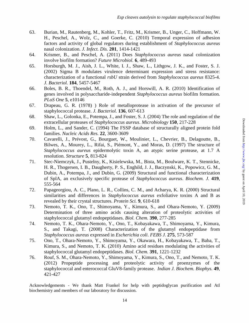
SspA (V8 protease) is required for S. aureus
biofilm formation- V8 (GluC) enzyme is a serine
protease that selectively cleaves peptides C-
terminal of glutamyl or aspartyl residues (48,49).
S. aureus V8 is highly homologous to S.
epidermidis Esp (59% identity and 78%
similarity), albeit that V8 harbors a C-terminal
extension of 52 amino acids that is absent in Esp
(Fig. 7A). Similar to Esp, purified V8 cleaved
GST-AM, GST-AMΔR1R2 and GST-GL (Fig. 7B).
In contrast to Esp, the V8 enzyme effectively
cleaved GST-GLΔR3 (Fig. 7B). This can be
explained by the protease activity of Esp, which,
unlike V8, cuts only at the C-terminal of glutamyl
but not of aspartyl (50,51). To analyze the
contribution of sspA towards S. aureus biofilm
formation, we transduced a bursa aurealis
insertional lesion in the sspA gene (28) into the
wild-type strain Newman (27). When analyzed for
biofilm formation on fibronectin matrix, the sspA
mutant was significantly impaired, as compared to
wild-type and similar to the biofilm defect of the
atl mutant (Fig. 7C). The culture supernatant of
the S. aureus Newman sspA mutant did not display
increased protease activity in the azocasein assay
when compared to wild-type (Fig. 7E). Thus, the
biofilm phenotype of the sspA mutant is not due to
by guest on April 10, 2019
http://ww
w.jbc.org/
Dow
nloaded from
Esp cleaves autolysin to regulate staphylococcal biofilms
7
an increase in extracellular protease activity, as
has been reported for the sspA deletion mutant of
S. aureus SH1000 (52). Moreover, the biofilm
phenotype of the S. aureus Newman sspA mutant
was restored following transformation with a
recombinant plasmid expressing wild-type sspA,
but not with vector (pWW412) control (Fig. 7D).
Incubation of S. aureus Newman with purified V8
protease reduced biofilm formation (Fig. 7C). V8
treatment did not improve biofilm formation of S.
aureus Newman atl or sspA mutants (Fig. 7C).
These results suggest that the expression and/or
activity of secreted V8 protease must be carefully
controlled during S. aureus biofilm formation,
because treatment with exogenous, active V8
protease cannot complement the sspA mutant
phenotype (Fig. 7C). Of note, neither V8 nor Esp
protease treatment affected biofilm formation of S.
epidermidis RP62a (Esp+), suggesting that the
biofilm program of this microbe is not controlled
by secreted serine proteases or their protease
sensitive substrates (Fig. 7C).
Crystallographic structure of Esp- Purified Esp
was crystallized and its three dimensional structure
determined using X-ray crystallography. Esp
displays a β-barrel fold assembled from two
discrete domains and a C-terminal α-helix, similar
to eukaryotic serine proteases of the chymotrypsin
family (Fig. 8AB) (53-55). Even though Esp
exhibits a highly conserved, compact β-barrel fold,
the five or more intra-domain disulfide bonds that
are responsible for the structural rigidity of
eukaryotic serine proteases are absent (54). Each
of the two Esp domains is comprised of six
antiparallel β-strands, and the solvent accessible
catalytic and substrate binding sites are situated at
the interface of the two domains. The N-terminal
domain (chymotrypsin nomenclature) is comprised
primarily of residues Gln77
-Ile183
, whereas the C-
terminal domain encompasses Ser184
-Ile264
. While
the position of the C-terminal α-helix (Asn266
-
Ile276
) is conserved with that of other serine
proteases, the N-terminal segment (Val67
-Gln76
)
contains a short β-strand that is associated with the
substrate binding S1 pocket and distinct from
eukaryotic serine proteases (Fig. 8B). In addition
to the conserved position of putative catalytic triad
residues (Ser235
, Asp159
and His117
), the substrate-
binding region (S1 pocket) and the oxyanion-hole,
which together constitute the critical functional
elements of activated serine proteases, are also
conserved in Esp (Fig. 8A). A search for structural
homologues of Esp identified S. aureus V8, a
serine protease with a Z-score of 39.7 and 59%
primary sequence identity (PDB ID: 1QY6)(33).
Structural comparison of Esp and V8- The
distances between Nε of the Esp active site His117
and Oγ of Ser235
and between Nδ of His117
and Oδ
of Asp159
are 2.8 and 2.6 Å, respectively. Such
short distances between the catalytic residues and
their relative positions are conserved among
canonical serine protease structures (56). A serine
residue that forms a hydrogen bond with the
catalytic Asp159
is sometimes referred to as the
fourth member of the catalytic triad of serine
proteases (57), however this residue is absent in
both Esp and V8; the corresponding space is
occupied by Tyr251
and Trp253
, respectively.
Interestingly, the highly conserved hydrophobic
residue (Trp or Phe) at position 215 of the serine
protease family has been replaced by a Gly (Gly252
Gly254
) in both Esp and V8.
A key element of serine protease catalytic
activity is the ‘oxyanion-hole’, which is
contributed by the right side wall of the S1 pocket
(54). The oxyanion-hole stabilizes the tetrahedral
transition intermediate of the substrate scissile
peptide bond, by compensating the negative
charge generated on its carbonyl oxygen during
acylation (54). The oxyanion-hole is formed by the
backbone peptide nitrogen atoms of the catalytic
Ser and two preceding residues in all serine
proteases. The oxyanion-hole is flexible and
disordered in the zymogen (pro-forms) serine
proteases. The conformation of Ser235
-Asn234
-
Gly233
-Gly232
-Val231
peptide segment in Esp is
rigid with conserved backbone angles, supporting
the presence of a functional oxyanion hole.
Further, the carbonyl oxygen of Gly232
in Esp is
suitably pointing outwards and away from the
hydroxyl group of the catalytic Ser235
. With a
distance of 4.2 Å between them, the catalytic
residue Ser235
is positioned for a nucleophilic
attack on the substrate scissile peptide bond’s
carbonyl carbon.
The substrate binding pocket of Esp- Perona
and Craik defined seven conserved loops (A, B, C,
D, Loop 1, Loop 2 and Loop 3) in serine proteases
that surround the S1 pocket (Fig. 8B), which
exhibits high specificity toward the substrate P1
residue (54). Structural integrity of the Loop 1 has
a direct impact on the catalytic efficiency of the
by guest on April 10, 2019
http://ww
w.jbc.org/
Dow
nloaded from

Esp cleaves autolysin to regulate staphylococcal biofilms
8
enzyme, while the Loop 2 residue composition
dictates the substrate P1 residue specificity and its
binding efficiency. Variations in lengths and
compositions of Loop 3 as well as A, B, C, and D
Loops dictate the residue identity of the substrate
at more distal positions on both sides of the P1
residue (54). Compared to all other eukaryotic
serine proteases, Loop 3 and Loop D are absent in
Esp and V8 and their Loop 1 and Loop 2 are also
considerably shorter. A single polar residue on
Loop 1 (Asp in trypsin), at the bottom of the S1
pocket, dictates primary substrate specificity for
serine proteases. We observe two polar residues
Ser229
and Thr230
at the Loop 1 position of Esp (V8
Ser231
and Thr232
). Nevertheless, these residues are
not suitably positioned for interaction with the P1
residue bound in the S1 pocket. Using molecular
modeling, Prasad and colleagues suggested that
Asn258
, positioned in Loop 2 of V8 (Esp Asn256
)
and pointing into the active site, may be the
determinant of substrate specificity (33).
DISCUSSION
When grown in liquid culture without rotation,
many bacterial pathogens, including S. aureus,
form biofilms on solid surfaces or at liquid-air
interfaces (58). Research over the past two
decades has identified bacterial genes and
mechanisms supporting biofilm growth, which can
be thought of as a developmental program with
three or more discrete steps (59,60). Biofilm
formation initially requires bacterial adhesion to
solid surfaces and this includes bacterial adhesion
to surfaces at liquid-air interfaces (61). Bacterial
replication into a biofilm is dependent on cell-cell
adhesions and on the establishment of an
extracellular matrix, which is often comprised of
DNA released from a subpopulation of biofilm
bacteria but may also involve the synthesis of
extracellular polysaccharides (61). Eventually,
biofilms must release planktonic cells for
dissemination in tissues of an infected host and/or
development of new biofilm structures (60). These
paradigms also appear to apply to the biofilms
formed by S. aureus (62), a pathogen that
colonizes human nares (1).
Iwase and colleagues reported that S.
epidermidis Esp, a secreted serine protease, can
disperse S. aureus biofilms (7). Furthermore,
colonization with Esp+ S. epidermidis strains was
associated with protection from S. aureus
colonization and administration of Esp+ S.
epidermidis into the nares of human volunteers
diminished S. aureus colonization (7). These
findings provide strong support for the model of S.
aureus biofilm formation in human nares, however
others have challenged this view and proposed that
S. aureus may replicate as planktonic bacteria in
the nasal cavity (63,64).
Here we investigated the molecular basis of S.
epidermidis Esp interference with S. aureus
biofilm formation. Our data suggest that Atl is the
premier target of Esp-mediated biofilm
interference. Esp treatment diminished Atl-
dependent release of extracellular DNA by
cleaving the AM and GL murein hydrolase
activities. Esp treatment did not affect biofilm
formation for atl and sspA mutants of S. aureus
Newman. X-ray crystallography revealed the
three-dimensional structure of Esp, which is
highly similar to that of S. aureus V8 (SspA)(33).
V8 protease also cleaved Atl AM and GL, and
blocked biofilm formation. These data suggest that
S. aureus biofilms are formed under conditions of
controlled secretion and proteolysis of autolysin, a
determinant for the release of DNA biofilm
matrix. This developmental program can be
perturbed by the Esp protease of S. epidermidis
and by the V8 protease.
Earlier work reported that sspA expression in S.
aureus SH1000, a variant of laboratory strain
8325-4 (RN6390B) in which the rsbU mutational
lesion has been repaired (65), is required for
biofilm formation when this strain is grown in 2%
tryptic soy broth supplemented with 0.2% glucose
but not when the strain is grown in TSB alone
(52). In S. aureus SH1000, mutations in sspA and
in other genes for extracellular serine proteases
(splABCDEF) trigger a relative increase in
extracellular protease activity (52,66), which is
associated with a reduction in biofilm formation.
This phenotype is abolished in a genetic
background where the structural gene for
aureolysin (aur) has been deleted (52); aureolysin
is a metalloproteinase that, following secretion
into the extracellular medium, activates the serine
proteases of S. aureus via removal of their pro-
peptides (67,68). Presumably, a cascade of
secretion reactions and the sequential activation of
extracellular proteases (aureolysin>-cysteine
proteases>serine proteases) controls the activity of
by guest on April 10, 2019
http://ww
w.jbc.org/
Dow
nloaded from
Esp cleaves autolysin to regulate staphylococcal biofilms
9
secreted Atl and the assembly or disassembly of
staphylococcal biofilms (62).
We also solved the three dimensional structure
of Esp. A search for structural homologues of Esp
using the DALI server (69) identified seven
structures with less than 2.0 Å rmsd value. S.
aureus V8 protease was the best fit with a Z-score
of 39.7 and 59% primary sequence identity (PDB
ID: 1QY6) (33). Staphylococcal epidermolytic
toxin A (ETA) (PDB ID: 1AGJ) was the second
best with Z-score of 29.9 and 28% sequence
identity (70). Staphylococcal secreted serine
proteases SplB (PDB ID: 1VID) and SplA (PDB
ID: 2W7U) displayed 32% and 28% identity,
respectively (71). Glutamyl-endopeptidase (PDB
ID: 1P3C, 26% identity) and exfoliative toxin B
(PDB ID: 1QTF, 29% identity), with rmsd less
than 2Å were also identified (72).
Nemoto and colleagues characterized S. aureus
glutamyl endopeptidase V8, and identified, in
addition to catalytic Ser237
(Esp Ser235
), the N-
terminal Val69
(Esp Val67
) residue as essential for
substrate cleavage (73,74). S. aureus V8 protease
with an N-terminal truncation to Ile70
(Esp Ile68
)
was inactive and mutants with an altered N-
terminal residue Val69
(even with conserved
substitutions) were also inactive, which is
indicative of a strict requirement of the N-terminal
Val residue for enzyme activity (75). Crystal
structures of Esp and V8 display identical
disposition for their N-termini, which associate
with respective S1 pockets more intimately than
other active serine proteases (Fig. 8C). The amino
terminal segment of Esp and V8 crosses over the
Loop 1 into the bottom of the S1 pocket, and the
N-terminal Val67
(V8 Val69
) amino group is
suitably positioned to act as an acceptor of the
negative charge of P1 residue side chain (33).
The N-terminal Val67
in Esp is positioned with its
α-amino group located adjacent to the conserved
Thr230
(V8 Thr232
) and Asn259
(V8 Asn261
), pointed
into S1 pocket, within hydrogen bonding distance.
Similarly, the His250
(V8 His252
) residue on Loop
2, conserved among glutamyl endopeptidases (73),
having hydrogen bonds with side chains of
conserved Tyr226
(V8 Tyr228
) and Thr230
(V8
Thr232
), is also suitably positioned to interact with
the substrate acidic P1 residue.
Nevertheless, there are some notable
differences between V8 and Esp in and around
their S1 pockets (Fig. 8D) that can be associated
with differences in substrate specificity. Extensive
mutational analysis of V8 and Esp by Nemoto and
colleagues localized the difference in their
specificities to Tyr251
(V8 Trp253
) and Asp255
(V8
Pro257
) residues on Esp Loop 2 (73). Substitutions
at these positions affected mainly the Km with
constant kcat values, suggesting these residues
affect only substrate binding affinities (73). The
Km value of native Esp harboring Tyr251
-Val254
-
Asp255
on loop 2 was larger than that of V8 with
Trp253
-Val256
-Pro257
, but with almost similar kcat
values (76). Tyr251
of Esp is hydrogen bonded with
the side chain of Glu223
, which is replaced by
Ala225
in V8. In addition, the Lys257
-Tyr258
-Asn259
-
Ser260
-Ser261
segment on Loop 2 of Esp is replaced
by Glu259
-Tyr260
-Asn261
-Gly262
-Ala263
in V8, all
side chains pointing out of the S1 pocket, but into
the known specificity determining secondary sites
of serine proteases. Other notable residue
differences between Esp and V8 in the vicinity of
the S1 pocket include Tyr92
(V8 Gln94
) and Tyr99
(V8 Thr101
) on Loop A. Thus, the S1 pockets of
Esp and V8 preferentially bind negatively charged
substrate side chains that are held in place by the
amino group of the N-terminal Val67
(V8 Val69
)
residue, and stabilized by conserved Thr230
, His250
and Asn259
in Esp. However, the specificity
differences between equally efficient Esp and V8
enzymes toward acidic P1 residue could be
assigned to the differences observed in Loop 2,
specially to the Asp255
(V8 Pro257
) present at the
bottom of S1 pocket and pointing toward the
catalytic site and other secondary residues on
either side of the substrate P1 residue to the
difference in Loop 2 and Loop D segments. These
features of staphylococcal serine proteases may
explain why Esp and V8 are able to cleave
multiple domains of Atl and, when added
exogenously during the early stages of biofilm
formation, can interfere with the establishment of
these structures. The V8 protease does contribute
to biofilm formation of S. aureus Newman
presumably by controlling the autolytic activity of
Atl-derived AM and GL enzymes. Thus, secreted
serine proteases can be viewed as biofilm
regulatory factors that impact the production of
biofilm matrix and the release of planktonic
bacteria to initiate invasive disease (Fig. 9). If so,
application of serine proteases (Esp or V8) as a
treatment of nasal colonization with S. aureus may
by guest on April 10, 2019
http://ww
w.jbc.org/
Dow
nloaded from
Esp cleaves autolysin to regulate staphylococcal biofilms
10
disperse planktonic staphylococci with invasive disease potential.
REFERENCES
1. Lowy, F. D. (1998) Staphylococcus aureus infections. New Engl. J. Med. 339, 520-532
2. Kluytmans, J., van Belkum, A., and Verburgh, H. (1997) Nasal carriage of
Staphylococcus aureus: epidemiology, underlying mechanisms, and associated risks.
Clin. Microbiol. Rev. 10, 505-520
3. Wertheim, H. F., Melles, D. C., Vos, M. C., van Leeuwen, W., van Belkum, A.,
Verbrugh, H. A., and Nouwen, J. L. (2005) The role of nasal carriage in Staphylococcus
aureus infections. Lancet Infect. Dis. 5, 751-762
4. Wertheim, H. F., Vos, M. C., Ott, A., van Belkum, A., Voss, A., Kluytmans, J. A., van
Keulen, P. H., Vandenbroucke-Grauls, C. M., Meester, M. H., and Verbrugh, H. A.
(2004) Risk and outcome of nosocomial Staphylococcus aureus bacteraemia in nasal
carriers versus non-carriers. Lancet 364, 703-705
5. Klevens, R. M., Edwards, J. R., and Gaynes, R. P. (2008) The impact of antimicrobial-
resistant, health care-associated infections on mortality in the United States. Clin. Infect.
Dis. 47, 927-930
6. Klevens, R. M., Morrison, M. A., Nadle, J., Petit, S., Gershman, K., Ray, S., Harrison, L.
H., Lynfield, R., Dumyati, G., Townes, J. M., Craig, A. S., Zell, E. R., Fosheim, G. E.,
McDougal, L. K., Carey, R. B., and Fridkin, S. K. (2007) Invasive methicillin-resistant
Staphylococcus aureus infections in the United States. JAMA 298, 1763-1771
7. Iwase, T., Uehara, Y., Shinji, H., Tajima, A., Seo, H., Takada, K., Agata, T., and
Mizunoe, Y. (2010) Staphylococcus epidermidis Esp inhibits Staphylococcus aureus
biofilm formation and nasal colonization. Nature 465, 346-349
8. Sadykov, M. R., and Bayles, K. W. (2012) The control of death and lysis in
staphylococcal biofilms: a coordination of physiological signals. Curr. Opin. Microbiol.
15, 211-215
9. Archer, N. K., Mazaitis, M. J., Costerton, J. W., Leid, J. G., Powers, M. E., and Shirtliff,
M. E. (2011) Staphylococcus aureus biofilms: properties, regulation, and roles in human
disease.Virulence 2, 445-459
10. Lower, S. K., Lamlertthon, S., Casillas-Ituarte, N. N., Lins, R. D., Yongsunthon, R.,
Taylor, E. S., DiBartola, A. C., Edmonson, C., McIntyre, L. M., Reller, L. B., Que, Y. A.,
Ros, R., Lower, B. H., and Fowler, V. G. J. (2011) Polymorphisms in fibronectin binding
protein A of Staphylococcus aureus are associated with infection of cardiovascular
devices.Proc. Natl. Acad. Sci. USA 108, 18372-18377
11. Geoghegan, J. A., Monk, I. R., O'Gara, J. P., and Foster, T. J. (2013) Subdomains N2N3
of fibronectin binding protein A mediate Staphylococcus aureus biofilm formation and
adherence to fibrinogen using distinct mechanisms.J. Bacteriol. 195, 2675-2683
12. Thompson, K. M., Abraham, N., and Jefferson, K. K. (2010) Staphylococcus aureus
extracellular adherence protein contributes to biofilm formation in the presence of serum.
FEMS Microbiol. Lett. 305, 143-147
13. Johnson, M., Cockayne, A., and Morrissey, J. A. (2008) Iron-regulated biofilm formation
in Staphylococcus aureus Newman requires ica and the secreted protein Emp. Infect.
Immun. 76, 1756-1765
14. Kaplan, J. B., Izano, E. A., Gopal, P., Karwacki, M. T., Kim, S., Bose, J. L., Bayles, K.
W., and Horswill, A. R. (2012) Low levels of β-lactam antibiotics induce extracellular
DNA release and biofilm formation in Staphylococcus aureus. mBio. 3, e00198-00112
by guest on April 10, 2019
http://ww
w.jbc.org/
Dow
nloaded from

Esp cleaves autolysin to regulate staphylococcal biofilms
11
15. Mann, E. E., Rice, K. C., Boles, B. R., Endres, J. L., Ranjit, D., Chandramohan, L.,
Tsang, L. H., Smeltzer, M. S., Horswill, A. R., and Bayles, K. W. (2009) Modulation of
eDNA release and degradation affects Staphylococcus aureus biofilm maturation. PLoS
One 4, e5822
16. Houston, P., Rowe, S. E., Pozzi, C., Waters, E. M., and O'Gara, J. P. (2011) Essential role
for the major autolysin in the fibronectin-binding protein-mediated Staphylococcus
aureus biofilm phenotype. Infect. Immun. 79, 1153-1165
17. Rice, K. C., Mann, E. E., Endres, J. L., Weiss, E. C., Cassat, J. E., Smeltzer, M. S., and
Bayles, K. W. (2007) The cidA murein hydrolase regulator contributes to DNA release
and biofilm development in Staphylococcus aureus. Proc. Natl. Acad. Sci. USA 104,
8113-8118
18. Ahn, J. S., Chandramohan, L., Liou, L. E., and Bayles, K. W. (2006) Characterization of
CidR-mediated regulation in Bacillus anthracis reveals a previously undetected role of S-
layer proteins as murein hydrolases. Mol. Microbiol. 62, 1158-1169
19. Oshida, T., Sugai, M., Komatsuzawa, H., Hong, Y. M., Suginaka, H., and Tomasz, A.
(1995) A Staphylococcus aureus autolysin that has an N-acetylmuramoyl-L-alanine
amidase domain and an endo-β-N-acetylglucosaminidase domain: cloning, sequence
analysis, and characterization. Proc. Natl. Acad. Sci. USA 92, 285-289
20. Baba, T., and Schneewind, O. (1998) Targeting of muralytic enzymes to the cell division
site of Gram-positive bacteria: repeat domains direct autolysin to the equatorial surface
ring of Staphylococcus aureus. EMBO J. 17, 4639-4646
21. Zoll, S., Schlag, M., Shkumatov, A. V., Rautenberg, M., Svergun, D. I., Götz, F., and
Stehle, T. (2012) Ligand-binding properties and conformational dynamics of autolysin
repeat domains in staphylococcal cell wall recognition. J. Bacteriol. 194, 3789-3802
22. Sugai, M., Yamada, S., Nakashima, S., Komatsuzawa, H., Matsumoto, A., Oshida, T.,
and Suginaka, H. (1997) Localized perforation of the cell wall by a major autolysin: atl
gene products and the onset of penicillin-induced lysis of Staphylococcus aureus. J.
Bacteriol. 179, 2958-2062
23. Yamada, S., Sugai, M., Komatsuzawa, H., Nakashima, S., Oshida, T., Matsumoto, A.,
and Suginaka, H. (1996) An autolysin ring associated with cell separation of
Staphylococcus aureus.J. Bacteriol. 178, 1565-1571
24. Schlag, M., Biswas, R., Krismer, B., Kohler, T., Zoll, S., Yu, W., Schwarz, H., Peschel,
A., and Götz, F. (2010) Role of staphylococcal cell wall teichoic acid in targeting the
major autolysin Atl. Mol. Microbiol. 75, 864-873
25. Sugai, M., Komatsuzawa, H., Akiyama, T., Hong, Y.-M., Oshida, T., Miyake, Y.,
Yamaguchi, T., and Suginaka, H. (1995) Identification of endo--N-
acetylglucosaminidase and N-acetylmuramyl-L-alanine amidase as cluster dispersing
enzymes in Staphylococcus aureus. J. Bacteriol. 177, 1491-1496
26. Sugimoto, S., Iwamoto, T., Takada, K., Okuda, K.-I., Tajima, A., Iwase, T., and
Mizunoe, Y. (2013) Staphylococcus epidermidis Esp egrades specific proteins associated
with Staphylococcus aureus biofilm formation and host-pathogen interaction. J.
Bacteriol. 195, 1645-1655
27. Baba, T., Bae, T., Schneewind, O., Takeuchi, F., and Hiramatsu, K. (2007) Genome
sequence of Staphylococcus aureus strain Newman and comparative analysis of
staphylococcal genomes. J. Bacteriol. 190, 300-310
by guest on April 10, 2019
http://ww
w.jbc.org/
Dow
nloaded from
Esp cleaves autolysin to regulate staphylococcal biofilms
12
28. Bae, T., Banger, A. K., Wallace, A., Glass, E. M., Aslund, F., Schneewind, O., and
Missiakas, D. M. (2004) Staphylococcus aureus virulence genes identified by bursa
aurealis mutagenesis and nematode killing. Proc. Natl. Acad. Sci. USA 101, 12312-
12317
29. Vengadesan, K., Macon, K., Sugumoto, S., Mizunoe, Y., Iwase, T., and Narayana, S. V.
(2013) Purification, crystallization and preliminary X-ray diffraction analysis of the
Staphylococcus epidermidis extracellular serine protease Esp. Acta Crystallogr. Sect. F
Struct. Biol. Cryst. Commun. 69, 49-52
30. Pflugrath, J. W. (1999) The finer things in X-ray diffraction data collection. Acta
Crystallogr. D Biol. Crystallogr. 55, 1718-1725
31. McCoy, A. J., Grosse-Kunstleve, R. W., Adams, P. D., Winn, M. D., Storoni, L. C., and
Read, R. J. (2007) Phaser crystallographic software. J. Appl. Crystallogr. 40, 658-674
32. Winn, M. D., Ballard, C. C., Cowtan, K. D., Dodson, E. J., Emsley, P., Evans, P. R.,
Keegan, R. M., Krissinel, E. B., Leslie, A. G., McCoy, A., McNicholas, S. J.,
Murshudov, G. N., Pannu, N. S., Potterton, E. A., Powell, H. R., Read, R. J., Vagin, A.
A., and Wilson, K. S. (2011) Overview of the CCP4 suite and current developments.
Acta Crystallogr. D Biol. Crystallogr. 67, 235-242
33. Prasad, L., Leduc, Y., Hayakawa, K., and Delbaere, L. T. (2004) The structure of a
universally employed enzyme: V8 protease from Staphylococcus aureus. Acta
Crystallogr. D Biol. Crystallogr. 60, 256-259
34. Emsley, P., Lohkamp, B., Scott, W. G., and Cowtan, K. (2010) Features and development
of Coot. Acta Crystallogr. D Biol. Crystallogr. 66, 486-501
35. Murshudov, G. N., Vagin, A. A., and Dodson, E. J. (1997) Refinement of
macromolecular structures by the maximum-likelihood method. Acta Crystallogr. D Biol.
Crystallogr. 53, 240-255
36. Laskowski, R. A., MacArthur, M. W., Moss, D. S., and Thornton, J. M. (1993)
PROCHECK: a program to check the stereochemical quality of protein structures. J.
Appl. Cryst. 26, 283-291
37. Park, D. W., Kim, S. S., Nam, M. K., Kim, G. Y., Kim, J., and Rhim, H. (2011)
Improved recovery of active GST-fusion proteins from insoluble aggregates:
solubilization and purification conditions using PKM2 and HtrA2 as model proteins.
BMB Reports 44, 279-284
38. Bose, J. L., Lehman, M. K., Fey, P. D., and Bayles, K. W. (2012) Contribution of the
Staphylococcus aureus Atl AM and GL murein hydrolase activities in cell division,
autolysis, and biofilm formation. PLoS One 7, e42244
39. Frankel, M. B., Hendrickx, A. P., Missiakas, D. M., and Schneewind, O. (2011) LytN, a
murein hydrolase in the cross-wall compartment of Staphylococcus aureus, is involved in
proper bacterial growth and envelope assembly. J. Biol. Chem. 286, 32593-32605
40. Charney, J., and Tomarelli, R.M. (1947) A colorimetric method for the determination of
the proteolytic activity of duodenal juice. J. Biol. Chem. 171, 501-505
41. Scriba, T. J., Sierro, S., Brown, E. L., Phillips, R. E., Sewell, A. K., and Massey, R. C.
(2008) The Staphyloccous aureus Eap protein activates expression of proinflammatory
cytokines. Infect. Immun. 76, 2164-2168
42. Heilmann, C., Hussain, M., Peters, G., and Götz, F. (1997) Evidence for autolysin-
mediated primary attachment of Staphylococcus epidermidis to a polystyrene surface.
Mol. Microbiol. 24, 1013-1024
by guest on April 10, 2019
http://ww
w.jbc.org/
Dow
nloaded from
Esp cleaves autolysin to regulate staphylococcal biofilms
13
43. Otto, M. (2012) Molecular basis of Staphylococcus epidermidis infections. Semin.
Immunopathol. 34, 201-214
44. Heilmann, C., Schweitzer, O., Gerke, C., Vanittanakom, N., Mack, D., and Götz, F.
(1996) Molecular basis of intercellular adhesion in the biofilm-forming Staphylococcus
epidermidis. Mol. Microbiol. 20, 1083-1091
45. Frankel, M. B., and Schneewind, O. (2012) Determinants of murein hydrolase targeting
to cross-wall of Staphylococcus aureus peptidoglycan. J. Biol. Chem. 287, 10460-10471
46. Schindler, C. A., and Schuhardt, V. T. (1964) Lysostaphin: a new bacteriolytic agent for
the Staphylococcus. Proc. Natl. Acad. Sci. USA 51, 414-421
47. Browder, H. P., Zygmunt, W. A., Young, J. R., and Tavormina, P. A. (1965)
Lysostaphin: enzymatic mode of action. Biochem. Biophys. Res. Com. 19, 383-389
48. Drapeau, G. R., Boily, Y., and Houmard, J. (1972) Purification and properties of an
extracellular protease of Staphylococcus aureus. J. Biol. Chem. 247, 67620-67626
49. Houmard, J., and Drapeau, G. R. (1972) Staphylococcal protease: a proteolytic enzyme
specific for glutamoyl bonds. Proc. Natl. Acad. Sci. USA 69, 3506-3509
50. Moon, J. L., Banbula, A., Oleksy, A., Mayo, J. A., and Travis, J. (2001) Isolation and
characterization of a highly specific serine endopeptidase from an oral strain of
Staphylococcus epidermidis. Biol. Chem. 382, 1095-1099
51. Dubin, G., Chmiel, D., Mak, P., Rakwalska, M., Rzychon, M., and Dubin, A. (2001)
Molecular cloning and biochemical characterisation of proteases from Staphylococcus
epidermidis. Biol. Chem. 382, 1575-1582
52. Boles, B. R., and Horswill, A. R. (2008) Agr-mediated dispersal of Staphylococcus
aureus biofilms. PLoS Pathog. 4, e1000052
53. Perona, J. J., Hedstrom, L., Rutter, W. J., and Fletterick, R. J. (1995) Structural origins of
substrate discrimination in trypsin and chymotrypsin. Biochemistry 34, 1489-1499
54. Perona, J. J., and Craik, C. S. (1995) Structural basis of substrate specificity in the serine
proteases. Protein Sci. 4, 337-360
55. Rühlmann, A., Kukla, D., Schwager, P., Bartels, K., and Huber, R. (1973) Structure of
the complex formed by bovine trypsin and bovine pancreatic trypsin inhibitor. Crystal
structure determination and stereochemistry of the contact region. J. Mol. Biol. 77, 417-
436
56. Volanakis, J. E., and Narayana, S. V. (1996) Complement factor D, a novel serine
protease. Protein Sci. 5, 553-564
57. McGrath, M. E., Vásquez, J. R., Craik, C. S., Yang, A. S., Honig, B., and Fletterick, R. J.
(1992) Perturbing the polar environment of Asp102
in trypsin: consequences of replacing
conserved Ser214
. Biochemistry 31, 3059-3064
58. Hall-Stoodley, L., Costerton, J. W., and Stoodley, P. (2004) Bacterial biofilms: from the
natural environment to infectious diseases. Nat. Rev. Microbiol. 2, 95-108
59. Vlamakis, H., Chai, Y., Beauregard, P., Losick, R., and Kolter, R. (2013) Sticking
together: building a biofilm the Bacillus subtilis way. Nat. Rev. Microbiol. 11, 157-168
60. Costerton, J. W., Stewart, P. S., and Greenberg, E. P. (1999) Bacterial biofilms: a
common cause of persistent infections. Science 284, 1318-1322
61. López, D., Vlamakis, H., and Kolter, R. (2010) Biofilms. Cold Spring Harb. Perspect.
Biol. 2, a000398
62. Boles, B. R., and Horswill, A. R. (2011) Staphylococcal biofilm disassembly. Trends
Microbiol. 19, 449-455
by guest on April 10, 2019
http://ww
w.jbc.org/
Dow
nloaded from
Esp cleaves autolysin to regulate staphylococcal biofilms
14
63. Burian, M., Rautenberg, M., Kohler, T., Fritz, M., Krismer, B., Unger, C., Hoffmann, W.
H., Peschel, A., Wolz, C., and Goerke, C. (2010) Temporal expression of adhesion
factors and activity of global regulators during establishment of Staphylococcus aureus
nasal colonization. J. Infect. Dis. 201, 1414-1421
64. Krismer, B., and Peschel, A. (2011) Does Staphylococcus aureus nasal colonization
involve biofilm formation? Future Microbiol. 6, 489-493
65. Horsburgh, M. J., Aish, J. L., White, I. J., Shaw, L., Lithgow, J. K., and Foster, S. J.
(2002) Sigma B modulates virulence determinant expression and stress resistance:
characterization of a functional rsbU strain derived from Staphylococcus aureus 8325-4.
J. Bacteriol. 184, 5457-5467
66. Boles, B. R., Thoendel, M., Roth, A. J., and Horswill, A. R. (2010) Identification of
genes involved in polysaccharide-independent Staphylococcus aureus biofilm formation.
PLoS One 5, e10146
67. Drapeau, G. R. (1978) ) Role of metalloprotease in activation of the precursor of
staphylococcal protease. J. Bacteriol. 136, 607-613
68. Shaw, L., Golonka, E., Potempa, J., and Foster, S. J. (2004) The role and regulation of the
extracellular proteases of Staphylococcus aureus. Microbiology 150, 217-228
69. Holm, L., and Sander, C. (1994) The FSSP database of structurally aligned protein fold
families. Nucleic Acids Res. 22, 3600-3609
70. Cavarelli, J., Prévost, G., Bourguet, W., Moulinier, L., Chevrier, B., Delagoutte, B.,
Bilwes, A., Mourey, L., Rifai, S., Piémont, Y., and Moras, D. (1997) The structure of
Staphylococcus aureus epidermolytic toxin A, an atypic serine protease, at 1.7 A
resolution. Structure 5, 813-824
71. Stec-Niemczyk, J., Pustelny, K., Kisielewska, M., Bista, M., Boulware, K. T., Stennicke,
H. R., Thogersen, I. B., Daugherty, P. S., Enghild, J. J., Baczynski, K., Popowicz, G. M.,
Dubin, A., Potempa, J., and Dubin, G. (2009) Structural and functional characterization
of SplA, an exclusively specific protease of Staphylococcus aureus. Biochem. J. 419,
555-564
72. Papageorgiou, A. C., Plano, L. R., Collins, C. M., and Acharya, K. R. (2000) Structural
similarities and differences in Staphylococcus aureus exfoliative toxins A and B as
revealed by their crystal structures. Protein Sci. 9, 610-618
73. Nemoto, T. K., Ono, T., Shimoyama, Y., Kimura, S., and Ohara-Nemoto, Y. (2009)
Determination of three amino acids causing alteration of proteolytic activities of
staphylococcal glutamyl endopeptidases. Biol. Chem. 390, 277-285
74. Nemoto, T. K., Ohara-Nemoto, Y., Ono, T., Kobayakawa, T., Shimoyama, Y., Kimura,
S., and Takagi, T. (2008) Characterization of the glutamyl endopeptidase from
Staphylococcus aureus expressed in Escherichia coli. FEBS J. 275, 573-587
75. Ono, T., Ohara-Nemoto, Y., Shimoyama, Y., Okawara, H., Kobayakawa, T., Baba, T.,
Kimura, S., and Nemoto, T. K. (2010) Amino acid residues modulating the activities of
staphylococcal glutamyl endopeptidases. Biol. Chem. 391, 1221-1232
76. Rouf, S. M., Ohara-Nemoto, Y., Shimoyama, Y., Kimura, S., Ono, T., and Nemoto, T. K.
(2012) Propeptide processing and proteolytic activity of proenzymes of the
staphylococcal and enterococcal GluV8-family protease. Indian J. Biochem. Biophys. 49,
421-427
Acknowledgements - We thank Matt Frankel for help with peptidoglycan purification and Atl
biochemistry and members of our laboratory for discussion.
by guest on April 10, 2019
http://ww
w.jbc.org/
Dow
nloaded from

Esp cleaves autolysin to regulate staphylococcal biofilms
15
FOOTNOTES *This work was supported by grants from the National Institute of Allergy and Infectious Diseases
(NIAID), Infectious Diseases Branch (AI38897 and AI52474 to O.S.). O.S. acknowledges membership
within and support from the Region V “Great Lakes” Regional Center of Excellence in Biodefense and
Emerging Infectious Diseases Consortium (NIH Award 1-U54-AI-057153). 1From the Department of Microbiology, University of Chicago, 920 East 58
th Street, Chicago, IL 60637,
USA 2UNESCO Regional Centre for Biotechnology, Biotech Science Cluster, 180 Udyog Vihar Phase 1,
Gurgaon-122016, Haryana, India 3Center for Biophysical Sciences and Engineering, School of Optometry, University of Alabama at
Birmingham, Birmingham, AL 35294, USA 4Competing interests: The authors declare no conflict of interests that could have impacted the
experimental work and interpretation of this study. 5The abbreviations used are: Atl, Staphylococcus aureus autolysin; AM, Atl derived N-acetylmuramoyl-
L-alanine amidase; DTT, dithiothreitol; EDTA, ethylenediaminetetraacetic acid; Esp, Staphylococcus
epidermidis extracellular serine protease; GL, Atl derived glucosaminidase; GST, glutathione S-
transferase; PBS, phosphate buffered saline; PEG, polyethylene glycol; PI, propidium iodide; PMSF,
phenylmethanesulfonyl fluoride; SspA (V8 protease), Staphylococcus aureus secreted serine protease A;
R1-3, repeat domains 1-3 of S. aureus Atl; TSA, tryptic soy agar; TSB, tryptic soy broth.
FIGURE LEGENDS
FIGURE 1. Purified, recombinant Esp displays protease activity, inhibits Staphylococcus aureus biofilm
formation and cleaves Atl. A. Diagram illustrating the primary structure of pro-Esp, Esp and the variant
EspS235A
that were purified from E. coli. Arrow indicates the thermolysin cleavage site. B. Purified pro-
Esp and Esp were separated by SDS-PAGE and stained with Coomassie Blue. C. Esp activity assay using
azocasein substrate and measuring product absorbance at 440 nm. Enzyme activity measurements were
averaged from three independent determinations and standard error of the means determined (brackets).
Statistical significance was determined with the two-tailed students’s t-test (*** denotes P<0.0001). D.
Purified pro-Esp, Esp or EspS235A
were incubated with S. aureus Newman during assembly of biofilms on
fibronectin-coated microtiter plates at 37°C with 5% CO2 over 24 hours. Following incubation, plates
were washed and stained with crystal violet to measure biofilm formation as absorbance at 595 nm.
Biofilm data were averaged from three independent determinations and standard error of the means
calculated (brackets). Statistical significance was assessed with the two-tailed student’s t-test in pairwise
comparison with mock treated samples (*** denotes P<0.0001, ** P<0.001). E. Mock, Esp or EspS235A
treated S. aureus Newman biofilms were dispersed and proteins analyzed by Coomassie-stained SDS-
PAGE. Protein species that were absent in Esp treated samples were identified by liquid-chromatography
tandem mass spectrometry (LC-MS/MS). Arrows identify the migratory position of Atl (AM).
FIGURE 2. Esp treatment of S. aureus Newman wild-type and atl mutant biofilms. A. Purified Esp or
mock treatment were added during S. aureus wild-type (wt) or atl mutant biofilm assembly on
fibronectin-coated microtiter plates at 37°C with 5% CO2 for 24 hours. Following incubation, plates were
washed and stained with crystal violet to measure biofilm formation as absorbance at 595 nm (A595).
Biofilm data were averaged from three independent determinations. Standard error of the means is
indicated as brackets. Statistical significance was assessed with the two-tailed student’s t-test (** denotes
P<0.001, * P<0.05). B. Mock or 25 µg/ml Esp were added to tryptic soy broth inoculated with S. aureus
Newman wt or atl mutant strains, incubated with rotation at 37°C and growth measured via absorbance at
600 nm (A600). C. Purified Esp or mock treatment were added during S. aureus Newman wt or atl mutant
biofilm assembly on fibronectin-coated microtiter plates at 37°C with 5% CO2 for 24 hours. Plates were
washed, viable staphylococci stained with SYTO 9, fluorescence captured via fluorescence microscopy.
by guest on April 10, 2019
http://ww
w.jbc.org/
Dow
nloaded from

Esp cleaves autolysin to regulate staphylococcal biofilms
16
D. Differential interference contrast (DIC) and fluorescence microscopy images acquired in panel (C)
were quantified with ImageJ. Data were averaged from three independent determinations. Standard error
of the means is indicated as brackets). Statistical significance was assessed with the two-tailed student’s t-
test (*** denotes P<0.0001).
FIGURE 3. Esp treatment of GST-Atl hybrids. A. Diagram illustrating the primary structure of
glutathione-S-transferase (GST) hybrids with Atl domains including GST-AM, GST-AMΔR1R2, GST-GL,
and GST-GLΔR3. B. Purified GST hybrids (5 µg) were incubated with 400 nM Esp (+) or mock treated (-)
for 20 min at 37°C. Proteins were separated on SDS-PAGE followed by Coomassie Blue staining. Black
arrowheads identify the migratory positions of GST-AM, GST-AMΔR1R2, GST-GL, and GST-GLΔR3. The
white arrowhead identifies an Esp-cleavage species of GST-GL, which had been cut after glutamyl 862
(E/VKTTQK), as identified by Edman degradation.
FIGURE 4. Peptidoglycan hydrolase and biofilm promoting activity of GST-Atl hybrids in the presence
or absence of Esp treatment. A. S. aureus Newman murein sacculi were obtained with a bead beater
instrument and extracted with detergent as well as hydrofluoric acid to remove membranes and wall
teichoic acids, respectively. Cleavage of peptidoglycan by 5 µg purified lysostaphin (L), GST-AM (AM),
GST-AMΔR1R2 (AMΔR1R2), GST-GL (GL) or GST-GLΔR3 (GLΔR3) in the presence (+) or absence (-) of 400
nM Esp was monitored by measuring absorbance at 600 nm (A600). Data represent averages of three
independent experimental determinations and the standard error of the means is indicated by brackets.
Statistical significance was assessed in pairwise comparison with the two-tailed student’s t-test (***
denotes P<0.0001, ** P<0.001, *P<0.05). B. Biofilm formation of the atl mutant on fibronectin-coated
microtiter plates at 37°C with 5% CO2 for 24 hours was analyzed in the presence or absence (mock) of 5
µg purified GST-AM (AM), GST-AMΔR1R2 (AMΔR1R2), GST-GL (GL), GST-GLΔR3 (GLΔR3) or 400 nM
Esp (+ or -). Following incubation, plates were washed and stained with crystal violet to measure biofilm
formation as absorbance at 595 nm (A595). Biofilm data were averaged from three independent
determinations. Standard error of the means is indicated as brackets. Statistical significance was assessed
with the two-tailed student’s t-test (** denotes P<0.001, * P<0.05).
FIGURE 5. Effect of Esp treatment on the staphylococcal cluster dispersing activity of GST-Atl hybrids.
(A) Overnight cultures of S. aureus Newman wild-type (wt) or atl variant were diluted to OD600 0.05 in 1
ml of TSB and incubated at 37°C for 2 hours in the presence or absence of 25 µg purified GST-AM
(AM), GST-AMΔR1R2 (AMΔR1R2), GST-GL (GL), GST-GLΔR3 (GLΔR3) or 400 nM Esp (+ or -).
Staphylococci were fixed with 4% paraformaldehyde, washed, suspended with 1 ml PBS and analyzed by
flow cytometry. B. Percent bacteria in large cell clusters were quantified for wild-type and atl mutant
staphylococci with or without Atl hybrids and Esp treatment.
FIGURE 6. Esp treatment and the release of extracellular DNA in S. aureus biofilms. A. Purified 400 nM
Esp or DNAse I were incubated with S.aureus Newman wild-type (wt) or its atl variant during biofilm
assembly on fibronectin-coated microtiter plates at 37°C with 5% CO2 for 24 hours. Following
incubation, plates were washed and stained with propidium iodide (PI) to reveal extracellular DNA or
SYTO 9 to reveal viable staphylococci and analyzed via DIC and fluorescence microscopy. B.
Fluorescence intensity staining of PI, SYTO 9 or PI/SYTO 9 staining in samples from panel (A) was
quantified with Image J. Data were averaged from three independent determinations and the standard
error of the means is indicated as brackets. Statistical significance was assessed in pairwise comparison
using the two-tailed student’s t-test (*** denotes P<0.0001, **P<0.001, * P<0.05).
FIGURE 7. S. aureus V8 protease and biofilm formation. A. Protein sequence alignment of mature Esp
and V8. B. GST-AM, GST-AMΔR1R2, GST-GL, and GST-GLΔR3 (5 µg) were incubated with 400 nM Esp
(Esp), V8 protease (V8) or mock treatment (-) for 20 min at 37°C. Proteins were separated on SDS-PAGE
followed by Coomassie Blue staining. Arrowheads identify the migratory positions of GST-AM, GST-
by guest on April 10, 2019
http://ww
w.jbc.org/
Dow
nloaded from

Esp cleaves autolysin to regulate staphylococcal biofilms
17
AMΔR1R2, GST-GL, and GST-GLΔR3. C. Purified Esp, V8 or mock treatment were added during biofilm
formation of S. epidermidis RP62a and S. aureus Newman wild-type, atl and sspA mutant strains on
fibronectin-coated microtiter plates at 37°C with 5% CO2 for 24 hours. Following incubation, plates were
washed, stained with crystal violet and biofilm formation measured as absorbance at 595 nm. D. S. aureus
Newman wild-type (wt) or its sspA mutant without plasmid (-) or with psspA or vector control (pWW412)
was incubated on fibronectin-coated microtiter plates at 37°C with 5% CO2 for 24 hours. Following
incubation, plates were washed and stained with crystal violet to measure biofilm formation as
absorbance at 595 nm (A595). Biofilm data were averaged from three independent determinations.
Standard error of the means is indicated as brackets. Statistical significance was assessed with the two-
tailed student’s t-test (** denotes P<0.001, * P<0.05, NS=no significant difference). E. The activity of
extracellular proteases secreted by S. aureus wild-type (wt), atl and sspA mutant cultures were quantified
with the azocasein assay and product cleavage measured as absorbance at 440 nm. Protease activity data
were averaged from three independent determinations. Standard error of the means is indicated as
brackets. Statistical significance was assessed with the two-tailed student’s t-test (NS=no significant
difference).
FIGURE 8. Comparison between Esp and V8 crystal structures. A. Ribbon representation of the refined
crystal structure of active Esp. The α-helices are represented in cyan color, β-strands in magenta and loop
regions are in light brown. The putative catalytic His117
, Asp159
and Ser235
residue side chains are
represented as green sticks. B. Superposition of Esp (PDB ID: 4JCN, represented in magenta color) and
pancreatic trypsin (PDB Code 1TRM, in cyan color) crystal structures. Surface loops (A, C, D, 1, 2 and 3)
that dictate substrate specificity for trypsin are labeled and the disulfide bonds (in yellow color) that hold
its structure together are also shown. C. Superposition of Esp (magenta) and V8 (ivory, PDB ID: 1QY6)
crystal structures. D. Significant residue differences observed between Esp (magenta) and V8 (ivory)
crystal structures are shown in their respective positions. Side chains are shown as sticks; green for V8
and magenta for Esp residues.
FIGURE 9. Model illustrating S. aureus atl-dependent biofilm formation and the impact of serine
proteases, i.e. S. epidermidis Esp or S. aureus V8 (SspA), on controlling Atl-activity and biofilm
disassembly. The model distinguishes five steps in the biofilm developmental process - attachment,
eDNA release, maturation, detachment, and dissemination. Three surface proteins (Eap, FnbA and FnbB)
are thought to promote S. aureus attachment to fibronectin (attachment). The secretion of Atl promotes
the release of eDNA as an extracellular matrix for biofilm formation (eDNA release). Activation of
secreted SspA (V8 protease) inactivates Atl, thereby promoting staphylococcal replication in the newly
formed matrix (biofilm maturation). The continued activation of SspA promotes the detachment of
staphylococcal cells from the biofilm (detachment). Detached staphylococci disseminate and adhere
elsewhere by binding to fibronectin and establishing another biofilm. S. aureus biofilm formation is
perturbed by the S. epidermidis secreted protease Esp. We propose that exuberant expression of S.
epidermidis Esp (unlike S. aureus SspA) perturbs biofilm formation of S. aureus.
by guest on April 10, 2019
http://ww
w.jbc.org/
Dow
nloaded from

Esp cleaves autolysin to regulate staphylococcal biofilms
18
Table 1. Data collection, processing and refinement statistics
Data collection Esp
Resolution range (Å) 41.9-1.8 (1.86-1.8)
Space group P21
Data processing
Unit-cell parameters
(a, b, c in Å; in ) 39.4, 60.4, 42.3; 98.6
Unique reflections 17,810
Multiplicity 5.2 (5.1)
Mean I/I (I) 20.2 (5.9)
Completeness (%) 97.4 (95.1)
*Rmerge (%) 3.9 (20.1)
Overall B factor from wilson (Å2) 23.8
Refinement
Rwork/Rfree (%) 17.3/19.9
Average B value (Å2) 22.4
r.m.s.d in bond lengths (Å) 0.02
r.m.s.d in bond angles (°) 2.05
No. of protein/solvent atoms 1667/185
Residues in favored/allowed
/disallowed regions in the
Ramachandran Plot (%)
83.1/16.9/0.0
PDB code ID: 4JCN *Rmerge = ∑hkl∑j |I(hkl)i – [I(hkl)]|⁄ ∑hkl∑i I(hkl), where I(hkl) are the intensities of
symmetry-related reflections and [I(hkl)] is the average intensity over all
observations.
Numbers in parentheses correspond to the values in the highest resolution shell.
by guest on April 10, 2019
http://ww
w.jbc.org/
Dow
nloaded from

Esp cleaves autolysin to regulate staphylococcal biofilms
19
Table 2. Esp substrates in the Staphylococcus aureus Newman biofilm
Protein Unique peptides/total Description
Atl 56/116 autolysin
Coa 51/209 coagulase
Geh 46/104 lipase
SpA 40/104 protein A
Eap 38/138 extracellular adherence protein
AM 35/87 amidase
FnbA 34/94 fibronectin binding protein A
LukS 25/36 leukocidin S
SdrE 24/31 Ser-Asp repeat protein E
ClpC 22/27 Clp protease subunit C
HlgA 21/46 γ-hemolysin subunit A
Sbi 19/29 staphylococcal binder of IgG
LukF 17/34 leukocidin F
HlgB 16/35 γ-hemolysin subunit B
FnbB 13/17 fibronectin binding protein B
Hla 13/16 α-hemolysin
ClfB 12/14 clumping factor B
HlgC 11/15 γ-hemolysin subunit C
ClfA 9/15 clumping factor A
by guest on April 10, 2019
http://ww
w.jbc.org/
Dow
nloaded from
Esp cleaves autolysin to regulate staphylococcal biofilms
20
Figure 1
by guest on April 10, 2019
http://ww
w.jbc.org/
Dow
nloaded from

Esp cleaves autolysin to regulate staphylococcal biofilms
21
Figure 2
by guest on April 10, 2019
http://ww
w.jbc.org/
Dow
nloaded from

Esp cleaves autolysin to regulate staphylococcal biofilms
22
Figure 3
by guest on April 10, 2019
http://ww
w.jbc.org/
Dow
nloaded from

Esp cleaves autolysin to regulate staphylococcal biofilms
23
Figure 4
by guest on April 10, 2019
http://ww
w.jbc.org/
Dow
nloaded from

Esp cleaves autolysin to regulate staphylococcal biofilms
24
Figure 5
by guest on April 10, 2019
http://ww
w.jbc.org/
Dow
nloaded from

Esp cleaves autolysin to regulate staphylococcal biofilms
25
Figure 6
by guest on April 10, 2019
http://ww
w.jbc.org/
Dow
nloaded from
Esp cleaves autolysin to regulate staphylococcal biofilms
26
Figure 7
by guest on April 10, 2019
http://ww
w.jbc.org/
Dow
nloaded from
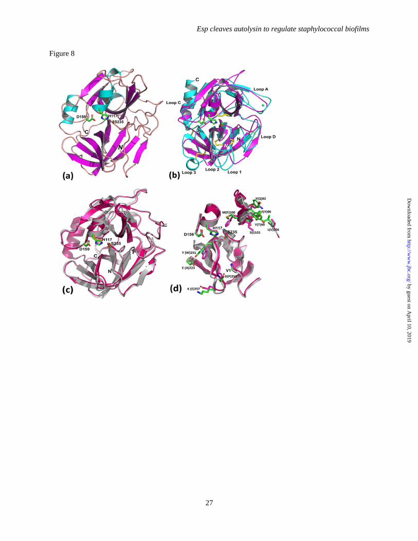
Esp cleaves autolysin to regulate staphylococcal biofilms
27
Figure 8
by guest on April 10, 2019
http://ww
w.jbc.org/
Dow
nloaded from

Esp cleaves autolysin to regulate staphylococcal biofilms
28
Figure 9
by guest on April 10, 2019
http://ww
w.jbc.org/
Dow
nloaded from
and Olaf SchneewindChen Chen, Vengadesan Krishnan, Kevin Macon, Kartik Manne, Sthanam V.L. Narayana
aureusSecreted proteases control autolysin-mediated biofilm growth of Staphylococcus
published online August 22, 2013J. Biol. Chem.
10.1074/jbc.M113.502039Access the most updated version of this article at doi:
Alerts:
When a correction for this article is posted•
When this article is cited•
to choose from all of JBC's e-mail alertsClick here
by guest on April 10, 2019
http://ww
w.jbc.org/
Dow
nloaded from