scrotal hemangioma - advanced radiology teaching · scrotal hemangioma joseph junewick, md facr...
TRANSCRIPT

Scrotal HemangiomaJoseph Junewick, MD FACR
09/16/2010
History7 month old male with scrotal mass.
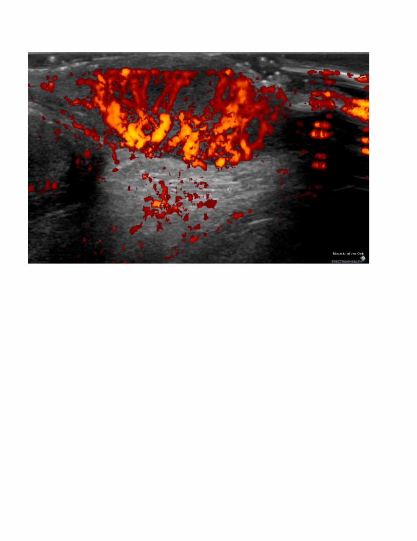
DiagnosisScrotal Hemangioma
DiscussionScrotal hemangiomas are extremely rare, comprising less than 1% of all hemangiomas. They usuallymanifest in infants, and occasionally in later childhood or adolescence. Most patients areasymptomatic, although on rare occasions a patient will present with dull, aching pain, heaviness,bleeding, and ulceration. These symptoms may lead to misdiagnosis of the lesion as a varicocele oringuinal hernia. Microscopic analysis reveals numerous vascular spaces that vary in size and arelined with benign epithelial cells. A lobular pattern of vessels may also be seen. The lesions may becharacterized as capillary, cavernous, or arteriovenous.
FindingsUS-Fusiform hypoechoic thickening of the scrotal skin which is markedly hyperemic on Dopplerimaging.
ReferenceAkbar SA, Sayyed TA, Jafri SZH, Hasteh F, Neill JSA. Multimodality Imaging of ParatesticularNeoplasms and Their Rare Mimics. RadioGraphics (2003); 23:1461-1476.

Sponsored By
DisclaimerThis teaching site is partially funded by an educational grant from GE Healthcare and Advanced Radiology Services, PC. The material on this site isindependently controlled by Advanced Radiology Services, PC, and GE Healthcare and Spectrum Health have no influence over the content of this siteContent Download AgreementThe cases and images on this website are owned by Spectrum Health. Permission is granted (for nonprofit educational purposes) to download and printmaterials to distribute for the purpose of facilitating the education of health professionals. The authors retain all rights to the material and users arerequested to acknowledge the source of the material. Site DisclaimerThis site is developed to reach healthcare professionals and medical students. Nothing this site should be considered medical advice.Only your own doctor can help you make decisions about your medical care. If you have a specific medical question or are seeking medical care, pleasecontact your physician.The information in this website is provided for general medical education purposes only and is not meant to substitute for the independent medicaljudgment of a physician relative to diagnostic and treatment options of a specific medical condition.The viewpoints expressed in these cases are those of the authors. They do not represent an endorsement. In no event will Advanced RadiologyAssociates, PC, Spectrum Health Hospitals (Helen Devos Children's Hospital) or GE Healthcare be liable for any decision made or action taken inreliance upon the information provided through this website.