screening of anti cancer drugs
TRANSCRIPT

1

“ Cancer is a disease which is characterized byuncontrolled proliferation of cells that havetransformed from the normal cells of the body. “
CAUSE: External Factors – chemicals, radiation, viruses, and lifestyle
Internal Factors – hormones, immune conditions, andinherited mutations
Theories
› Cellular change/mutation theories
› Carcinogens
› Oncogenes/ protooncogenes
2

3

Although 92 approved anticancer drugs are available today for thetreatment of more than 200 different tumor entities, effectivetherapies for most of these tumors are lacking.
Out of the 92 registered drugs, 17 are considered by oncologists tobe more broadly applicable and 12 additional agents are perceivedas having certain advantages in some clinical settings
They are mostly cytotoxic in nature and act by a very limitednumber of molecular mechanisms.
Thus, the need for novel drugs to treat malignant disease requiringsystemic therapy is still pressing.
A preselection, called the screening process, is therefore required.
The aim of screening efforts is to identify products that will produceantitumor effects matching the activity criteria used to define whichcompounds can progress to the next stage in the preclinicaldevelopment program.
4

Development of multidrug resistance in patients.
Long-term treatment with cancer drugs is also
associated with severe side effects.
Cytotoxic drugs have the potential to be very harmful to
the body unless they are very specific to cancer cells.
New drugs that will be more selective for cancer cells
5

INVITRO METHODS INVIVO METHODS
6

In Vitro:
1. Tetrazolium salt assay.
2. Sulphorhodamine B assay.
3. 3H-Thymidine uptake.
4. Dye exclusion test.
5. Clonogenic test.
6. Cell counting assay.
7.Morphological assay
In Vivo:
1. Carcinogen induced models
2. Viral infection models
3. Transplantation Models
4. Genetically Engineered Mouse Models
5. In vivo hollow fibre assay
7

8

This assay is a sensitive, quantitative and reliablecolorimetric assay that measures viability, proliferationand activation of cells.
The assay is based on the capacity of mitochondrialdehydrogenase enzymes in living cells to convert theyellow water-soluble substrate 3-(4,5-dimethylthiazol-2-yl)-2,5-diphenyl tetrazolium bromide (MTT) into a darkblue formazan product which is insoluble in water.
9

The amount of formazan produced is directly
proportional to the cell number in range of cell lines.
10
MTT Formazan
metabolically active Cell
Insoluble
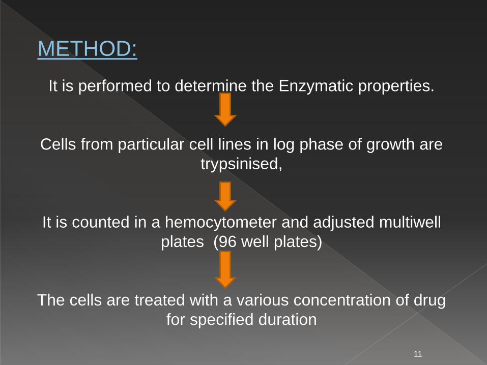
It is performed to determine the Enzymatic properties.
Cells from particular cell lines in log phase of growth are
trypsinised,
It is counted in a hemocytometer and adjusted multiwell
plates (96 well plates)
The cells are treated with a various concentration of drug
for specified duration
11
METHOD:

After MTT dye is added in each well and plates are incubated at 37° C for 4 hrs in a CO2 incubator.
The plates are taken out from the incubator and dark-
blue colored formazan crystal are thoroughly
dissolved in DMSO in room temperature.
The plates are then read on a ELISA reader at 570
nm
To calculate the percent cell viability with respect to
control is calculated .
12

HemocytometerThe most common routine method for cell
counting which is efficient and accurate is with the use
of a hemocytometer.
13

14
% cell viability =(OD of treated cells/ OD ofcontrol cells) × 100

The Sulphorhodamine B assay measures whole-culture protein content, which should be proportional to the cell
number.
Cell culture are stained with a protein staining dye, Sulphorhodamine B.
SRB is a bright pink anionic dye that binds to basic amino acid of cell.
Unbound dye is then removed by washing with acetic acid.
15

During the dead cell either lyse or are lost during
procedure, the amount of SRB binding is proportional
to the number of live cells left in a culture after drug
exposure.
16

Large-scale, morphological changes that occur at
the cell surface, or in the cytoskeleton, can be
followed and related to cell viability.
Damage can be identified by large decreases in
volume secondary to losses in protein and
intracellular ions due to altered permeability to
sodium or potassium.
Necrotic cells: nuclear swelling, chromatin
flocculation, loss of nuclear basophilia
Apoptotic cells: cell shrinkage, nuclear
condensation, nuclear fragmentation
17
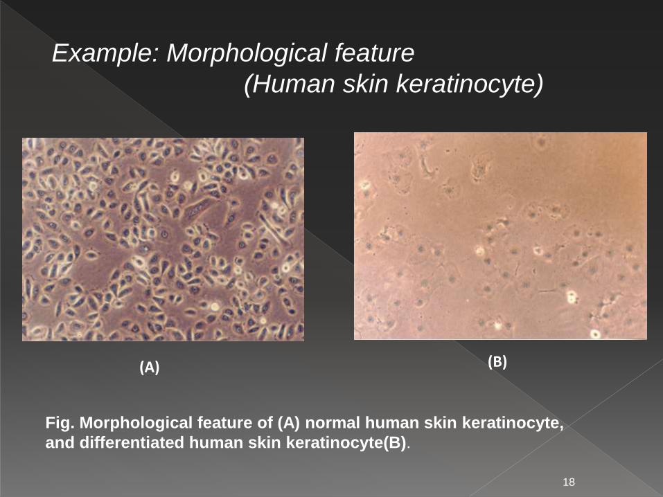
Example: Morphological feature
(Human skin keratinocyte)
(A) (B)
Fig. Morphological feature of (A) normal human skin keratinocyte,
and differentiated human skin keratinocyte(B).
18

Example: Morphological feature
(Human skin fibroblasts)
(A) (B)
Fig. Morphological feature of (A) normal
human skin fibroblasts,
and aging human skin fibroblasts(B).
19

This assay is based on the structural integrity of the
cells.
Live cells possess intact cell membranes that exclude
certain dyes, such as tryphan blue, Eosin, or propidium,
whereas dead cells would have lost membrane integrity.
Hence they would take up the dyes while the live cells
exclude it.METHOD:
1. Cell lines are counted, cultured and innoculated in 96
well plates as above.
20

2. Cells were incubated with different concentrations of test
compounds for 4days.
3. Number of cultured cells in different wells were counted
using hemocytometer after staining with suitable dyes.
%cell viability = no.of viable cell
Total no.of cells (viable+dead)×100
21

Untreated Live cells Treated cells
Tryphan Blue Dye
22

Advantages:
• Reduce the usage of animals.
• Less time consuming, cost effective & easy to manage
• Able to process a larger number of compounds quickly
with minimum quantity.
• Range of concentrations used are comparable to that
expected for in vivo studies.
Disadvantages:
• Difficulty in Maintaining of cultures.
• Show Negative results for the compounds which gets
activated after body metabolism and vice versa.
Impossible to ascertain the Pharmacokinetics
23

24

1. Chemical carcinogen model
DMBA induced mouse skin papillomas
Two stage experimental carcinogenesis
› Initiator – DMBA (dimethylbenz[a]anthracene),
› Promotor – TPA (12-O-tetradecanoyl-phorbol-13-
acetate)
Mice : Single dose – 2.5 µg of DMBA , 5 to 10 μg of TPA
in 0.2 ml of acetone twice weekly.
Papilloma begins to appear after 8 to 10 wks - Tumor
incidence & multiplicity of treatment group is compared
with DMBA control group
25

Mice are topically applied a single dose of 2.5 µg DMBA in
acetone, followed by 5-10 µg of TPA in 0.2 ml acetone
twice weekly on the same site starting one week after
DMBA application.
Percent tumor incidence and multiplicity of treatment
groups is compared with DMBA control group.
Drug under test can be administered either topically or oral
route.
26
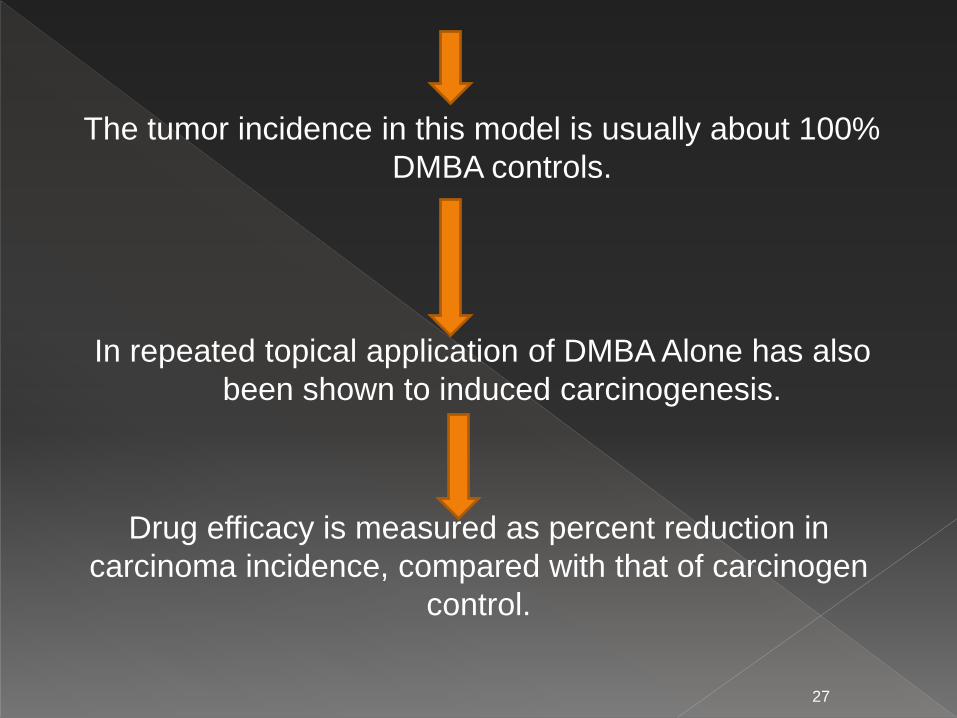
The tumor incidence in this model is usually about 100%
DMBA controls.
In repeated topical application of DMBA Alone has also
been shown to induced carcinogenesis.
27
Drug efficacy is measured as percent reduction in
carcinoma incidence, compared with that of carcinogen
control.

Mouse skin papillomas
RAT MAMMARY GLAND CA
28

Mouse Mammary Tumor Virus (MMTV) was the firstmouse virus, isolated at Jackson labs as the “non-chromosomal factor” that caused mammary tumors inthe C3H strain of mice.
Some viruses cause cancer via random integration incertain cells
Some viruses carry cellular oncogenes
› Abelson murine leukemia virus – Abl
› Moloneymurine sarcoma virus – Raf
Engineered viruses now used routinely in the laboratoryto induce cancer.
29

Tumor cells or tissues (mouse or human) transplanted
into a host mouse.
Ectopic – Implanted into a different organ than the
original (typically subcutaneous or kidney capsule)
Orthotopic – Implanted into the analogous organ of the
original tumor.
Advantages :
› Typically cheap, fast & easy to use.
› Not covered by patents
30

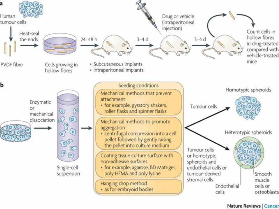
In vivo screening tool implemented in 1995 by NCI.
12 human tumor cell lines (lung, breast, colon,melanoma, ovary, and glioma.
Cells suspended into hollow polyvinylidene fluoridefibers implanted IP or SC in lab mice
After in vivo drug treatment, fibers are removed andanalyzed in vitro
Antitumor (growth inhibitory) activity assessed
31
32

Subcutaneous Hollow Fibre implants
33

2-D and 3-D Cell-based Assays in
Drug Screening
Currently, pharmaceutical firms spend a large amount of
money on the compound efficacy and cytotoxicity test.
There is still a 78% failure rate for all drugs, which may be
devastating to developing companies.
Effective compounds in vitro may be non-effective in vivo
for many reasons, including differences between in vitro
and in vivo target biology, interrelated biochemical
mechanism, metabolism, poor penetration into solid
tissues, etc.
34
Currently, almost all cell-based assays or biosensors are
developed in 2-D culture systems, although conventional 2-
D cultures usually suffer from contact inhibition and a loss
of native cell morphology and functionality.
In comparison with 2-D cultures, 3-D cell models create a
more realistic representation of real human tissues, which
is critical to many important cell functions, including
morphogenesis, cell metabolism, gene expression,
differentiation and cell-cell interactions.
35
1.Ashish A., Sonia SY, Mark AH, and Minas TC., Pressure
Related apoptosis in Neuronal Cel Lines., Journal of
Neuroscience Research 2000 60: 495-503
2.Essentials of medical pharmacology ,by KD Tripathi, 7th
edition ,page no :857
3.https://www.slideshare.net/ashwinisomayaji7/screening-
of-anticancer-drugs
4.https://en.wikipedia.org/wiki/Chemotherapy.
5. http://www.embl-heidelberg.de/
ExternalInfo/karsenti/countingcells. html
36

37