screening of analgesics & drugs used in arthritis & neuropathic pain
TRANSCRIPT

screening of analgesics & drugs used in arthritis &
neuropathic pain
Made by: Meenakshi Gupta

ANALGESICS

INTRODUCTIONPain is a complex unpleasant phenomenon
composed of sensory experiences originating from damaged tissue or abnormal physiological condition.
Analgesics are those agents which selectively relieve pain by acting in the CNS or peripheral pain mechanisms without significantly altering consciousness. Analgesics may be narcotic or non- narcotics.
Narcotic analgesic Non-narcotic analgesic
Act centrally Act peripherallyCause addiction Do not cause addictionProduce CNS depression Do not produce CNS
depressionShow no anti-inflammatory effect
Show anti-inflammatory effect
e.g. Morphine, tramadol, pethidine
e.g. Diclofenac, ibuprofen, aspirin


EVALUATION TECHNIQUESIN-VIVO MODELS:Pain state models using thermal
stimuli The tail-flick model Paw-withdrawal test Hot-plate test Pain- state models using cold stimuli
Pain state models using mechanical stimuli
Strain gauges Von- frey filaments Inclined plane test

Pain state models using electrical stimuli
Electrical stimulation of the tail Grid-shock test Stimulation of the tooth pulp Monkey-shock titration test Stimulation of the limbs.
Pain state models using chemical stimuli
Formalin test Acetic acid induced writhing test Stimulation of hollow organs
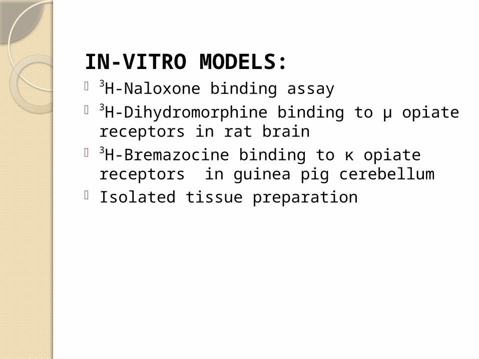
IN-VITRO MODELS:• 3H-Naloxone binding assay • 3H-Dihydromorphine binding to µ opiate
receptors in rat brain• 3H-Bremazocine binding to κ opiate receptors
in guinea pig cerebellum• Isolated tissue preparation

IN-VIVO MODELS:The tail- flick model
Principle: The application of thermal stimulation to the tail of an animal provokes the withdrawal of the tail by a brief vigorous movement. The withdrawal of the tail from the heat source is referred to as ‘tail-flick latency’.
Procedure: In this model, timer is started at the same time as the application of the
heat source
Time taken by the rat to withdraw the tail is recorded. Usually withdrawal time is within 2 to 10s

The lengthening of this reaction time by the animal seen after the administration of drug is interpreted as an analgesic action
Merits:Effective for screening morphine like analgesicsSimple technique & does not require any special
skillResults are quiet accurate & less time consuming
Demerits:Tail- flick response is prone to habituationTruly efficient only for revealing the activity of
opioid analgesics It is advisable not to prolong the exposure to
radiant heat beyond 20s.

Modification:1. Ultra- sound induced tail-flick procedure2. Ear-withdrawal by applying radiant heat3. The tail flick formalin test in rodents

The hot plate methodPrinciple: A plate heated to a constant temperature produces two behavioral components that can be measured in terms of their reaction time, namely paw licking & jumping. Both are considered to be supra-spinally integrated responses.
Procedure:Introduction of rat or mouse into an open-ended cylindrical open space with a floor consisting of metallic plate that is heated by a thermode or a
boiling liquid.
The paw licking behaviour is affected only by opioids whereas the jumping reaction is increased
equally by less powerful analgesics especially when the temperature is 50 degree or less.

The specificity & sensitivity of the test can be increased by measuring the reaction time of the first evoked behaviour. Chaotic defensive mechanisms in rats includes sniffing, licking fore paws & hind paws, straightening, stamping of feet.

Mechanical method:Procedure:
The preferred sites for applying nociceptive mechanical stimuli are the hind paws and
the tail.a pressure of increasing intensity is
applied to a punctiform area on the hind paw or on the tail
The paw or tail is jammed between a plane surface & a blunt point mounted on
top of a system of cogwheels with a cursor that can be displaced along the
length of graduated beam

Disadvantages:Sometimes difficult to measure the intensity
of stimulus with precision.
These devices permits the application of increasing measurable pressures and the
interruption of test, when threshold is reached
The measured parameter is the threshold for the appearance of a given behaviour
When the pressure increases, the reflex withdrawal of the paw, or a complex
movement of the animal to release its trapped limb & finally a vocal reaction is
observed.

A non negligible level of the variability of responses.
Repetition of a mechanical stimulus can produce a diminution or conversely an increase in the sensitivity of the stimulated part of the body.

Electrical stimulation of tail
Principle: Electrical stimuli of gradually increasing intensity can be applied in sequence through subcutaneous electrodes placed in tail of the rat or the mouse. When such gradually increasing intensity of electrical stimuli are applied from constant voltages, one can observe a reflex movement of tail & vocalisation.
Morphine or morphine like drugs are effective in this model.
There may be chances of death of the animal due to the electrical stimulation.
Modifications: ultrasonic stimulation of the tail may be used in placed of electrical stimulation. This method is fast, simple & precise.

Grid-shock modelProcedure:
Male mice weighing around 18-20g are individually placed in clear plastic chambers
The floor of the box is wired with tightly strung stainless steel wire, space about
1mm apart
The stimulus is given in the form of square wave pulses, 30 cycles per second.
The output of the stimulator has to be connected to alternate wires of the grid. A
fixed resistance is placed in series with grid and in parallel to oscilloscope.

The behavior is accurately reflected on the oscilloscope by marked fluctuations of the displaced pulse and defined as pain threshold response. Pain threshold are determined in individual mouse twice before administration of test drug and 15, 30, 60, 90 & 120 minutes after dosing.
Modification: fractional escape procedure
With increasing shock intensities, the mice flinch, exhibit a starling reaction, increased
locomotion & attempt to jump

Electrical stimulation of the tooth pulp
Principle: This method is based on the stimulation of the tooth pulp of the animal by applying the electrical current. Stimulation of the tooth pulp produces characteristic reactions such as licking, biting, chewing & head flick due to induction of pain.Procedure: Rabbits of either sex are anesthetized with 15 mg/kg
thiopental
Pump chambers are exposed close to the gingival line in the lateral margins of the two front upper incisors with a high speed dental-drills. On the day of experiment, clamping
electrodes are placed into the drilled holesAfter an accommodation period of 30 min, stimulation
is started to determine the threshold value. The stimulus of frequency of 50 Hz is applied for the
duration of 1s. The electrical current is started with 0.2 mA & increased till licking occurs.

Monkey shock titration testProcedure:
Doses of 3.0 mg/kg i.m. morphine, 1.7 mg/kg i.m. methadone & 10 mg/kg i.m. pentazocine were found to be effective. It may be used for the final evaluation of a new compound before administration to man.
Monkeys are seated in restraining chairs. Electrical current is delivered by shockers through electrodes coupled to two test tube clamps, which are attached to the shaved portion of the
tail.
The current ranges from 0 to 4 mA through 29 progressive steps. The monkey presses the bar to interrupt the shock. A stable baseline shock level is established for each monkey
on the day prior to drug administration
After drug administration, shock titration activity is rated according to the change in maximum level of median shock
intensity attained for the drugs as compared to control levels.


Formalin test
Principle: chemical stimulation involving the administration of algogenic agents represents a slow, progressive and irreversible form of stimulation.Procedure:
Painful behavior can be assessed on the four-level scale related to posture: 0 denotes normal posture; 1denotes the injected paw remains on the ground but not supporting the animal; 2 denotes the injected paw clearly raised and 3 denotes the injected paw being licked, nibbled & shaken.
Formalin is injected into the front paw and reaction is recorded as excessive licking & biting of the paw.
Analgesic response or protection is indicated, if both paws rest on the floor

Acetic-acid induced writhingPain is often induced in rats or mice by injecting certain irritants such as phenyl quinone or acetic acid into the peritoneal cavity. The animal reacts with a characteristic stretching behaviour which is called writhing. The i.p. administration of agents that irritate serous membrane elicit abdominal contractions.
Modifications: Use of phenyl benzoquinone & acetyl choline Use of substance P & capsaicin in rats in knee joints Intraplantar injection of LPS, bradykinin, carragen- an, TNF-α etc.

IN-VITRO MODELS:3H-Dihydromorphine binding to µ
receptors Purpose and rationaleµ Receptors are considered to mediate the supra-spinal activity of opioids. 3H-Dihydromorphine (3H-DHM)exhibits some selectivity for the µ receptor, a high affinity opiate binding site.Tissue preparation: Male Wistar rats are sacrificed by decapitation & whole brain homogenated.Assay
• 850 µl tissue suspension• 80 µl distilled water• 20 µl vehicle, or levallorphan, or appropriate concentration of drug• 50 µl [3H]DHM.
Evaluation• Specific binding is defined as the difference between total binding
and binding in the presence of 0.1 mM levallorphan. • IC50 values are calculated from the percent specific binding at
each drug concentration.

NEUROPATHIC PAIN

‘The most terrible of all tortures, which a nerve wound may inflict’. Neuropathic pain is characterized by the sensory abnormalities such as unpleasant abnormal sensation (dysesthesia), an increased response to painful stimuli (hyperalgesia), and pain in response to a stimulus that does not normally provoke pain (allodynia).
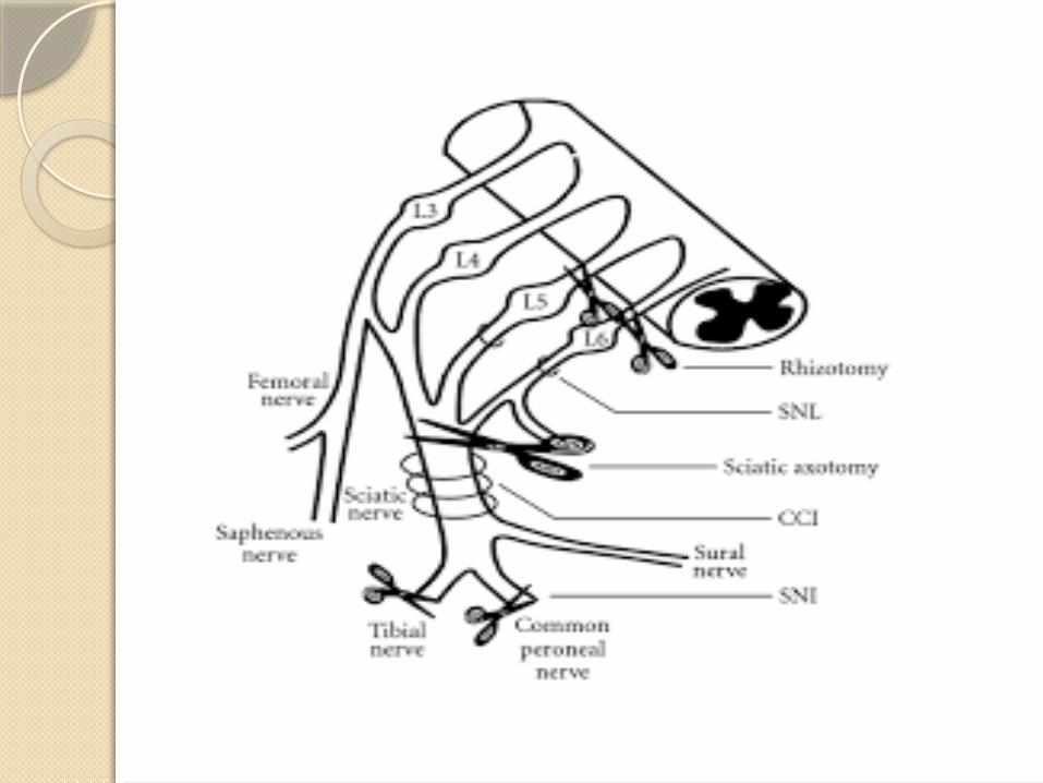
Classification of models: a. Central pain models: Weight drop model Excitotoxic spinal cord injuryb. Peripheral nerve injury models: Chronic constriction injury model Partial sciatic nerve ligation L5/L6 spinal nerve ligation modelc. Cancer pain models: Cancer invasion pain model Vincristine induced peripheral neuropathic paind. Cellular models: Primary culture of sensory models

Weight drop modelIn this model, the spinal cord at lower thoracic-lumbar level is exposed and a constant weight is dropped over the nerve to produce an injury, which is characterized by severe paraplegia and complete segmental necrosis.Procedure:
After anesthesia, laminectomy is performed at the vertebral lower thoracic-lumbar region (T-10) level to expose dorsal
spinal cord
A brass guide tube (15 cm in length) is positioned perpendicularly above the exposed cord, and a cylindrical 10 g steel weight (2 mm in diameter) with a rounded tip is
suspended within the tube
The weight is allowed to drop on the exposed cord, at the T12–13 segmental level, to produce cord injury.

The hypersensitivity to light mechanical stimulation of the skin has been noted to develop within 1 day of injury and parallels the allodynia experienced by patients rapidly following spinal
injury
Alternatively, a longitudinal incision is made on the midline of the back to expose T8 vertebrae by dissecting the
paravertebral muscles. It is followed by a four-level T6–T7 laminectomy to expose the spinal cord, and injury is
produced by extradural compression of the spinal cord using an aneurysm clip with a closing force of 24 g

Excitotoxic spinal cord injury
Intraspinal injections of a-amino-3-hydroxy-5-methyl-4-isoxazolepropionic acid (AMPA) metabotropic receptor agonist quisqualic acid (QUIS) have been made to simulate injury-induced elevations of excitatory amino acids.
Procedure:
Intraspinal or intrathecal injections of glutamate,N-methyl-D-aspartic acid, kainic acid, dynorphin A, serotonin, and tryptamine have also been reported to produce SCI-related pain behaviors.
The unilateral injections are made between the dorsal vein and dorsal root entry zone at depths ranging from 300 to
1200 mm below the surface of the spinal cord, at the levels ranging from T10 to L4
The intraspinal injection of QUIS has been made to produce excitotoxic injury resulting in neuronal loss in specific regions of the spinal gray matter and produces ‘spontaneous’ and/or ‘evoked’ pain behaviors

Chronic constriction injury model
The behavioral signs of spontaneous pain guarding, excessive licking, and avoidance of placing weight on the injury side, mechanical and thermal hyperalgesia, chemical hyper-reactivity and cold allodynia have been noted to develop within a week.
Under anesthesia, about 3-cm long blunt dissection is made into the skin exposing the common sciatic nerve at the level of
the mid-thigh level and loose ligation of nerve with three ligatures (chromic silk, Ethicon), at about 0.5 mm spacing,
proximal to trifurcation of the nerve
This constriction of the sciatic nerve is associated with intraneural edema, focal ischemia, and Wallerian degeneration

Partial sciatic nerve ligation
The left hind leg of rat is shaved and dissection is made to expose the sciatic nerve at the upper-thigh level.
The dorsal one-third to half of the sciatic nerve is tightly ligated with a silk suture just distal to the point at which posterior biceps
semitendinosusnerve branches off
The behavioral signs of spontaneous pain in the form of paw guarding and licking on the injury side have been reported. The
behavioral alterations like cold allodynia, chemical hyper-reactivity, and mechanical hyperalgesia have been noted to
occur within 1 week after the surgery and most of the changes persist for 6 weeks


Vincristine induced peripheral neuropathic painProcedure: Administration of multiple doses of vincristine (20, 75, 100 or 200 µg/kg i.v.) by tail vein injection, followed by 500µl of saline to prevent vessel deterioration, was reported to result in rapid onset painful neuropathy. The dosage regimen of 75 lg/kg i.v. has been preferred because of maximal hyperalgesia and relative absence of motor impairment in rat.

ARTHRITIS

Canine anterior cruciate ligament transection model
Principle:
Procedure:
ACL transection in the dog knee & joint instability
Cartilage erosion, fibrillation & formation of osteophytes
Dog anaesthetized with 30 mg/kg sodium pentobarbital i.v. followed by continuous inhalation of halothane+ nitrous oxide+
oxygenAfter shaving & sterilizing the knee joint externally, it is bent in
a bent position at 90º
Scalpel blade is inserted deep into the joint space diagonally posterior to the ACL and parallel to the lateral border of the
patellar ligament

Evaluation:Microscopic inspection of cartilage & osteophytes are recorded.
By the rotation of blade, ACL is dissected & blade withdrawn & the wound is closed
After 8-12 weeks, fibrillation & erosion of cartillage
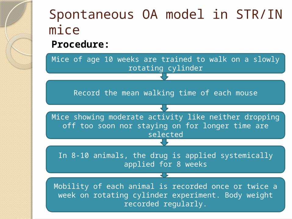
Spontaneous OA model in STR/IN miceProcedure:Mice of age 10 weeks are trained to walk on a slowly rotating
cylinder
Record the mean walking time of each mouse
Mice showing moderate activity like neither dropping off too soon nor staying on for longer time are selected
In 8-10 animals, the drug is applied systemically applied for 8 weeks
Mobility of each animal is recorded once or twice a week on rotating cylinder experiment. Body weight recorded regularly.

Evaluation: mean walking time decreases with age & disease progression.
At the end of experiment, animals are sacrificed, both knees are dissected, fixed, decalcified & embedded in defined orientation
for histology.

Thank you