scleroderma-associated interstitial lung disease (ssc-ild)
TRANSCRIPT
Systemic Sclerosis-Associated Interstitial Lung Disease
(SSc-ILD)
SC-US-68902
Objectives
• Review the epidemiology and burden of SSc-ILD
• Highlight the clinical presentation of SSc-ILD, including the clinical, biological, and radiographic features associated with SSc-ILD progression
• Describe the underlying pathogenesis in SSc-ILD, which is characterized by the interplay between fibrosis, autoimmunity, inflammation, and vascular injury
• Discuss best practices for diagnosing SSc-ILD, including the tools and tests utilized to diagnose SSc-ILD and assess disease severity
2
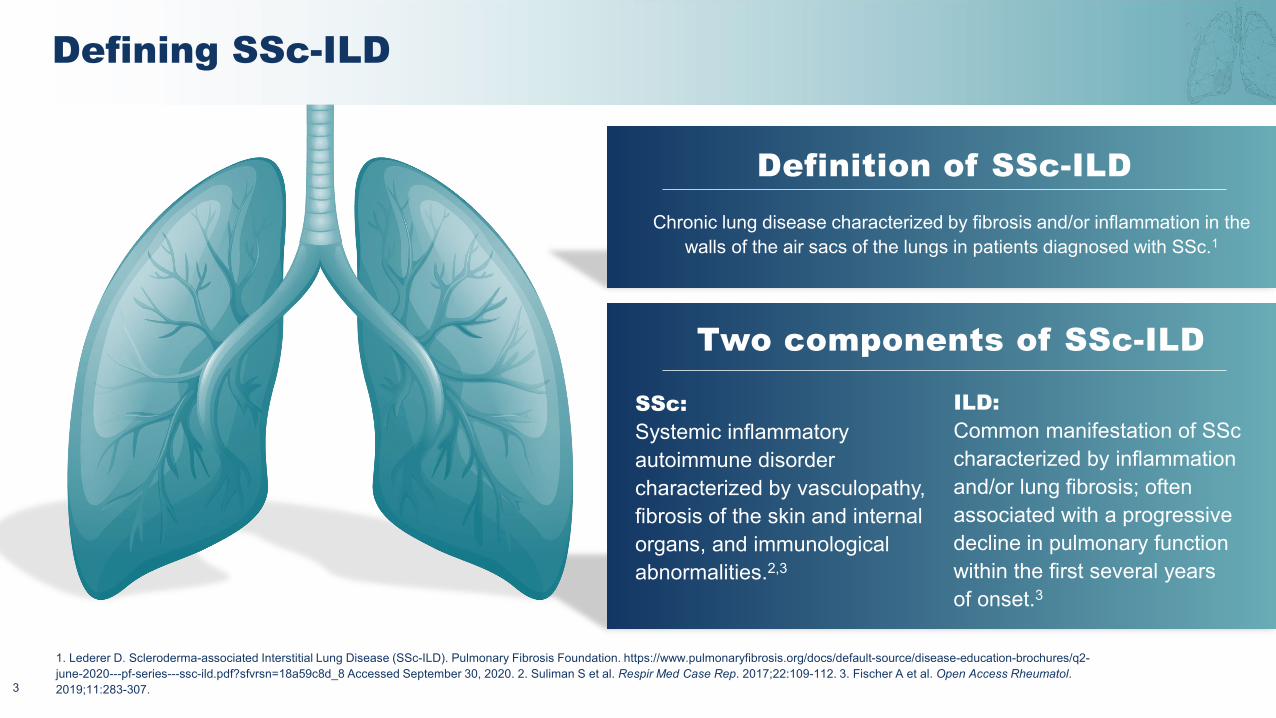
Defining SSc-ILD
3
Definition of SSc-ILDChronic lung disease characterized by fibrosis and/or inflammation in the
walls of the air sacs of the lungs in patients diagnosed with SSc.1
Two components of SSc-ILD
SSc: Systemic inflammatory autoimmune disorder characterized by vasculopathy, fibrosis of the skin and internal organs, and immunological abnormalities.2,3
ILD: Common manifestation of SSc characterized by inflammation and/or lung fibrosis; often associated with a progressive decline in pulmonary function within the first several years of onset.3
1. Lederer D. Scleroderma-associated Interstitial Lung Disease (SSc-ILD). Pulmonary Fibrosis Foundation. https://www.pulmonaryfibrosis.org/docs/default-source/disease-education-brochures/q2-june-2020---pf-series---ssc-ild.pdf?sfvrsn=18a59c8d_8 Accessed September 30, 2020. 2. Suliman S et al. Respir Med Case Rep. 2017;22:109-112. 3. Fischer A et al. Open Access Rheumatol. 2019;11:283-307.
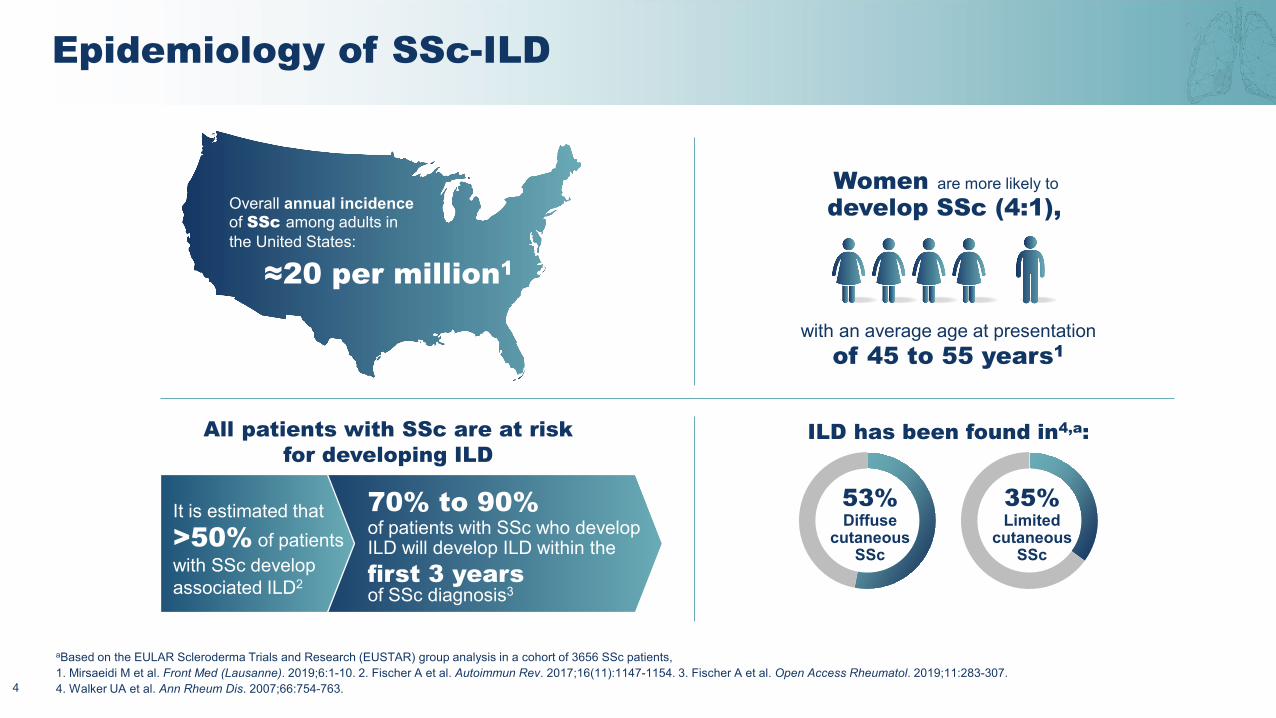
Epidemiology of SSc-ILD
4
Overall annual incidence of SSc among adults in the United States:
≈20 per million1
Women are more likely to develop SSc (4:1),
with an average age at presentationof 45 to 55 years1
It is estimated that >50% of patients with SSc develop associated ILD2
70% to 90%of patients with SSc who develop ILD will develop ILD within the
ILD has been found in4,a:
53%Diffuse
cutaneousSSc
35%Limited
cutaneousSSc
All patients with SSc are at risk for developing ILD
first 3 years of SSc diagnosis3
Based on the EULAR Scleroderma Trials and Research (EUSTAR) group analysis in a cohort of 3656 SSc patients,(Lausanne)
Walker UA et al.
a
1. Mirsaeidi M et al. Front Med . 2019;6:1-10. 2. Fischer A et al. Autoimmun Rev. 2017;16(11):1147-1154. 3. Fischer A et al. Open Access Rheumatol. 2019;11:283-307. 4. Ann Rheum Dis. 2007;66:754-763.
Risk of Mortality With SSc-ILD
5
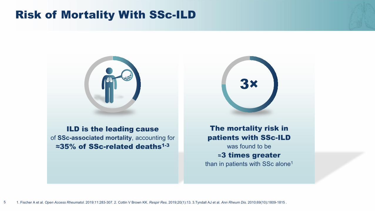
ILD is the leading cause of SSc-associated mortality, accounting for
≈35% of SSc-related deaths1-3
The mortality risk in patients with SSc-ILD
was found to be ≈3 times greater
than in patients with SSc alone1
3×
1. Fischer A et al. Open Access Rheumatol. 2019;11:283-307. 2. Cottin V Brown KK. Respir Res. 2019;20(1):13. 3.Tyndall AJ et al. Ann Rheum Dis. 2010;69(10):1809-1815 .
Burden of SSc
6
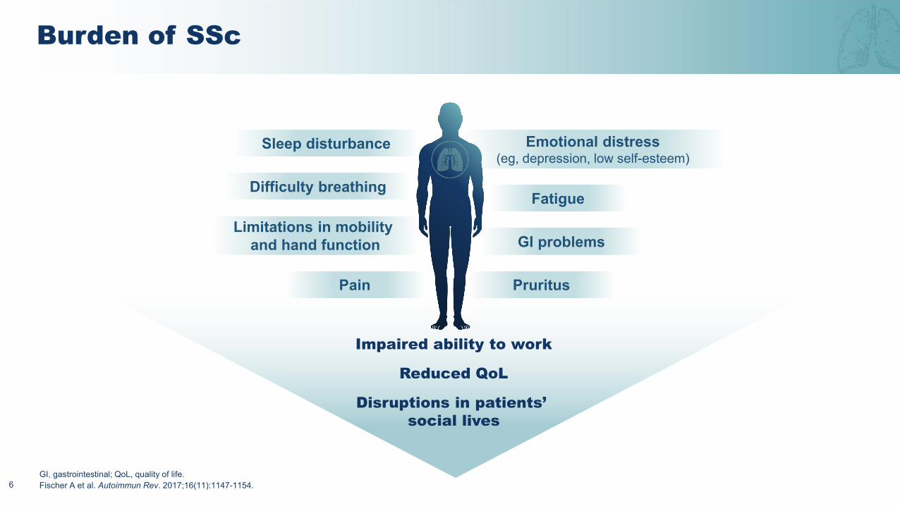
Sleep disturbance
Difficulty breathing
Limitations in mobility and hand function
Pain
Emotional distress(eg, depression, low self-esteem)
Fatigue
GI problems
Pruritus
Impaired ability to work
Reduced QoL
Disruptions in patients’ social lives
GI, gastrointestinal; QoL, quality of life.Fischer A et al. Autoimmun Rev. 2017;16(11):1147-1154.
Distinct ILD Clinical Phenotypes Exist in SSc
7
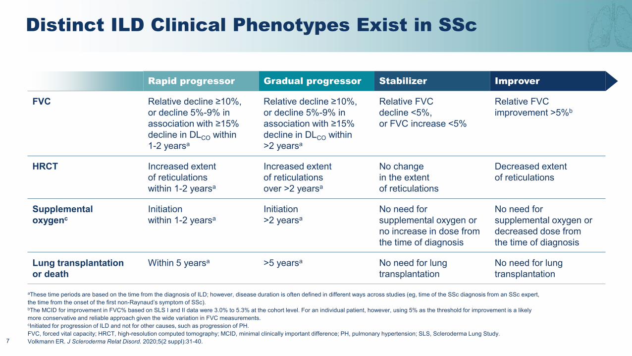
Rapid progressor Gradual progressor Stabilizer Improver
FVC Relative decline ≥10%, or decline 5%-9% in association with ≥15% decline in DLCO within 1-2 yearsa
Relative decline ≥10%, or decline 5%-9% in association with ≥15% decline in DLCO within >2 yearsa
Relative FVC decline <5%, or FVC increase <5%
Relative FVC improvement >5%b
HRCT Increased extent of reticulations within 1-2 yearsa
Increased extent of reticulations over >2 yearsa
No change in the extent of reticulations
Decreased extent of reticulations
Supplemental oxygenc
Initiation within 1-2 yearsa
Initiation >2 yearsa
No need for supplemental oxygen or no increase in dose from the time of diagnosis
No need for supplemental oxygen or decreased dose from the time of diagnosis
Lung transplantation or death
Within 5 yearsa >5 yearsa No need for lung transplantation
No need for lung transplantation
aThese time periods are based on the time from the diagnosis of ILD; however, disease duration is often defined in different ways across studies (eg, time of the SSc diagnosis from an SSc expert, the time from the onset of the first non-Raynaud’s symptom of SSc).bThe MCID for improvement in FVC% based on SLS I and II data were 3.0% to 5.3% at the cohort level. For an individual patient, however, using 5% as the threshold for improvement is a likely more conservative and reliable approach given the wide variation in FVC measurements.cInitiated for progression of ILD and not for other causes, such as progression of PH.FVC, forced vital capacity; HRCT, high-resolution computed tomography; MCID, minimal clinically important difference; PH, pulmonary hypertension; SLS, Scleroderma Lung Study.Volkmann ER. J Scleroderma Relat Disord. 2020;5(2 suppl):31-40.
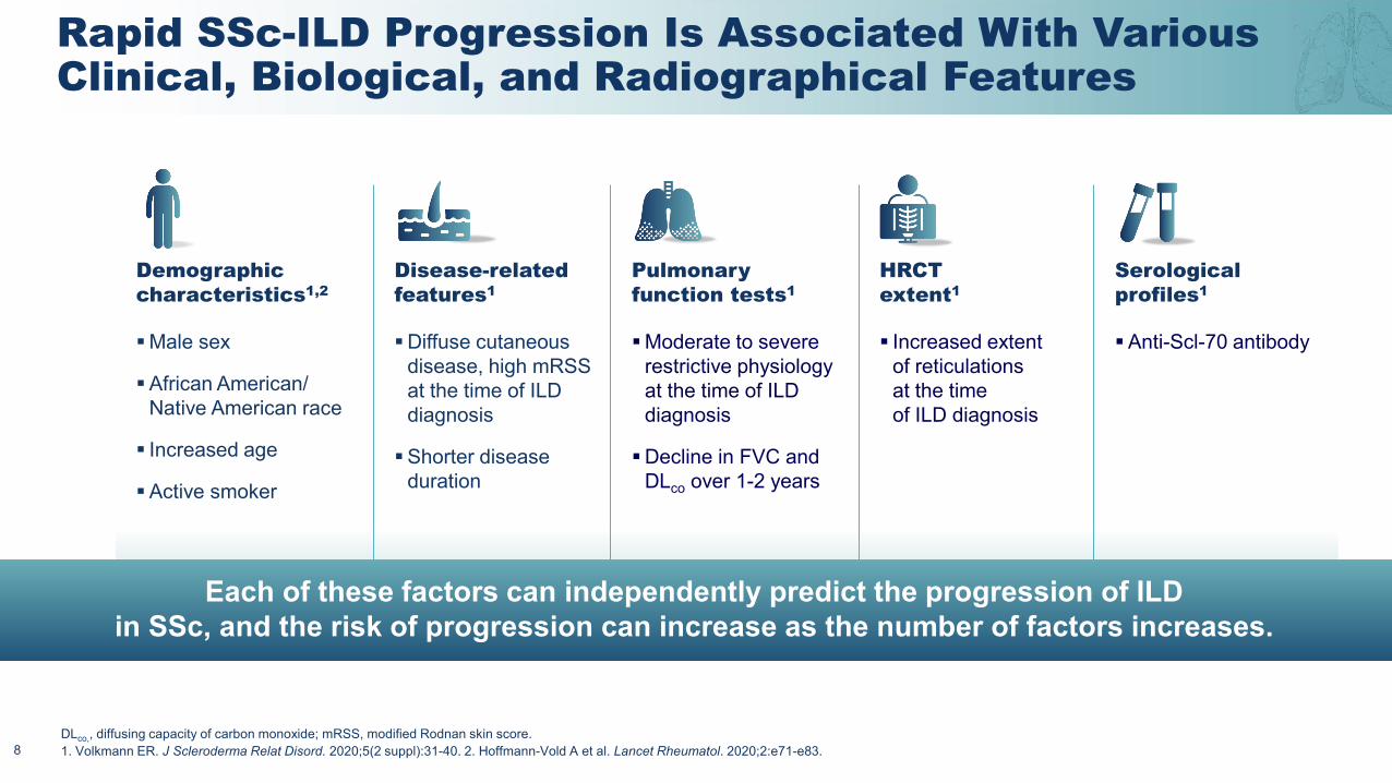
Demographic characteristics1,2
Disease-related features1
Pulmonary function tests1
HRCT extent1
Serological profiles1
Male sex
African American/Native American race
Increased age
Active smoker
Diffuse cutaneous disease, high mRSSat the time of ILD diagnosis
Shorter disease duration
Moderate to severe restrictive physiology at the time of ILD diagnosis
Decline in FVC and DLco over 1-2 years
Increased extent of reticulations at the time of ILD diagnosis
Anti-Scl-70 antibody
Rapid SSc-ILD Progression Is Associated With Various Clinical, Biological, and Radiographical Features
DLco,, diffusing capacity of carbon monoxide; mRSS, modified Rodnan skin score. 1. Volkmann ER. J Scleroderma Relat Disord. 2020;5(2 suppl):31-40. 2. Hoffmann-Vold A et al. Lancet Rheumatol. 2020;2:e71-e83. 8
Each of these factors can independently predict the progression of ILD in SSc, and the risk of progression can increase as the number of factors increases.
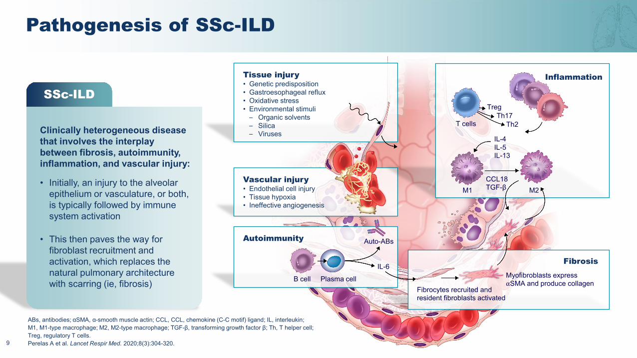
Tissue injury• Genetic predisposition• Gastroesophageal reflux• Oxidative stress• Environmental stimuli
– Organic solvents– Silica– Viruses
Vascular injury• Endothelial cell injury• Tissue hypoxia• Ineffective angiogenesis
Fibrosis
Fibrocytes recruited and resident fibroblasts activated
Myofibroblasts express ⍺SMA and produce collagen
Inflammation
T cells
TregTh17
Th2
IL-4IL-5IL-13
M1CCL18TGF-β M2
Pathogenesis of SSc-ILD
9
SSc-ILD
Clinically heterogeneous disease that involves the interplay between fibrosis, autoimmunity, inflammation, and vascular injury:
• Initially, an injury to the alveolar epithelium or vasculature, or both, is typically followed by immune system activation
• This then paves the way for fibroblast recruitment and activation, which replaces the natural pulmonary architecture with scarring (ie, fibrosis)
ABs, antibodies; SMA, smooth muscle actin; CCL, CCL, chemokine (C-C motif) ligand; IL, interleukin; M1, M1-type macrophage; M2, M2-type macrophage; TGF-β, transforming growth factor β; Th, T helper cell; Treg, regulatory T cells.
α α-
Perelas A et al. Lancet Respir Med. 2020;8(3):304-320.
Autoimmunity
B cell Plasma cell
Auto-ABs
IL-6
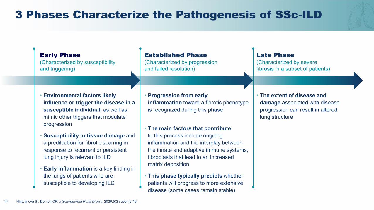
3 Phases Characterize the Pathogenesis of SSc-ILD
10
Established Phase (Characterized by progression and failed resolution)
• Progression from early inflammation toward a fibrotic phenotype is recognized during this phase
• The main factors that contribute to this process include ongoing inflammation and the interplay between the innate and adaptive immune systems; fibroblasts that lead to an increased matrix deposition
• This phase typically predicts whether patients will progress to more extensive disease (some cases remain stable)
• The extent of disease and damage associated with disease progression can result in altered lung structure
Late Phase (Characterized by severe fibrosis in a subset of patients)
Nihtyanova SI, Denton CP. J Scleroderma Relat Disord. 2020;5(2 suppl):6-16.
Early Phase (Characterized by susceptibility and triggering)
• Environmental factors likely influence or trigger the disease in a susceptible individual, as well as mimic other triggers that modulate progression
• Susceptibility to tissue damage and a predilection for fibrotic scarring in response to recurrent or persistent lung injury is relevant to ILD
• Early inflammation is a key finding in the lungs of patients who are susceptible to developing ILD
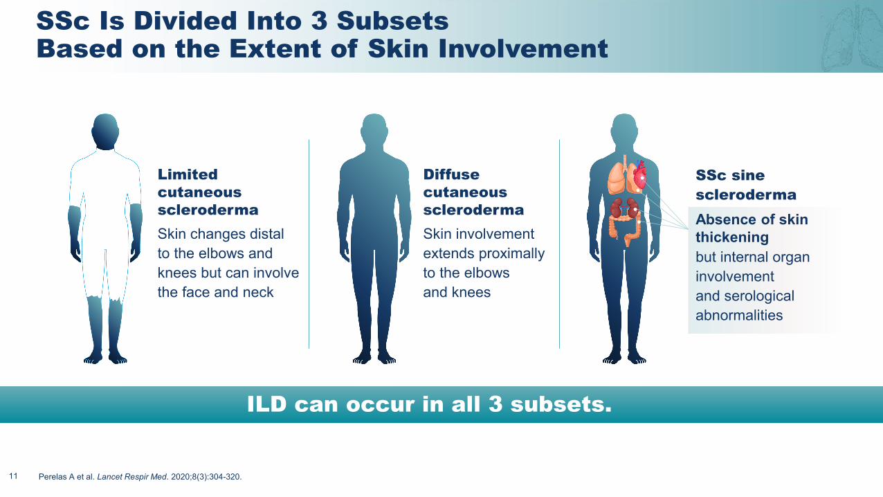
SSc Is Divided Into 3 Subsets Based on the Extent of Skin Involvement
11
ILD can occur in all 3 subsets.
Limited cutaneous sclerodermaSkin changes distal to the elbows and knees but can involve the face and neck
Diffuse cutaneous sclerodermaSkin involvement extends proximally to the elbows and knees
SSc sine sclerodermaAbsence of skin thickening but internal organ involvement and serological abnormalities
Perelas A et al. Lancet Respir Med. 2020;8(3):304-320.
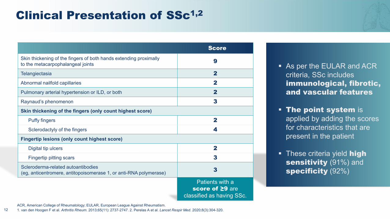
Patients with a score of ≥9 are
classified as having SSc.
Clinical Presentation of SSc1,2
12
Score
Skin thickening of the fingers of both hands extending proximally to the metacarpophalangeal joints 9
Telangiectasia 2Abnormal nailfold capillaries 2Pulmonary arterial hypertension or ILD, or both 2Raynaud’s phenomenon 3Skin thickening of the fingers (only count highest score)
Puffy fingers 2Sclerodactyly of the fingers 4
Fingertip lesions (only count highest score)
Digital tip ulcers 2Fingertip pitting scars 3
Scleroderma-related autoantibodies (eg, anticentromere, antitopoisomerase 1, or anti-RNA polymerase) 3
As per the EULAR and ACR criteria, SSc includes immunological, fibrotic, and vascular features
The point system is applied by adding the scores for characteristics that are present in the patient
These criteria yield high sensitivity (91%) and specificity (92%)
ACR, American College of Rheumatology; EULAR, European League Against Rheumatism.Arthritis Rheum. 2013;65(11): 2737-2747. 2. Perelas A et al. Lancet Respir Med. 2020;8(3):304-320.1. van den Hoogen F et al.
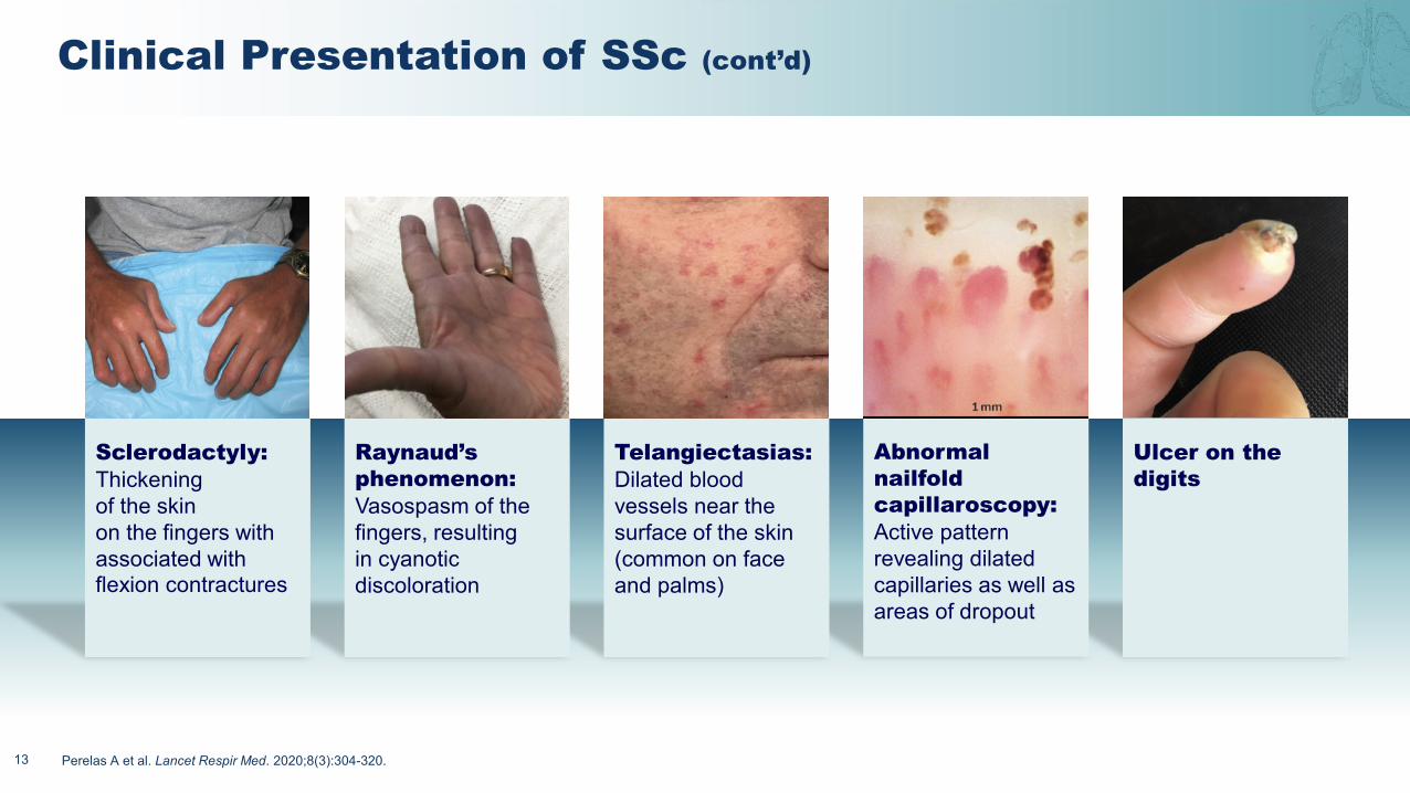
Ulcer on the digits
Abnormal nailfold capillaroscopy:Active pattern revealing dilatedcapillaries as well as areas of dropout
Telangiectasias: Dilated blood vessels near the surface of the skin (common on face and palms)
Raynaud’s phenomenon: Vasospasm of the fingers, resulting in cyanotic discoloration
Clinical Presentation of SSc (cont’d)
13
Sclerodactyly: Thickening of the skin on the fingers with associated with flexion contractures
Perelas A et al. Lancet Respir Med. 2020;8(3):304-320.
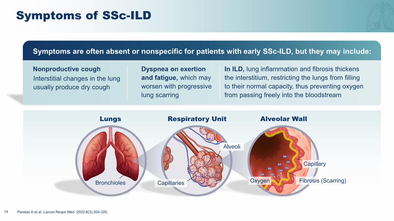
Symptoms of SSc-ILD
14
Nonproductive cough Interstitial changes in the lung usually produce dry cough
Dyspnea on exertion and fatigue, which may worsen with progressive lung scarring
In ILD, lung inflammation and fibrosis thickens the interstitium, restricting the lungs from filling to their normal capacity, thus preventing oxygen from passing freely into the bloodstream
Symptoms are often absent or nonspecific for patients with early SSc-ILD, but they may include:
Lungs Respiratory Unit Alveolar Wall
Bronchioles Capillaries Oxygen Fibrosis (Scarring)
Capillary
Alveoli
Perelas A et al. Lancet Respir Med. 2020;8(3):304-320.
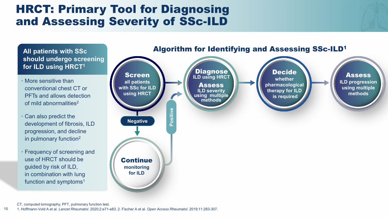
HRCT: Primary Tool for Diagnosing and Assessing Severity of SSc-ILD
15
Screenall patients
with SSc for ILDusing HRCT
DiagnoseILD using HRCT
AssessILD severity
using multiple methods
Decidewhether
pharmacological therapy for ILD
is required
AssessILD progression using multiple
methods
Continuemonitoring
for ILDPo
sitiv
e
Negative
• More sensitive than conventional chest CT or PFTs and allows detection of mild abnormalities2
• Can also predict the development of fibrosis, ILD progression, and decline in pulmonary function2
• Frequency of screening and use of HRCT should be guided by risk of ILD, in combination with lung function and symptoms1
All patients with SSc should undergo screening for ILD using HRCT1
Algorithm for Identifying and Assessing SSc-ILD1
CT, computed tomography; PFT, pulmonary function test. 1. Hoffmann-Vold A et al. Lancet Rheumatol. 2020;2:e71-e83. 2. Fischer A et al. Open Access Rheumatol. 2019;11:283-307.
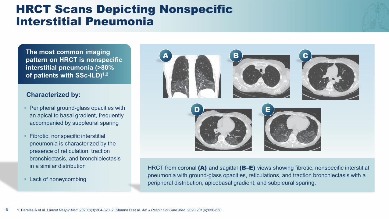
HRCT Scans Depicting Nonspecific Interstitial Pneumonia
16
A B C
D E
HRCT from coronal (A) and sagittal (B–E) views showing fibrotic, nonspecific interstitial pneumonia with ground-glass opacities, reticulations, and traction bronchiectasis with a peripheral distribution, apicobasal gradient, and subpleural sparing.
Peripheral ground-glass opacities with an apical to basal gradient, frequently accompanied by subpleural sparing
Fibrotic, nonspecific interstitial pneumonia is characterized by the presence of reticulation, traction bronchiectasis, and bronchiolectasisin a similar distribution
Lack of honeycombing
The most common imaging pattern on HRCT is nonspecific interstitial pneumonia (>80% of patients with SSc-ILD)1,2
Characterized by:
1. Perelas A et al. Lancet Respir Med. 2020;8(3):304-320. 2. Khanna D et al. Am J Respir Crit Care Med. 2020;201(6):650-660.
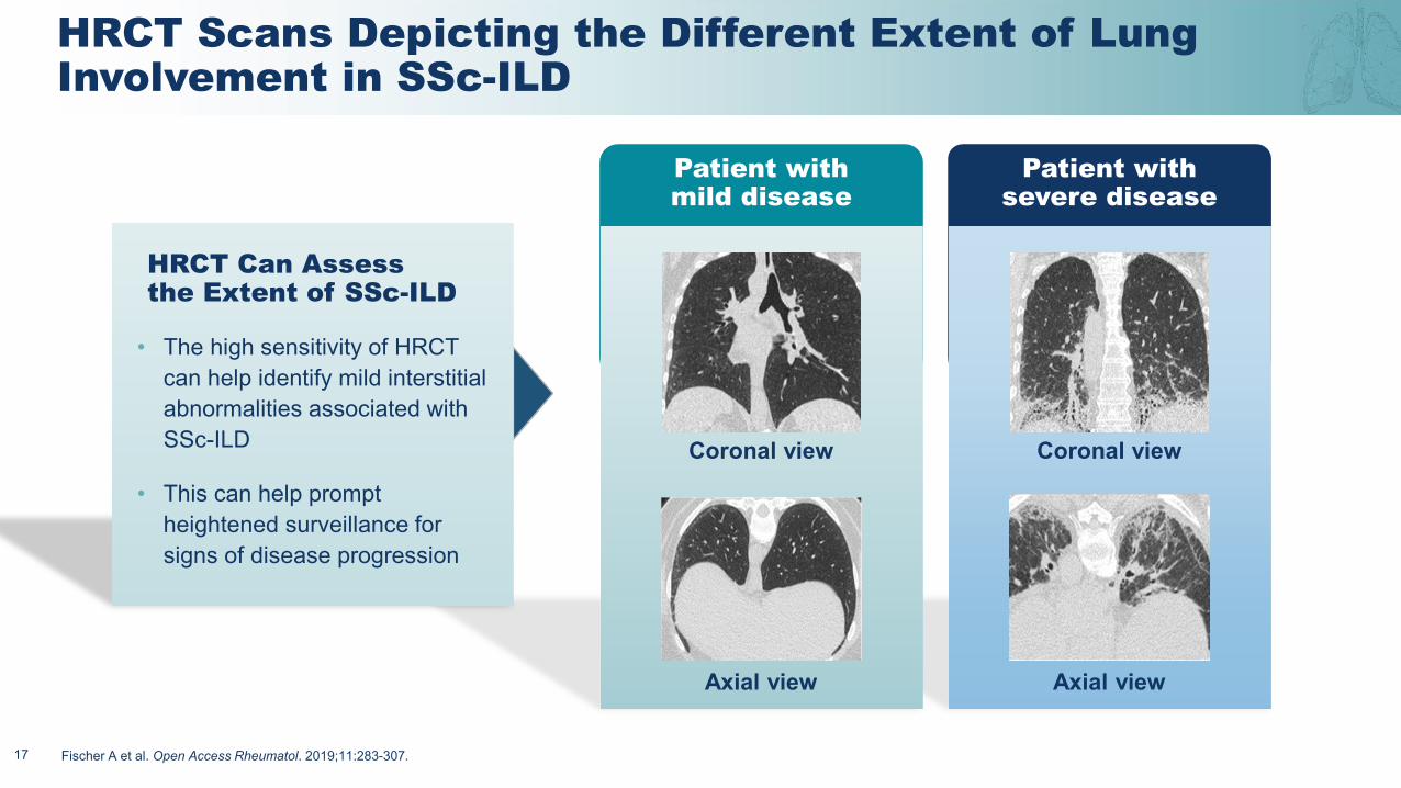
HRCT Scans Depicting the Different Extent of Lung Involvement in SSc-ILD
17
Axial view
Coronal view Coronal view
Axial view
Patient with mild disease
Patient with severe disease
• The high sensitivity of HRCT can help identify mild interstitial abnormalities associated with SSc-ILD
• This can help prompt heightened surveillance for signs of disease progression
HRCT Can Assess the Extent of SSc-ILD
Fischer A et al. Open Access Rheumatol. 2019;11:283-307.
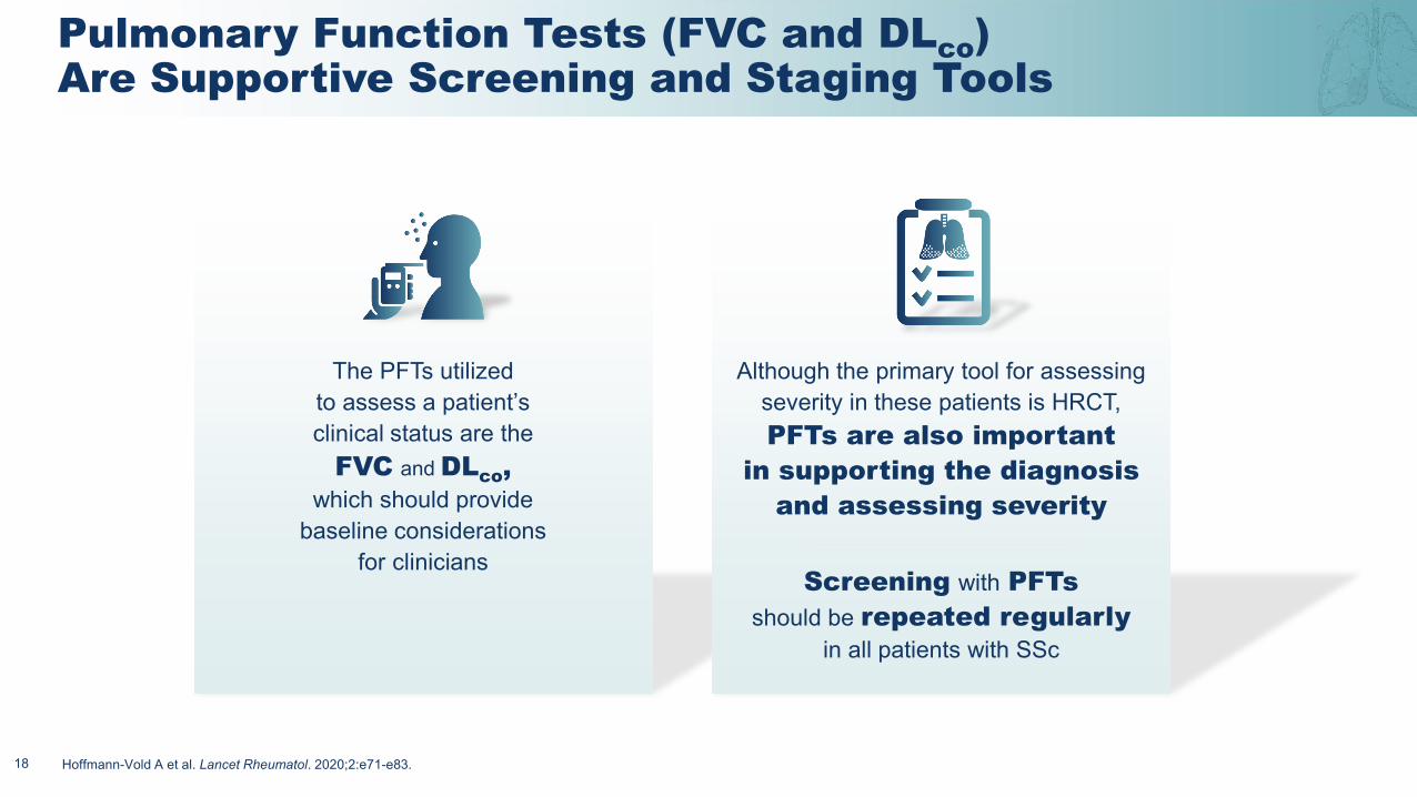
Pulmonary Function Tests (FVC and DLco) Are Supportive Screening and Staging Tools
18
The PFTs utilized to assess a patient’s clinical status are the
FVC and DLco, which should provide
baseline considerations for clinicians
Screening with PFTsshould be repeated regularly
in all patients with SSc
Although the primary tool for assessingseverity in these patients is HRCT, PFTs are also important
in supporting the diagnosis and assessing severity
Hoffmann-Vold A et al. Lancet Rheumatol. 2020;2:e71-e83.
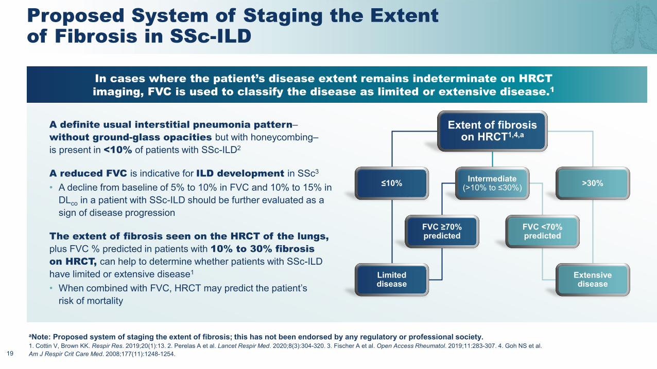
Proposed System of Staging the Extent of Fibrosis in SSc-ILD
19
Extent of fibrosis on HRCT1,4,a
≤10% Intermediate(>10% to ≤30%) >30%
FVC ≥70%predicted
FVC <70%predicted
Limited disease
Extensive disease
A definite usual interstitial pneumonia pattern–without ground-glass opacities but with honeycombing–is present in <10% of patients with SSc-ILD2
A reduced FVC is indicative for ILD development in SSc3
• A decline from baseline of 5% to 10% in FVC and 10% to 15% in DLco in a patient with SSc-ILD should be further evaluated as a sign of disease progression
The extent of fibrosis seen on the HRCT of the lungs, plus FVC % predicted in patients with 10% to 30% fibrosis on HRCT, can help to determine whether patients with SSc-ILD have limited or extensive disease1
• When combined with FVC, HRCT may predict the patient’s risk of mortality
In cases where the patient’s disease extent remains indeterminate on HRCT imaging, FVC is used to classify the disease as limited or extensive disease.1
aNote: Proposed system of staging the extent of fibrosis; this has not been endorsed by any regulatory or professional society.1. Cottin V, Brown KK. Respir Res. 2019;20(1):13. 2. Perelas A et al. Lancet Respir Med. 2020;8(3):304-320. 3. Fischer A et al. Open Access Rheumatol. 2019;11:283-307. 4. Goh NS et al. Am J Respir Crit Care Med. 2008;177(11):1248-1254.
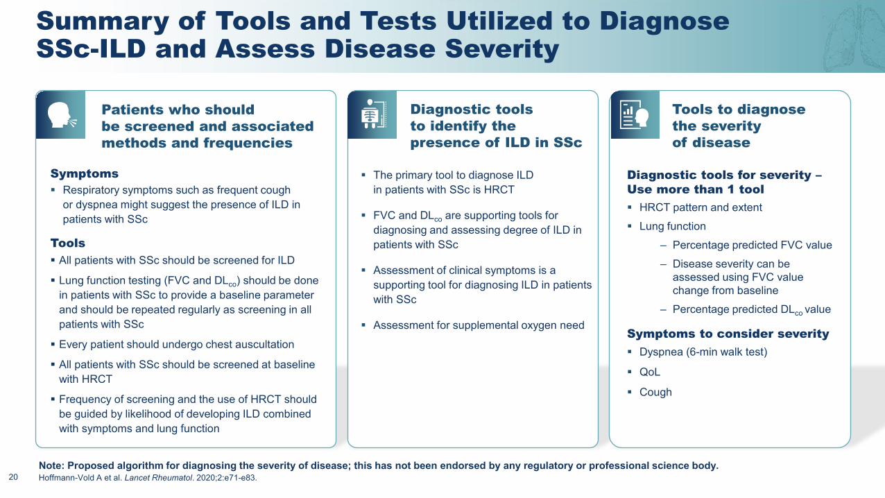
Tools All patients with SSc should be screened for ILD
Lung function testing (FVC and DLco) should be done in patients with SSc to provide a baseline parameter and should be repeated regularly as screening in all patients with SSc
Every patient should undergo chest auscultation
All patients with SSc should be screened at baseline with HRCT
Frequency of screening and the use of HRCT should be guided by likelihood of developing ILD combined with symptoms and lung function
Summary of Tools and Tests Utilized to Diagnose SSc-ILD and Assess Disease Severity
20
Patients who should be screened and associated methods and frequencies
Symptoms Respiratory symptoms such as frequent cough
or dyspnea might suggest the presence of ILD in patients with SSc
Diagnostic tools to identify the presence of ILD in SSc
The primary tool to diagnose ILD in patients with SSc is HRCT
FVC and DLco are supporting tools for diagnosing and assessing degree of ILD in patients with SSc
Assessment of clinical symptoms is a supporting tool for diagnosing ILD in patients with SSc
Assessment for supplemental oxygen need
Tools to diagnose the severity of disease
Diagnostic tools for severity –Use more than 1 tool HRCT pattern and extent Lung function
– Percentage predicted FVC value– Disease severity can be
assessed using FVC value change from baseline
– Percentage predicted DLco value
Symptoms to consider severity Dyspnea (6-min walk test)
QoL
Cough
Note: Proposed algorithm for diagnosing the severity of disease; this has not been endorsed by any regulatory or professional science body.Hoffmann-Vold A et al. Lancet Rheumatol. 2020;2:e71-e83.
Created by popcornartsfrom the Noun Project
Summary
21
SSc-ILD is a clinically heterogeneous disease characterized by a complex interplay between autoimmunity, vasculopathy, and fibrosis, yielding a significant burden on patients
HRCT is the primary tool for diagnosing and assessing degree of disease severity, with nonspecific interstitial pneumonia being the most common imaging pattern on HRCT
The clinical presentations of SSc-ILDare distinct, and should be recognized and monitored appropriately
Various clinical, biological, and radiographic features can drive the progression of SSc-ILD